1. Oculus BateriaA study of bacteria found around the eye area using culturing methods.
Eyelashes have been proven to house small arachnids. This well-
known fact led to the curiosity of bacteria also living around the
eye area. Through culturing methods, multiple bacterial colonies
were found to be present and growing on the untainted
eyelash. These colonies were analyzed by shape, elevation, size,
texture, appearance, pigmentation, and optical density. With the
results, it was concluded that neither E. coli nor staphylococcus are
present around the human eye.
• To learn how to effectively prepare agar plates.
• To discover if bacteria are present around the human eye.
• 1 Eppendorf Tube
• 2 Eyelashes
• 9 Growth Plates
• 1 Magnetic Stirrer
• 25-µL Volumetric Pipette
• 200-mL Erlenmeyer Flasks
• Autoclave
• DI Water
• Stirring Hot Plate
• Tin Foil
• Tape
• Wax Pen
• Glass Spreader
• Bunsen Burner
• Gloves
• Glass Petri Dish with
Alcohol
PROCEDURE
1. Nine nutrient agar (NA) plates were prepared. Three as only
NA, three as NA with staphylococcus, and three as NA with
levine without lactose.
2. Eyelash solution was prepared. Two eyelashes plucked from
donor were placed in an Eppendorf tube with DI water and
shaken well.
3. 25 µL of eyelash solution was pipetted onto each agar plate
and spread evenly with a sterile glass spreader.
4. These agar plates were covered, labeled, and placed in a 37℃
incubator (plates were placed upside down to keep
condensation off the growing bacteria.).
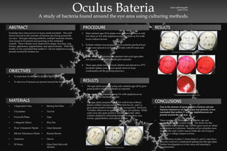
• Due to the absence of gram-negative bacteria and any
bacteria whatsoever on the B plates (see picture), it was
concluded that neither E. coli nor staphylococcus are
present around the eye area.
• These results may differ based on gender, age and
environment of the eyelash donor. The donor for this
experiment was a female, age 17, who grew up in an urban
environment in California. Samples of her eyelashes were
taken in the early winter season while she was living in
dorms on a college campus in Iowa.
• The difference in plate C3 from plates C1 and C2 may have
been the result of a contaminant getting into the agar plate.
Further investigation as to the cause and meaning is
recommended.
Clara Lalhmangaihi
Sydni Crow
• The agar plates prepared using only nutrient agar (NA) grew
gram-positive bacterial colonies as expected.
• The agar plates prepared using NA with staphylococcus
grew no visible bacterial colonies.
• The agar plates prepared using NA with levine without
lactose yielded varying results. The first two (C1 and C2)
grew gram-positive bacterial colonies as expected. However,
*Colonies observed on agar plate C3 were not uniform. While
some were also gram positive, circular, and dull, other
colonies displayed contrasting features of elevation, size,
texture, pigmentation, and optical density.
RESULTS
RESULTSABSTRACT
OBJECTIVES
MATERIALS CONCLUSIONS
Bacterial growth in nutrient agar
No bacterial growth in nutrient agar with staphylococcus
Bacterial growth in nutrient agar with levine*