
Recommended
More Related Content
What's hot
What's hot (20)
Viewers also liked
Viewers also liked (20)
Similar to Bacteria Presentation Slideshow
Similar to Bacteria Presentation Slideshow (20)
Recently uploaded
Recently uploaded (20)
Bacteria Presentation Slideshow
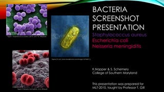
- 1. BACTERIA SCREENSHOT PRESENTATION Staphylococcus aureus Escherichia coli Neisseria meningiditis K.Napper & S. Schemery College of Southern Maryland This presentation was prepared for MLT-2010, taught by Professor T. Gill Figure 1 Staphylococcus aureus. (2014, June 19). Wikipedia Figure 2 E.coli (www.visualphotos.com/image/1x3746017.). Figure 3 Neisseria meningitidis. (n.d.). Bioquell
- 2. STAPHYLOCOCCUS AUREUS Figure 5 Staphylococcus aureus (Pictures.life.ku.dk, 2014)Figure 4 Staphylococcus aureus. (2014, June 19). Wikipedia
- 3. STAPHYLOCOCCUS AUREUS Figure 6 Staphylococcus aureus Bacteriainphotos
- 4. STAPHYLOCOCCUS AUREUS Cellular Morphology • Gram positive cocci in grape like clusters. Colony Morphology • Grows well on blood agar and abundant growth in 18-24 hrs • Large (6-8 mm) smooth, entire, slightly raised, translucent, and pigmented colonies • Off-white, gray, cream-yellow, yellow, yellow-orange, or orange pigment • Colonies surrounded by multiple zones of hemolysis, resembling targets • On MSA plates colonies will have a yellow pigment and the pink medium will turn yellow Figure 7 Staphylococcus aureus(Hogan,2011) Figure 9 Physiological tests (Web.cn.edu, 2014) Figure 8 Staphylococcus aureus (Pictures.life.ku.dk, 2014)
- 5. STAPHYLOCOCCUS AUREUS Distinguishing Media • Blood Agar – for hemolysis detection • Mannitol Salt Agar (MSA)-for mannitol fermentation Differential and Susceptibility testing • Catalase positive • Coagulase positive • Commercial latex agglutination testing • Most strains susceptible to penicillin but some are resistant • Methicillin-resistant MRSA and Vancomycin-resistant strains VRSA
- 6. STAPHYLOCOCCUS AUREUS Indigenous locations • Often found in low numbers as indigenous microflora of the skin. • 20% -30% of the population are “staph carriers” – nasal passages are colonized with S. aureus. As long as the S. aureus is localized to the nasal passages of the carrier it poses no threat, but the bacteria can be transmitted to others. Infections caused by bacteria • Skin and soft tissue-Furnicle, Cabuncles, Wound infections ( surgical/ traumatic), Cellulitis, Impetigo • Musculoskeltal- Ostemyelitis, Arthritis • Cardiovascular- Endocarditis • Genitourinary Tract- Renal carbuncle, Lower urinary tract infection • Other infections cause by toxins- Toxic shock syndrome (TSS), Scalded skin syndrome, Food poisoning (gastroenteritis)
- 7. ESCHERICHIA COLI Figure 11 E. coli on MAC(Bacteriainphotos.com, 2014)Figure 10 E coli (www.visualphotos.com/image/1x3746017).
- 8. ESCHERICHIA COLI Figure 12 Escherichia coli Bacteriainphotos
- 9. ESCHERICHIA COLI Cellular morphology • Gram negative bacilli motile by peritrichous flagella Colony Morphology • On blood agar colonies are smooth, dull gray, and 2-3 mm in diameter • On MAC colonies are lactose- positive colonies producing pink to red, flat, dry, and 2-3 mm in diameter. The colonies are usually surrounded by a darker pink area of precipitated bile salts • On HE and XLD agar colonies have a yellow pigment. • Foul odor Figure 13 E. coli(News, 2014) Figure 14 E. coli on TSA (Catalog.hardydiagnostics.com, 2014 Figure 15 E. coli on MAC(Bacteriainphotos.com, 2014)
- 10. ESCHERICHIA COLI Distinguishing Media • Blood agar – for hemolysis detection • MAC – lactose fermentation detection • HE • XLD Differential and Susceptibility testing • A/A reaction on TSI and KIA slants with or without gas • Indole positive • MR positive • Voges-Proskauer negative • Citrate negative • Oxidase negative • Nitrate positive • PYR negative • MUG positive IF SUSCEPTIBLE: Cephalosporins I, II, III Aminoglycosides Nitrofurantoin Trimethoprim- sulfamethoxazole Doxycycline
- 11. ESCHERICHIA COLI Indigenous locations • Commonly found in the indigenous gastrointestinal flora of humans and animals Infections caused by bacteria • The strains and serotypes of E. coli found in the GI tracts of humans and animals are opportunistic pathogens and can be problematic when entering other sites than the GI tract (bladder, bloodstream, wounds) • Most common cause of UTIs and common cause of septicemia and nosocomial infections • Enterovirulent E. coli or diarrheagenic E. coli serotypes cause GI disease when they are ingested by humans.
- 12. NEISSERIA MENINGITIDIS Figure 17 N. meningiditis on Choc (Cdc.gov, 2014 Figure 16 Neisseria meningitidis. (n.d.). Bioquell
- 13. NEISSERIA MENINGITIDIS Figure 18 Neisseria meningitigis Bacteriainphotos
- 14. NEISSERIA MENINGITIDIS Cellular morphology • Encapsulated gram negative diplococcus • Grows best at 35-37C in moist 5%-7% CO2 environment • On Blood agar, Chocolate agar, or Thayer-Martin medium colonies are gray, low, convex, and glistening, with a smooth, moist entire edge. • Heavily encapsulated strains may be mucoid. • N. meningiditis colonies are about 1 mm larger than N. gonorrhoeae after 18-24 hrs. of incubation. • Older colonies become gummy due to autolysis and release of cellular DNA. Colony morphology Figure 20 N. meningiditis(Textbookofbacteriology.net, 2014) Figure 19 N. meningiditis on Choc (Cdc.gov, 2014 Figure 21 N. menigniditis on BA(Cdc.gov, 2014)
- 15. NEISSERIA MENINGITIDIS Distinguishing Media • Will grow on Blood agar incubated in non-CO2 incubator where as N. gonorrhoeae will not. • Thayer- Martin Differential and Susceptibility testing • Carbohydrate utilization or chromogenic enzyme substrate test • Will acidify media containing glucose and maltose • Oxidase positive • IF SUSCEPTIBLE: penicillin G ampicillin
- 16. NEISSERIA MENINGITIDIS Indigenous locations • Strict human pathogen • Colonizes the nasopharyngeal mucosa of some individuals; N. meningiditis carriers • May also colonizes the genital tract and conjunctiva Infections causes by bacteria • N. meningiditis may be transmitted from person to person by transfer of respiratory secretions or aerosolized droplets • Menigococcemia (N. meningiditis in blood stream) • Petechiae • Meningococcus meningitis • Rare types of infections- genital tract infection, proctitis, and conjunctivitis
- 17. REFERENCES• Bacteriainphotos.com (2014). Escherichia coli colonies on macconkey agar, e.coli lactose positive colonies. [online] Retrieved from: http://www.bacteriainphotos.com/Escherichia%20coli%20on%20MacConkey%20agar.html [Accessed: 22 Jun 2014]. • Catalog.hardydiagnostics.com (2014). Tryptic soy agar (tsa). [online] Retrieved from: https://catalog.hardydiagnostics.com/cp_prod/Content/hugo/TrypticSoyAgar.htm [Accessed: 22 Jun 2014]. • Cdc.gov (2014). Cdc - meningitis - lab manual - identification and characterization of neisseria meningitidis - chapter 7. [online] Retrieved from: http://www.cdc.gov/meningitis/lab-manual/chpt07-id-characterization-nm.html [Accessed: 22 Jun 2014]. • E. Coli. (Color Enhanced Shadow cast Transmission Electron Micrograph (TEM) Of The Bacteria Escherichia Coli. E. Coli Is Found In The Human Intestinal Tract And Is Normally Non pathological. Certain Strains Can Cause Sev | Stock Photos | Royalty Free | Royalty Free Photos. (n.d.). Retrieved March 17, 2015, from http://www.visualphotos.com/image/1x3746017/e-coli-color-enhanced-shadow-cast-transmission-electron-micrograph-tem-of-the-bacteria-escherichia-coli-e-coli-is-found-in- the-human-intestinal-tract-and-is-normally-non-pathological-certain-strains-can-cause-sev • Engelkirk, P. & Duben-Englekirk, J. (2008). Laboratory diagnostics of infectious diseases: essentials of diagnostic microbiolgy. Batlimore: Lippincott Williams & Wilkins, a Wolters Kluer business. • Escherichia coli. Bacteriainphotos, Retrived June 25, 2014, from http://www.bacteriainphotos.com/bacteria%20photo%20gallery.html#ecoli • Hogan, G. (2011). Staphylococcus aureus. [online] Retrieved from: http://www.microbeworld.org/index.php?option=com_jlibrary&view=article&id=7611 [Accessed: 22 Jun 2014]. • Neisseria meningitidis. (n.d.). Bioquell. Retrieved June 22, 2014, from http://www.bioquell.com/technology/microbiology/neisseria-meningitidis • Neisseria Meningitidis. Bacteriainphotos. Retrieved June 25, 2014, from http://www.bacteriainphotos.com/Meningococcus.html • News, B. (2014). Local food distributor might be behind e. coli outbreak at trader joe's. [online] Retrieved from: http://sfappeal.com/2013/11/local-food-distributor- might-be-behind-e-coli-outbreak-at-trader-joes/ [Accessed: 22 Jun 2014]. • Pictures.life.ku.dk (2014). Staphylococcus aureus. [online] Retrieved from: http://pictures.life.ku.dk/atlas/microatlas/food/bacteria/Staphylococcus_aureus/pop1.html [Accessed: 22 Jun 2014]. • Textbookofbacteriology.net (2014). The normal bacterial flora of humans. [online] Retrieved from: http://textbookofbacteriology.net/normalflora.html [Accessed: 22 Jun 2014]. • Staphylococcus aureus. Bacteriainphotos . Retrieved June 25, 2014, from http://www.bacteriainphotos.com/beta_hemolysis_on_agar.html • Staphylococcus aureus. (2014, June 19). Wikipedia. Retrieved June 22, 2014, from http://en.wikipedia.org/wiki/Staphylococcus_aureus • Web.cn.edu (2014). Physiological tests. [online] Retrieved from: http://web.cn.edu/stkarr/physiolo.htm [Accessed: 22 Jun 2014].
Editor's Notes
- Staphylococcus aureus. (2014, June 19). Wikipedia. Retrieved June 22, 2014, from http://en.wikipedia.org/wiki/Staphylococcus_aureus Water E. coli Lawsuit Settlement. (n.d.). Water E. coli Lawsuit. Retrieved June 22, 2014, from http://www.pritzkerlaw.com/ecoli/water-ecoli.html Neisseria meningitidis. (n.d.). Bioquell. Retrieved June 22, 2014, from http://www.bioquell.com/technology/microbiology/neisseria-meningitidis
- Pictures.life.ku.dk (2014). Staphylococcus aureus. [online] Retrieved from: http://pictures.life.ku.dk/atlas/microatlas/food/bacteria/Staphylococcus_aureus/pop1.html [Accessed: 22 Jun 2014]. Staphylococcus aureus. (2014, June 19). Wikipedia. Retrieved June 22, 2014, from http://en.wikipedia.org/wiki/Staphylococcus_aureus
- Staphylococcus aureus. Bacteriainphotos . Retrieved June 25, 2014, from http://www.bacteriainphotos.com/beta_hemolysis_on_agar.html
- Pictures.life.ku.dk (2014). Staphylococcus aureus. [online] Retrieved from: http://pictures.life.ku.dk/atlas/microatlas/food/bacteria/Staphylococcus_aureus/pop1.html [Accessed: 22 Jun 2014]. Web.cn.edu (2014). Physiological tests. [online] Retrieved from: http://web.cn.edu/stkarr/physiolo.htm [Accessed: 22 Jun 2014]. Hogan, G. (2011). Staphylococcus aureus. [online] Retrieved from: http://www.microbeworld.org/index.php?option=com_jlibrary&view=article&id=7611 [Accessed: 22 Jun 2014]. Engelkirk, P. & Duben-Englekirk, J. (2008). Laboratory diagnostics of infectious diseases: essentials of diagnostic microbiolgy. Batlimore: Lippincott Williams & Wilkins, a Wolters Kluer business. Pgs 217-221
- Engelkirk, P. & Duben-Englekirk, J. (2008). Laboratory diagnostics of infectious diseases: essentials of diagnostic microbiolgy. Batlimore: Lippincott Williams & Wilkins, a Wolters Kluer business. Pgs 217-221
- Engelkirk, P. & Duben-Englekirk, J. (2008). Laboratory diagnostics of infectious diseases: essentials of diagnostic microbiolgy. Batlimore: Lippincott Williams & Wilkins, a Wolters Kluer business Pgs 217-221
- Bacteriainphotos.com (2014). Escherichia coli colonies on macconkey agar, e.coli lactose positive colonies. [online] Retrieved from: http://www.bacteriainphotos.com/Escherichia%20coli%20on%20MacConkey%20agar.html [Accessed: 22 Jun 2014 Water E. coli Lawsuit Settlement. (n.d.). Water E. coli Lawsuit. Retrieved June 22, 2014, from http://www.pritzkerlaw.com/ecoli/water-ecoli.html
- Escherichia coli. Bacteriainphotos, Retrived June 25, 2014, from http://www.bacteriainphotos.com/bacteria%20photo%20gallery.html#ecoli
- News, B. (2014). Local food distributor might be behind e. coli outbreak at trader joe's. [online] Retrieved from: http://sfappeal.com/2013/11/local-food-distributor-might-be-behind-e-coli-outbreak-at-trader-joes/ [Accessed: 22 Jun 2014]. Bacteriainphotos.com (2014). Escherichia coli colonies on macconkey agar, e.coli lactose positive colonies. [online] Retrieved from: http://www.bacteriainphotos.com/Escherichia%20coli%20on%20MacConkey%20agar.html [Accessed: 22 Jun 2014]. Catalog.hardydiagnostics.com (2014). Tryptic soy agar (tsa). [online] Retrieved from: https://catalog.hardydiagnostics.com/cp_prod/Content/hugo/TrypticSoyAgar.htm [Accessed: 22 Jun 2014]. Engelkirk, P. & Duben-Englekirk, J. (2008). Laboratory diagnostics of infectious diseases: essentials of diagnostic microbiolgy. Batlimore: Lippincott Williams & Wilkins, a Wolters Kluer business pgs 303-306
- Engelkirk, P. & Duben-Englekirk, J. (2008). Laboratory diagnostics of infectious diseases: essentials of diagnostic microbiolgy. Batlimore: Lippincott Williams & Wilkins, a Wolters Kluer business. Pgs 303-306
- Engelkirk, P. & Duben-Englekirk, J. (2008). Laboratory diagnostics of infectious diseases: essentials of diagnostic microbiolgy. Batlimore: Lippincott Williams & Wilkins, a Wolters Kluer business. Pgs 303-306
- Cdc.gov (2014). Cdc - meningitis - lab manual - identification and characterization of neisseria meningitidis - chapter 7. [online] Retrieved from: http://www.cdc.gov/meningitis/lab-manual/chpt07-id-characterization-nm.html [Accessed: 22 Jun 2014]. Neisseria meningitidis. (n.d.). Bioquell. Retrieved June 22, 2014, from http://www.bioquell.com/technology/microbiology/neisseria-meningitidis
- Neisseria Menigitidis. Bacteriainphotos. Retrieved June 25, 2014, from http://www.bacteriainphotos.com/Meningococcus.html
- Textbookofbacteriology.net (2014). The normal bacterial flora of humans. [online] Retrieved from: http://textbookofbacteriology.net/normalflora.html [Accessed: 22 Jun 2014]. Cdc.gov (2014). Cdc - meningitis - lab manual - identification and characterization of neisseria meningitidis - chapter 7. [online] Retrieved from: http://www.cdc.gov/meningitis/lab-manual/chpt07-id-characterization-nm.html [Accessed: 22 Jun 2014]. Engelkirk, P. & Duben-Englekirk, J. (2008). Laboratory diagnostics of infectious diseases: essentials of diagnostic microbiolgy. Batlimore: Lippincott Williams & Wilkins, a Wolters Kluer business.
- Engelkirk, P. & Duben-Englekirk, J. (2008). Laboratory diagnostics of infectious diseases: essentials of diagnostic microbiolgy. Batlimore: Lippincott Williams & Wilkins, a Wolters Kluer business. Pgs 278-282
- Engelkirk, P. & Duben-Englekirk, J. (2008). Laboratory diagnostics of infectious diseases: essentials of diagnostic microbiolgy. Batlimore: Lippincott Williams & Wilkins, a Wolters Kluer business. Pgs 279-282