SlideShare a Scribd company logo
1 of 53
Presented by: Capt Alauddin
MD, Ph-A, Radiology and Imaging
Infections of the musculoskeletal system
can be subdivided into 3 categories:
(a) those involving bones (osteomyelitis)
(b) those involving joints (infectious
arthritis) and
(c) those involving soft tissues (cellulitis).
Entry of infectious organism
into bones may be from:
• Hematogenous spread,
• From the contiguous soft
tissues or
• Direct implantation
secondary to trauma or
surgery.
Etiology : In children, the
etiology is typically
hematogenous
In infants, as diaphyseal
vessels extending through
the cartilaginous growth
plate reach the epiphysis,
so there is increased
frequency of epiphyseal
and joint infections.
In early childhood, these
diaphyseal vessels
terminate in the
metaphysis-forming
venous sinusoidal lakes
with a slow, turbulent
flow which allows blood
borne organisms to seed,
proliferate, and cause
metaphyseal infection.
In adults, the
hematogenous spread is
less common and the
bacterial access is usually
due to direct inoculation
from penetrating trauma,
surgery, or adjacent
contaminated soft tissue.
Different imaging techniques for
diagnosis of MSK infections include
• Radiographs
• Ultrasonography (USG)
• Computed tomography (CT)
• Magnetic resonance imaging (MRI)
and
• Functional Imaging( Bone
scintigraphy).
Soft-Tissue Infection :
Cellulitis: A spreading inflammatory
reaction of infectious origin
occurring along the skin,
subcutaneous and fascial planes with
edema and skin changes.
The common pathogens associated
with cellulitis are Streptococcus
pyogenes or Staphylococcus aureus.
Clinical presentations :
• Skin erythema without a well-defined border
• Increased skin temperature
• Swelling of the affected area
• Regional lymphadenopathy
• Systemic features such as fever and rigors may also be present.
Predisposing factors :
• Poor general health
• Skin laceration or ulceration
• Venepuncture
• Eczema and
• Immunosuppression
Radiographic features:
Ultrasound:
Diffuse swelling, hyperechogenicity of the skin, and
subcutaneous tissues to a variable degree and presence of
subcutaneous edema.
Cellulitis(Cont)
Transverse Ultrasound of the right
leg showing hyperechoic
subcutaneous fat lobules separated
by hypoechoic fluid-filled areas
which appears as branching,
anechoic striations typically known
as "cobblestone" appearance
This is suggestive of cellulitis.
Cellulitis(Cont)
In Doppler imaging
the presence of hyperemia
is the diagnostic of cellulitis.
Cellulitis(Cont)
CT of cellulitis shows
subcutaneous fat stranding with
thickening of overlying skin.
Cellulitis(Cont)
On MRI there is T2 hyperintensity of the affected area with diffuse
linear or ill-defined soft-tissue thickening with corresponding T1
hypointensity and postcontrast enhancement.
Necrotizing Fasciitis :
An aggressive infection of the skin
and soft tissue with necrosis of the
muscle, fascia, and subcutaneous
tissues. This infection typically
spreads rapidly along the fascial
plane, which has a poor blood
supply.
Necrotizing Fasciitis :
C/F: Pain, Fever, And Sepsis which are out of proportion.
Subtypes : Necrotizing fasciitis encompasses all soft-tissue infections
deep to the hypodermis,
2 subtypes depending upon the anatomical site,
• Fournier gangrene for the perineum and
• Ludwig angina for the submandibular region.
Necrotizing Fasciitis (cont):
Has an association with pre-existing
comorbidities, such as diabetes,
vascular disease, immunosuppression,
obesity, and drug abuse .
Etiology: Direct inoculation of bacteria
through a breach in the skin is seen in
the majority of cases, gram-positive
cocci such as Staph. aureus and
Streptococci are responsible in single
site infection.
Necrotizing Fasciitis :
Radiological features :
USG : Diffuse thickening of the
subcutaneous tissue, subcutaneous
hyperemia, perifascial fluid,
distorted appearance of the visual
field when there is presence of air
in soft tissue.
Necrotizing Fasciitis (Cont):
MRI :
T2 : high signal intensity,
T1 : low signal intensity and
Post Contrast: variable
enhancement.
Presence of low signal foci in
all sequences and blooming
artifact on the GRE in the
soft tissue is suggestive of
free air and is diagnostic of
necrotizing fasciitis
Pyomyositis :
Pyomyositis (also known as tropical myositis, pyogenic
myositis , suppurative myositis ) is a primary infection of skeletal
muscle and often associated with abscess formation.
Pyomyositis :
C/F : Pain localised to one or more muscles (although in most
cases it is in a single muscle),
variable degrees of systemic inflammatory manifestations
Predisposing conditions:
• Diabetes,
• Malnutrition,
• Human immunodeficiency virus (HIV) infection,
• Immunodeficiency,
• Drug abuse,
• Malignancy, or trauma
Pyomyositis :
stages: there are three stages-
a. invasive stage is characterized by nonspecific muscle edema
b. suppurative stage is characterized by intramuscular abscess
formation
c. late stage is characterized by septicemia and multiorgan failure
with a significantly high mortality rate
Pyomyositis :
Radiological features:
On USG, the affected muscle
shows low attenuation
changes and the collection
may have heterogeneous
contents with postacoustic
enhancement
Pyomyositis (cont):
(A) USG of the triceps
muscle, showing edema,
fusiform enlargement , and
ill-defined fluid collection
interspersed between the
muscle fibers.
(B) Ultrasonography image 2
weeks later after antibiotic
therapy shows complete
resolution.
Pyomyositis (cont):
MRI: shows only nonspecific muscle edema with high T2 and
isointense T1 signal change in the early stages.
Infectious Tenosynovitis :
Infection of the space between the
inner visceral layer adherent to the
tendon and an outer parietal layer
of tendon sheath with
inflammatory reaction and pus
formation
. Risk factors:
• Immunosuppression
• Diabetes
• Smoking
Infectious Tenosynovitis (cont) :
Most common pathogen is Staph.
aureus, less commonly by fungus
and MTB.
It commonly affects the flexor
tendons of the hands and wrist as
flexor tendon sheaths
communicate with adjacent bursae
and hence the direct spread of
infection is more common with
flexor tendons
Infectious Tenosynovitis :
Four physical exam signs are
collectively known as the Kanavel
signs:
• Fusiform swelling to the affected
digit
• Digit held in flexion at rest
• Tenderness with percussion
/palpation of flexor sheath
• Pain with passive extension of the
affected digit
Infectious Tenosynovitis (cont):
Radiological features:
USG demonstrate thickening of
the tendon and tendon sheath
with hyperemia on Doppler
imaging.
Infectious Tenosynovitis
(cont):
MRI: Shows tendon and
tendon sheath thickening
and the presence or
absence of fluid. It is
particularly useful to
depict the extent and
anatomical relations of
the tenosynovitis .
Septic Bursitis:
Inflammation of various
bursae due to chronic
repetitive strain or
mechanical reasons
characterized by sterile
inflammation of the bursal
wall with accumulation of
free fluid in the bursa.
Septic Bursitis:
Etiology: Staph. aureus is the most common
pathogen involved.
Superficial bursae are more commonly involved
due to the chance of direct inoculation due to
trauma, e. g: Olecranon bursa, prepatellar and
infrapatellar bursae.
Infection of deep bursae is uncommon and is a
result of either surgery, contiguous or
hematogenous spread, e. g trochanteric and
subacromial bursae.
Septic Bursitis(cont):
Radiological features: USG studies
show extensive inflammatory
changes of the bursa. Bursal wall
thickening with hyperemia on
Doppler imaging, peribursal
inflammation, and mixed
echogenic fluid with internal
debris are seen.
Septic Bursitis(cont):
MRI demonstrates thickened
bursae with fluid distention and
inflammatory edema of the
adjacent peribursal soft tissues
Osteomyelitis:
Osteomyelitis is defined as
inflammation of the bone &
medullary cavity due to infection
characterized by progressive
inflammatory destruction and new
apposition of bone.
Predisposing factors:
• Trauma,
• Bacteremia,
• Adjacent soft tissue infection,
• Surgery, or foreign bodies
Osteomyelitis:
Age of onset: Bone infections show a bimodal age distribution,
occurring most commonly in people younger than 20 or older
than 50 years of age.
Etiology: In children and adults, Staphylococcus is more common
and is seen in 90–95% of cases. In neonates and infants,
Streptococcus is more common. other microorganisms
responsible are Escherichia coli, Pseudomonas, and Klebsiella.
The location of osteomyelitis within a bone varies with age, on
account of changes in vascularisation of different parts of the
bone
• Neonates: metaphysis and/or epiphysis
• Children: metaphysis
• Adults: epiphyses and subchondral regions
Osteomyelitis(cont):
Types : Waldvogel classification system based on pathogenesis
dividing the osteomyelitis into three separate groups
1. Hematogenous
2. Secondary to contiguous spread from a focus of infection
3. Associated with vascular insufficiency
Traditionally, it may be acute, subacute, or chronic, depending
on its clinical course, histologic findings, and disease duration
Acute osteomyelitis typically presents 2 weeks after bone
infection and chronic osteomyelitis 6 or more weeks after
bone infection. Some studies describe an additional
subacute phase, with 1–3 months of symptoms.
Osteomyelitis:
Radiological features :
Plain radiographs are usually the first technique of imaging. Destructive bone
changes do not occur until 7–10 days of onset of infection.
On radiographs taken after this time period, a number of changes may be
noted:
• Regional osteopenia
• Periosteal reaction/thickening (periostitis): variable; may appear
aggressive, including the formation of a codman's triangle
• Focal bony lysis or cortical loss
• Endosteal scalloping
• Loss of trabecular bone architecture
• New bone apposition
• Peripheral sclerosis
Acute Osteomyelitis:
Radiological features :
Plain X-ray of the proximal
humerus AP view showing ill-
defined bone lucency and
osseous destruction involving
the medial aspect of the
metaphysis in a child.
Osteomyelitis:
Radiological features :
Plain X-ray of the proximal
humerus AP view showing
proximal humeral metaphyseal
lucent focus with periosteal
reaction in diaphysis suggest
osteomyelitis.
Enlarged soft tissue shadow
around right shoulder joint with
increased density, that suggests
associated joint effusion.
Few radiological features
associated with osteomyelitis:
Periosteal Reaction : Periosteal
reaction is a nonspecific
radiographic finding that
indicates new bone formation in
reaction to the abnormal
stimulants. Periosteal reactions
may be broadly characterised as
benign or aggressive.
Periosteal Reaction (cont):
• nonaggressive types of
periosteal reaction include
noninterrupted, smooth, thick
or thin, and undulating
• aggressive types of periosteal
reaction include interrupted,
lamellated/onion skinning,
sunburst, Codman triangle,
and spiculated
Subperiosteal Abscess :
subperiosteal abscess is
most commonly found in
pediatric forms of
osteomyelitis, as these
patients are known to
have looser adherence of
the periosteum to the
underlying cortex.
Brodie’s Abscess : Intraosseous
abscess cavity filled with pus,
with a rim of granulation tissue.
First described by Brodie.
Intraosseous abscesses occur
most often in children, have a
predilection for the metaphysis
of long bones, and are observed
in the subacute or chronic stage
of osteomyelitis when the
organism has reduced
virulence.
Bony sequestrum:
piece of devitalized bone that
has become separated from the
surrounding bone during the
process of necrosis.
presence of a sequestrum is
definitive for chronic OM. The
presence of dead bone usually
with fistulous tracts secondary
to infection confirms the
presence of chronic
osteomyelitis.
Involucrum:
describes the formation of a
spherical capsule of viable new
bone around an area of
sequestered and necrotic bone.
The involucrum can be viewed
as a response to wall-off the
necrotic, infected sequestrum.
Involucrum:
The inner lining of the
involucrum faces the
sequestrum and consists of
granulation tissue
The outer layer of the
involucrum consists of
expansile, coarse, woven bone,
which is typically sclerotic in the
mature stage.
Cloaca: The term cloaca is used
to indicate an opening or
rupture of the bony cortex
overlying an area of
osteomyelitis that allows
granulation tissue and/or
intramedullary pus to be
discharged out of the bone.
Musculoskeletal  infection by capt alauddin.pptx

More Related Content

Similar to Musculoskeletal infection by capt alauddin.pptx

septic arthritis.pptx
septic arthritis.pptxseptic arthritis.pptx
septic arthritis.pptxFuadNaji1
 
SEPTIC ARTHRITIS-2023.pptx esp. For DCM STUDENTS
SEPTIC ARTHRITIS-2023.pptx esp. For DCM STUDENTSSEPTIC ARTHRITIS-2023.pptx esp. For DCM STUDENTS
SEPTIC ARTHRITIS-2023.pptx esp. For DCM STUDENTSMarkone7
 
5-OSTEOMYELITIS and SEPTIC ARTHRITIS.ppt
5-OSTEOMYELITIS and SEPTIC ARTHRITIS.ppt5-OSTEOMYELITIS and SEPTIC ARTHRITIS.ppt
5-OSTEOMYELITIS and SEPTIC ARTHRITIS.pptKeyaArere
 
Infection of bone
Infection of boneInfection of bone
Infection of boneOM VERMA
 
Acute musculoskeletal infections in children
Acute musculoskeletal infections in childrenAcute musculoskeletal infections in children
Acute musculoskeletal infections in childrenrangaraya medical college
 
Pyogenic bone and joint infections
Pyogenic bone and joint infectionsPyogenic bone and joint infections
Pyogenic bone and joint infectionsBajanagaraju
 
Acute and Chronic Osteomyelitis - Infection of Bone
Acute and Chronic Osteomyelitis - Infection of BoneAcute and Chronic Osteomyelitis - Infection of Bone
Acute and Chronic Osteomyelitis - Infection of BoneRahul Singh
 
Shah alam sir om (2)
Shah alam sir om (2)Shah alam sir om (2)
Shah alam sir om (2)wasek_bd
 
spread of oral infections
spread of oral infectionsspread of oral infections
spread of oral infectionsipshadhali
 

Similar to Musculoskeletal infection by capt alauddin.pptx (20)

mss(0).pptx
mss(0).pptxmss(0).pptx
mss(0).pptx
 
bone diseases.pptx
bone diseases.pptxbone diseases.pptx
bone diseases.pptx
 
septic arthritis.pptx
septic arthritis.pptxseptic arthritis.pptx
septic arthritis.pptx
 
osteomyelitis.ppt
osteomyelitis.pptosteomyelitis.ppt
osteomyelitis.ppt
 
Septicarthritis
Septicarthritis Septicarthritis
Septicarthritis
 
Acute osteomyelitis
Acute osteomyelitisAcute osteomyelitis
Acute osteomyelitis
 
SEPTIC ARTHRITIS-2023.pptx esp. For DCM STUDENTS
SEPTIC ARTHRITIS-2023.pptx esp. For DCM STUDENTSSEPTIC ARTHRITIS-2023.pptx esp. For DCM STUDENTS
SEPTIC ARTHRITIS-2023.pptx esp. For DCM STUDENTS
 
Osteomyelitis In Adults
Osteomyelitis In AdultsOsteomyelitis In Adults
Osteomyelitis In Adults
 
Orthopedics 5th year, 3rd lecture (Dr. Omar Barawi)
Orthopedics 5th year, 3rd lecture (Dr. Omar Barawi)Orthopedics 5th year, 3rd lecture (Dr. Omar Barawi)
Orthopedics 5th year, 3rd lecture (Dr. Omar Barawi)
 
Osteomyelitis
OsteomyelitisOsteomyelitis
Osteomyelitis
 
5-OSTEOMYELITIS and SEPTIC ARTHRITIS.ppt
5-OSTEOMYELITIS and SEPTIC ARTHRITIS.ppt5-OSTEOMYELITIS and SEPTIC ARTHRITIS.ppt
5-OSTEOMYELITIS and SEPTIC ARTHRITIS.ppt
 
Infection of bone
Infection of boneInfection of bone
Infection of bone
 
Acute musculoskeletal infections in children
Acute musculoskeletal infections in childrenAcute musculoskeletal infections in children
Acute musculoskeletal infections in children
 
Osteomyelitis.ppt
Osteomyelitis.pptOsteomyelitis.ppt
Osteomyelitis.ppt
 
Pyogenic bone and joint infections
Pyogenic bone and joint infectionsPyogenic bone and joint infections
Pyogenic bone and joint infections
 
Acute and Chronic Osteomyelitis - Infection of Bone
Acute and Chronic Osteomyelitis - Infection of BoneAcute and Chronic Osteomyelitis - Infection of Bone
Acute and Chronic Osteomyelitis - Infection of Bone
 
Septic arthritis
Septic arthritisSeptic arthritis
Septic arthritis
 
Shah alam sir om (2)
Shah alam sir om (2)Shah alam sir om (2)
Shah alam sir om (2)
 
spread of oral infections
spread of oral infectionsspread of oral infections
spread of oral infections
 
bone and joint infections.pptx
bone and joint infections.pptxbone and joint infections.pptx
bone and joint infections.pptx
 

Recently uploaded

Interdisciplinary_Insights_Data_Collection_Methods.pptx
Interdisciplinary_Insights_Data_Collection_Methods.pptxInterdisciplinary_Insights_Data_Collection_Methods.pptx
Interdisciplinary_Insights_Data_Collection_Methods.pptxPooja Bhuva
 
Understanding Accommodations and Modifications
Understanding  Accommodations and ModificationsUnderstanding  Accommodations and Modifications
Understanding Accommodations and ModificationsMJDuyan
 
Unit 3 Emotional Intelligence and Spiritual Intelligence.pdf
Unit 3 Emotional Intelligence and Spiritual Intelligence.pdfUnit 3 Emotional Intelligence and Spiritual Intelligence.pdf
Unit 3 Emotional Intelligence and Spiritual Intelligence.pdfDr Vijay Vishwakarma
 
TỔNG ÔN TẬP THI VÀO LỚP 10 MÔN TIẾNG ANH NĂM HỌC 2023 - 2024 CÓ ĐÁP ÁN (NGỮ Â...
TỔNG ÔN TẬP THI VÀO LỚP 10 MÔN TIẾNG ANH NĂM HỌC 2023 - 2024 CÓ ĐÁP ÁN (NGỮ Â...TỔNG ÔN TẬP THI VÀO LỚP 10 MÔN TIẾNG ANH NĂM HỌC 2023 - 2024 CÓ ĐÁP ÁN (NGỮ Â...
TỔNG ÔN TẬP THI VÀO LỚP 10 MÔN TIẾNG ANH NĂM HỌC 2023 - 2024 CÓ ĐÁP ÁN (NGỮ Â...Nguyen Thanh Tu Collection
 
Basic Civil Engineering first year Notes- Chapter 4 Building.pptx
Basic Civil Engineering first year Notes- Chapter 4 Building.pptxBasic Civil Engineering first year Notes- Chapter 4 Building.pptx
Basic Civil Engineering first year Notes- Chapter 4 Building.pptxDenish Jangid
 
REMIFENTANIL: An Ultra short acting opioid.pptx
REMIFENTANIL: An Ultra short acting opioid.pptxREMIFENTANIL: An Ultra short acting opioid.pptx
REMIFENTANIL: An Ultra short acting opioid.pptxDr. Ravikiran H M Gowda
 
Jamworks pilot and AI at Jisc (20/03/2024)
Jamworks pilot and AI at Jisc (20/03/2024)Jamworks pilot and AI at Jisc (20/03/2024)
Jamworks pilot and AI at Jisc (20/03/2024)Jisc
 
This PowerPoint helps students to consider the concept of infinity.
This PowerPoint helps students to consider the concept of infinity.This PowerPoint helps students to consider the concept of infinity.
This PowerPoint helps students to consider the concept of infinity.christianmathematics
 
FSB Advising Checklist - Orientation 2024
FSB Advising Checklist - Orientation 2024FSB Advising Checklist - Orientation 2024
FSB Advising Checklist - Orientation 2024Elizabeth Walsh
 
Beyond_Borders_Understanding_Anime_and_Manga_Fandom_A_Comprehensive_Audience_...
Beyond_Borders_Understanding_Anime_and_Manga_Fandom_A_Comprehensive_Audience_...Beyond_Borders_Understanding_Anime_and_Manga_Fandom_A_Comprehensive_Audience_...
Beyond_Borders_Understanding_Anime_and_Manga_Fandom_A_Comprehensive_Audience_...Pooja Bhuva
 
Graduate Outcomes Presentation Slides - English
Graduate Outcomes Presentation Slides - EnglishGraduate Outcomes Presentation Slides - English
Graduate Outcomes Presentation Slides - Englishneillewis46
 
Sociology 101 Demonstration of Learning Exhibit
Sociology 101 Demonstration of Learning ExhibitSociology 101 Demonstration of Learning Exhibit
Sociology 101 Demonstration of Learning Exhibitjbellavia9
 
Towards a code of practice for AI in AT.pptx
Towards a code of practice for AI in AT.pptxTowards a code of practice for AI in AT.pptx
Towards a code of practice for AI in AT.pptxJisc
 
HMCS Max Bernays Pre-Deployment Brief (May 2024).pptx
HMCS Max Bernays Pre-Deployment Brief (May 2024).pptxHMCS Max Bernays Pre-Deployment Brief (May 2024).pptx
HMCS Max Bernays Pre-Deployment Brief (May 2024).pptxEsquimalt MFRC
 
Google Gemini An AI Revolution in Education.pptx
Google Gemini An AI Revolution in Education.pptxGoogle Gemini An AI Revolution in Education.pptx
Google Gemini An AI Revolution in Education.pptxDr. Sarita Anand
 
Salient Features of India constitution especially power and functions
Salient Features of India constitution especially power and functionsSalient Features of India constitution especially power and functions
Salient Features of India constitution especially power and functionsKarakKing
 
Sensory_Experience_and_Emotional_Resonance_in_Gabriel_Okaras_The_Piano_and_Th...
Sensory_Experience_and_Emotional_Resonance_in_Gabriel_Okaras_The_Piano_and_Th...Sensory_Experience_and_Emotional_Resonance_in_Gabriel_Okaras_The_Piano_and_Th...
Sensory_Experience_and_Emotional_Resonance_in_Gabriel_Okaras_The_Piano_and_Th...Pooja Bhuva
 
How to Give a Domain for a Field in Odoo 17
How to Give a Domain for a Field in Odoo 17How to Give a Domain for a Field in Odoo 17
How to Give a Domain for a Field in Odoo 17Celine George
 
Holdier Curriculum Vitae (April 2024).pdf
Holdier Curriculum Vitae (April 2024).pdfHoldier Curriculum Vitae (April 2024).pdf
Holdier Curriculum Vitae (April 2024).pdfagholdier
 
How to Manage Global Discount in Odoo 17 POS
How to Manage Global Discount in Odoo 17 POSHow to Manage Global Discount in Odoo 17 POS
How to Manage Global Discount in Odoo 17 POSCeline George
 

Recently uploaded (20)

Interdisciplinary_Insights_Data_Collection_Methods.pptx
Interdisciplinary_Insights_Data_Collection_Methods.pptxInterdisciplinary_Insights_Data_Collection_Methods.pptx
Interdisciplinary_Insights_Data_Collection_Methods.pptx
 
Understanding Accommodations and Modifications
Understanding  Accommodations and ModificationsUnderstanding  Accommodations and Modifications
Understanding Accommodations and Modifications
 
Unit 3 Emotional Intelligence and Spiritual Intelligence.pdf
Unit 3 Emotional Intelligence and Spiritual Intelligence.pdfUnit 3 Emotional Intelligence and Spiritual Intelligence.pdf
Unit 3 Emotional Intelligence and Spiritual Intelligence.pdf
 
TỔNG ÔN TẬP THI VÀO LỚP 10 MÔN TIẾNG ANH NĂM HỌC 2023 - 2024 CÓ ĐÁP ÁN (NGỮ Â...
TỔNG ÔN TẬP THI VÀO LỚP 10 MÔN TIẾNG ANH NĂM HỌC 2023 - 2024 CÓ ĐÁP ÁN (NGỮ Â...TỔNG ÔN TẬP THI VÀO LỚP 10 MÔN TIẾNG ANH NĂM HỌC 2023 - 2024 CÓ ĐÁP ÁN (NGỮ Â...
TỔNG ÔN TẬP THI VÀO LỚP 10 MÔN TIẾNG ANH NĂM HỌC 2023 - 2024 CÓ ĐÁP ÁN (NGỮ Â...
 
Basic Civil Engineering first year Notes- Chapter 4 Building.pptx
Basic Civil Engineering first year Notes- Chapter 4 Building.pptxBasic Civil Engineering first year Notes- Chapter 4 Building.pptx
Basic Civil Engineering first year Notes- Chapter 4 Building.pptx
 
REMIFENTANIL: An Ultra short acting opioid.pptx
REMIFENTANIL: An Ultra short acting opioid.pptxREMIFENTANIL: An Ultra short acting opioid.pptx
REMIFENTANIL: An Ultra short acting opioid.pptx
 
Jamworks pilot and AI at Jisc (20/03/2024)
Jamworks pilot and AI at Jisc (20/03/2024)Jamworks pilot and AI at Jisc (20/03/2024)
Jamworks pilot and AI at Jisc (20/03/2024)
 
This PowerPoint helps students to consider the concept of infinity.
This PowerPoint helps students to consider the concept of infinity.This PowerPoint helps students to consider the concept of infinity.
This PowerPoint helps students to consider the concept of infinity.
 
FSB Advising Checklist - Orientation 2024
FSB Advising Checklist - Orientation 2024FSB Advising Checklist - Orientation 2024
FSB Advising Checklist - Orientation 2024
 
Beyond_Borders_Understanding_Anime_and_Manga_Fandom_A_Comprehensive_Audience_...
Beyond_Borders_Understanding_Anime_and_Manga_Fandom_A_Comprehensive_Audience_...Beyond_Borders_Understanding_Anime_and_Manga_Fandom_A_Comprehensive_Audience_...
Beyond_Borders_Understanding_Anime_and_Manga_Fandom_A_Comprehensive_Audience_...
 
Graduate Outcomes Presentation Slides - English
Graduate Outcomes Presentation Slides - EnglishGraduate Outcomes Presentation Slides - English
Graduate Outcomes Presentation Slides - English
 
Sociology 101 Demonstration of Learning Exhibit
Sociology 101 Demonstration of Learning ExhibitSociology 101 Demonstration of Learning Exhibit
Sociology 101 Demonstration of Learning Exhibit
 
Towards a code of practice for AI in AT.pptx
Towards a code of practice for AI in AT.pptxTowards a code of practice for AI in AT.pptx
Towards a code of practice for AI in AT.pptx
 
HMCS Max Bernays Pre-Deployment Brief (May 2024).pptx
HMCS Max Bernays Pre-Deployment Brief (May 2024).pptxHMCS Max Bernays Pre-Deployment Brief (May 2024).pptx
HMCS Max Bernays Pre-Deployment Brief (May 2024).pptx
 
Google Gemini An AI Revolution in Education.pptx
Google Gemini An AI Revolution in Education.pptxGoogle Gemini An AI Revolution in Education.pptx
Google Gemini An AI Revolution in Education.pptx
 
Salient Features of India constitution especially power and functions
Salient Features of India constitution especially power and functionsSalient Features of India constitution especially power and functions
Salient Features of India constitution especially power and functions
 
Sensory_Experience_and_Emotional_Resonance_in_Gabriel_Okaras_The_Piano_and_Th...
Sensory_Experience_and_Emotional_Resonance_in_Gabriel_Okaras_The_Piano_and_Th...Sensory_Experience_and_Emotional_Resonance_in_Gabriel_Okaras_The_Piano_and_Th...
Sensory_Experience_and_Emotional_Resonance_in_Gabriel_Okaras_The_Piano_and_Th...
 
How to Give a Domain for a Field in Odoo 17
How to Give a Domain for a Field in Odoo 17How to Give a Domain for a Field in Odoo 17
How to Give a Domain for a Field in Odoo 17
 
Holdier Curriculum Vitae (April 2024).pdf
Holdier Curriculum Vitae (April 2024).pdfHoldier Curriculum Vitae (April 2024).pdf
Holdier Curriculum Vitae (April 2024).pdf
 
How to Manage Global Discount in Odoo 17 POS
How to Manage Global Discount in Odoo 17 POSHow to Manage Global Discount in Odoo 17 POS
How to Manage Global Discount in Odoo 17 POS
 

Musculoskeletal infection by capt alauddin.pptx

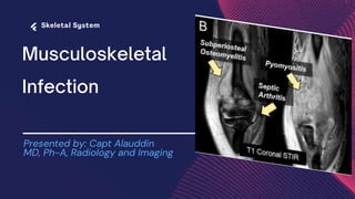
  • 1. Presented by: Capt Alauddin MD, Ph-A, Radiology and Imaging
  • 2. Infections of the musculoskeletal system can be subdivided into 3 categories: (a) those involving bones (osteomyelitis) (b) those involving joints (infectious arthritis) and (c) those involving soft tissues (cellulitis).
  • 3. Entry of infectious organism into bones may be from: • Hematogenous spread, • From the contiguous soft tissues or • Direct implantation secondary to trauma or surgery.
  • 4. Etiology : In children, the etiology is typically hematogenous In infants, as diaphyseal vessels extending through the cartilaginous growth plate reach the epiphysis, so there is increased frequency of epiphyseal and joint infections.
  • 5. In early childhood, these diaphyseal vessels terminate in the metaphysis-forming venous sinusoidal lakes with a slow, turbulent flow which allows blood borne organisms to seed, proliferate, and cause metaphyseal infection.
  • 6. In adults, the hematogenous spread is less common and the bacterial access is usually due to direct inoculation from penetrating trauma, surgery, or adjacent contaminated soft tissue.
  • 7.
  • 8. Different imaging techniques for diagnosis of MSK infections include • Radiographs • Ultrasonography (USG) • Computed tomography (CT) • Magnetic resonance imaging (MRI) and • Functional Imaging( Bone scintigraphy).
  • 9. Soft-Tissue Infection : Cellulitis: A spreading inflammatory reaction of infectious origin occurring along the skin, subcutaneous and fascial planes with edema and skin changes. The common pathogens associated with cellulitis are Streptococcus pyogenes or Staphylococcus aureus.
  • 10. Clinical presentations : • Skin erythema without a well-defined border • Increased skin temperature • Swelling of the affected area • Regional lymphadenopathy • Systemic features such as fever and rigors may also be present.
  • 11. Predisposing factors : • Poor general health • Skin laceration or ulceration • Venepuncture • Eczema and • Immunosuppression
  • 12. Radiographic features: Ultrasound: Diffuse swelling, hyperechogenicity of the skin, and subcutaneous tissues to a variable degree and presence of subcutaneous edema.
  • 13. Cellulitis(Cont) Transverse Ultrasound of the right leg showing hyperechoic subcutaneous fat lobules separated by hypoechoic fluid-filled areas which appears as branching, anechoic striations typically known as "cobblestone" appearance This is suggestive of cellulitis.
  • 14. Cellulitis(Cont) In Doppler imaging the presence of hyperemia is the diagnostic of cellulitis.
  • 15. Cellulitis(Cont) CT of cellulitis shows subcutaneous fat stranding with thickening of overlying skin.
  • 16. Cellulitis(Cont) On MRI there is T2 hyperintensity of the affected area with diffuse linear or ill-defined soft-tissue thickening with corresponding T1 hypointensity and postcontrast enhancement.
  • 17. Necrotizing Fasciitis : An aggressive infection of the skin and soft tissue with necrosis of the muscle, fascia, and subcutaneous tissues. This infection typically spreads rapidly along the fascial plane, which has a poor blood supply.
  • 18. Necrotizing Fasciitis : C/F: Pain, Fever, And Sepsis which are out of proportion. Subtypes : Necrotizing fasciitis encompasses all soft-tissue infections deep to the hypodermis, 2 subtypes depending upon the anatomical site, • Fournier gangrene for the perineum and • Ludwig angina for the submandibular region.
  • 19. Necrotizing Fasciitis (cont): Has an association with pre-existing comorbidities, such as diabetes, vascular disease, immunosuppression, obesity, and drug abuse . Etiology: Direct inoculation of bacteria through a breach in the skin is seen in the majority of cases, gram-positive cocci such as Staph. aureus and Streptococci are responsible in single site infection.
  • 20. Necrotizing Fasciitis : Radiological features : USG : Diffuse thickening of the subcutaneous tissue, subcutaneous hyperemia, perifascial fluid, distorted appearance of the visual field when there is presence of air in soft tissue.
  • 21. Necrotizing Fasciitis (Cont): MRI : T2 : high signal intensity, T1 : low signal intensity and Post Contrast: variable enhancement. Presence of low signal foci in all sequences and blooming artifact on the GRE in the soft tissue is suggestive of free air and is diagnostic of necrotizing fasciitis
  • 22. Pyomyositis : Pyomyositis (also known as tropical myositis, pyogenic myositis , suppurative myositis ) is a primary infection of skeletal muscle and often associated with abscess formation.
  • 23. Pyomyositis : C/F : Pain localised to one or more muscles (although in most cases it is in a single muscle), variable degrees of systemic inflammatory manifestations Predisposing conditions: • Diabetes, • Malnutrition, • Human immunodeficiency virus (HIV) infection, • Immunodeficiency, • Drug abuse, • Malignancy, or trauma
  • 24. Pyomyositis : stages: there are three stages- a. invasive stage is characterized by nonspecific muscle edema b. suppurative stage is characterized by intramuscular abscess formation c. late stage is characterized by septicemia and multiorgan failure with a significantly high mortality rate
  • 25. Pyomyositis : Radiological features: On USG, the affected muscle shows low attenuation changes and the collection may have heterogeneous contents with postacoustic enhancement
  • 26. Pyomyositis (cont): (A) USG of the triceps muscle, showing edema, fusiform enlargement , and ill-defined fluid collection interspersed between the muscle fibers. (B) Ultrasonography image 2 weeks later after antibiotic therapy shows complete resolution.
  • 27. Pyomyositis (cont): MRI: shows only nonspecific muscle edema with high T2 and isointense T1 signal change in the early stages.
  • 28. Infectious Tenosynovitis : Infection of the space between the inner visceral layer adherent to the tendon and an outer parietal layer of tendon sheath with inflammatory reaction and pus formation . Risk factors: • Immunosuppression • Diabetes • Smoking
  • 29. Infectious Tenosynovitis (cont) : Most common pathogen is Staph. aureus, less commonly by fungus and MTB. It commonly affects the flexor tendons of the hands and wrist as flexor tendon sheaths communicate with adjacent bursae and hence the direct spread of infection is more common with flexor tendons
  • 30. Infectious Tenosynovitis : Four physical exam signs are collectively known as the Kanavel signs: • Fusiform swelling to the affected digit • Digit held in flexion at rest • Tenderness with percussion /palpation of flexor sheath • Pain with passive extension of the affected digit
  • 31. Infectious Tenosynovitis (cont): Radiological features: USG demonstrate thickening of the tendon and tendon sheath with hyperemia on Doppler imaging.
  • 32. Infectious Tenosynovitis (cont): MRI: Shows tendon and tendon sheath thickening and the presence or absence of fluid. It is particularly useful to depict the extent and anatomical relations of the tenosynovitis .
  • 33. Septic Bursitis: Inflammation of various bursae due to chronic repetitive strain or mechanical reasons characterized by sterile inflammation of the bursal wall with accumulation of free fluid in the bursa.
  • 34. Septic Bursitis: Etiology: Staph. aureus is the most common pathogen involved. Superficial bursae are more commonly involved due to the chance of direct inoculation due to trauma, e. g: Olecranon bursa, prepatellar and infrapatellar bursae. Infection of deep bursae is uncommon and is a result of either surgery, contiguous or hematogenous spread, e. g trochanteric and subacromial bursae.
  • 35. Septic Bursitis(cont): Radiological features: USG studies show extensive inflammatory changes of the bursa. Bursal wall thickening with hyperemia on Doppler imaging, peribursal inflammation, and mixed echogenic fluid with internal debris are seen.
  • 36. Septic Bursitis(cont): MRI demonstrates thickened bursae with fluid distention and inflammatory edema of the adjacent peribursal soft tissues
  • 37. Osteomyelitis: Osteomyelitis is defined as inflammation of the bone & medullary cavity due to infection characterized by progressive inflammatory destruction and new apposition of bone. Predisposing factors: • Trauma, • Bacteremia, • Adjacent soft tissue infection, • Surgery, or foreign bodies
  • 38. Osteomyelitis: Age of onset: Bone infections show a bimodal age distribution, occurring most commonly in people younger than 20 or older than 50 years of age. Etiology: In children and adults, Staphylococcus is more common and is seen in 90–95% of cases. In neonates and infants, Streptococcus is more common. other microorganisms responsible are Escherichia coli, Pseudomonas, and Klebsiella.
  • 39. The location of osteomyelitis within a bone varies with age, on account of changes in vascularisation of different parts of the bone • Neonates: metaphysis and/or epiphysis • Children: metaphysis • Adults: epiphyses and subchondral regions
  • 40. Osteomyelitis(cont): Types : Waldvogel classification system based on pathogenesis dividing the osteomyelitis into three separate groups 1. Hematogenous 2. Secondary to contiguous spread from a focus of infection 3. Associated with vascular insufficiency Traditionally, it may be acute, subacute, or chronic, depending on its clinical course, histologic findings, and disease duration
  • 41. Acute osteomyelitis typically presents 2 weeks after bone infection and chronic osteomyelitis 6 or more weeks after bone infection. Some studies describe an additional subacute phase, with 1–3 months of symptoms.
  • 42. Osteomyelitis: Radiological features : Plain radiographs are usually the first technique of imaging. Destructive bone changes do not occur until 7–10 days of onset of infection. On radiographs taken after this time period, a number of changes may be noted: • Regional osteopenia • Periosteal reaction/thickening (periostitis): variable; may appear aggressive, including the formation of a codman's triangle • Focal bony lysis or cortical loss • Endosteal scalloping • Loss of trabecular bone architecture • New bone apposition • Peripheral sclerosis
  • 43. Acute Osteomyelitis: Radiological features : Plain X-ray of the proximal humerus AP view showing ill- defined bone lucency and osseous destruction involving the medial aspect of the metaphysis in a child.
  • 44. Osteomyelitis: Radiological features : Plain X-ray of the proximal humerus AP view showing proximal humeral metaphyseal lucent focus with periosteal reaction in diaphysis suggest osteomyelitis. Enlarged soft tissue shadow around right shoulder joint with increased density, that suggests associated joint effusion.
  • 45. Few radiological features associated with osteomyelitis: Periosteal Reaction : Periosteal reaction is a nonspecific radiographic finding that indicates new bone formation in reaction to the abnormal stimulants. Periosteal reactions may be broadly characterised as benign or aggressive.
  • 46. Periosteal Reaction (cont): • nonaggressive types of periosteal reaction include noninterrupted, smooth, thick or thin, and undulating • aggressive types of periosteal reaction include interrupted, lamellated/onion skinning, sunburst, Codman triangle, and spiculated
  • 47. Subperiosteal Abscess : subperiosteal abscess is most commonly found in pediatric forms of osteomyelitis, as these patients are known to have looser adherence of the periosteum to the underlying cortex.
  • 48. Brodie’s Abscess : Intraosseous abscess cavity filled with pus, with a rim of granulation tissue. First described by Brodie. Intraosseous abscesses occur most often in children, have a predilection for the metaphysis of long bones, and are observed in the subacute or chronic stage of osteomyelitis when the organism has reduced virulence.
  • 49. Bony sequestrum: piece of devitalized bone that has become separated from the surrounding bone during the process of necrosis. presence of a sequestrum is definitive for chronic OM. The presence of dead bone usually with fistulous tracts secondary to infection confirms the presence of chronic osteomyelitis.
  • 50. Involucrum: describes the formation of a spherical capsule of viable new bone around an area of sequestered and necrotic bone. The involucrum can be viewed as a response to wall-off the necrotic, infected sequestrum.
  • 51. Involucrum: The inner lining of the involucrum faces the sequestrum and consists of granulation tissue The outer layer of the involucrum consists of expansile, coarse, woven bone, which is typically sclerotic in the mature stage.
  • 52. Cloaca: The term cloaca is used to indicate an opening or rupture of the bony cortex overlying an area of osteomyelitis that allows granulation tissue and/or intramedullary pus to be discharged out of the bone.

Editor's Notes

  1. Normally the subcutaneous tissue is hypoechoic with few hyperechoic strands (representing connective tissue). Above this, there is a narrow, relatively hyperechoic epidermal-dermal layer. Muscular fascia lies deep to the subcutaneous layer.  Transverse plane USG images of the right upper arm showing diffuse subcutaneus edema in a case of Cellulitis of right upper limb.
  2. Short-axis color Doppler ultrasound image of leg shows (diffuse thickened hyperechoic subcutaneous tissue and skin with cobblestone appearance and) hyperemia (arrows).
  3. Axial post contrast ct scan of abdomen showing There is diffuse fat infiltration with edema in subcutaneous layer along abdominal wall with skin thickening which is in favour of cellulitis.
  4. Axial T1 And T2 weighted MRI images of brain shows Thickening and edema of preseptal orbital soft tissue on right side suggestive of orbital cellulitis.
  5. A, Axial T2-weighted fat-suppressed image of proximal right lower leg shows increased T2 signal intensity of soft tissues and superficial fascia (arrowheads) . B, Gadolinium-enhanced fat-suppressed T1- weighted image shows variable enhancement (arrowheads). No definitive muscle edema or enhancement is yet appreciated. No T1- or T2-hypointense air locules are seen, so this is suggestive of early necrotizing fasciitis.
  6. Sonographic image of the left (A) and right (B) thigh in the transverse plane. The right thigh musculature is hypoechoic with increased layer thickness. This is a case of Pyomyositis
  7. suppurative stage of pyomyositis as there is intramuscular abscess formation
  8. Coronal non contrast T1 and saggital T2 w MRI image showing Extensive left thigh intermuscular heterogenous loculated signal change areas which is iso in T1 and hyper in T2 involving mainly the anterior muscle groups ( V. Lateralis) in a case of Pyomyositis.
  9. (A) Transverse USG image. (B) Longitudinal USG image of the of the hand showing significantly thickened tendon sheath (arrow) with an associated abscess formation along the tendon sheath (arrowhead). (C) Longitudinal USG image with Doppler, showing significant hyperemia. The appearances are representative of infective tenosynovitis.
  10. (A) axial STIR MRI images of the wrist showing Foreign body within the flexor tendon sheath (black arrow) causing septic flexor tenosynovitis (white arrow)
  11. usg of shoulder shows : fluid distending the bursa more than 1.5 mm. The tendon fibres are poorly characterized due to anisotropy , all these features are suggestive of septic bursitis.
  12. Saggital STIR image of elbow joint showing Olecranon bursal fluid collection with inflammatory edema of the adjacent peribursal soft tissues which elicits high signal at STIR WI.
  13. (A) Longitudinal and (B) axial USG images of leg showing subperiosteal collection (white arrows). (C) Axial PD FS image showing subperiosteal fluid signal suggestive of a collection (white arrow). (D) A repeat USG image after 4 weeks showing periosteal consolidation representing healing.
  14. (A) Anteroposterior radiograph view of the knee showing a well-defined lucency in the proximal tibia (white arrow), representing an abscess. (B) Coronal STIR image showing an interosseous collection (arrow) with surrounding bone marrow edema, (C) corresponding coronal T1FS mri image with contrast, showing rim enhancement
  15. A patient with chronic osteomyelitis of the tibia. (D) Axial unenhanced CT image of left leg showing hyperdense area which is separated from rest of the part of tibia by hypodense rim suggestive of devitalized bone (sequestrum )(black arrow)
  16. (B) axial STIR, and (C) axial postcontrast-enhanced T1 image of left leg showing cortical bone defect with enhancement of the granulation tissue (white arrow) which is suggestive of inner lining of involucrum .
  17. Fluoroscopic spot radiographs of distal femur showing A fistulous tract in the soft tissue extending into a bony defect and subsequently terminated into the round radiolucent area in the distal femur suggestive cloaca formation