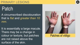
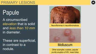
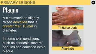
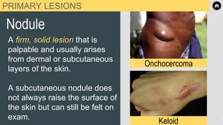
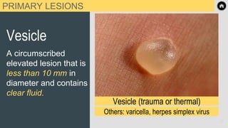
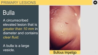
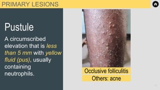
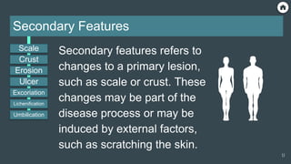
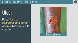
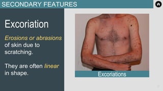
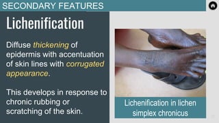
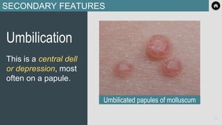
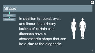
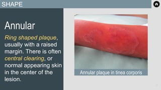
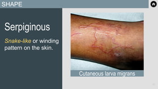
MODULE 1
Tropical Dermatology
Terminology
1
2
How to navigate this presentation Key to module icons
Return to interactive Table of Contents.
Click any page to progress to the next page.
Additional navigation can be found at the
bottom of each slide in the Google Slide
toolbar, e.g., use the arrows to move forward
and back.
Clicking on the button in Canvas
will advance you to the next page in Canvas.
Tips and Tricks
Treatment
Diagnosis / Bedside
Diagnostics
Geography
Aetiology
Key Clinical Features
Specific terms are used in
dermatology to describe lesions
and rashes.
This terminology will be used
extensively throughout the
course, therefore knowledge of it
is essential.
Terminology
Primary
Lesions
Secondary
Features
Shape
Distribution
3
Specific terms are used in
dermatology to describe
lesions and rashes.
This terminology will be
used extensively
throughout the course,
therefore knowledge of it
is essential.
Return to this
page at any
time to navigate
to any section
or specific
condition
Primary
Lesions
Secondary
Features Shape Distribution
Scale
Macule
Patch
Papule
Plaque
Nodule
Pustule
Ulcer
Erosion
Crust
Annular Seborrheic
Bulla
Vesicle
Wheal
Excoriation
Lichenification
Umbilication
Serpiginous Photodistributed
Sporotrichoid
After reviewing terminology
and passing the quiz, you
may move on to the next
modules and complete them
in any order you wish. 4
Interactive Table of Contents
Terminology
> Visit sections by clicking buttons below.
Section 1: Primary Lesions
Terminology
5
Primary Skin Lesions
“Primary lesion” refers to
the form or structure of
an individual skin lesion.
They are described by
these nine terms.
Macule
Patch
Papule
Plaque
Nodule
Pustule
Bulla
Vesicle
Wheal
6
PRIMARY LESIONS
Macule
A circumscribed
discolouration that is flat and
less than 10 mm in diameter.
There may be a change in
colour or texture, but
macules are not raised above
the surface of the skin.
Pityriasis versicolor (scaly)
wikipedia.org
Vitiligo
wikipedia.org
7
PRIMARY LESIONS
Patch
A circumscribed discolouration
that is flat and greater than 10
mm.
It is essentially a large macule.
There may be a change in
colour or texture, but patches
are not raised above the
surface of the skin.
Patches of vitiligo
8
PRIMARY LESIONS
Papule
A circumscribed
elevation that is solid
and less than 10 mm
in diameter.
These are superficial,
in contrast to a
nodule. Other examples: scabies, papular
pruritic eruption of HIV, insect bites
Molluscum
Neurofibromas in neurofibromatosis
9
PRIMARY LESIONS
Plaque
A circumscribed slightly
raised elevation that is
greater than 10 mm in
diameter.
In some skin conditions,
such as psoriasis, several
papules can coalesce into a
plaque.
Tinea corporis
Psoriasis 10
PRIMARY LESIONS
Nodule
A firm, solid lesion that is
palpable and usually arises
from dermal or subcutaneous
layers of the skin.
A subcutaneous nodule does
not always raise the surface of
the skin but can still be felt on
exam.
Keloid
Onchocercoma
11
PRIMARY LESIONS
Vesicle
A circumscribed
elevated lesion that is
less than 10 mm in
diameter and contains
clear fluid.
Vesicle (trauma or thermal)
Others: varicella, herpes simplex virus
12
PRIMARY LESIONS
Bulla
A circumscribed
elevated lesion that is
greater than 10 mm in
diameter and contains
clear fluid.
A bulla is a large
vesicle. l
Bullous Impetigo 13
PRIMARY LESIONS
Pustule
A circumscribed
elevation that is less
than 5 mm with yellow
fluid (pus), usually
containing
neutrophils. Occlusive folliculitis
Others: acne
14
PRIMARY LESIONS
Wheal
A transient circumscribed
papule or plaque, usually with
erythema (redness) and
peripheral pallor.
Wheals change rapidly in size
and shape due to edema in the
upper dermis and are typically
present for only a few hours.
Urticaria (hives)
15
Section 2: Secondary Features
Terminology
16
Secondary Features
Secondary features refers to
changes to a primary lesion,
such as scale or crust. These
changes may be part of the
disease process or may be
induced by external factors,
such as scratching the skin.
Scale
Ulcer
Erosion
Crust
Excoriation
Lichenification
Umbilication
17
SECONDARY FEATURES
Scale
Shedding of the top layer of
skin (stratum corneum) which
appears as dry flakes.
In inflammatory conditions of
the skin, such as psoriasis,
scale is due to increased
production of skin cells.
Psoriasis plaque with dry scale
18
SECONDARY FEATURES
Crust
A collection of dried
serum, blood, or pus
on the surface of
skin.
Papules with overlying crust
19
SECONDARY FEATURES
Erosion
A focal loss of
superficial epidermis.
Because erosions are
superficial, they heal
without scarring. Erosion
20
SECONDARY FEATURES
Ulcer
Focal loss of
epidermis and some
dermis that heals with
scarring.
Tropical ulcer
21
SECONDARY FEATURES
Excoriation
Erosions or abrasions
of skin due to
scratching.
They are often linear
in shape. Excoriations
22
SECONDARY FEATURES
Lichenification
Diffuse thickening of
epidermis with accentuation
of skin lines with corrugated
appearance.
This develops in response to
chronic rubbing or
scratching of the skin.
Lichenification in lichen
simplex chronicus 23
SECONDARY FEATURES
Umbilication
This is a central dell
or depression, most
often on a papule.
Umbilicated papules of molluscum
24
Section 3: Shape
Terminology
25
Shape
In addition to round, oval,
and linear, the primary
lesions of certain skin
diseases have a
characteristic shape that can
be a clue to the diagnosis.
Annular
Serpiginous
26
SHAPE
Annular
Ring shaped plaque,
usually with a raised
margin. There is often
central clearing, or
normal appearing skin
in the center of the
lesion.
Annular plaque in tinea corporis
27
SHAPE
Serpiginous
Snake-like or winding
pattern on the skin.
Cutaneous larva migrans
28
Section 4: Distribution
Terminology
29
Distribution
The distribution of skin lesions
refers to where they are located
on the skin. Some skin diseases
have a characteristic distribution,
such as pityriasis versicolor that
affects seborrheic (oily) parts of
the skin.
Seborrheic
Photodistributed
Sporotrichoid
30
DISTRIBUTION
Seborrheic
This is the oily parts of the
skin such as the central
face, mid chest, and central
upper back.
Pityriasis versicolor on the upper back
31
DISTRIBUTION
Photodistributed
Sun exposed areas of
the skin, such as the
face, upper chest,
and dorsal forearms.
Pellagra in sun-exposed
parts of the skin 32
DISTRIBUTION
Sporotrichoid
Skin lesions, usually
nodules, following the
lymphatic drainage. It
usually occurs on the
limbs. Sporotrichoid spread
33
This course has been created by:
⮚ Alexia P. Knapp, MD, MS, FAAD, CTropMed®
⮚ Karolyn Wanat, MD, FAAD, CTropMed®
34
End of Presentation
Thank you!
35

Module_1_Terminology.pdf terminology vvv

  • 1.
  • 2.
    2 How to navigatethis presentation Key to module icons Return to interactive Table of Contents. Click any page to progress to the next page. Additional navigation can be found at the bottom of each slide in the Google Slide toolbar, e.g., use the arrows to move forward and back. Clicking on the button in Canvas will advance you to the next page in Canvas. Tips and Tricks Treatment Diagnosis / Bedside Diagnostics Geography Aetiology Key Clinical Features
  • 3.
    Specific terms areused in dermatology to describe lesions and rashes. This terminology will be used extensively throughout the course, therefore knowledge of it is essential. Terminology Primary Lesions Secondary Features Shape Distribution 3 Specific terms are used in dermatology to describe lesions and rashes. This terminology will be used extensively throughout the course, therefore knowledge of it is essential.
  • 4.
    Return to this pageat any time to navigate to any section or specific condition Primary Lesions Secondary Features Shape Distribution Scale Macule Patch Papule Plaque Nodule Pustule Ulcer Erosion Crust Annular Seborrheic Bulla Vesicle Wheal Excoriation Lichenification Umbilication Serpiginous Photodistributed Sporotrichoid After reviewing terminology and passing the quiz, you may move on to the next modules and complete them in any order you wish. 4 Interactive Table of Contents Terminology > Visit sections by clicking buttons below.
  • 5.
    Section 1: PrimaryLesions Terminology 5
  • 6.
    Primary Skin Lesions “Primarylesion” refers to the form or structure of an individual skin lesion. They are described by these nine terms. Macule Patch Papule Plaque Nodule Pustule Bulla Vesicle Wheal 6
  • 7.
    PRIMARY LESIONS Macule A circumscribed discolourationthat is flat and less than 10 mm in diameter. There may be a change in colour or texture, but macules are not raised above the surface of the skin. Pityriasis versicolor (scaly) wikipedia.org Vitiligo wikipedia.org 7
  • 8.
    PRIMARY LESIONS Patch A circumscribeddiscolouration that is flat and greater than 10 mm. It is essentially a large macule. There may be a change in colour or texture, but patches are not raised above the surface of the skin. Patches of vitiligo 8
  • 9.
    PRIMARY LESIONS Papule A circumscribed elevationthat is solid and less than 10 mm in diameter. These are superficial, in contrast to a nodule. Other examples: scabies, papular pruritic eruption of HIV, insect bites Molluscum Neurofibromas in neurofibromatosis 9
  • 10.
    PRIMARY LESIONS Plaque A circumscribedslightly raised elevation that is greater than 10 mm in diameter. In some skin conditions, such as psoriasis, several papules can coalesce into a plaque. Tinea corporis Psoriasis 10
  • 11.
    PRIMARY LESIONS Nodule A firm,solid lesion that is palpable and usually arises from dermal or subcutaneous layers of the skin. A subcutaneous nodule does not always raise the surface of the skin but can still be felt on exam. Keloid Onchocercoma 11
  • 12.
    PRIMARY LESIONS Vesicle A circumscribed elevatedlesion that is less than 10 mm in diameter and contains clear fluid. Vesicle (trauma or thermal) Others: varicella, herpes simplex virus 12
  • 13.
    PRIMARY LESIONS Bulla A circumscribed elevatedlesion that is greater than 10 mm in diameter and contains clear fluid. A bulla is a large vesicle. l Bullous Impetigo 13
  • 14.
    PRIMARY LESIONS Pustule A circumscribed elevationthat is less than 5 mm with yellow fluid (pus), usually containing neutrophils. Occlusive folliculitis Others: acne 14
  • 15.
    PRIMARY LESIONS Wheal A transientcircumscribed papule or plaque, usually with erythema (redness) and peripheral pallor. Wheals change rapidly in size and shape due to edema in the upper dermis and are typically present for only a few hours. Urticaria (hives) 15
  • 16.
    Section 2: SecondaryFeatures Terminology 16
  • 17.
    Secondary Features Secondary featuresrefers to changes to a primary lesion, such as scale or crust. These changes may be part of the disease process or may be induced by external factors, such as scratching the skin. Scale Ulcer Erosion Crust Excoriation Lichenification Umbilication 17
  • 18.
    SECONDARY FEATURES Scale Shedding ofthe top layer of skin (stratum corneum) which appears as dry flakes. In inflammatory conditions of the skin, such as psoriasis, scale is due to increased production of skin cells. Psoriasis plaque with dry scale 18
  • 19.
    SECONDARY FEATURES Crust A collectionof dried serum, blood, or pus on the surface of skin. Papules with overlying crust 19
  • 20.
    SECONDARY FEATURES Erosion A focalloss of superficial epidermis. Because erosions are superficial, they heal without scarring. Erosion 20
  • 21.
    SECONDARY FEATURES Ulcer Focal lossof epidermis and some dermis that heals with scarring. Tropical ulcer 21
  • 22.
    SECONDARY FEATURES Excoriation Erosions orabrasions of skin due to scratching. They are often linear in shape. Excoriations 22
  • 23.
    SECONDARY FEATURES Lichenification Diffuse thickeningof epidermis with accentuation of skin lines with corrugated appearance. This develops in response to chronic rubbing or scratching of the skin. Lichenification in lichen simplex chronicus 23
  • 24.
    SECONDARY FEATURES Umbilication This isa central dell or depression, most often on a papule. Umbilicated papules of molluscum 24
  • 25.
  • 26.
    Shape In addition toround, oval, and linear, the primary lesions of certain skin diseases have a characteristic shape that can be a clue to the diagnosis. Annular Serpiginous 26
  • 27.
    SHAPE Annular Ring shaped plaque, usuallywith a raised margin. There is often central clearing, or normal appearing skin in the center of the lesion. Annular plaque in tinea corporis 27
  • 28.
    SHAPE Serpiginous Snake-like or winding patternon the skin. Cutaneous larva migrans 28
  • 29.
  • 30.
    Distribution The distribution ofskin lesions refers to where they are located on the skin. Some skin diseases have a characteristic distribution, such as pityriasis versicolor that affects seborrheic (oily) parts of the skin. Seborrheic Photodistributed Sporotrichoid 30
  • 31.
    DISTRIBUTION Seborrheic This is theoily parts of the skin such as the central face, mid chest, and central upper back. Pityriasis versicolor on the upper back 31
  • 32.
    DISTRIBUTION Photodistributed Sun exposed areasof the skin, such as the face, upper chest, and dorsal forearms. Pellagra in sun-exposed parts of the skin 32
  • 33.
    DISTRIBUTION Sporotrichoid Skin lesions, usually nodules,following the lymphatic drainage. It usually occurs on the limbs. Sporotrichoid spread 33
  • 34.
    This course hasbeen created by: ⮚ Alexia P. Knapp, MD, MS, FAAD, CTropMed® ⮚ Karolyn Wanat, MD, FAAD, CTropMed® 34
  • 35.