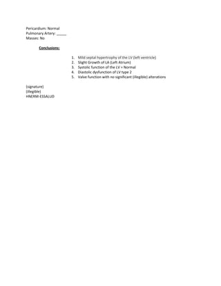
Subject: 016704 (41521)

                                            ADMISSION NOTE

ADMISSION DATE: 29/Nov/2011 8:30 p.m.                  HOSPITAL’S ADMISSION DATE: 28/Nov/2011

NAME:

AGE: 71                SEX: FEMALE                     TELEPHONE:

ADDRESS:

CLINICAL HISTORY:      IDDM (+) INSULIN NPH 20-20UI
                       HTN (+) ENALAPRIL, ATENOLOL
                       CABG 3 YEARS AGO
                       CARDIAC CATHETERIZATION 2011??

MEDICATION:            INSULIN NPH 20-20
                       ENALAPRIL 20mg/12h
                       ATENOLOL

ALLERGIES:             NONE

DESCRIPTION:
               Patient indicates that she has been suffering from chest oppression and chest pains for
               the last 2 years when making moderate efforts. She now states that this occurs when
               she makes light efforts, this being why she is seeking emergency services. She notes that
               the pain has been the oppressive type, with an irradiation towards the back. She took
               ISB, but saw no apparent improvement; thus, she arrived to Emergency.

BP:            113 / 46        FC:     61      FR:     18      TEM:

PHYSICAL EXAMINATION
TESTS:, PATIENTS’S REGULAR GENERAL APPEARANCE, GOOD HYDRATION AND GOOD NUTRITION
CONDITIONS, <2SEC CAPILLARY REFILL, STC IN MODERATE AMOUNT
THORAX- LUNGS: NORMAL BREATH SOUNDS IN BOTH LUNG FIELDS, NO RSA, NO GALLOP, JVP (-)HJR (-)
CV: RCRR NO MURMURS JVP (-) HJR (-)
ABDOMEN: B/D NO PAIN IN THE DEEP OR SUPERFICIAL PALPATION
NEUROLOGICAL: WELL ORIENTED IN TIME AND SPACE, NO SIGNS OF MENINGEAL OR FOCAL
NEUROLOGIC SIGNS

EKG:           RS: FC60X       AXIS: 60O, NO SIGNS OF ACUTE ISCHEMIA

CPK-MB: 1.68 TROPONIN: 0.003      TOTAL CPK: 0      HB:11.5
GLUCOSE: 366       CREATININE: 1.23      HEMOGRAM: NORMAL

OTHER EXAMINATIONS:
DIAGNOSTIC IMPRESSION
                               1. UNSTABLE ANGINA
2. ECOC REVASCULARIZED
                            3. UNCONTROLLED DM
                            4. HTN

WORK PLAN

UCICOR PROFILE, INVASIVE STRATIFICATION. REVIEW OLD HISTORY.
016170 (41521)

                                           RELEASE REPORT

The Head of Cardiology Services at the Edgardo Rebagliati Martins Hospital reports that a 71-year old
female patient by the name of … … …          was admitted for cardiology services on 29/Nov/2011.

During her hospitalization:

Patient indicates that she has been suffering from chest oppression and chest pains for the last 2 years
when making moderate efforts. She now states that this occurs when she makes light efforts, this being
why she is seeking emergency services. She notes that the pain has been the oppressive type, with an
irradiation towards the back. She took ISB, but saw no apparent improvement; thus, she arrived to
Emergency.

Cardiac catheterism was conducted on Nov/6/11. Internal mammary artery bypasses graft to Anterior
Descending Artery. Saphenous vein bypass graft to marginal branches and saphenous vein bypass graft
to Posterior Descending Artery: all permeable. Poor Anterior Descending Artery distal bed: poor distal
bed. Marginal branches: thin vessel. Conclusion: Continue with medical treatment.

Pharmacological and Supplementary Treatment          Aspirin                    Clopidogrel
Irbesartan               Elantana                    Bisoprolol                 Hydrochlorothiazide
Atorvastatin             Cephalexin                  Insulin NPH


Main Diagnosis of Release       Unstable angina         120.0

Secondary Diagnosis             NIDDM                   E11

                                HTN                     I10

Condition at Release:   IMPROVED

Appointment: Outpatient.

The staff and management of this Service thank you for allowing us to take care of you, and for any
suggestions and opinions you may wish to pass on in the requested survey.


                                  Edgardo Rebagliati National Hospital
                                              ESSALUD
                                _________________________________
CATHETERIZATION REPORT 1031

Name:                                                              Insurance No.

Age:      71            Gender: Female                             Catheterization Date: 5/Dec/2011

Type:     Left          Surgeons:                       MR3 Paredes

Technique: SELDINGER’S APPROACH THROUGH RIGHT FEMORAL ARTERY INTRODUCER 6Fr

Dominance: Right                                        Cylinder: AI

Right Coronary Artery: Blood vessel of good size mildly diffused atheromatosis.

Posterior descending artery: Beginning of OT, opacified with venous bypass. Poor distal bed.

Left Main Coronary Artery: Long, trifurcated. 70% ostium lesion.

Posterior descending artery: Segmentation lesion in middle third, up to 95%. Flow involved in distal
third. Poor distal bed.


Diagonal:               1° DG thin vessel, with ostium lesion of up to 95%.

Circumflex:             Vessel with a moderate caliber. SLAS, thin distal bed.

Marginal:               1° opacified through venous bypass, thin vessel.

                        2° - 3° Mg: thin, SLAS

Collateral and bypasses:VENOUS BYPASS: Ao-DP: PERMEABLE

                        VENOUS BYPASS: Ao-Mg, PERMEABLE

                        BYPASS: AMI – AD: PERMEABLE

Ventriculography:       Not performed.

Issues:                 None.


Ao Pre Pressure:        145 / 72

LV Pre Pressure :       0/0

Ao Post Pressure :      0/0

LV Post Pressure :      0/0

Average blood pressure :        0

Ejection fraction :     0%
CONCLUSIONS:           Atheromatous coronary artery disease, LMCAD (Left Main Coronary Artery
Disease), and LAD (Anterior Descending Artery) . Permeable coronary bypasses.

Suggestions:          Medical Treatment
Patient 016704 (4152)

                           “EDGARDO REBAGLIATI MARTINS” National Hospital

                                        CARDIOLOGY SERVICE
                                 ADULT ECHOCARDIOGRAPHIC REPORT
Name:                                                                        Date: 30/Nov/2011
Age: 71                                 SI:
Ultrasonic Access:               Acceptable

Medication                                           Aortic root     :       30 mm
IV Septum       :         13 mm                      Aortic valves :         18 mm
LV Diastole     :         41 mm                      LV outflow tract :         mm
LV Systole      :         26 mm                      Aortic ring     :          mm
LV Posterior wall :       10 mm                      Right Ventricular:      29 mm
Left atrium     :         38 mm (23) m2              Right atrium :             mm (14) cm2

Systolic Function                                    Diastolic Function
Ejection fraction:        6.5 %                      Flow velocity E :       70 cm/sec
Diastolic Vol. :            ml                       Flow velocity A :       80 cm/sec
Systolic Vol.    :          ml                       Slowdown time E         :      280 m/sec
Ejection Vol. :             ml                       Tissue velocity E:      5 9 cm/sec
dp /dt           :         mm/Hg/sec                 Tissue velocity A:      6 10 cm/sec

DOPPLER VALVULAR ASSESSMENT

MITRAL DIASTOLIC: Max. Vel.:__ Max.Slope:___ Average Slope:____ HPT:_____ Mitral Area: Normal
MITRAL SYSTOLIC: Failure: Negative
AORTIC SYSTOLIC: Max. Slope: 1.3 Medium Slope: 6.76 TSV: _____ Valve Area: Normal
AORTIC DIASTOLIC: HPT:___ deceleration slope:____ Failure Grade: No
SYSTOLIC TRICUSPID: VD-AD Slope: 29 mmHg                     Systolic pressure PA: 29 mmHg
SYSTOLIC PULMONARY: Max. Vel. ________________ cm/sec RV-LAD Slope:_________mmHg
SHUNTS: No

DESCRIPTION
Heart valves: Damaged

left ventricular contractile pattern                 Right ventricular contractile pattern (NO)

                      BASAL               MEDIAL             APICAL
Anterior Septal                                                                   0 : not tested
Posterior Septal                                                                  1 : normal
Anterior              1                   1                  1                    2 : hypokinesia
Inferior                                                                          3 : akinesia
Lateral                                                                           4 : dyskinesia
Posterior                                                                         5 : aneurysm

Ascending Aorta: ____
Pericardium: Normal
Pulmonary Artery: _____
Masses: No

       Conclusions:

                          1.   Mild septal hypertrophy of the LV (left ventricle)
                          2.   Slight Growth of LA (Left Atrium)
                          3.   Systolic function of the LV = Normal
                          4.   Diastolic dysfunction of LV type 2
                          5.   Valve function with no significant (illegible) alterations

(signature)
(illegible)
HNERM-ESSALUD

Modelos de informes médicos

  • 1.
    Subject: 016704 (41521) ADMISSION NOTE ADMISSION DATE: 29/Nov/2011 8:30 p.m. HOSPITAL’S ADMISSION DATE: 28/Nov/2011 NAME: AGE: 71 SEX: FEMALE TELEPHONE: ADDRESS: CLINICAL HISTORY: IDDM (+) INSULIN NPH 20-20UI HTN (+) ENALAPRIL, ATENOLOL CABG 3 YEARS AGO CARDIAC CATHETERIZATION 2011?? MEDICATION: INSULIN NPH 20-20 ENALAPRIL 20mg/12h ATENOLOL ALLERGIES: NONE DESCRIPTION: Patient indicates that she has been suffering from chest oppression and chest pains for the last 2 years when making moderate efforts. She now states that this occurs when she makes light efforts, this being why she is seeking emergency services. She notes that the pain has been the oppressive type, with an irradiation towards the back. She took ISB, but saw no apparent improvement; thus, she arrived to Emergency. BP: 113 / 46 FC: 61 FR: 18 TEM: PHYSICAL EXAMINATION TESTS:, PATIENTS’S REGULAR GENERAL APPEARANCE, GOOD HYDRATION AND GOOD NUTRITION CONDITIONS, <2SEC CAPILLARY REFILL, STC IN MODERATE AMOUNT THORAX- LUNGS: NORMAL BREATH SOUNDS IN BOTH LUNG FIELDS, NO RSA, NO GALLOP, JVP (-)HJR (-) CV: RCRR NO MURMURS JVP (-) HJR (-) ABDOMEN: B/D NO PAIN IN THE DEEP OR SUPERFICIAL PALPATION NEUROLOGICAL: WELL ORIENTED IN TIME AND SPACE, NO SIGNS OF MENINGEAL OR FOCAL NEUROLOGIC SIGNS EKG: RS: FC60X AXIS: 60O, NO SIGNS OF ACUTE ISCHEMIA CPK-MB: 1.68 TROPONIN: 0.003 TOTAL CPK: 0 HB:11.5 GLUCOSE: 366 CREATININE: 1.23 HEMOGRAM: NORMAL OTHER EXAMINATIONS: DIAGNOSTIC IMPRESSION 1. UNSTABLE ANGINA
  • 2.
    2. ECOC REVASCULARIZED 3. UNCONTROLLED DM 4. HTN WORK PLAN UCICOR PROFILE, INVASIVE STRATIFICATION. REVIEW OLD HISTORY.
  • 3.
    016170 (41521) RELEASE REPORT The Head of Cardiology Services at the Edgardo Rebagliati Martins Hospital reports that a 71-year old female patient by the name of … … … was admitted for cardiology services on 29/Nov/2011. During her hospitalization: Patient indicates that she has been suffering from chest oppression and chest pains for the last 2 years when making moderate efforts. She now states that this occurs when she makes light efforts, this being why she is seeking emergency services. She notes that the pain has been the oppressive type, with an irradiation towards the back. She took ISB, but saw no apparent improvement; thus, she arrived to Emergency. Cardiac catheterism was conducted on Nov/6/11. Internal mammary artery bypasses graft to Anterior Descending Artery. Saphenous vein bypass graft to marginal branches and saphenous vein bypass graft to Posterior Descending Artery: all permeable. Poor Anterior Descending Artery distal bed: poor distal bed. Marginal branches: thin vessel. Conclusion: Continue with medical treatment. Pharmacological and Supplementary Treatment Aspirin Clopidogrel Irbesartan Elantana Bisoprolol Hydrochlorothiazide Atorvastatin Cephalexin Insulin NPH Main Diagnosis of Release Unstable angina 120.0 Secondary Diagnosis NIDDM E11 HTN I10 Condition at Release: IMPROVED Appointment: Outpatient. The staff and management of this Service thank you for allowing us to take care of you, and for any suggestions and opinions you may wish to pass on in the requested survey. Edgardo Rebagliati National Hospital ESSALUD _________________________________
  • 4.
    CATHETERIZATION REPORT 1031 Name: Insurance No. Age: 71 Gender: Female Catheterization Date: 5/Dec/2011 Type: Left Surgeons: MR3 Paredes Technique: SELDINGER’S APPROACH THROUGH RIGHT FEMORAL ARTERY INTRODUCER 6Fr Dominance: Right Cylinder: AI Right Coronary Artery: Blood vessel of good size mildly diffused atheromatosis. Posterior descending artery: Beginning of OT, opacified with venous bypass. Poor distal bed. Left Main Coronary Artery: Long, trifurcated. 70% ostium lesion. Posterior descending artery: Segmentation lesion in middle third, up to 95%. Flow involved in distal third. Poor distal bed. Diagonal: 1° DG thin vessel, with ostium lesion of up to 95%. Circumflex: Vessel with a moderate caliber. SLAS, thin distal bed. Marginal: 1° opacified through venous bypass, thin vessel. 2° - 3° Mg: thin, SLAS Collateral and bypasses:VENOUS BYPASS: Ao-DP: PERMEABLE VENOUS BYPASS: Ao-Mg, PERMEABLE BYPASS: AMI – AD: PERMEABLE Ventriculography: Not performed. Issues: None. Ao Pre Pressure: 145 / 72 LV Pre Pressure : 0/0 Ao Post Pressure : 0/0 LV Post Pressure : 0/0 Average blood pressure : 0 Ejection fraction : 0%
  • 5.
    CONCLUSIONS: Atheromatous coronary artery disease, LMCAD (Left Main Coronary Artery Disease), and LAD (Anterior Descending Artery) . Permeable coronary bypasses. Suggestions: Medical Treatment
  • 6.
    Patient 016704 (4152) “EDGARDO REBAGLIATI MARTINS” National Hospital CARDIOLOGY SERVICE ADULT ECHOCARDIOGRAPHIC REPORT Name: Date: 30/Nov/2011 Age: 71 SI: Ultrasonic Access: Acceptable Medication Aortic root : 30 mm IV Septum : 13 mm Aortic valves : 18 mm LV Diastole : 41 mm LV outflow tract : mm LV Systole : 26 mm Aortic ring : mm LV Posterior wall : 10 mm Right Ventricular: 29 mm Left atrium : 38 mm (23) m2 Right atrium : mm (14) cm2 Systolic Function Diastolic Function Ejection fraction: 6.5 % Flow velocity E : 70 cm/sec Diastolic Vol. : ml Flow velocity A : 80 cm/sec Systolic Vol. : ml Slowdown time E : 280 m/sec Ejection Vol. : ml Tissue velocity E: 5 9 cm/sec dp /dt : mm/Hg/sec Tissue velocity A: 6 10 cm/sec DOPPLER VALVULAR ASSESSMENT MITRAL DIASTOLIC: Max. Vel.:__ Max.Slope:___ Average Slope:____ HPT:_____ Mitral Area: Normal MITRAL SYSTOLIC: Failure: Negative AORTIC SYSTOLIC: Max. Slope: 1.3 Medium Slope: 6.76 TSV: _____ Valve Area: Normal AORTIC DIASTOLIC: HPT:___ deceleration slope:____ Failure Grade: No SYSTOLIC TRICUSPID: VD-AD Slope: 29 mmHg Systolic pressure PA: 29 mmHg SYSTOLIC PULMONARY: Max. Vel. ________________ cm/sec RV-LAD Slope:_________mmHg SHUNTS: No DESCRIPTION Heart valves: Damaged left ventricular contractile pattern Right ventricular contractile pattern (NO) BASAL MEDIAL APICAL Anterior Septal 0 : not tested Posterior Septal 1 : normal Anterior 1 1 1 2 : hypokinesia Inferior 3 : akinesia Lateral 4 : dyskinesia Posterior 5 : aneurysm Ascending Aorta: ____
  • 7.
    Pericardium: Normal Pulmonary Artery:_____ Masses: No Conclusions: 1. Mild septal hypertrophy of the LV (left ventricle) 2. Slight Growth of LA (Left Atrium) 3. Systolic function of the LV = Normal 4. Diastolic dysfunction of LV type 2 5. Valve function with no significant (illegible) alterations (signature) (illegible) HNERM-ESSALUD