Master Presentation
•Download as PPTX, PDF•
1 like•349 views

This document summarizes a master's thesis on segmenting the left ventricle boundary in tagged magnetic resonance images. The student developed a two-step algorithm using semantic descriptors of tissue and prior shape knowledge to automatically segment the left ventricle boundary. Results showed high accuracy of the automatic segmentation compared to manual segmentation, with mean errors less than 0.2 degrees for global rotation and 1 degree for regional rotation. This promising algorithm could serve as a support tool for clinical analysis of heart function. Future work further validates the approach on additional image slices and pathological cases.

Recommended
Recommended
More Related Content
Viewers also liked
Viewers also liked (12)
Master Presentation
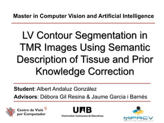
- 1. Master in Computer Vision and Artificial Intelligence LV Contour Segmentation in TMR Images Using Semantic Description of Tissue and Prior Knowledge Correction Student: Albert AndaluzGonzález Advisors: Débora Gil Resina & Jaume Garcia iBarnés
- 2. Index Introduction Our Contribution Results Conclusions ●○○○○○○○○○○○○○○○○○○○○○○○
- 3. Clinical problem ♥ The problem: 30% of global deaths are caused by heart diseases Treatment: diagnose of the heart function Method: Extraction of clinical scores from regional wall motion Requirements: accurate contour estimation ●●○○○○○○○○○○○○○○○○○○○○○○
- 4. Contour Segmentation Manual: slow Inter/intra observer variability -> requires high expertise. Automatic Objective for all subjects Requires accurate estimation of the LV boundaries ●●●○○○○○○○○○○○○○○○○○○○○○
- 5. Tagged Magnetic Resonance LV(*) TMR in SA view Long Axis (LA) Base Mid Short Axis (SA) cuts Apex (*)LV = LeftVentricle ●●●●○○○○○○○○○○○○○○○○○○○○
- 6. Problems in Tagged Magnetic Resonance Blood pool and tissue appearance is similar at time=0 … Tag pattern misleads some common image descriptors… Contours LV Boundaries …and few algorithms for TMR automatic segmentation currently exist ●●●●●○○○○○○○○○○○○○○○○○○○
- 7. Introduction Our Contribution Results Conclusions ●●●●●●○○○○○○○○○○○○○○○○○○
- 8. Our Contribution Goal: Algorithm for automatic segmentation for the extraction of clinical regional scores of the LV Semantic definition of the LV Two steps: Shape approximation Shape correction ●●●●●●●○○○○○○○○○○○○○○○○○
- 10. Internal energy: avoids deviation from anatomical LV shapes●●●●●●●●○○○○○○○○○○○○○○○○
- 11. External energy computation Define contour for image potential LV Contour extraction Distance map to the LV contours ●●●●●●●●●○○○○○○○○○○○○○○○
- 12. Semantic descriptors Amplitude1 Amplitude 2 Motion module Original t=0 t=1 … t=end ●●●●●●●●●●○○○○○○○○○○○○○○
- 13. Energy definition Amplitude 2 Motion module Amplitude1 * * mean mean mean * * * * * * mean * * * * ●●●●●●●●●●●○○○○○○○○○○○○○
- 14. Contour extraction 2 Detection 3 Selection 1 Clustering from E1|E2 K-Means Contours+ Binary Morphology Region area Filtering Epicardium (outerboundary of the LV) Endocardium (innerboundary of the LV) ●●●●●●●●●●●●○○○○○○○○○○○○
- 15. Distance map Epicardium Endocardium Distance to LV contours Distance map Vector Field ●●●●●●●●●●●●●○○○○○○○○○○○
- 16. Shape Correction Converged snake shape is corrected using PCA and GOPA: Incoming shape Mean shape Variation modes PCA eigenvalues then If for Correction applied No correction needed Externalenergy Converged snake Correction ●●●●●●●●●●●●●●○○○○○○○○○○
- 17. Introduction Our Contribution Results Conclusions ●●●●●●●●●●●●●●●○○○○○○○○○
- 18. Test set 29 real cases from 15 healthy patients 2 cuts (basal and mid) 2 energies for automatic segmentation TMR sequences from the Clinica La Creu Blanca ●●●●●●●●●●●●●●●●○○○○○○○○
- 20. distance between contours
- 21. Accuracy of Clinical Scores for clinicialaplicability
- 22. Compare global & regional rotation auto vs manual Validation protocol ●●●●●●●●●●●●●●●●○○○○○○○○
- 23. Segmentation error Accuracy (pixels) Base Accuracy (pixels) Externalenergy Manual Mid Automatic ●●●●●●●●●●●●●●●●●●○○○○○○
- 24. Rotation (low error) Regional LV Contours IS AS I A Global score LV IL AL Manual Automatic ●●●●●●●●●●●●●●●●●●●○○○○○
- 25. Rotation (mild error) Regional LV Contours IS AS I A Global score LV IL AL Manual Automatic ●●●●●●●●●●●●●●●●●●●●○○○○
- 26. Absolute error (in degrees) ●●●●●●●●●●●●●●●●●●●●●○○○
- 27. Introduction Our Contribution Results Conclusions ●●●●●●●●●●●●●●●●●●●●●●○○
- 29. < 1º mean regional rotationPromising support tool for clinical analysis ●●●●●●●●●●●●●●●●●●●●●●●○
- 30. Future work Validate in apical cuts Use Mean shift for improved clustering Validate our method in pathologic patients ●●●●●●●●●●●●●●●●●●●●●●●●
- 31. “Coronary heart disease is a silent disease and the first manifestation frequently is sudden death.” Dr. Herman K.Hellerstein - Cardiologist 1916- 1993 (Ohio, USA)
Editor's Notes
- There are severalreason f
- There are severalreason f
- There are severalreasonforblablaWewillnow show visuallythegeometry of theheart and theinnergroundsfortheissuespresententher
- The vertical plane divides the heart in two parts. Whereas the three horizontal cuts (the sort axis) cretae the base mid and apex cutsThe videos show the evolution at those sections of the myocardium motion.
- Aswehaveseen, theimages look blurry. Indeed, blablabla,Moreover, tagpatternsblabla. Thegreenboundaries are manual segmentations done bymedicalexperts. NoticehowusingcontourdetectiontecniquesdoesnotdetectedtheseboundariesIN fact, fewalgorithmfow
- Wehaveseemthelimitations, nowletsmoveouttoourcontribution
- Classicsnakes are curves thatdeformundertheinfluence of twoenergies. Theexternalenergyattractsthesnakestowardsthe LV contours, whereastheinternalenergyavoids…
- Asforthesemanticdescriptors, foreachframe of thetaggedsequence, we compute the amplitude3 of thegaborfiltger in twodirection. Wealso compute themotion module of theimage. Thus, allthreefeatureslackthetaggedgrid
- We define twoenergyes. Thefirstisobtained as follows. Wecomptuethe mean of everysequence of imagedescitpros. Thethreemeans are combiendinto a single energypotential, namely E1In thesecondeneryg, foreachframewe combine alltheedescriptorsinto a single one and compute the mean. Weget a new sequence, whichisavergaged , into a global mean image
- Theimagepotentialisclusteredby pixel intentsyusingkmenas. Next, we use commoncontourdetectionstodetectthecanditateregions. Finally, weselectedthe externa landinternalboundaries of the LV byusingregionareafiltering.
- Foreachboundary, we compute thedistancemaptothecontours. Next, weextractthe vector fieldwhcihatttractsthesnakestowardsthedesiredcontours.
- Wehaveseenhowourmethodwork. Now, letsmoveontotheresults
- The test set balbalb
- Tovalidateiourmethod, wedefinedtwocritera:Thesegmentation error isthedistancebetwweemourautomawticsegmentation and manual shapesbymedicalexperts.Moreover, oneachboundary, we compute the global and regional rotationforboth cases fortestingtheaccuracy
- In vertical, we can seethetwo short axis cuts,. Eachcolumnrepresents a diferentenedy,. The manual and automaticaproximations are shown. Thetablesrefertothewhole set, values show similar behaviour in bouth cases
- In the case of a goodaproxiamtion, the manual and automatic curves are veryclose, which shows thatouraproximationbehavessimilarytothe manual (groundtruth) segmentation,. Thisfactisfurtherconfirmedbythe global score, whichiscomputed in thewhole disc.
- Whenthereis considerable deviation, the scores diverge in some sector (A, AS). In therestits more orlessthesmaAlkso, thegtlobal scores are afected in themiddle of thesystoliccycle. However, thedifferenceisnotthatgreat
- Bothenergies show similar behaviour. In both cases, the mean rotationisbelow 1 degree in both cases forregionalscores. In the CASE of global scores, the mean rotationisbelwo 0.2 degreesw
- There are severalreason f
- Acceptablerotationvalues showbebelow 1 degreese
- Beforeendingthispresentation, I wouldliketoquote a cardiologistfromthestaes. He saidthat
- Whatwehaveseekwithourworkistobecomethefirstmanifestation