This document describes a study using molecular mechanics Poisson-Boltzmann surface area (MM/PBSA) methods to analyze the binding energetics and identify interaction hot spots in complexes between a camel single chain antibody (cAb-Lys3) and two lysozyme antigens (hen egg white lysozyme and turkey egg white lysozyme). The study involves molecular dynamics simulations of the complexes followed by MM/PBSA free energy decomposition to determine the contribution of individual amino acids to complex formation. Key hot spot residues in the antibody that make important contributions to binding are identified. The study provides insights that could inform rational design of small molecule mimics of the antibody.
![PROTEINS: Structure, Function, and Bioinformatics 67:418–434 (2007)
Protein–Protein Recognition and Interaction Hot Spots in
an Antigen–Antibody Complex: Free Energy Decomposition
Identifies ‘‘Efficient Amino Acids’’
Virginie Lafont,1,2 Michael Schaefer,3 Roland H. Stote,4 Daniele Altschuh,2 and Annick Dejaegere1*
`
1
´ ´ ´
Structural Biology and Genomics Department, UMR 7104, Institut de Genetique et de Biologie Moleculaire et Cellulaire, CNRS/
INSERM/ULP, F-67404 Illkirch Cedex, France
2
UMR7175-LC1 CNRS/ULP, ESBS, Parc d’Innovation, 67412 Illkirch Cedex, France
3
´
Laboratoire de Chimie Biophysique, Institut le Bel, Universite Louis Pasteur, 67000 Strasbourg, France
4
´ ´
Laboratoire de Biophysicochime Moleculaire, Institut de Chimie, LC3-UMR7177, Universite Louis Pasteur,
67070 Strasbourg Cedex, France
ABSTRACT The molecular mechanics Poisson– tions is difficult.1 There are several examples of natural
Boltzmann surface area (MM/PBSA) method was molecules that bind at protein interfaces2 and a growing
applied to the study of the protein–protein complex number of synthetic compounds that are aimed at per-
between a camelid single chain variable domain turbing protein–protein interactions (for a recent review,
(cAb-Lys3) and hen egg white lysozyme (HEL), and see Ref. 3). Progress in developing synthetic compounds
between cAb-Lys3 and turkey egg white lysozyme will benefit greatly from improved understanding of the
(TEL). The electrostatic energy was estimated by energetics and dynamics of protein–protein recognition.
solving the linear Poisson–Boltzmann equation. A Our current understanding of protein–protein interac-
free energy decomposition scheme was developed to tions is based on structural analysis of protein–protein
determine binding energy hot spots of each complex. complexes mostly determined by crystallography, on vari-
The calculations identified amino acids of the anti- ous biophysical characterizations of protein–protein com-
body that make important contributions to the inter- plexes that give insight into the kinetics and thermody-
action with lysozyme. They further showed the influ- namics of protein–protein association,4 and on biochemi-
ence of small structural variations on the energetics cal analyses that help identify amino acids important for
of binding and they showed that the antibody amino recognition.
acids that make up the hot spots are organized in An important concept that has emerged in the last
such a way as to mimic the lysozyme substrate. years is the notion of interaction energy ‘‘hot spots,’’ which
Through further analysis of the results, we define is the idea that a handful of amino acids at the binding
the concept of ‘‘efficient amino acids,’’ which can pro- interface make a dominant contribution to the binding af-
vide an assessment of the binding potential of a par- finity.5,6 Mutation studies, mostly alanine scanning stud-
ticular hot spot interaction. This information, in ies, have indeed shown7 that only a few mutations [native
turn, can be useful in the rational design of small ? Ala] affect the free energy of binding by more than
molecules that mimic the antibody. The implications 2 kcal/mol. This has led to a distinction between the func-
of using free energy decomposition to identify tional epitope (i.e. ‘‘hot spot residues) and the structural
regions of a protein–protein complex that could be epitope (all residues that participate in the interface).8
targeted by small molecules inhibitors are discussed.
Proteins 2007;67:418–434. V 2007 Wiley-Liss, Inc.
C
The Supplementary Material referred to in this article can be found
Key words: MM/PBSA; molecular dynamics; macro- at http://www.interscience.wiley.com/jpages/0887-3585/suppmat/
molecular electrostatics; protein engi- Grant sponsor: French Ministry of Defense, Centre National de la
neering; drug design; antibodies; ´
Recherche Scientifique, Institut National de la Sante et de la Recher-
´ ´
che Medicale, and Universite Louis Pasteur, Strasbourg, France.
camelids
Virginie Lafont’s current address is Department of Biology, Johns
Hopkins University, 3400 North Charles Street, Baltimore, MD
INTRODUCTION 21218-2685.
Michael Schafer’s current address is Novartis Pharma AG, Kly-
For a wide range of biological processes, including anti- beckstrasse 141, 4057 Basel, Switzerland.
body–antigen interactions, hormone–receptor interac- *Correspondence to: Annick Dejaegere. Structural Biology and
tions, signal transduction, and regulation, the formation ´ ´
Genomics Department, UMR 7104, Institut de Genetique et de Biolo-
´
gie Moleculaire et Cellulaire, CNRS/INSERM/ULP, BP 10142,
of stable protein–protein complexes is of fundamental im- F-67404 Illkirch Cedex, France. E-mail: annick@igbmc.u-strasbg.fr
portance. Interfering with or modulating the formation of Received 20 May 2006; Revised 6 September 2006; Accepted 15 Sep-
these complexes therefore offers attractive opportunities tember 2006
for therapeutic intervention. However, the development of Published online 26 January 2007 in Wiley InterScience (www.
small molecules that modulate protein–protein interac- interscience.wiley.com). DOI: 10.1002/prot.21259
V 2007 WILEY-LISS, INC.
C](https://image.slidesharecdn.com/lafontproteins2007-120227133851-phpapp02/85/Lafont-proteins-2007-1-320.jpg)
![PROTEINS: Structure, Function, and Bioinformatics 67:418–434 (2007)
Protein–Protein Recognition and Interaction Hot Spots in
an Antigen–Antibody Complex: Free Energy Decomposition
Identifies ‘‘Efficient Amino Acids’’
Virginie Lafont,1,2 Michael Schaefer,3 Roland H. Stote,4 Daniele Altschuh,2 and Annick Dejaegere1*
`
1
´ ´ ´
Structural Biology and Genomics Department, UMR 7104, Institut de Genetique et de Biologie Moleculaire et Cellulaire, CNRS/
INSERM/ULP, F-67404 Illkirch Cedex, France
2
UMR7175-LC1 CNRS/ULP, ESBS, Parc d’Innovation, 67412 Illkirch Cedex, France
3
´
Laboratoire de Chimie Biophysique, Institut le Bel, Universite Louis Pasteur, 67000 Strasbourg, France
4
´ ´
Laboratoire de Biophysicochime Moleculaire, Institut de Chimie, LC3-UMR7177, Universite Louis Pasteur,
67070 Strasbourg Cedex, France
ABSTRACT The molecular mechanics Poisson– tions is difficult.1 There are several examples of natural
Boltzmann surface area (MM/PBSA) method was molecules that bind at protein interfaces2 and a growing
applied to the study of the protein–protein complex number of synthetic compounds that are aimed at per-
between a camelid single chain variable domain turbing protein–protein interactions (for a recent review,
(cAb-Lys3) and hen egg white lysozyme (HEL), and see Ref. 3). Progress in developing synthetic compounds
between cAb-Lys3 and turkey egg white lysozyme will benefit greatly from improved understanding of the
(TEL). The electrostatic energy was estimated by energetics and dynamics of protein–protein recognition.
solving the linear Poisson–Boltzmann equation. A Our current understanding of protein–protein interac-
free energy decomposition scheme was developed to tions is based on structural analysis of protein–protein
determine binding energy hot spots of each complex. complexes mostly determined by crystallography, on vari-
The calculations identified amino acids of the anti- ous biophysical characterizations of protein–protein com-
body that make important contributions to the inter- plexes that give insight into the kinetics and thermody-
action with lysozyme. They further showed the influ- namics of protein–protein association,4 and on biochemi-
ence of small structural variations on the energetics cal analyses that help identify amino acids important for
of binding and they showed that the antibody amino recognition.
acids that make up the hot spots are organized in An important concept that has emerged in the last
such a way as to mimic the lysozyme substrate. years is the notion of interaction energy ‘‘hot spots,’’ which
Through further analysis of the results, we define is the idea that a handful of amino acids at the binding
the concept of ‘‘efficient amino acids,’’ which can pro- interface make a dominant contribution to the binding af-
vide an assessment of the binding potential of a par- finity.5,6 Mutation studies, mostly alanine scanning stud-
ticular hot spot interaction. This information, in ies, have indeed shown7 that only a few mutations [native
turn, can be useful in the rational design of small ? Ala] affect the free energy of binding by more than
molecules that mimic the antibody. The implications 2 kcal/mol. This has led to a distinction between the func-
of using free energy decomposition to identify tional epitope (i.e. ‘‘hot spot residues) and the structural
regions of a protein–protein complex that could be epitope (all residues that participate in the interface).8
targeted by small molecules inhibitors are discussed.
Proteins 2007;67:418–434. V 2007 Wiley-Liss, Inc.
C
The Supplementary Material referred to in this article can be found
Key words: MM/PBSA; molecular dynamics; macro- at http://www.interscience.wiley.com/jpages/0887-3585/suppmat/
molecular electrostatics; protein engi- Grant sponsor: French Ministry of Defense, Centre National de la
neering; drug design; antibodies; ´
Recherche Scientifique, Institut National de la Sante et de la Recher-
´ ´
che Medicale, and Universite Louis Pasteur, Strasbourg, France.
camelids
Virginie Lafont’s current address is Department of Biology, Johns
Hopkins University, 3400 North Charles Street, Baltimore, MD
INTRODUCTION 21218-2685.
Michael Schafer’s current address is Novartis Pharma AG, Kly-
For a wide range of biological processes, including anti- beckstrasse 141, 4057 Basel, Switzerland.
body–antigen interactions, hormone–receptor interac- *Correspondence to: Annick Dejaegere. Structural Biology and
tions, signal transduction, and regulation, the formation ´ ´
Genomics Department, UMR 7104, Institut de Genetique et de Biolo-
´
gie Moleculaire et Cellulaire, CNRS/INSERM/ULP, BP 10142,
of stable protein–protein complexes is of fundamental im- F-67404 Illkirch Cedex, France. E-mail: annick@igbmc.u-strasbg.fr
portance. Interfering with or modulating the formation of Received 20 May 2006; Revised 6 September 2006; Accepted 15 Sep-
these complexes therefore offers attractive opportunities tember 2006
for therapeutic intervention. However, the development of Published online 26 January 2007 in Wiley InterScience (www.
small molecules that modulate protein–protein interac- interscience.wiley.com). DOI: 10.1002/prot.21259
V 2007 WILEY-LISS, INC.
C](https://image.slidesharecdn.com/lafontproteins2007-120227133851-phpapp02/75/Lafont-proteins-2007-1-2048.jpg)
![INTERACTION HOT SPOTS IN ANTIGEN–ANTIBODY COMPLEX 425
relative contribution of lysozyme to the total binding free
energy is only about 15% (cf. Table II, À7 kcal/mol from a
total of À52 kcal/mol for HEL, and À6 kcal/mol out of a
total of À38 kcal/mol for TEL). The observation that the
difference in total contribution to binding between the two
partners is directly linked to the difference in electrostatic
interaction also results from the fact that, by definition
(see Methods), the van der Waals interactions are shared
equally between the two proteins, and that the difference
in buried surface area between two proteins in a complex
is very small.68 Electrostatic desolvation, on the other
hand, is strongly dependent on the individual structure
and charge distribution of each partner, and large differ-
ences can be observed for two proteins in a complex as
demonstrated here. Although the absolute values of the
antibody and lysozyme contributions to binding differ
between the HEL and TEL complexes (see Table II), the
relative magnitudes of the individual contributions are
very similar.
Comparison of the Different Structures
We studied three experimental structures of the cAb-
Lys3 in complex with HEL, and two of the complex with
TEL. The three HEL complex structures differ signifi-
cantly by a rigid body rotation of one protein with respect
to the other42 and both the sequence and the internal
structure of the lysozyme monomer are different in the
TEL and in the HEL complex structures. The RMS differ-
ences between the different protein–protein complexes (in
both the crystal structures and structures obtained from
the MD trajectory) are given in Table 1 of Supplementary
Material.
With these differences, it is possible to test the robust-
ness of the free energy decomposition scheme with respect
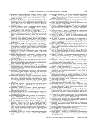
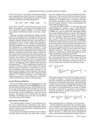
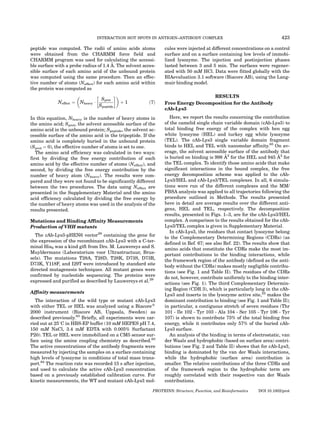
Fig. 3. (a) Net binding energy contribution by residue of the HEL lyso- to changes in sequence and in conformation space. The
zyme for the cAb-Lys3/hen egg white lysozyme complex. The values
reported are averages of the total contribution DG [cf. Eq. (3)]. The results
short MD simulations that were run for each structure
are presented for residues with a contribution different from zero. The were used to generate ensembles of conformations and
averages were calculated over all structures extracted from the trajecto- thus to reduce statistical error. We are then able to obtain
ries of the different initial structures (MEL AL HSD, MEL AL HSE, MEL a more general understanding of the recognition process
BM, and JTT). Units: kcal/mol. (b) Projection of the total energetic contri-
bution of each residue onto the lysozyme surface. The residues are col- in that our results are less dependent on the specific
ored from green (favorable contributions) to red (unfavorable contribu- three-dimensional structure.
tions). The values range from À7.74 to þ7.13 kcal/mol. The picture was A comparison of the results for the two lysozymes (see
prepared using the program GRASP66. The residues with the most favor-
able or unfavorable contributions are noted. [Color figure can be viewed
Fig. 5) shows that the interaction energy hot spots are the
in the online issue, which is available at www.interscience.wiley.com.] same in the two complexes; similar results are obtained
for the antibody (see Fig. 1 of Supplementary Material).
However, some charged amino acids of the interface, such
as Asp 48 and Arg 73 (HEL) or Lys 73 (TEL), show signifi-
complexed and uncomplexed lysozyme may modulate the cantly different binding energy contributions for the five
magnitude of this term, the desolvation effect explains, in structures of the complex (three structures for HEL and
large part, why the electrostatic contribution is large and two structures for TEL). These differences can be
positive for lysozyme (about þ36 Æ 6 kcal/mol, average of explained either structurally or based on the lysozyme
HEL and TEL; see Table II). sequences. For example, Asp 48 of lysozyme can form
This is an important difference with the cAb-Lys3, favorable interactions with Tyr 106 of cAb-Lys3, but it is
where such large positive contributions were not situated in a loop and shows larger structural fluctuations
observed. As a result, although unfavorable electrostatic than other amino acids. Hence its contribution varies
interactions are compensated by the favorable nonpolar considerably between the different structures. Likewise,
ones, the contribution of lysozyme to the binding ener- Arg 73 (HEL) or Lys 73 (TEL) are in a region of the pro-
getics is small when compared with that of cAb-Lys3. The tein–protein interface that shows significant structural
PROTEINS: Structure, Function, and Bioinformatics DOI 10.1002/prot](https://image.slidesharecdn.com/lafontproteins2007-120227133851-phpapp02/85/Lafont-proteins-2007-8-320.jpg)
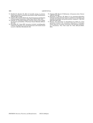
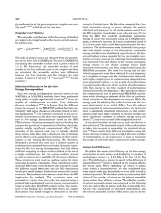
![426 LAFONT ET AL.
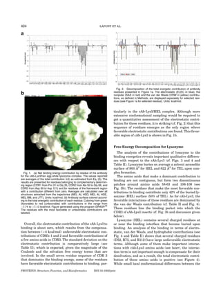
TABLE II. Individual Contributions to the Binding Energy for the cAb-Lys3/HEL Complex (top part) and the
cAb-Lys3/TEL Complex (lower part)
ELEC SAS VDW TOTAL
CHICKEN
CDR1 {P31-G35} 1.3 Æ 1.2 À0.1 Æ 0.04 À2.9 Æ 0.9 À1.8 Æ 2.2
CDR2 {A50-G66} 2.6 Æ 2.6 À0.7 Æ 0.1 À6.4 Æ 1.2 À4.6 Æ 3.9
CDR3 {D99-D121} À3.8 Æ 5.5 À3.4 Æ 0.2 À26.9 Æ 2.0 À34.0 Æ 7.7
HOT SPOT {T101-Y107} À6.4 Æ 4.6 À2.8 Æ 0.2 À23.6 Æ 1.9 À32.8 Æ 6.7
FRAME {all without CDRs} 0.3 Æ 5.3 À0.7 Æ 0.3 À4.0 Æ 1.9 À4.4 Æ 7.4
cAb-Lys3 {all antibody} 0.4 Æ 4.5 À5.0 Æ 0.2 À40.2 Æ 3.6 À44.8 Æ 8.3
Lysozyme: N46, I58, N59, W62, À1.8 Æ 2.1 À1.9 Æ 0.1 À20.6 Æ 1.1 À24.3 Æ 3.3
W63, N103, A107, W108, V109
Lysozyme: D48, D52, R61, R73, R112 29.5 Æ 6.3 À1.5 Æ 0.1 À9.6 Æ 1.7 18.4 Æ 8.1
HEL {all lysozyme} 37.4 Æ 7.0 À4.5 Æ 0.3 À40.2 Æ 3.6 À7.2 Æ 10.9
Total 37.8 Æ 8.6 À9.5 Æ 0.5 À80.4 Æ 7.2 À52.0 Æ 16.2
TURKEY
CDR1 {P31-G35} 2.6 Æ 0.8 À0.2 Æ 0.03 À2.1 Æ 0.5 0.3 Æ 1.3
CDR2 {A50-G66} 3.7 Æ 1.2 À0.7 Æ 0.04 À5.0 Æ 0.4 À2.1 Æ 1.7
CDR3 {D99-D121} 1.8 Æ 3.9 À3.4 Æ 0.2 À26.9 Æ 1.7 À28.5 Æ 5.8
HOT SPOT {T101-Y107} 1.1 Æ 2.9 À2.8 Æ 0.1 À23.8 Æ 1.7 À25.5 Æ 4.7
FRAME {all without CDR’s} 0.4 Æ 0.6 À0.4 Æ 0.1 À2.0 Æ 0.3 À2.0 Æ 1.0
cAb-Lys3 {all antibody} 8.5 Æ 4.7 À4.7 Æ 0.2 À36.0 Æ 2.0 À32.3 Æ 6.8
Lysozyme: N46, I58, N59, W62, W63, 0.02 Æ 1.9 À2.1 Æ 0.1 À20.0 Æ 1.2 À22.08 Æ 3.2
N103, A107, W108, V109
Lysozyme: D48, D52, R61, R73, R112 30.1 Æ 3.3 À1.3 Æ 0.1 À8.8 Æ 0.7 20.0 Æ 4.1
TEL {all lysozyme} 34.3 Æ 3.3 À4.2 Æ 0.2 À36.0 Æ 2.0 À5.8 Æ 5.5
Total 42.8 Æ 7.4 À8.9 Æ 0.3 À72.1 Æ 3.9 À38.1 Æ 11.6
All contributions are averages over the structures extracted from several molecular dynamics trajectories, as described in the Methods section.
The contributions for CDRs (Complementary Determining Regions) 1 to 3, for the hot spot residues (T101 to Y107 for cAb-Lys3), for the frame-
work (residues not in the CDR regions of cAb-Lys3, FRAME), for the entire antibody (cAb-Lys3), for lysozyme (HEL or TEL), for the hydrophobic
patches in lysozyme, and for charged residues in lysozyme are obtained by summing over the specified residues. All energies are in kcal/mol.
Fig. 5. Comparison of results obtained for the two complexes cAb-
Fig. 4. Decomposition of the total energetic contribution of lysozyme Lys3/hen egg white lysozyme (red) and the cAb-Lys3/turkey egg white
residues presented in Figure 3a. The electrostatic (ELEC in blue), the lysozyme (green). The contributions plotted are averages of total contri-
nonpolar (SAS in red) and the van der Waals (VDW in yellow) contribu- butions of lysozyme residues. The two lysozymes differ by only seven
tions are displayed separately for selected residues (see Fig. 3a for residues. The sequence used in this plot is the one for the chicken egg-
selected residue). Units: kcal/mol. [Color figure can be viewed in the white lysozyme. Amongst the residues shown in the graph, the following
online issue, which is available at www.interscience.wiley.com.] sequence differences in the turkey egg-white lysozyme are found: Q41H,
R73K, V99A, and D101G. The averages were calculated over all trajecto-
ries generated for each complex. The standard deviation for each residue
is shown. [Color figure can be viewed in the online issue, which is avail-
able at www.interscience.wiley.com.]
variations. These results underline the need to exercise
caution when interpreting the results from a free energy
decomposition for a single crystallographic structure. The For example, in the cAb-Lys3/HEL complex, the confor-
conformational sampling applied in this study yields a mational sampling clearly helps discriminating the true
mean value with a standard deviation for each contribu- hot spot residues from amino acids that have favorable
tion; the standard deviation is a useful first indication of but highly fluctuating contributions (see Fig. 1 of Supple-
the dependence of the results on small structural changes. mentary Material). These results suggest that, whenever
PROTEINS: Structure, Function, and Bioinformatics DOI 10.1002/prot](https://image.slidesharecdn.com/lafontproteins2007-120227133851-phpapp02/85/Lafont-proteins-2007-9-320.jpg)
![428 LAFONT ET AL.
In contrast to the unfavorable electrostatic contribu-
tions calculated for Arg 112 and Asp 52 of lysozyme, a
small favorable contribution is found for Trp 63 of lyso-
zyme, which interacts with Tyr 103 main chain from cAb-
Lys3. Thus, even though the electrostatic interaction from
the main chain of Tyr 103 of cAb-Lys3 is smaller than the
interaction made by its side chain (cf. Fig. 7), the main
chain H-bond yields a favorable binding energy contribu-
tion both on cAb-Lys3 and on lysozyme. Two additional
amino acids of lysozyme make favorable (although smaller
than those found in cAb-Lys3) electrostatic contributions:
Asn 59 (HEL: À1.7 kcal/mol) and Ala 107 (HEL: À0.6 kcal/
mol), see Figure 4. An analysis of the structures shows
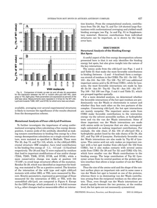
Fig. 7. Decomposition of the electrostatic contributions into side chain that Asn 59 and Ala 107 are engaged in hydrogen bonds
and main chain components for the antibody in the cAb-Lys3/hen egg
white lysozyme complex. The values represented are averages of the
with Ala 104 of cAb-Lys3 (cf. Figure 9), and this buried
electrostatic contribution of backbone (yellow) and side chain (blue) of network of H-bonds is particularly favorable.
antibody residues. (see Fig. 1a for selected residues). The average was
calculated over all the trajectories done with the different initial structures
(MEL AL HSD, MEL AL HSE, MEL BM, and JTT). Units: kcal/mol. Comparison of Experimental and
[Color figure can be viewed in the online issue, which is available at www. Computational Data
interscience.wiley.com.]
The conclusions of the calculations were challenged
with both existing and new mutational studies of cAb-
Even though van der Waals interactions have an impor- Lys3. While the existing studies mainly focus on residues
tant contribution to hot spots, some important electro- identified here as ‘‘hot spot,’’ we chose to focus on four
static interactions can also be identified. The electrostatic positions with a calculated small contribution to binding
contribution is favorable for cAb-Lys3 residues Tyr 103 - energy.
Ala 104 - Ser 105 - Tyr 106, which are involved in a hydro- Existing experimental studies include replacements of
gen bond network with several residues from lysozyme. residues Thr 101 (by L,S,P) and Ser 105 (by N,H,Q,A,
The most favorable electrostatic interactions are made by G,P,M) from CDR3 of cAb-Lys3 for a quantitative struc-
the two tyrosine residues; they can be further decomposed ture-activity study,53 and replacements of residues from
into side chain and main chain contributions. As can be CDR1 and CDR2 to revert to the germ line sequence.54
seen from Figure 7, the dominant contributions are made The replacements at positions 101 and 105 had varied
by the side chain for Tyr 103 (the main chain also makes a effects, the largest losses in binding affinity being 120-fold
favorable, but less important, contribution) and by the for the T100P change, and 100-fold for the S105Q
main chain for Tyr 106. Examination of the structures change.53 In particular, the mutation S105A resulted in a
indicates that the Tyr 103 side chain hydroxyl group 10-fold loss in experimental binding affinity (i.e. a change
makes an H-bond with Arg 112 from lysozyme, while the of 1.4 kcal/mol in binding affinity), which is in reasonable
Tyr 106 main chain NH makes an H-bond with the car- agreement with the computed binding free energy contri-
boxyl group of Asp 52 from lysozyme. The favorable contri- bution of À4.8 Æ 2.3 kcal/mol for the serine as a whole (i.e.
bution of these H-bonds is directly linked to the strong including main chain contributions that are not lost upon
electrostatic potential created by the charged amino acids mutation to Ala). Multiple mutations were introduced in
on lysozyme. Remarkably, the free energy decomposition CDR1 and CDR2 so that the influence of individual
for lysozyme (see Figs. 3 and 4) yields unfavorable electro- changes is not known.54 Thermodynamic data however
static contributions to the binding free energy for Arg 112 show a role of residues from CDR1 and CDR2 for
and Asp 52. This is due to the large desolvation penalty enthalpic stabilization of the bound complex during matu-
upon binding for the charged side chains of these residues. ration,54 which is in agreement with the results of the
The fact that electrostatic interactions are not always present calculations that show that CDR1 and CDR2
favorable to binding has been noted in other free energy make favorable van der Waals interactions with lysozyme
decomposition studies.13,19,21,58 In this particular exam- and thus play a role in stabilizing the complex.
ple, it can be seen that Arg 112 and Asp 52, although not To investigate whether positions with small, but non-
favorable themselves, create a favorable environment for zero, computed contributions can nevertheless influence
Tyr 103 and Tyr 106 on the cAb-Lys3, and thus have an binding, we targeted residues Thr 28, Asp 73, and Tyr
important role in the formation of the complex. The other 118. The residue Ile 29, which is identified as important in
charged amino acid on lysozyme that has a large unfavor- CDR1, was taken as a positive control. None of the muta-
able desolvation contribution is Arg 73. Arg 73 is not tions at positions 28, 73, and 118 had a measurable effect
involved in hydrogen bonds with amino acids on cAb- on binding kinetics (see Figure 6), while the mutation at
Lys3, and its binding energy contribution shows larger position I29 (I29T) increased koff 3- to 4-fold, thus result-
fluctuations than those of Arg 112 and Asp 52 (see ing in a loss of binding affinity. These experimental
Fig. 3a). results support the validity of our computational ap-
PROTEINS: Structure, Function, and Bioinformatics DOI 10.1002/prot](https://image.slidesharecdn.com/lafontproteins2007-120227133851-phpapp02/85/Lafont-proteins-2007-11-320.jpg)
![INTERACTION HOT SPOTS IN ANTIGEN–ANTIBODY COMPLEX 429
proach. In particular, the mutations data support our face area (hydrophobic solvation) terms as a sum over
simplified conformational averaging scheme, and show individual amino acid contributions (as described in Mate-
that the three amino acids tested, which showed large rial and Methods). The decomposition is performed in
fluctuations in the computed contribution, do not influ- such a way that the sum of all individual contributions
ence binding, while their contribution was non negligible corresponds to the total free energy (cf. Eq. 1). Decompos-
if only one experimental X-ray structure is used. ing into individual amino acid contributions implies that
pairwise interactions (such as van der Waals and electro-
static) are divided equally between the interacting amino
Experimental vs. Computational Hot Spots
acids i.e. if for example the van der Waals interaction
The comparison of the experimental data to the compu- energy between amino acids A and B amounts to À4 kcal/
tational results raises some important issues. Amino acids mol, a contribution of À2 kcal/mol will be attributed to A
that are found to play an important role in binding are of- and À2 kcal/mol to B, and likewise for electrostatic inter-
ten referred to in the computational literature as ‘‘binding actions. It must be noted that in the continuum descrip-
energy hot spots,’’ irrespective of whether the quantity tion of the solvent that is used here, desolvation (whether
calculated is the change in binding free energy upon electrostatic or hydrophobic) is a self-energy term and is
mutating the residue to alanine, or a related but different thus naturally decomposed in individual contribu-
quantity. In this paper, we used the term hot spot for tions.57,71
amino acids that make significant individual contribu- The earlier-mentioned differences between computa-
tions (À1.5 kcal/mol) to the total binding free energy, as tional free energy decomposition and experimental ala-
described in Material and Methods. It is therefore useful nine scanning data should be kept in mind when compar-
to clarify the relationship between ‘‘hot spots’’ as dis- ing computational and experimental hot spot residues.
cussed in the present computations and ‘‘hot spots’’
obtained by alanine scanning experiments.
Experimentally, binding energy hot spots are often
defined as residues that, when mutated to alanine, give
rise to a significant drop in the binding affinity (typically,
changes in binding free energy larger than 1.5 kcal/mol).7
As discussed in Ref. 69, mutations to alanine can affect
the binding affinity by both changing the structure and/or
free energy of the unbound partners as well as those of
the bound complex. This combination of effects compli-
cates the interpretation of alanine scanning experiments,
and consequently their exploitation in, for example,
designing small molecule inhibitors that mimic important
interactions. An advantage of computational approaches
is that the energetic contributions can be separated in a
manner that is not attainable experimentally. For exam-
ple, as done in the present study, it is possible to analyze
the energetic contribution of an amino acid without
resorting to explicit mutation to alanine.
This is an important aspect of the present calculations,
which is to identify binding energy hot spots and, thus,
free energy decomposition is used rather than explicit
mutation to alanine. Individual amino acid contributions
to binding presented in Figs. 1, 2, 3, 4, 7 are therefore not
the same quantity as the change in free energy upon
mutating the residue to alanine. If a direct comparison to
Ala-scanning experiments was desired, it would be possi-
ble to calculate free energy changes upon mutating a resi-
due to alanine by constructing the mutant and reevaluat-
ing the free energy of binding.11,70 A full estimate of the
free energy change should also consider the structural
consequences of mutating a specific amino acid to alanine,
Fig. 8. Bar graph of the amino acid efficiency. Each free energy contri-
both in the complex and in the unbound partners. bution (Electrostatic (ELE), nonpolar solvation (SAS), van der Waals
Another difference with experimental alanine scanning energy (VDW), and total DG (TOT)) of amino acids is divided by the num-
data is that single amino acid free energy contributions to ber of nonhydrogen atoms. The results are presented for the residues
binding, as computed here, include contributions to the having the most efficient amino-acids in the interaction for the antibody
(top graph) and for the lysozyme (bottom graph). See Methods for details.
binding energy from main chain atoms. They are obtained [Color figure can be viewed in the online issue, which is available at
by writing the total electrostatic, van der Waals and sur- www.interscience.wiley.com.]
PROTEINS: Structure, Function, and Bioinformatics DOI 10.1002/prot](https://image.slidesharecdn.com/lafontproteins2007-120227133851-phpapp02/85/Lafont-proteins-2007-12-320.jpg)
![430 LAFONT ET AL.
Fig. 9. Structural comparison of the lysozyme–cAb-Lys 3 complex and lysozyme-N-acetyl glucosamine
(NAG) ring complex. The similarity between the hydrogen bond network formed between A107 and N59 of ly-
sozyme and A104 of cAb-Lys3 (upper picture) and the network formed between A107 and N59 of lysozyme
and NAG is shown. This network involves efficient amino acids of the lysozyme-cAb-Lys3 complex, and shows
the energetic mimicry of the substrate (see text for details). [Color figure can be viewed in the online issue,
which is available at www.interscience.wiley.com.]
The two approaches nevertheless give convergent infor- nine will be detrimental to the binding affinity (unless the
mation i.e. when the free energy decomposition identifies contribution is made by main chain atoms). Significant
a residue as making a large favorable contribution to correlations between experimental free energy changes
binding, one can expect that mutating this residue to ala- upon mutating a residue to alanine and free energy
PROTEINS: Structure, Function, and Bioinformatics DOI 10.1002/prot](https://image.slidesharecdn.com/lafontproteins2007-120227133851-phpapp02/85/Lafont-proteins-2007-13-320.jpg)