1. Pioneering In Vivo Tools for the Study of the Axonal Proteome
Objectives
Future MethodsBackgroundAbstract
Sciatic Nerve Sympathetic Nerve
The neocortex underlies extraordinary perceptive, cognitive, and motor
capabilities, the implementation of which depends on the exact
formation of axonal connectivity during development. Miswiring of the
neocortex can ultimately alter executive functioning and behavior, with
potentially pathological consequences. The genes and molecules
behind the wiring of the neocortex are being unraveled. One potential
mechanism of this complicated process is the synthesis of proteins
within the tips of the growing axons (growth cones). Axonally
synthesized proteins are expected to be able to respond to the cellular
environment to guide axons to their target destination. Thus far, we
have been unable to attain a clear view of the local axonal proteins
within axons and axonal growth cone in the in vivo brain due to
technical limitations. We seek to pioneer a new technique to probe the
local axon proteins in vivo. By tagging the proteins within a neuron’s
growth cone, we can separate them from those proteins of the cells
surrounding the axonal growth path. These selected proteins can then
be isolated, captured, and analyzed using mass spectrometry for
protein identification – allowing us to determine what proteins are
present during an axon’s development in the intact brain. A procedure
such as this could yield results that enable future research on different
mental disorders such as the autism spectrum and fragile X syndrome
to study the differences in expressed genes between a healthy brain
and those with abnormalities.
Conclusions and Significance
A A
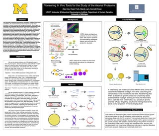
B
APEX labels endogenous
proteins in vivo in a mouse
brain. We capture proteins
via biotinylation (explained
below) and pull down for
proteomic analysis.
This method for determining the protein mechanisms behind axonal growth
can be later tested in vivo on transgenic mice containing our APEX
peroxidase Beta-actin 3’UTR construct. The eventual results from these mice
could further our understanding of axonal growth and the genes involved in
the growth process. With a better understanding of the proteins involved in
the innervation of the brain of normal and disordered mice, we may gain
valuable insight into the physical differences that arise in cognitive disorders,
and how to properly diagnose and potentially treat them.
We aim to extract proteins out of the growth cone of
mice neuronal axons. We are pioneering new methods that will
allow us to tag the newly formed proteins inside the axonal
growth cone, isolate them, and identify them. This in vitro
testing will likely lead to later in vivo testing and discovery of
the axon growth cone’s proteome.
Objective 1: Direct APEX expression to the growth cone.
Beta-actin is an important structural protein vital for the
growth and expansion of cells. The 3’ untranslated region
(UTR) has a zip code that directs transcode towards the
axonal; growth cone. Employing the Beta-Actin 3’ untranslated
region, the RNA strand is transcribed from our initial plasmid.
Once made, this RNA strand is directed to the axonal growth
cone where it is translated into our APEX peroxidase protein.
Objective 2: Transform neuronal cultures with the APEX β-actin
plasmid.
We will synthesize the APEX β-actin plasmid and verify it
with DNA sequencing by using a combination of PCR,
restriction digestion, recombination, and Gibson assembly to
build the required DNA vectors for the neuron. When this is
completed we will be able to add biotin-phenol. Due to the
proximal nature or APEX’s protein labeling, APEX only needs to
be expressed in the growth cone. After biotinylating, we will
isolate the tagged proteins using beads and identify the axonal
growth cone’s proteome. Eventually, this technique will be
applied in vivo towards identifying discrepancies in disease
models and developmental stages.
We expect to discover proteins related to growth and
axonal path finding. These findings would allow us to better
understand how the brain is wired and organized. Discovering
the proteins involved in the propagation of axons would also
provide insight into the mechanisms behind the disorders that
occur when the brain is incorrectly wired and certain proteins
are not present or in some way dysfunctional. Many of the the
current brain diseases and maladies are thought to arise from
the miswiring of the brain during development. Understanding
the molecular mechanisms that enable the brain to connect
itself may provide insights into the origins of humanity’s
complex and sophisticated cerebrum.
APEX catalyzes the creation of short-lived
biotin-phenol derivatives for endogenous
protein labeling.
Methods
In vitro testing will enable us to test different time points and
concentrations before we begin more time consuming and
complicated in vivo procedures and tests. Ascertaining that
APEX is directed to growth cones and functionally labels
proteins before we begin working on mice and their brains
allows us to be more certain about our procedure and its
potential efficacy for getting results and making discoveries
about axonal projection in the developing brain.