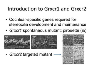
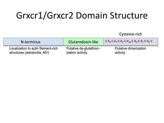
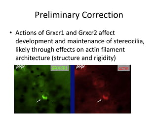
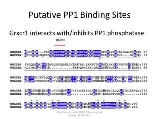
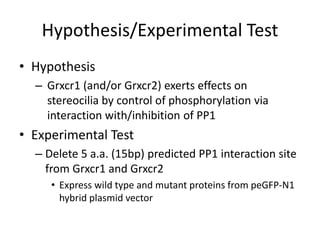
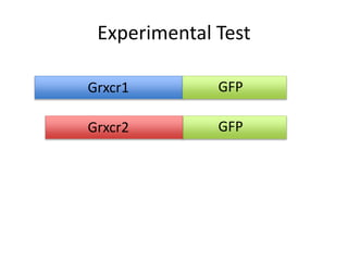
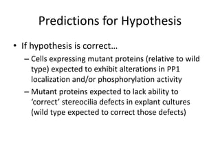
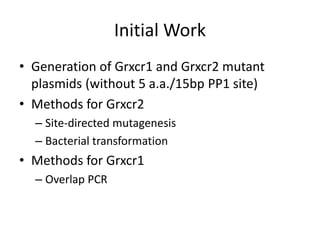
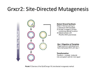
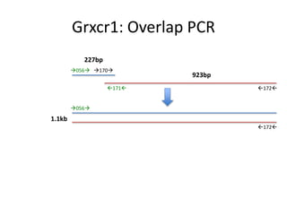
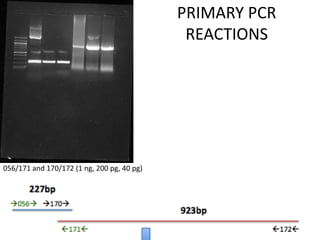
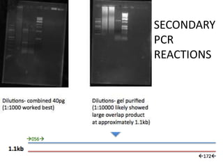
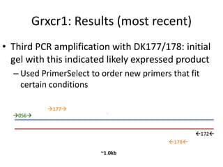
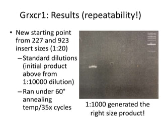
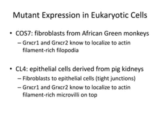
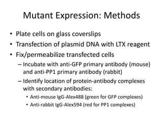
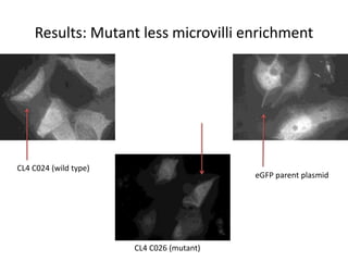
2. Introduction to Grxcr1 and Grxcr2
• Cochlear-specific genes required for
stereocilia development and maintenance
• Grxcr1 spontaneous mutant: pirouette (pi)
• Grxcr2 targeted mutant
4. Preliminary Correction
• Actions of Grxcr1 and Grxcr2 affect
development and maintenance of stereocilia,
likely through effects on actin filament
architecture (structure and rigidity)
6. Putative PP1 Binding Sites
Grxcr1 interacts with/inhibits PP1 phosphatase
Hendricks, A., et al. (2009). Chemistry and
Biology, 16:365-371.
RKVRF
7. Hypothesis/Experimental Test
• Hypothesis
– Grxcr1 (and/or Grxcr2) exerts effects on
stereocilia by control of phosphorylation via
interaction with/inhibition of PP1
• Experimental Test
– Delete 5 a.a. (15bp) predicted PP1 interaction site
from Grxcr1 and Grxcr2
• Express wild type and mutant proteins from peGFP-N1
hybrid plasmid vector
9. Predictions for Hypothesis
• If hypothesis is correct…
– Cells expressing mutant proteins (relative to wild
type) expected to exhibit alterations in PP1
localization and/or phosphorylation activity
– Mutant proteins expected to lack ability to
‘correct’ stereocilia defects in explant cultures
(wild type expected to correct those defects)
10. Initial Work
• Generation of Grxcr1 and Grxcr2 mutant
plasmids (without 5 a.a./15bp PP1 site)
• Methods for Grxcr2
– Site-directed mutagenesis
– Bacterial transformation
• Methods for Grxcr1
– Overlap PCR
18. Grxcr1: Results
• Grxd1 #4 replaced faulty wild type
Grxcr1/C023 clone
• Considerable attempts to improve the
specificity/yield of the products:
– Altered annealing temperatures and cycling
conditions during PCR
– Different amounts of templates
– Purification of initial smaller products
19. Grxcr1: Results (most recent)
• Third PCR amplification with DK177/178: initial
gel with this indicated likely expressed product
– Used PrimerSelect to order new primers that fit
certain conditions
177
178
~1.0kb
20. Grxcr1: Results (repeatability!)
• New starting point
from 227 and 923
insert sizes (1:20)
–Standard dilutions
(initial product
above from
1:10000 dilution)
–Ran under 60°
annealing
temp/35x cycles 1:1000 generated the
right size product!
21. Mutant Expression in Eukaryotic Cells
• COS7: fibroblasts from African Green monkeys
– Grxcr1 and Grxcr2 know to localize to actin
filament-rich filopodia
• CL4: epithelial cells derived from pig kidneys
– Fibroblasts to epithelial cells (tight junctions)
– Grxcr1 and Grxcr2 know to localize to actin
filament-rich microvilli on top
22. Mutant Expression: Methods
• Plate cells on glass coverslips
• Transfection of plasmid DNA with LTX reagent
• Fix/permeabilize transfected cells
– Incubate with anti-GFP primary antibody (mouse)
and anti-PP1 primary antibody (rabbit)
– Identify location of protein-antibody complexes
with secondary antibodies:
• Anti-mouse IgG-Alex488 (green for GFP complexes)
• Anti-rabbit IgG-Alex594 (red for PP1 complexes)
23. Mutant Expression: Results
• Expected:
– Localization of wild type Grxcr2-GFP (and Grxcr1-
GFP) to microvilli of CL4/filopodia of Cos7 cells
• Experimental Questions:
– Localization of mutant proteins?
– Altered localization of PP1 in mutant expressing
cells?
25. Results: PP1 similar in WT/mutant
C024 GFP C026 GFP eGFP GFP
C026 PP1C024 PP1 eGFP PP1
26. In Conclusion/Future Work
• Grxcr2 mutant sequence confirmed
• Grxcr1 mutant clone yet to be generated
• Continue to experiment with Grxcr1 mutant to
replicate initial overlap
• Continue to follow up on transfected cells (i.e.
laser scope microscope for fine sections)
– Top of cell vs. inside of cell
– (Small region of N-terminus needed for localization)
27. Acknowledgments
• Dr. Kohrman
• Cathy, Catherine, Jessica
• Kresge Hearing Research Institute (KHRI)
• National Institute on Deafness and Other
Communication Disorders (NIDCD)
Editor's Notes
Grxcr1: left picture is wild type (normal stereocilia) and right picture is mutant (affected stereocilia) MUTATION ARISES NATURALLY (not from mutagens)
Grxcr2: top row is wild type and bottom row is mutant (MUTATION ELIMINATES GENE FUNCTION => KNOCKOUTS)
N-terminus: start of amino acid chain (specific to structures rich in actin, such as stereocilia and microvilli)
De-glutathionylation: modifies protein by removing glutathione molecule (via breakage of disulfide linkage)
Cysteine: sulfur-containing amino acid
Dimerization: forming dimers (two identical molecules)
---------------------------------------------------------------------------------------------------------------------------------------------
Glutaredoxin distantly related, don’t have activity
Cysteine-rich (groups of 4) primitive eukaryotes, binding DNA or other proteins (dimers of 2 identical proteins or 2 Grxcr1/cr2 heterodimers)
PROVIDE STRUCTURE AND RIGIDITY TO STEREOCILIA
VERY LITTLE ACTIN IN MUTANT EXCEPT THOSE EXPRESSED IN WT THROUGH TRANSFECTION (mutant cannot correct this deficit)
GFP HYBRID GREEN
RED ACTIN FILAMENT (PP1)
PP1 phosphatase: removes phosphates from Ser (serine) and Thr (threonine) residues
Protein phosphorylation controls some aspects of actin filament dynamics (previous slide)
Red line (RKVRF) is PP1 binding site/motif (5 amino acids => 15 nucleotides/base pairs)
N-terminus 1.5 lines
Glutaredoxin 2.5 lines
Cysteine-rich last line
Phosphorylation: addition of phosphate group
eGFP: enhanced green fluorescent protein
Grxcr1: 950 base pairs (insert size)
Grxcr2: 789 base pairs (little smaller)
GFP attached to C-terminus (end of amino acid chain)
Follow up from earlier (stereocilia correction)
Site-directed mutagenesis: site-specific/targeted mutation
Bacterial transformation: bacterial cells take up DNA molecules
Overlap PCR: overlap extension PCR cloning is a simple and reliable way to create recombinant plasmids (clone an insert into a plasmid)
In-vitro reaction
Denaturation (DNA template), annealing (melting) primers with mutations, extension (of primers with DNA polymerase)
DpnI digestion of template
Transform mutated molecule into competent cells for nick repair
Oligo(nucleotide) => short sequence that matches mutation region
High-fidelity low error rate: high match with minimum mistakes (in extension process)
Extend 3’ ends => Goal is complete circle (circular map) with break in it
Nicked vector DNA incorporating desired mutations transformed into ultracompetent cells
Dpn-I will distinguish mutant from WT because will cut WT and not mutant (resistance)
TOP SEQ IS WT
Cr2 mutant sequence alignment- without 5 amino acids (PP1 binding site/motif)
(reverse primer DK172)
(forward primer DK056)
Primary PCRs (2 separate reactions) to generate 2 overlapping DNA fragments/products, then a secondary PCR to produce the single larger product (anneal together)
Overlap PCR involves denaturation, annealing (melting), and extension:
(vector and insert are mixed, denatured, and annealed) => extension (DNA polymerase uses vector as a template until polymerase reaches 5’ end of the insert)
After several PCR cycles, new plasmid with two nicks (one on each strand) gets accumulated as a product
New plasmid can be transformed into E. coli after parental plasmid is destroyed by DpnI digest
Primary PCR reactions- 2 sets of primers and 3 dilutions each
Secondary PCR reaction (056172)- combined 20 microliters of 40pg from initial reactions and ran dilutions (1:100, 1:1000, 1:10000); background/artifacts in 1:1000
Purified 1:1000 and ran dilutions (1:100, 1:1000, 1:10000)- overlap product visible/evident/present
Annealing temps (65 vs. 60) and cycling conditions (3-step vs. 2-step-no annealing and 30x repetitions vs. 35x)
Dilutions of templates (10^-2 to 10^-6)- 1:100, 1:1000, 1:10000, 1:100000, 1:1000000
PCR column purification (cleaning up product via mini centrifuge) vs. gel purification (cut out gel product to be purified and similar to PCR purification, except melt the gel first)
20-folds dilution of 227 because more product
Standard dilutions => 1:100, 1:1000, 1:10000
SHOW RECENT IMAGE BUT NOT REPEATABLE
REPEATABILITY IS IMPORTANT BECAUSE NEED TO GENERATE ENOUGH PRODUCT TO EFFICIENTLY ISOLATE AND TRANSFECT INTO CLONE
-DMEM vs. (alpha)MEM
-Fibroblasts to epithelial cells over few days
-Microvilli serves as proxy model for stereocilia
Anti-GFP primary antibody in mouse and then anti-mouse secondary antibody (green for GFP)
Anti-PP1 primary antibody in rabbit and then anti-rabbit secondary antibody (red for PP1)
Strong microvilli expression in wild type (along edges)
Stability unaffected (medium/low/high levels) -> rule out trivial explanation (I.e. protein misfolding)
Mutant weaker expression than WT and eGFP similar to mutant
MUTANT LESS MICROVILLI ENRICHMENT
WT STRONG MICROVILLI ENRICHMENT
EXPECTED MUTANT AND WT TO BE SAME
ALTERED LOCALIZATION OF MUTANT
MUTANT PROTEIN NOT ENRICHED IN MICROVILLI
GFP and PP1 in WT/mutant/eGFP parent plasmid
Mutants would be different without PP1 interaction (increased PP1 enrichment– filopodia and microvilli – in wild type)
Can’t separate localization from PP1 => difficult to extract relevance of PP1 itself (difficult to distinguish defect itself)
Structure-function analysis > small region necessary for localization (unclear whether or not it affects PP1 activity)
PP1 SIMILAR IN WT AND MUTANT
SMALL REGION IN N-TERMINUS (5 A.A.) NECESSARY FOR LOCALIZATION
SHOW PICTURES OF WT AND MUTANT TO SHOW THAT LASER MICROSCOPE WOULD ALLOW FOR HIGHER RESOLUTION (LOOKING AT TOP OF CELL VS. INSIDE OF CELL)