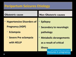
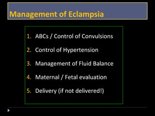
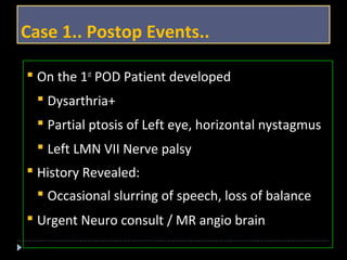
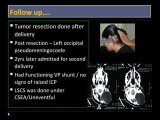
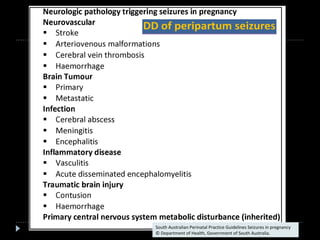
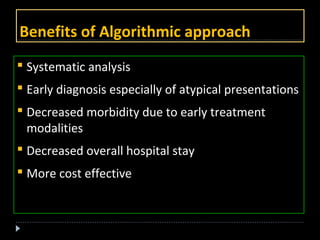
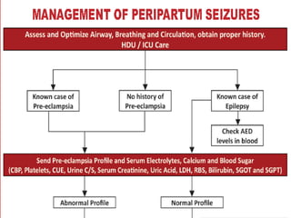
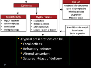
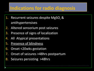
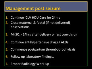
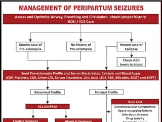
Greetings from 
Prerna Anesthesia & Critical Care Services 
& Fernandez Hospital 
Hyderabad
2 Isolation Rooms with 
separate AHUs 
220 Bed Tertiary care Perinatal Centre 
 ADR – 7500 + 
 Six bed dedicated Maternal CCU 
 Six Bed HDU (LW) 
 4 Bed Step Down Unit 
 22 Bed NICU 
 Fetal Medicine Unit 
 Dedicated Obstetric Medicine Unit 
 Critical Care Outreach teams /MOEWS / SMS 
220 Bed Tertiary care Perinatal Centre 
 ADR – 7500 + 
 Six bed dedicated Maternal CCU 
 Six Bed HDU (LW) 
 4 Bed Step Down Unit 
 22 Bed NICU 
 Fetal Medicine Unit 
 Dedicated Obstetric Medicine Unit 
 Critical Care Outreach teams /MOEWS / SMS 
Counseling Room with 
A-V Facility
Algorithmic approach to 
Peripartum seizures 
Dr. Kousalya Chakravarthy 
Consultant Prerna Anaesthesia & Critical Care Services 
Asst Prof Anaesthesia; Niloufer Hospital 
Osmania Medical College; Hyderabad. INDIA
Definition 
 Seizures can be defined as “abnormal 
electrical activity associated with certain 
behavioural and neurologic effects
Peripartum Seizures Etiology 
Obstetric cause Non Obstetric causes 
 Hypertensive Disorders of 
Pregnancy (HDP) 
 Eclampsia 
 Severe Pre eclampsia 
with HELLP 
 Epilepsy 
 Secondary to neurologic 
pathology 
 Metabolic derangements 
as a result of critical 
illness
Case 1 
 Primigravida /38 wks / normotensive / PROM 
 Patient in labor requested epidural analgesia 
 Developed GTCS when epidural dose was being 
administered!! 
 BP : 160/100mmHg post seizure 
 ? Intrapartum eclampsia 
MgSO4 given, Pre eclampsia (PE) Profile sent
? Cause of convulsions 
 There is a dictum that new onset of 
convulsions in a pregnant patient should be 
assumed to be caused by eclampsia unless 
proved otherwise!
Management of Eclampsia 
1. ABCs / Control of Convulsions 
2. Control of Hypertension 
3. Management of Fluid Balance 
4. Maternal / Fetal evaluation 
5. Delivery (if not delivered!)
Magnesium Sulphate Regime 
IV Infusion 
1 
 Loading dose 
 20% solution, 4 gm 
 Slow IV, rate not more than 0.5 - 1 gm / min 
 Maintenance regime 
 As infusion, 1-2 gm / hr
Control of Blood Pressure 
IV Labetolol 
 20mg stat over 10min 
 Intervals ½ hr: 20-20-40-40-80mg 
 Max 220mg(2-3g/kg) 
 2mg/ml infusion 
2 
Maintenance 
 SR Nifedipine 10mg BD 
 T. Labetolol 100 – 200 mg bid or tid max 600mg/day
3 
4 Maternal Evaluation 
Fluid volume regimes 
 75 -80ml of fluid /hr balancing input and output 
MINI PE* 
MINI PE* 
PROFILE 
PE* PROFILE PROFILE 
EXTENDED PE* 
PROFILE 
5 Evaluation of Fetus 
 CTG, Ultrasound 
*Pre eclampsia profile
Case 1….Cont…. 
 Emergency LSCS in view of fetal compromise 
 PE Profile was Normal 
 Intra operatively epidural activated 
- While giving epidural patient threw a GTCS 
- Accompanied by vomiting
Protocol for Recurrent Seizures 
Loading Dose of MgSO4 
4 gm,20% Max 1 gm/min . Maintenance of 1gm/ hour 
Second episode of Seizures 
Draw a sample for Serum Magnesium 
2 gm of MgSO4 IV, Increase maintenance to 2gm / hr 
Third episode of Seizures 
Check The S Magnesium reports, if wt > 70 kg 
2 gm of MgSO4 IV 20% can be repeated again…. 
Clinical guideline for the management of a woman with 
eclampsia and/or Severe pre eclampsia /august 2012
Protocol for Recurrent Seizures 
Recurrence of Seizures 
Midazolam 
Dose 0.1 mg / kg body weight, slow IV 
Still recurrence 
Phenytoin Sodium 
Loading Dose: 15 mg / kg body weight 
(1000 mg in 100 ml of NS over 45 mins) 
Maintenance dose of 5 mg / kg / day 
(100 mg 8th hourly Slow infusion) 
AIRWAY 
has to be 
maintained
Status epilepticus 
All simultaneously 
Maintain 
oxygenation 
Protect airway 
Terminate seizure 
activity 
Propofol / 
Thiopentone sodium / 
Succinyl choline 
RSI if need arises! 
Oxygen by mask 
Guedel’s Airway 
Endotracheal 
intubation Midazolam 2- 5 mg IV 
Clonazepam 1 mg IV, 
over 2 to 5 min, not 
exceeding 0.5 mg / 
min 
Repeat once 15 minutes later if status epilepticus continues
But Our Dilemma.. 
 Uneventful antenatal period 
 Successive normal BP 
recordings 
 Normal PE Profile 
 Not suggestive of 
Eclampsia!! 
 Both episodes of GTCS coincided with epidural 
dosing of the drug!! 
 ? Local Anesthetic Toxicity
Case Details… 
Intra Operatively: 
 Regained consciousness in 3 minutes. 
 Epidural block: Dermatomal level of T6 was 
present 
 Surgery done under EA uneventfully – 
which rules out LA toxicity!
Case 1.. Postop Events.. 
 On the 1st POD Patient developed 
 Dysarthria+ 
 Partial ptosis of Left eye, horizontal nystagmus 
 Left LMN VII Nerve palsy 
 History Revealed: 
 Occasional slurring of speech, loss of balance 
 Urgent Neuro consult / MR angio brain
Follow up…. 
 Tumor resection done after 
delivery 
 Post resection – Left occipital 
pseudomeningocoele 
 2yrs later admitted for second 
delivery 
 Had Functioning VP shunt / no 
signs of raised ICP 
 LSCS was done under 
CSEA/Uneventful
South Australian Perinatal Practice Guidelines Seizures in pregnancy 
© Department of Health, Government of South Australia.
Literature review 
Maria Hirsch, CRNA, DNAPAANA Journal ; October 2011; Vol. 79, No. 5
Our Statistics Over 7 Yrs
Algorithmic approach……. 
 We derived an algorithm keeping in mind 
 Eclampsia should be considered in all cases 
 Eclampsia may not be the cause in all 
 Systematic approach needed for 
Further investigations 
Radio diagnosis 
Follow up
Benefits of Algorithmic approach 
 Systematic analysis 
 Early diagnosis especially of atypical presentations 
 Decreased morbidity due to early treatment 
modalities 
 Decreased overall hospital stay 
 More cost effective
 Atypical presentations can be 
 Focal deficits 
 Refractory seizures 
 Altered sensorium 
 Seizures >7days of delivery
Atypical presentation …… 
 Rule out 
 Cerebro vascular 
compromise 
 SOL - Brain 
 Infectious diseases 
 Drug toxicity 
 Metabolic causes 
 Further 
investigations 
 ABG with Lactate 
 S.Ca++, Mg++ levels 
 Radio diagnosis 
 Neuro consultation/ 
Neuro ICU
Indications for radio diagnosis 
1. Recurrent seizures despite MgSO4 & 
antihypertensives 
2. Altered sensorium post seizures 
3. Presence of signs of localization 
4. All Atypical presentations 
5. Presence of blindness 
6. Onset <20wks gestation 
7. Onset of seizures >48hrs postpartum 
8. Seizures persisting >48hrs
Role of radio imaging 
To R/o 
 CSVT 
 Cerebral infarcts 
 Leucoencephalopathy (PRES) 
 Mass lesions (IC-SOL) 
 Aneurysm / Bleeding
Case 2 
 Booked patient 
 Term / BP: 130/90 
 Proteinuria 1+ 
 Admitted with severe 
headache…..EXCRUCIATING 
 Pleading for pain relief 
 GCS: 15 
 Pupils NSRL/ VII CN Palsy 
 ? Eclampsia ? IC bleed ? IC-SOL
CT Brain 
Bullet in the Brain!!!!
Case 2.... 
Emg LSCS & Emg Craniotomy same sitting
CT Vs MRI 
Which is a better radiological 
tool in pregnancy?
 A patient complains of postural headache, after an 
uneventful epidural . 
 Conservative treatment started 
 With worsening of headache, plain CT scan brain done 
 CT SCAN WAS NORMAL 
 Epidural blood patch was given. 
 After few hours, had a GCTS and later was pronounced 
dead!! 
Autopsy revealed bilateral subdural hematoma!!! 
Fahad Aziz, MD New york Medical JournaNovember 2 2010l
CT & MRI of a patient with Intra cerebral bleed & PRES 
CCTT B BRRAAININ MMRRI IB BRRAAININ 
MRI showing thicker and better delineated PRES & 
haematoma
CT vs. MRI 
 CT is faster /readily available can be used in acute 
conditions with unstable patients 
 Deep CSVT is likely to be missed out on a plain CT 
 Contrast CT brain carries a risk of AKI in hypertensive 
disorders of pregnancy 
 Even the contrast CT brain can miss out deep seated 
CSVT 
 Smaller haemorrhages may be missed on CT scans CT and MR imaging of chronic subdural haematomas: a comparative 
study SWISS MED WKLY 2 010 ; 14 0 ( 2 3 – 2 4 ) : 3 3 5 – 3 4 0
MRI preferred over CT scan 
 Decreased risk of radiation ( antenatal) 
 High sensitivity and specificity 
 Cytotoxic edema better diagnosed 
 MR venogram brain is diagnostic of CSVT 
DW MR images with T2 weighted FLAIR can be 
extremely helpful in evaluation of women with new 
onset peripartum seizures. 
Brain MRI in peripartum seizures: usefulness of combined T2 and diffusion weighted MR 
imaging Journal of the Neurological Sciences Volume 166, Issue 2, Pages 122–125, July 1, 1999
Case 3.. Postpartum seizures 
 Primi / Post LSCS/ 1st POD 
 Antenatal period - uneventful 
 C/o ? Loss of vision since 1hour (not able to see!) 
 Vitals –stable 
 BP : 140/90mmHg/ 
 Had vomiting 
 GTCS while shifting to ICU! 
 ? Cause of the seizures 
 ? Eclampsia
Postpartum seizures D.D
Case 3.. Postpartum seizures 
MR Venogram was done to R/O CSVT 
Posterior Reversible Encephalopathy Syndrome
Postpartum seizures
Anaesthetic Technique ?? 
VS 
Regional techniques 
SAB / EA / CSEA 
General anaesthesia
General anaesthesia -Issues of Concern 
 Level of consciousness 
 Hypertension – Laryngo sympathetic response 
 Sublingual Hematoma – difficult laryngoscopy 
 Difficulty in Airway assessment 
 RSI – Succinyl choline vs. Serum Potassium 
 Intra op hypertension 
 Increased intra operative Blood loss 
 Delayed recovery
Regional techniques in peripartum 
 Epilepsy per se is not a CI for RA 
 Subarachnoid block is safe in HDP 
 HELLP syndrome, altered coagulation profile, low GCS 
score mandate GA 
Issues of concern with RA: 
 Un co-operative patient 
 Sacral Oedema- difficult landmarks 
 Difficult technique 
 Deranged Platelet & Coagulation profile
Management post seizure 
1. Continue ICU/ HDU Care for 24hrs 
2. Close maternal & foetal (if not delivered) 
observations 
3. MgSO4 - 24hrs after delivery or last convulsion 
4. Continue antihypertensive drugs / AEDs 
5. Commence postpartum thromboprophylaxis 
6. Follow up laboratory findings, 
7. Proper Radiology Work-up
To Summarize 
 The most common cause of ‘New-onset’ seizures in 
pregnancy is eclampsia 
 But..... 
 Not all first time seizures occurring in the third 
trimester are preeclampsia / eclampsia!! 
 Atypical presentations should have a proper 
workup and managed accordingly
Conclusion 
 Unusual types of cerebrovascular pathology is 
relatively common in pregnancy and the dictum 
that all peripartum seizures should be regarded as 
eclampsia until proved otherwise.. 
Should be wisely judged!
Thank you
Kausalaya chakravarthy

Kausalaya chakravarthy

  • 1.
    Greetings from PrernaAnesthesia & Critical Care Services & Fernandez Hospital Hyderabad
  • 2.
    2 Isolation Roomswith separate AHUs 220 Bed Tertiary care Perinatal Centre  ADR – 7500 +  Six bed dedicated Maternal CCU  Six Bed HDU (LW)  4 Bed Step Down Unit  22 Bed NICU  Fetal Medicine Unit  Dedicated Obstetric Medicine Unit  Critical Care Outreach teams /MOEWS / SMS 220 Bed Tertiary care Perinatal Centre  ADR – 7500 +  Six bed dedicated Maternal CCU  Six Bed HDU (LW)  4 Bed Step Down Unit  22 Bed NICU  Fetal Medicine Unit  Dedicated Obstetric Medicine Unit  Critical Care Outreach teams /MOEWS / SMS Counseling Room with A-V Facility
  • 3.
    Algorithmic approach to Peripartum seizures Dr. Kousalya Chakravarthy Consultant Prerna Anaesthesia & Critical Care Services Asst Prof Anaesthesia; Niloufer Hospital Osmania Medical College; Hyderabad. INDIA
  • 4.
    Definition  Seizurescan be defined as “abnormal electrical activity associated with certain behavioural and neurologic effects
  • 5.
    Peripartum Seizures Etiology Obstetric cause Non Obstetric causes  Hypertensive Disorders of Pregnancy (HDP)  Eclampsia  Severe Pre eclampsia with HELLP  Epilepsy  Secondary to neurologic pathology  Metabolic derangements as a result of critical illness
  • 6.
    Case 1 Primigravida /38 wks / normotensive / PROM  Patient in labor requested epidural analgesia  Developed GTCS when epidural dose was being administered!!  BP : 160/100mmHg post seizure  ? Intrapartum eclampsia MgSO4 given, Pre eclampsia (PE) Profile sent
  • 7.
    ? Cause ofconvulsions  There is a dictum that new onset of convulsions in a pregnant patient should be assumed to be caused by eclampsia unless proved otherwise!
  • 8.
    Management of Eclampsia 1. ABCs / Control of Convulsions 2. Control of Hypertension 3. Management of Fluid Balance 4. Maternal / Fetal evaluation 5. Delivery (if not delivered!)
  • 9.
    Magnesium Sulphate Regime IV Infusion 1  Loading dose  20% solution, 4 gm  Slow IV, rate not more than 0.5 - 1 gm / min  Maintenance regime  As infusion, 1-2 gm / hr
  • 10.
    Control of BloodPressure IV Labetolol  20mg stat over 10min  Intervals ½ hr: 20-20-40-40-80mg  Max 220mg(2-3g/kg)  2mg/ml infusion 2 Maintenance  SR Nifedipine 10mg BD  T. Labetolol 100 – 200 mg bid or tid max 600mg/day
  • 11.
    3 4 MaternalEvaluation Fluid volume regimes  75 -80ml of fluid /hr balancing input and output MINI PE* MINI PE* PROFILE PE* PROFILE PROFILE EXTENDED PE* PROFILE 5 Evaluation of Fetus  CTG, Ultrasound *Pre eclampsia profile
  • 12.
    Case 1….Cont…. Emergency LSCS in view of fetal compromise  PE Profile was Normal  Intra operatively epidural activated - While giving epidural patient threw a GTCS - Accompanied by vomiting
  • 13.
    Protocol for RecurrentSeizures Loading Dose of MgSO4 4 gm,20% Max 1 gm/min . Maintenance of 1gm/ hour Second episode of Seizures Draw a sample for Serum Magnesium 2 gm of MgSO4 IV, Increase maintenance to 2gm / hr Third episode of Seizures Check The S Magnesium reports, if wt > 70 kg 2 gm of MgSO4 IV 20% can be repeated again…. Clinical guideline for the management of a woman with eclampsia and/or Severe pre eclampsia /august 2012
  • 14.
    Protocol for RecurrentSeizures Recurrence of Seizures Midazolam Dose 0.1 mg / kg body weight, slow IV Still recurrence Phenytoin Sodium Loading Dose: 15 mg / kg body weight (1000 mg in 100 ml of NS over 45 mins) Maintenance dose of 5 mg / kg / day (100 mg 8th hourly Slow infusion) AIRWAY has to be maintained
  • 15.
    Status epilepticus Allsimultaneously Maintain oxygenation Protect airway Terminate seizure activity Propofol / Thiopentone sodium / Succinyl choline RSI if need arises! Oxygen by mask Guedel’s Airway Endotracheal intubation Midazolam 2- 5 mg IV Clonazepam 1 mg IV, over 2 to 5 min, not exceeding 0.5 mg / min Repeat once 15 minutes later if status epilepticus continues
  • 16.
    But Our Dilemma..  Uneventful antenatal period  Successive normal BP recordings  Normal PE Profile  Not suggestive of Eclampsia!!  Both episodes of GTCS coincided with epidural dosing of the drug!!  ? Local Anesthetic Toxicity
  • 17.
    Case Details… IntraOperatively:  Regained consciousness in 3 minutes.  Epidural block: Dermatomal level of T6 was present  Surgery done under EA uneventfully – which rules out LA toxicity!
  • 18.
    Case 1.. PostopEvents..  On the 1st POD Patient developed  Dysarthria+  Partial ptosis of Left eye, horizontal nystagmus  Left LMN VII Nerve palsy  History Revealed:  Occasional slurring of speech, loss of balance  Urgent Neuro consult / MR angio brain
  • 20.
    Follow up…. Tumor resection done after delivery  Post resection – Left occipital pseudomeningocoele  2yrs later admitted for second delivery  Had Functioning VP shunt / no signs of raised ICP  LSCS was done under CSEA/Uneventful
  • 21.
    South Australian PerinatalPractice Guidelines Seizures in pregnancy © Department of Health, Government of South Australia.
  • 23.
    Literature review MariaHirsch, CRNA, DNAPAANA Journal ; October 2011; Vol. 79, No. 5
  • 24.
  • 25.
    Algorithmic approach……. We derived an algorithm keeping in mind  Eclampsia should be considered in all cases  Eclampsia may not be the cause in all  Systematic approach needed for Further investigations Radio diagnosis Follow up
  • 26.
    Benefits of Algorithmicapproach  Systematic analysis  Early diagnosis especially of atypical presentations  Decreased morbidity due to early treatment modalities  Decreased overall hospital stay  More cost effective
  • 28.
     Atypical presentationscan be  Focal deficits  Refractory seizures  Altered sensorium  Seizures >7days of delivery
  • 29.
    Atypical presentation ……  Rule out  Cerebro vascular compromise  SOL - Brain  Infectious diseases  Drug toxicity  Metabolic causes  Further investigations  ABG with Lactate  S.Ca++, Mg++ levels  Radio diagnosis  Neuro consultation/ Neuro ICU
  • 30.
    Indications for radiodiagnosis 1. Recurrent seizures despite MgSO4 & antihypertensives 2. Altered sensorium post seizures 3. Presence of signs of localization 4. All Atypical presentations 5. Presence of blindness 6. Onset <20wks gestation 7. Onset of seizures >48hrs postpartum 8. Seizures persisting >48hrs
  • 31.
    Role of radioimaging To R/o  CSVT  Cerebral infarcts  Leucoencephalopathy (PRES)  Mass lesions (IC-SOL)  Aneurysm / Bleeding
  • 32.
    Case 2 Booked patient  Term / BP: 130/90  Proteinuria 1+  Admitted with severe headache…..EXCRUCIATING  Pleading for pain relief  GCS: 15  Pupils NSRL/ VII CN Palsy  ? Eclampsia ? IC bleed ? IC-SOL
  • 33.
    CT Brain Bulletin the Brain!!!!
  • 34.
    Case 2.... EmgLSCS & Emg Craniotomy same sitting
  • 35.
    CT Vs MRI Which is a better radiological tool in pregnancy?
  • 36.
     A patientcomplains of postural headache, after an uneventful epidural .  Conservative treatment started  With worsening of headache, plain CT scan brain done  CT SCAN WAS NORMAL  Epidural blood patch was given.  After few hours, had a GCTS and later was pronounced dead!! Autopsy revealed bilateral subdural hematoma!!! Fahad Aziz, MD New york Medical JournaNovember 2 2010l
  • 37.
    CT & MRIof a patient with Intra cerebral bleed & PRES CCTT B BRRAAININ MMRRI IB BRRAAININ MRI showing thicker and better delineated PRES & haematoma
  • 38.
    CT vs. MRI  CT is faster /readily available can be used in acute conditions with unstable patients  Deep CSVT is likely to be missed out on a plain CT  Contrast CT brain carries a risk of AKI in hypertensive disorders of pregnancy  Even the contrast CT brain can miss out deep seated CSVT  Smaller haemorrhages may be missed on CT scans CT and MR imaging of chronic subdural haematomas: a comparative study SWISS MED WKLY 2 010 ; 14 0 ( 2 3 – 2 4 ) : 3 3 5 – 3 4 0
  • 39.
    MRI preferred overCT scan  Decreased risk of radiation ( antenatal)  High sensitivity and specificity  Cytotoxic edema better diagnosed  MR venogram brain is diagnostic of CSVT DW MR images with T2 weighted FLAIR can be extremely helpful in evaluation of women with new onset peripartum seizures. Brain MRI in peripartum seizures: usefulness of combined T2 and diffusion weighted MR imaging Journal of the Neurological Sciences Volume 166, Issue 2, Pages 122–125, July 1, 1999
  • 40.
    Case 3.. Postpartumseizures  Primi / Post LSCS/ 1st POD  Antenatal period - uneventful  C/o ? Loss of vision since 1hour (not able to see!)  Vitals –stable  BP : 140/90mmHg/  Had vomiting  GTCS while shifting to ICU!  ? Cause of the seizures  ? Eclampsia
  • 41.
  • 42.
    Case 3.. Postpartumseizures MR Venogram was done to R/O CSVT Posterior Reversible Encephalopathy Syndrome
  • 43.
  • 44.
    Anaesthetic Technique ?? VS Regional techniques SAB / EA / CSEA General anaesthesia
  • 45.
    General anaesthesia -Issuesof Concern  Level of consciousness  Hypertension – Laryngo sympathetic response  Sublingual Hematoma – difficult laryngoscopy  Difficulty in Airway assessment  RSI – Succinyl choline vs. Serum Potassium  Intra op hypertension  Increased intra operative Blood loss  Delayed recovery
  • 46.
    Regional techniques inperipartum  Epilepsy per se is not a CI for RA  Subarachnoid block is safe in HDP  HELLP syndrome, altered coagulation profile, low GCS score mandate GA Issues of concern with RA:  Un co-operative patient  Sacral Oedema- difficult landmarks  Difficult technique  Deranged Platelet & Coagulation profile
  • 47.
    Management post seizure 1. Continue ICU/ HDU Care for 24hrs 2. Close maternal & foetal (if not delivered) observations 3. MgSO4 - 24hrs after delivery or last convulsion 4. Continue antihypertensive drugs / AEDs 5. Commence postpartum thromboprophylaxis 6. Follow up laboratory findings, 7. Proper Radiology Work-up
  • 50.
    To Summarize The most common cause of ‘New-onset’ seizures in pregnancy is eclampsia  But.....  Not all first time seizures occurring in the third trimester are preeclampsia / eclampsia!!  Atypical presentations should have a proper workup and managed accordingly
  • 51.
    Conclusion  Unusualtypes of cerebrovascular pathology is relatively common in pregnancy and the dictum that all peripartum seizures should be regarded as eclampsia until proved otherwise.. Should be wisely judged!
  • 52.

Editor's Notes

  • #21 After 1yr the patient came back with second pregnancy
  • #46 mgso4