1. Changes in Expression of Calbindin in the Mouse Retina Following
a Blast-Induced Model of Traumatic Brain Injury
Introduction
Recent wars and increases in recreational and industrial accidents have raised
awareness of traumatic brain injury (TBI) as an serious public health concern.
Even though there are often clear behavioral and cognitive deficits following
TBI, most imaging studies of patients show no obvious anatomical pathology.1
This suggests that there may be subtle and cellular aspects of pathology.
Excitotoxicity due to increased levels of intracellular calcium ions following TBI
affects patients at the cellular and molecular level. 2 One mechanism used to
regulate intracellular calcium levels involves the calcium binding protein,
calbindin, which acts as a calcium buffer. Any alteration in levels of calbindin
may influence the regulation of intracellular calcium in neurons. In this study,
we are using the previously established rodent model3 of blast-induced
traumatic brain injury to evaluate changes in calbindin regulation. While
elevation in calcium ions have been reported in various brain regions4 following
TBI, few studies to date have examined the effects of TBI in the retina. We
hypothesized that the retina, an accessible part of the central nervous system,
would also be vulnerable to damage sustained following TBI. Although many of
the animal models used to study TBI are invasive and require the use of
anesthetics, it has been reported that anesthetics can influence vulnerability
following injury. The goals of the this project were to determine the extent of
vulnerability following single blast exposure in animals treated with commonly
used anesthetics, assessing changes in calbindin regulation following TBI in
animals following a single blast, and investigating whether these changes
persist.
Methods
The BU Institutional Animal Care and Use Committee have approved all of
these studies. Adult C57BL/6 mice were inserted into a custom-made Cranium
Only Blast Injury Apparatus (COBIA) as described and characterized by
Kuehn et al. (2011). The central component of the COBIA is a single-shot,
powder-actuated tool. The blast was generated by firing .22 caliber crimped
brass blank cartridge onto the head of animals. For each experiment, mice
were divided into sham (control) and blasted groups with two post-blast
survival time points (48 hours and two weeks).
Following specific survival times retinas were fixed using 4%
paraformaldehyde and cryoprotected in phosphate buffer (pH 7.4) containing
30% sucrose. Immunocytochemistry was performed using a primary antibody
directed against calbindin overnight (goat anti Calbindin, 1:500, Santa Cruz
Biotechnology) prior to incubation in a fluorescently conjugated secondary
antibody diluted 1:500 for two hours at room temperature.
Immunostained retinas were imaged using an Olympus DSU microscope.
Images were analyzed with image J software (Rasband, W.S., ImageJ, U.S.
National Institutes of Health, Bethesda, Maryland, USA, http://imagej.nih.gov)
and inverted such that signal appeared black.
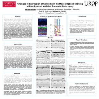
Figure 2. At one week following blast without drugs, there is still a slight increase
in calbindin levels in the blasted mice (asterisk, B) in comparison to sham mice (A).
At two weeks (C,D) following blast without drugs, there are no clear differences in
levels of calbindin between the sham and blasted mice. Scale bar = 20 um.
Figure 1. In mice blasted without drugs, at 48 hours following blast exposure
calbindin expression in sham mice (A) was lower than calbindin expression in
animals exposed to blast (B). In contrast, there were high levels of calbindin
expression in both sham and blast exposed mice at 48 hours following blast
exposure in mice that were administered drugs (C and D). Scale bar = 20 µm.
Conclusions
• In the absence of drugs, there is increased expression of
calbindin in the retinas of blast exposed mice in comparison to
sham mice after a 48-hour recovery. This suggests that there may
be a limited window for prophylaxis
• The use of anesthetics increased the levels of calbindin in sham
mice to levels seen in the blast exposed mice, suggesting that the
presence of anesthetics can confound results.
• In the absence of drugs, the levels of calbindin expression were
somewhat higher in blast mice versus sham mice following a one
week recovery period. This slight increase in calbindin in mice
blasted without drugs seen at one week was not apparent in mice
allowed to recover for two weeks.
Acknowledgements
I would love to thank everyone who has worked in this wonderful lab
including those who worked here last year. Namely: Andrea Foster,
Shama Patel, Bryan Duong, Eric Schmidt, Madeline Tenenbaum,
Danica Smith, Sara Mansuri, Biraaj Mahajan, and last but absolutely not
least Gloria DeWalt, Todd Blute, and of course William D. Eldred.
I would also like to thank the Boston University Biology Department and
UROP
References
1.Lee, B., Newberg, A.”Neuroimaging in Traumatic Brain Imaging.” NeuroRx. 2 (2005):
372-383.
2. Werner, C., and Engelhard, K. “Pathophysiology of traumatic brain injury.” British
Journal of Anesthesia. 99 (2007): 4-9.
3. Kuehn, R., Simard, P.F., Driscoll, I., Keledjian, K., Ivanova, S., Tosun, C., Williams, A.,
Bochicchio, Gerzanich, V., Simard, J.M. “Rodent Model of Direct Cranial Blast Injury.” 28
(2011) 10:2155-2169.
4. Sun DA1, Deshpande LS, Sombati S, Baranova A, Wilson MS, Hamm RJ, DeLorenzo
RJ. “Traumatic brain injury causes a long-lasting calcium (Ca2+)-plateau of elevated
intracellular Ca levels and altered Ca2+ homeostatic mechanisms in hippocampal
neurons surviving brain injury.” European Journal of neuroscience. 27 (2008) 7:1659-
1672.
5. Hoon, M., Okawa, H., Santina, L.D., Wong, R.O.L. “Functional Architecture of the
Retina: Development and Disease.” Progress in retinal and eye research 42 (2014): 44–
84.
Results
Traumatic brain injury is a growing public health concern due, in part, to the
rise in military conflicts. Although there are clear cognitive deficits following TBI,
few studies have examined the sensitivity of the retina to injury. As an easily
accessible extension of the central nervous system, we sought to determine
whether the retina could be used to indirectly evaluate calcium homeostasis, as
alterations in calcium levels can contribute to the pathology of TBI. Using a
previously validated blast-induced TBI model we evaluated the expression of
the calcium binding protein, calbindin, using immunocytochemistry in the
retinas of adult C57BL/6 mice 48 hours following blast exposure. Emerging
evidence indicates that anesthetics can affect the outcome of TBI, therefore we
examined mice blasted in the presence and absence of ketamine and xylazine.
Preliminary results showed increased calbindin expression in blast exposed
mice in the absence of drugs. No difference in calbindin expression was
observed in anesthetized animals indicating the use of anesthetics does affect
results. To determine whether the elevation in calbindin expression persisted
beyond 48 hours, we examined calbindin levels one and two weeks post blast
exposure in the absence of anesthetics. A slight increase in calbindin levels
was observed one week following blast exposure and no clear difference
following two weeks indicating the acute sensitivity of calbindin expression to
injury. Additional work examining the duration of acute vulnerability of calbindin
expression is needed.
Abstract Anatomy of the Mammalian Retina
Adapted from Hoon, et al. 2014
Karla Brandao, Gloria DeWalt, Madeline Tenenbaum, Dominique Thompson,
Todd A. Blute, and William D. Eldred
Department of Biology, Boston University, Boston, MA
Future Directions
• Evaluation of additional calcium binding proteins like calretinin and
parvalbumin to determine whether there are changes in expression
similar to calbindin.
• The results reported are based on preliminary studies following
exposure to a single blast. Additional work to determine whether
similar changes would result following more blast exposure are
necessary.
• Additional work exploring the biochemical and cellular changes
following blast-induced TBI could offer insight in therapeutics
aimed at mitigating cellular pathology that results from excitoxicity.
• Further research could lead to development of retinograms to
increase speed and accuracy of TBI diagnosis
OPL
INL
IPL
OPL
INL
IPL
GCL GCL
OPL
INL
IPL
GCL
OPL
INL
IPL
GCL
A B
C D
*
Sham-
Sham-
Blast -
Blast -