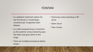
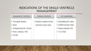
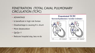
FONTAN PROCEDURE
DR JULIETH NACHONE KABIRIGI : PEDIATRIC CARDIOLOGIST 20TH FEBRUARY 2024
CATHOLIC UNIVERSITY OF HEALTH AND ALLIED SCIENCES,MWANZA, TANZANIA
BUGANDO MEDICAL CENTRE, MWANZA, TANZANIA
HIGHLIGHTS
• OVERVIEW OF FONTAN PROCEDURE
• CLINICAL SIGNIFICANCY
• INDICATIONS
• PRINCIPLES OF MANAGEMENT
• DRIVING FORCES
• FONTAN CIRCULATION
• COMPLICATIONS AND ITS MANAGEMENT
FONTAN
• Is a palliative treatment option for
the functional or morphologic
univentricular Congenital heart
disease
• Is a total cavopulmonary connection
as the systemic venous blood by pass
the heart and goes direct to the
lungs
• There are multiple procedures before
fontan
• Pulmonary artery banding or BT-
shunt
• Glenn shunt
• Then Fontan
CLINICAL SIGNIFICANCE
• Previously around 1940s , single
ventricle were considered fatal
• But currently there about 50,000-
70,000 people are living with Fontan
worldwide1
• Thirty year survive is > 80% at
30years
• 95% of free transplant survival at 5-
10years
• 1. AHA 2019
INDICATIONS OF THE SINGLE VENTRICLE
MANAGEMENT
EXAMPLES OF SV PHYSIOLOGY
SINGLE VEMTRICLE PHYSIOLOGY LESIONS
MORE EXAMPLES
PRINCIPLES OF MANAGEMENT OF SV
GLENN SHUNT
• Direct blood from Superior vena cava to
Right Pulmonary artery
• Overall survival is 90% post Glenn
• Driving forces
• 1.Gravitation force
• 2. 6-9mo as the baby can be sit without
support
• After some years if the baby has
desaturation continue to Fontan
• Because the lower body grows fast than the
upper body
CAUSES OF CYANOSIS AFTER GLENN
• Development of veno-venous circulation
• Is the most important factor by
development of collaterals on IVC and SVC
• RA is 5cmHg where SVC and SVC connected
with the same low pressure
• When SVC is connected to RPA after quiet
some time few years they dev cyanosis
• The collaterals develop from IVC to SVC
following the development of High pressure
in the connected PAs
• After 7years of Age ,the lower body develop
faster than the upper body hence more
blood from IVC than SVC as a result of more
deoxygenated blood to the Systemic
compare to SVC blood to PA
• Solution to severe cyanosis is to connect IVC
to PA and then is FONTAN,
• Sepateted circulation no mixing SO2 100%
• QP:QS=1
DRIVING FORCES
• Normal individual venous return to the
heart is depend on
• Negative intrathoracic pressure
• RV contractility and relaxation, such blood
from systemic veins
• Gravity
• Muscle pump
• That’s why we do Glenn at least from 6-9mo
when the baby is able to sit, to allow flow
from SVC to RPA by gravitation drive
• With time after 7years of age, these children
need to perform Fontan procedure because
of increase cyanosis after prolong exposure
of SVC to PAP which is 3x the SVC
• So all blood bypass the lungs and and go to
IVC, at this point we need FONTAN to direct
all of the systemic venous blood to PAs
PRINCIPLES OF FONTAN CIRCULATION
• Low PVR (is single and most important factor is to avoid PHTN
• Example Tricuspid atresia type IC,IIC, Unrestrictive VSD, Large PDA, Obstructed
TAPVR, Severe Aortic regurgitation,
• Single ventricle to systemic circulation
• Make sure that no obstruction to systemic outflow obstruction
• Good size PAs branches no obstruction
1. PROTECT PULMONARY CIRCULATION
• Pulmonary stenosis vulvular and subvulvular
• Restrictive VSD (PA from Hyperplastic ventricle)
• PA Banding
2.SYSTEMIC OUTFLOW OBSTRUCTION
• Aortic stenosis (valvular or subvulvular)
• Restrictive VSD (Aorta from hypoplastic
chamber)
• Coarctation/Hypoplastic arch
• QN? Do you wish to do PAB in this patient?
• Management: DAMUS KAYE
PROCEDURE+BT-Shunt
• WHY Can we connect ascending aorta to
the dominant Ventricle?
DAMUS KAYE STANSEL (DKS)
• For the favor of Coronaries, you cant
cut Ascending Aorta to the dominant
Ventricle because you want to
maintain coronary supply to the
ventricles
• DKS You connect the pulmonary root
to the aorta to maintain Aortic
circulation PLUS BTS
• Later on proceed to GLENN
HYPOPLASTIC LEFT HEART SYNDROME
• Severe MS/Mitral atresia
• Restrictive VSD
• Dominant RV
• PDA supply Ascending and
descending Aorta
• WHAT are factors are aligned with
our principles of management?
NORWOOD PROCEDURE
FONTAN PHYSIOLOGY
• No mixing except for fenestration
• Circulation in series
• Decrease volume overload
• Increase afterload
• Systemic venous congestion
FONTAN CIRCULATION
ESSENCE OF FONTAN CIRCULATION
• One functional single ventricle to systemic circulation
• Systemic venous return flow without pumping ventricle to the lungs
• No desaturated blood to the systemic circulation(SO2 is 100%)
• Qp;Qs=1
• Is a PALLIATIVE PROCEDURE
FONTAN CIRCULATION PARADOXICAL
• In normal circulation, Systemic venous pressure is 5-7mmHg
• Mean Pulmonary arterial pressure of at least 15mmHg in order the pulmonary
vascular network to be patent
• PARADOXICAL IN FONTAN is the existence of 3x normal of systemic venous
hypertension (15mmHg)
COMPONENTS FAVORING FONTAN
TEN COMMANDMENTS CURRENT ONE
• Low Pulmonary vascular resistance
(PVR)
• Normal systolic function
• Lack of AV Valve insufficiency
FACTORS ASSOCIATED WITH POOR OUTCOME
• Increase Meam PAP >15mmHg
• Increase PVR >2
• Systemic ventricular dysfunction
• AV valve regurgitation
HOW TO IMAGE FONTAN PATIENTS
• Know what type of Fontan repair done
• Read the surgical reports
• Anatomy of Fontan circulation
• IVC and SVC driving force is inspiration
• Tunnel (intra or extracardiac)
• Connection with PA
• PA Branches
• Pulmonary veins
FENETRATION (TOTAL CAVAL PULMONARY
CIRCULATION (TCPC)
• ADVANTAGE
• Is beneficial in high risk fontan
• Disadvantage is causing R-L shunt
• More desaturation
• Qp;Qs<1
• Reduces hospital stay, less re do
FOLLOW UP COMPLICATIONS
1.CARDIAC TYPE
Arrhythmia
2. EXTRACARDIAC TYPE
Thromboembolic
Lymphatic system PLE (13%)
Plastic bronchitis
LIVER FAILURE
• 1,ATRIAL ARRHYTHMIA 40%
• Is a life threatening complication from
sustained arrhythmia
• Tachycardia increase Pulmonary venous
atrial pressure, reducing Pulmonary blood
flow, reduce Cardiac output
• Pre load reduction, failure of fontan
• Rx Beta blocker Bisoprol, Amiodarone if is
recurrent
• EPS is under research
LYMPHATIC ABNOMALITIES
• Due to increase venous pressure,
decrease and impaired thoracic duct
drainage
• PLE 13% Loss of protein in vascular
bed
• Massive edema,
• Fat malabsorption
• Hypocalcemia
• Hypomagnesemia
• Immunodeficiency
• TREATMENT
• Low fat diet
• Low salt
• High calories
• Diuretics
• Fenestration and HEART
TRANSPLANT
FONTAN AND LIVER PROBLEM
• Hepatocellular carcinoma
• Is from chronic elevation of hepatic
venous pressure lead into chronic
liver pathology
• The Guideline recommend the
annual screening for Hepatic
adenoma
• Liver imaging
• Alfa fetal protein
• THANKS

JULIETH KABIRIGI,PAED.CARDIOLOGIST-CUHAS

  • 1.
    FONTAN PROCEDURE DR JULIETHNACHONE KABIRIGI : PEDIATRIC CARDIOLOGIST 20TH FEBRUARY 2024 CATHOLIC UNIVERSITY OF HEALTH AND ALLIED SCIENCES,MWANZA, TANZANIA BUGANDO MEDICAL CENTRE, MWANZA, TANZANIA
  • 2.
    HIGHLIGHTS • OVERVIEW OFFONTAN PROCEDURE • CLINICAL SIGNIFICANCY • INDICATIONS • PRINCIPLES OF MANAGEMENT • DRIVING FORCES • FONTAN CIRCULATION • COMPLICATIONS AND ITS MANAGEMENT
  • 3.
    FONTAN • Is apalliative treatment option for the functional or morphologic univentricular Congenital heart disease • Is a total cavopulmonary connection as the systemic venous blood by pass the heart and goes direct to the lungs • There are multiple procedures before fontan • Pulmonary artery banding or BT- shunt • Glenn shunt • Then Fontan
  • 4.
    CLINICAL SIGNIFICANCE • Previouslyaround 1940s , single ventricle were considered fatal • But currently there about 50,000- 70,000 people are living with Fontan worldwide1 • Thirty year survive is > 80% at 30years • 95% of free transplant survival at 5- 10years • 1. AHA 2019
  • 5.
    INDICATIONS OF THESINGLE VENTRICLE MANAGEMENT
  • 6.
    EXAMPLES OF SVPHYSIOLOGY
  • 7.
  • 8.
  • 9.
  • 10.
    GLENN SHUNT • Directblood from Superior vena cava to Right Pulmonary artery • Overall survival is 90% post Glenn • Driving forces • 1.Gravitation force • 2. 6-9mo as the baby can be sit without support • After some years if the baby has desaturation continue to Fontan • Because the lower body grows fast than the upper body
  • 11.
    CAUSES OF CYANOSISAFTER GLENN • Development of veno-venous circulation • Is the most important factor by development of collaterals on IVC and SVC • RA is 5cmHg where SVC and SVC connected with the same low pressure • When SVC is connected to RPA after quiet some time few years they dev cyanosis • The collaterals develop from IVC to SVC following the development of High pressure in the connected PAs • After 7years of Age ,the lower body develop faster than the upper body hence more blood from IVC than SVC as a result of more deoxygenated blood to the Systemic compare to SVC blood to PA • Solution to severe cyanosis is to connect IVC to PA and then is FONTAN, • Sepateted circulation no mixing SO2 100% • QP:QS=1
  • 12.
    DRIVING FORCES • Normalindividual venous return to the heart is depend on • Negative intrathoracic pressure • RV contractility and relaxation, such blood from systemic veins • Gravity • Muscle pump • That’s why we do Glenn at least from 6-9mo when the baby is able to sit, to allow flow from SVC to RPA by gravitation drive • With time after 7years of age, these children need to perform Fontan procedure because of increase cyanosis after prolong exposure of SVC to PAP which is 3x the SVC • So all blood bypass the lungs and and go to IVC, at this point we need FONTAN to direct all of the systemic venous blood to PAs
  • 13.
    PRINCIPLES OF FONTANCIRCULATION • Low PVR (is single and most important factor is to avoid PHTN • Example Tricuspid atresia type IC,IIC, Unrestrictive VSD, Large PDA, Obstructed TAPVR, Severe Aortic regurgitation, • Single ventricle to systemic circulation • Make sure that no obstruction to systemic outflow obstruction • Good size PAs branches no obstruction
  • 14.
    1. PROTECT PULMONARYCIRCULATION • Pulmonary stenosis vulvular and subvulvular • Restrictive VSD (PA from Hyperplastic ventricle) • PA Banding
  • 15.
    2.SYSTEMIC OUTFLOW OBSTRUCTION •Aortic stenosis (valvular or subvulvular) • Restrictive VSD (Aorta from hypoplastic chamber) • Coarctation/Hypoplastic arch • QN? Do you wish to do PAB in this patient? • Management: DAMUS KAYE PROCEDURE+BT-Shunt • WHY Can we connect ascending aorta to the dominant Ventricle?
  • 16.
    DAMUS KAYE STANSEL(DKS) • For the favor of Coronaries, you cant cut Ascending Aorta to the dominant Ventricle because you want to maintain coronary supply to the ventricles • DKS You connect the pulmonary root to the aorta to maintain Aortic circulation PLUS BTS • Later on proceed to GLENN
  • 17.
    HYPOPLASTIC LEFT HEARTSYNDROME • Severe MS/Mitral atresia • Restrictive VSD • Dominant RV • PDA supply Ascending and descending Aorta • WHAT are factors are aligned with our principles of management?
  • 18.
  • 19.
    FONTAN PHYSIOLOGY • Nomixing except for fenestration • Circulation in series • Decrease volume overload • Increase afterload • Systemic venous congestion
  • 20.
  • 21.
    ESSENCE OF FONTANCIRCULATION • One functional single ventricle to systemic circulation • Systemic venous return flow without pumping ventricle to the lungs • No desaturated blood to the systemic circulation(SO2 is 100%) • Qp;Qs=1 • Is a PALLIATIVE PROCEDURE
  • 22.
    FONTAN CIRCULATION PARADOXICAL •In normal circulation, Systemic venous pressure is 5-7mmHg • Mean Pulmonary arterial pressure of at least 15mmHg in order the pulmonary vascular network to be patent • PARADOXICAL IN FONTAN is the existence of 3x normal of systemic venous hypertension (15mmHg)
  • 23.
    COMPONENTS FAVORING FONTAN TENCOMMANDMENTS CURRENT ONE • Low Pulmonary vascular resistance (PVR) • Normal systolic function • Lack of AV Valve insufficiency
  • 24.
    FACTORS ASSOCIATED WITHPOOR OUTCOME • Increase Meam PAP >15mmHg • Increase PVR >2 • Systemic ventricular dysfunction • AV valve regurgitation
  • 25.
    HOW TO IMAGEFONTAN PATIENTS • Know what type of Fontan repair done • Read the surgical reports • Anatomy of Fontan circulation • IVC and SVC driving force is inspiration • Tunnel (intra or extracardiac) • Connection with PA • PA Branches • Pulmonary veins
  • 26.
    FENETRATION (TOTAL CAVALPULMONARY CIRCULATION (TCPC) • ADVANTAGE • Is beneficial in high risk fontan • Disadvantage is causing R-L shunt • More desaturation • Qp;Qs<1 • Reduces hospital stay, less re do
  • 27.
    FOLLOW UP COMPLICATIONS 1.CARDIACTYPE Arrhythmia 2. EXTRACARDIAC TYPE Thromboembolic Lymphatic system PLE (13%) Plastic bronchitis LIVER FAILURE • 1,ATRIAL ARRHYTHMIA 40% • Is a life threatening complication from sustained arrhythmia • Tachycardia increase Pulmonary venous atrial pressure, reducing Pulmonary blood flow, reduce Cardiac output • Pre load reduction, failure of fontan • Rx Beta blocker Bisoprol, Amiodarone if is recurrent • EPS is under research
  • 28.
    LYMPHATIC ABNOMALITIES • Dueto increase venous pressure, decrease and impaired thoracic duct drainage • PLE 13% Loss of protein in vascular bed • Massive edema, • Fat malabsorption • Hypocalcemia • Hypomagnesemia • Immunodeficiency • TREATMENT • Low fat diet • Low salt • High calories • Diuretics • Fenestration and HEART TRANSPLANT
  • 29.
    FONTAN AND LIVERPROBLEM • Hepatocellular carcinoma • Is from chronic elevation of hepatic venous pressure lead into chronic liver pathology • The Guideline recommend the annual screening for Hepatic adenoma • Liver imaging • Alfa fetal protein
  • 30.

Editor's Notes

  • #5 This is basically depends on advances in surgical techniques Pre operative care Better patient selection But regardless of all these 50% of survivals have adverse events before reaching adulthood
  • #18 QN? Systemic outflow obstruction, LOCS, PAP,Pulmonar venous HTN,Restrictive VSD,Circulation is not align with Systemic circulation
  • #22 Bacause all desaturated blood goes to the lungs, QP:QS is 100%
  • #28 Thrombosis us MRI, use WARFARIN, HOWEVER LARGE THROMBUS IS FATAL REGARDLESS OF INTERVENTION, may nee EMBOLOCTOMY