Etiologia de la celulitis y Predicción clínica de la enfermedad Estreptocócic...Alex Castañeda-Sabogal
Etiologia de la celulitis. Estudio prospectivo y predicción clínica de la infeccion por Estreptococcus basado en la frecuencia encontrada de las especies de estreptococo
Etiologia de la celulitis y Predicción clínica de la enfermedad Estreptocócic...Alex Castañeda-Sabogal
Etiologia de la celulitis. Estudio prospectivo y predicción clínica de la infeccion por Estreptococcus basado en la frecuencia encontrada de las especies de estreptococo
Prevalence of Cryptosporidiosis Among Selected Group of Sudanese Patients by Mosab Nouraldein Mohammed Hamad in Advancements in Bioequivalence & Bioavailability
Kathryn Maitland describes the challenges faced with oxygen therapy as an emergency intervention in critical illness in African children.
Where Kathryn works, in East Africa, there is no access to intensive care. Caring for critically ill children is all done in the Emergency Department.
70% of the global burden of disease and deaths from pneumonia occurs in Southeast Asia and Sub-Saharan Africa. The WHO has published guidelines as to what classifies as pneumonia, severe pneumonia, and very severe pneumonia.
These classifications rely on clinical signs. However, Kathryn in her research has discovered that these classifications are rarely correlated with the actual underlying disease process.
Clinical signs are non-specific for the diagnosis of pneumonia. Oxygen is recommended for severe and very severe pneumonia.
This has led to calls to prioritise oxygen delivery in African hospitals. However, it has not led to change from a health department or funding viewpoint.
There are also oxygen delivery practicalities to consider. Often there is only one source of oxygen on a ward (if at all) with patients clustered around it.
The production of Oxygen may only happen in a few places.
Poor cylinder quality leads to leaks and therefore, low supply.
Concentrators are useful however they need regular servicing. They also rely on power, and in a region that experiences regular power outages, this can be problematic. When the power goes off, there is no oxygen available.
Kathryn asks – do all children actually need oxygen? There is still however a hidden burden of hypoxia.
Outside of Africa, Kathryn discusses the current state of equipoise on oxygen therapy.
Moreover, oxygen can be harmful if given inappropriately. This leads to concerns more broadly on the harms of oxygen therapy.
Kathryn concludes her talk by looking to the future. She discusses ongoing research and the implications for future practice in resource poor settings, and indeed the world.
The International Journal of Engineering and Science (The IJES)theijes
The International Journal of Engineering & Science is aimed at providing a platform for researchers, engineers, scientists, or educators to publish their original research results, to exchange new ideas, to disseminate information in innovative designs, engineering experiences and technological skills. It is also the Journal's objective to promote engineering and technology education. All papers submitted to the Journal will be blind peer-reviewed. Only original articles will be published.
The papers for publication in The International Journal of Engineering& Science are selected through rigorous peer reviews to ensure originality, timeliness, relevance, and readability.
The Significance of Bacterial and Fungal Coinfection in the Setting of Viral ...Texas Children's Hospital
Keystone ECMO meeting 2018: To better characterize the frequency of bacterial and/or fungal coinfections in patients with viral pneumonias placed on ECMO and to understand their impact on mortality.
Newer diagnostic methods in tuberculosis detectionApollo Hospitals
One-third of the world's population has been infected with Mycobacterium tuberculosis, with new infections occurring in about 1% of the population each year. However 90–95% of infections remain asymptomatic. Thus early diagnosis of tuberculosis and drug resistance improves survival and helps to promote contact tracing, implementation of institutional cross-infection procedures, and other public-health actions. There have been many advances and modifications to the methodology for tuberculosis diagnosis some of which are very promising. But these advances have not kept pace with the explosion of tuberculosis or the outbreak of drug resistant tuberculosis. This review describes some of the newer advances in tuberculosis diagnostics and the challenges they face.
Implementation and evaluation of anursing assessmentstandin.docxwilcockiris
Implementation and evaluation of a
nursing assessment/standing orders–
based inpatient pneumococcal
vaccination program
Carl Eckrode, MPH, RRT-NPS,b Nancy Church, RN, MT,a and Woodruff J. English III, MDa
Portland and Gresham, Oregon
Background: Pneumococcal vaccination is recommended for patients aged 65 years and greater; inpatient vaccination has been
suggested as means to increase vaccination rates is this population. Our hospital implemented an inpatient pneumococcal vacci-
nation program, and expanded the population of interest to include patients aged 2 to 64 years with risk factors for pneumococcal
bacteremia. We studied the outcomes of this program to determine if the rate of pneumococcal vaccination opportunities and
pneumococcal vaccination rate could be significantly increased through the application of an in-hospital pneumococcal vaccina-
tion program, based on standing orders and assessment by Registered Nurses, when compared to our previous method of physi-
cian assessment and written vaccination order for each patient.
Methods: Subjects were inpatients admitted to non-intensive care units of our hospital from August to December of 2004. Cases
were aged greater than 65 years, or were greater than 2 years of age with selected risk factors. Patients with previous pneumococcal
vaccination with the past five years, in terminal or comfort care, those allergic to vaccine components, patients who received organ
or bone marrow transplants in the year prior to the study, and those physicians barred them from the vaccination protocol were
excluded. Program effectiveness was evaluated through retrospective evaluation of medical records to determine if subjects had
been evaluated for vaccination eligibility, and if subjects were eligible, whether or not they had received pneumococcal vaccination.
Results: Overall vaccination opportunity rate after implementation of the standing orders-based program increased form 8.6% to
59.1%, and overall vaccination rates improved form 0% to 15.4%. The study found a statistically significant difference in the rate
of pneumococcal vaccination opportunities (x2 = 182.46, p = .00) and the pneumococcal vaccination rate (x2 = 56, p = .00)
between the two methods of assessment and vaccination; these results are attributable to the study intervention.
Conclusions: The study program contributed to increased overall vaccination opportunity and vaccination rates, when compared
to the previous method. The overall rates of vaccination attained by this program were often lower than those reported in the ex-
isting literature for other program designs; however, this may be due to an unusually high rate of vaccination refusal. (Am J Infect
Control 2007;35:508-15.)
The significance of invasive pneumococcal disease
cannot be understated, because disease caused by
Streptococcus pneumoniae has been reported to be
responsible for an estimated 36% of community-
acquired pneumonia, an estimated 50% of nosocomial
pneumonias,.
What next for prevention of pneumococcal disease in light of serotype replacement? Is there a pathway to licensure for novel pneumococcal vaccines?
https://www.meningitis.org/mrf-conference-2017
A randomised, double-blind clinical trial was undertaken in order to assess the effectiveness of probiotics in
the prevention of necrotising enterocolitis (NEC) in newborns weighing <1500 g.
Like personalized medicine, personalized vaccinology aims to provide the right vaccine, to the right patient, at the right time, to achieve protection from disease, while being safe (i.e., free from unintended side effects). Starting with these lines, this presentation will provide overall information related to the vaccinomoic along with the suitable examples and thus will be helpful for the students to understand the basics related to the same.
2. serotype, whilst only eight methods had 70% sensitivity to detect minor serotypes. For the
field samples, the overall sensitivity ranged from 74.2% to 95.8% (reference method
93.8%), and PPV from 82.2% to 96.4% (reference method 99.6%). The microarray had the
highest sensitivity (95.8%) and high PPV (93.7%). The major limitation of this study is that
not all of the available alternative serotyping methods were included.
Conclusions
Most methods were able to detect the dominant serotype in a sample, but many performed
poorly in detecting the minor serotype populations. Microarray with a culture amplification
step was the top-performing method. Results from this comprehensive evaluation will inform
future vaccine evaluation and impact studies, particularly in low-income settings, where
pneumococcal disease burden remains high.
Introduction
Streptococcus pneumoniae (the pneumococcus) is a dominant cause of childhood illness and
death worldwide. It has been estimated to cause ~800,000 deaths in children aged under 5 y
annually, most due to pneumonia and most in low-income countries [1]. Pneumococci com-
monly colonise the nasopharynx of healthy children. Although carriage is usually asymptom-
atic, it can cause local inflammation and is considered a prerequisite for pneumococcal disease
[2]. Pneumococci are diverse, with 97 serotypes identified to date. Understanding carriage is
important for understanding pneumococcal population biology and transmission, assessing
vaccine impact, and evaluating the performance of new vaccines.
Pneumococcal conjugate vaccines (PCVs) are effective at preventing pneumonia and inva-
sive pneumococcal disease, and are currently being introduced in many resource-poor coun-
tries (e.g., by Gavi, the Vaccine Alliance). PCVs reduce nasopharyngeal carriage of the
pneumococcal serotypes they contain (vaccine types). Although transmission and disease due
to vaccine types is reduced, non-vaccine types fill the ecological niche, becoming more com-
mon in carriage and disease [3–5]. This phenomenon of serotype replacement, which may be
more pronounced in low-income settings (because of higher rates, load, and diversity of pneu-
mococcal carriage [6] and higher rates of disease), represents a significant risk to the global
pneumococcal immunisation strategy. It is difficult to monitor the serotypes causing pneumo-
nia or invasive disease because of the insensitivity of blood culture for detecting pneumococcal
pneumonia, inconsistencies in clinical practice (e.g., performing of lumbar punctures), and
limitations on diagnostic laboratory capacity, particularly in resource-poor settings [7–9]. Fur-
thermore, any improvement in laboratory or clinical diagnostic practices over time in vacci-
nated populations would lead to enhanced detection of non-vaccine types. This increase may
not reflect true replacement, and may cause unnecessary concerns about vaccine effectiveness
and inappropriate changes to vaccine policy.
Carriage studies offer a practical approach for monitoring serotype replacement and can
help in assessing the impact of PCVs and other pneumococcal vaccines [8,10]. Pneumococcal
carriage is an important endpoint for efficacy trials of new pneumococcal vaccines such as
common protein, combination (PCV + common protein), and whole-cell vaccines [11]. Tradi-
tional serotyping methods involve typing a small number of colonies, frequently missing car-
riage of multiple serotypes and providing no quantitative data. The gold-standard serotyping
Serotyping Methods for Pneumococcal Carriage Studies
PLOS Medicine | DOI:10.1371/journal.pmed.1001903 November 17, 2015 2 / 30
org), Grant 52099. This project was also supported
by the Murdoch Childrens Research Institute (www.
mcri.edu.au) and the Victorian Government's
Operational Infrastructure Support Program. The
funders had no role in the study design, data
collection and analysis, decision to publish, or
preparation of the manuscript; except for the specific
roles of DCHB, DH and KPK as described in the
author contributions. All authors have read, and
confirm that they meet, ICMJE criteria for authorship.
Competing Interests: I have read the journal's policy
and the authors of the manuscript have the following
competing interests: DCHB and DH are previous
employees of the Bill and Melinda Gates Foundation,
which provided funding for this project. KPK was
employed by the Bill and Melinda Gates Foundation
towards the completion of the project, but was not
involved in funding the project. KPK is a member of
the Editorial Board of PLOS Medicine. CS and EMD
were awarded the Robert Austrian Award which is
funded by Pfizer. Authors have served on advisory
boards for Merck (CS, KPK, EKM), GSK (KPK,
EKM), and Pfizer (KPK). Research group authors
have a scientific and/or commercial interest in the
performance of their methods, so blinded testing was
conducted.
Abbreviations: CDC, US Centers for Disease
Control and Prevention; CFU, colony-forming units;
ESI-MS, electrospray ionisation mass spectrometry;
HBA, horse blood agar; IQR, interquartile range; MFI,
mean fluorescence intensity; MLST, multilocus
sequence typing; mPCR, multiplex PCR; PBS,
phosphate-buffered saline; PCV, pneumococcal
conjugate vaccine; PPV, positive predictive value;
RFLP, restriction fragment length polymorphism;
RLB, reverse line blot; SSI, Statens Serum Institut;
STGG, skim milk-tryptone-glucose-glycerol; ULT,
ultra-low temperature.
3. method (the Quellung reaction) was developed in the early 1900s (see [12]) and is performed
by testing colonies with a set of antisera and visualising the bacteria with a microscope. It is
laborious and requires a complete set of type-specific antisera, and is therefore mainly per-
formed by reference laboratories. Alternate serotyping methods have been developed, but few
data formally comparing the performance of these methods to the gold-standard Quellung
reaction, or to each other, are available [13].
A pneumococcal serotyping method suitable for use in carriage studies should have high
sensitivity (including the ability to detect multiple serotypes), detect most or all serotypes, be
suitable to scale up for large projects, and be practical for resource-poor countries. We estab-
lished the PneuCarriage project, a large, international multi-centre study, with the aim of iden-
tifying the best pneumococcal serotyping methods for carriage studies.
We compared 20 different pneumococcal serotyping methods from 15 research groups
using 81 laboratory-prepared samples. We then tested the five top-performing methods using
260 nasopharyngeal samples collected from children in six high-burden low- and middle-
income countries. All samples were subjected to comprehensive conventional microbiology to
thoroughly characterise the pneumococcal content, and the results were used to assess the per-
formance of the new methods to identify the best pneumococcal serotyping methods. Sensitiv-
ity and positive predictive value (PPV) were selected as the key parameters for method
evaluation, as these measures best convey the overall ability of a method to correctly detect
pneumococcal serotypes present in a sample.
Methods
All research involving human participants was approved by the relevant institutional review
board or an equivalent committee, specifically the Bangladesh Institute of Child Health Ethics
Review Committee; the Fiji National Research Ethics Review Committee; the Gambian Gov-
ernment/Medical Research Council Laboratory Joint Ethics Committee and the Medical
Research Council (UK) Gambia Scientific Coordinating Committee; the Kenya Medical
Research Institute National Ethical Review Committee and Scientific Steering Committee; the
Government of Papua New Guinea Medical Research Advisory Committee and the Papua
New Guinea Institute of Medical Research Institutional Review Board; the Human Research
Ethics Committee of the University of the Witwatersrand, the Ethics Committee of Stellen-
bosch University, and the Medicine Control Council of South Africa; and the Clinical Science
Review Committee of the Division of AIDS, US National Institutes for Health. Written
informed consent was obtained from the parents/guardians of all participants.
Spiked Samples
Fifteen pneumococcal isolates were used to prepare 81 laboratory-prepared (“spiked”) samples
in skim milk-tryptone-glucose-glycerol (STGG) medium [14,15]. The 15 isolates used to con-
struct the spiked samples were derived from four geographical areas (Bangladesh, Fiji, South
Africa, and the United States). Isolates were selected following a review of the literature, and
the selection was informed by our international steering committee. The selection included
serotypes both common and rare in carriage and a mix of vaccine and non-vaccine types
(Table 1). Fourteen of the isolates were derived from the nasopharynx. The serotype 5 isolate
was sourced from invasive disease (site not known) because of its relative rarity in carriage. Iso-
lates were serotyped by latex agglutination and Quellung reaction. Quellung reactions were
independently confirmed by the Microbiological Diagnostic Unit, University of Melbourne,
Australia. Multilocus sequence typing (MLST) was performed as previously described [16]. All
isolates had a 80% survival (mean 96.8% [95% CI: 91.1%, 102.5%]) and were able to be
Serotyping Methods for Pneumococcal Carriage Studies
PLOS Medicine | DOI:10.1371/journal.pmed.1001903 November 17, 2015 3 / 30
4. serotyped by Quellung reaction and latex agglutination when cultured following a freeze–thaw
step. To create the spiked samples, fresh overnight pneumococcal cultures were recovered
from horse blood agar (HBA) plates, resuspended in physiological saline, and used to inoculate
5 ml of STGG medium. Once inoculated, tubes were immediately placed at ultra-low tempera-
ture (ULT) (−70°C) for 24 h. Spiked samples were then thawed, vortexed, and dispensed
into 60μl aliquots in 0.5ml screw-capped tubes. Aliquots were immediately frozen at ULT and
held at that temperature until use (24 h).
Viable counts were performed in saline to assess the concentration of the inocula, and the
associated load (and proportion) of each serotype in the spiked samples was determined. Sam-
ples containing one serotype were inoculated with low numbers of pneumococci (i.e., they had
loads similar to those of “minor” serotypes in samples with more than one serotype; see
below); however, the serotype contained within them represented 100% of the pneumococcal
content. As such, the viable count data from samples with one serotype were included when
calculating the mean load of the minor type, but excluded when calculating the mean percent
of the minor serotypes.
Spiked samples consisted of medium alone (n = 4) or contained one, two, three, or four
serotypes (n = 7, 38, 26, and 6, respectively). Samples inoculated with pneumococci contained
a mean of 2.4 serotypes (95% CI: 2.2, 2.6), with a mean load of 2.11 × 10⁵
colony-forming units
(CFU)/ml (95% CI: 1.68 × 10⁵
, 2.55 × 10⁵
). Samples inoculated with more than one serotype
contained a “major type” (the serotype with the highest percent abundance in each sample)
and one or more “minor types” (the other serotypes present in each sample). Major types ran-
ged from 67.78% to 98.71% (median 93.69%, interquartile range [IQR]: 88.67, 96.12) of the
total pneumococcal content in each sample, with a load ranging from 3.08 × 10⁴
to 5.70 × 10⁵
CFU/ml (median 1.56 × 10⁵
, IQR: 5.73 × 10⁴
, 3.28 × 10⁵
). The minor types ranged from 1.29%
to 14.49% (median 4.96%, IQR: 3.05, 6.92) of the total pneumococcal content in each sample,
Table 1. Isolates used to create spiked samples.
Isolate Site of
Isolation
Country of
Origin1
Serotype by Quellung
Reaction
Multilocus Sequence
Type (ST)
Year of Sample
Collection
Child Age at Sample
Collection (in Months)
PMP812 Invasive2
Bangladesh 5 289 2006 3
PMP825 Nasopharynx Bangladesh 12F 10232 2005 3
PMP817 Nasopharynx Bangladesh 15A 6332 2006 3
PMP818 Nasopharynx Bangladesh 20 5392 2006 3
PMP492 Nasopharynx Fiji 6B 4781 2007 17
PMP847 Nasopharynx Fiji 8 404 2007 17
PMP284 Nasopharynx Fiji 29 9987 2006 18
PMP241 Nasopharynx South Africa 4 5410 2005 9
PMP228 Nasopharynx South Africa 6A 1447 2007 7
PMP221 Nasopharynx South Africa 23F 242 2007 7
PMP219 Nasopharynx South Africa 14 10231 2007 7
PMP849 Nasopharynx United States 1 227 2007 363
PMP846 Nasopharynx United States 9V 1269 2007 7
PMP843 Nasopharynx United States 19F 3040 2006 66
PMP845 Nasopharynx United States 38 10230 2006 94
1
Isolates were kindly provided by Prof. Samir Saha (Bangladesh), Assoc. Prof Fiona Russell (Fiji), Dr. Peter Adrian and Prof. Shabir Madhi (South Africa),
and Prof. Kate O’Brien (United States).
2
Site of isolation not known.
doi:10.1371/journal.pmed.1001903.t001
Serotyping Methods for Pneumococcal Carriage Studies
PLOS Medicine | DOI:10.1371/journal.pmed.1001903 November 17, 2015 4 / 30
5. with the load ranging from 6.95 × 10²
to 3.89 × 10⁴
CFU/ml (median 8.19 × 10³
, IQR:
2.95 × 10³
, 1.85 × 10⁴
).
Each set of spiked samples contained randomly selected aliquots shipped frozen on dry ice
to the research groups. One set was tested by conventional serotyping (the reference method,
see below) in the project laboratory at the Murdoch Childrens Research Institute.
Field Samples
The 260 nasopharyngeal (“field”) samples were from 260 children aged 24 mo in six low-
and middle-income countries (Table 2). Samples were collected and stored at ULT using proto-
cols consistent with WHO guidelines [14,17]. The frozen samples were shipped on dry ice to
the project laboratory in Melbourne. Samples were dispensed into aliquots and stored as for
the spiked samples.
Reference Serotyping Method
Conventional methods were used to identify and serotype the pneumococci carried in the sam-
ples. Sample aliquots were thawed and mixed, and 50 μl was used to conduct a viable count by
serial dilution and plating on selective medium (HBA plates containing 5 μg/ml gentamicin).
After 36–44 h of incubation at 35–37°C in 5% CO2, up to 120 well-separated alpha-haemolytic
colonies were randomly selected. To do this, the culture plate was evenly divided into eight sec-
tions, and a section was chosen from a previously prepared random list. The outermost colony
in the designated sector was then picked and designated the “first” colony. The remaining colo-
nies on the whole plate were then picked. If not all of these colonies were required, sections were
prioritised from a list of randomly generated numbers. If not all the colonies in a particular sec-
tion were required, the operator selected colonies from the outer edge of the plate working
towards the centre. Throughout the random colony selection process, only well-separated
alpha-haemolytic colonies were chosen. An example of each morphological variant was also
subcultured if not chosen during random selection. Selected colonies were then subcultured
onto HBA plates. Subcultured colonies underwent pneumococcal identification using optochin,
and then bile and Phadebact Pneumococcus (MKL Diagnostics) tests, as appropriate. Pneumo-
coccal isolates were serotyped by Quellung reaction and/or latex agglutination (see below).
Latex agglutination was conducted using the Denka Seiken kit [23] according to the manufac-
turer’s instructions [24], except that we used 15 μl of reagent for each test. If all reactions were
negative, testing was repeated using 30 μl. For the 72 tests not included in the Denka Seiken kit
(seven types [13, 37, 42, 43, 44, 45, and 48] and 65 factors), we used latex reagents prepared in
house [25,26] using antisera from Statens Serum Institut (SSI) Diagnostica. A negative control
reagent was prepared using normal rabbit serum (Antibodies Australia). Pneumococcal isolates
were serotyped by Quellung reaction [27,28] using polyclonal antisera from the SSI.
The primary colony (i.e., the first randomly selected colony for which a serotype was
obtained) was serotyped with both the Quellung reaction and latex agglutination. Subsequent
colonies were serotyped with latex agglutination, using the minimum number of reactions
needed to confirm whether the serotype was the same. Any different serotypes detected were
fully serotyped by latex agglutination and Quellung reaction. Laboratory staff were fully
blinded during sample processing and participated in an external quality assurance program
during the course of the project (RCPA Quality Assurance Program; http://www.rcpaqap.com.
au).
When used to examine spiked samples, the reference method had no false positives and
four false negatives (serotypes 14 and 23F were not detected once, and 6B not detected twice),
all occurring when the target was present as a minor serotype.
Serotyping Methods for Pneumococcal Carriage Studies
PLOS Medicine | DOI:10.1371/journal.pmed.1001903 November 17, 2015 5 / 30
6. Alternate Serotyping Methods—Overview
To identify serotyping methods for testing, we conducted literature reviews and contacted
pneumococcal researchers, gave presentations at international meetings to explain the project,
and circulated a project fact sheet. We identified and contacted 29 research groups. Fifteen
research groups joined the study. Of the remaining 14 groups, 11 had methods that were no
longer in use, had been superseded by other approaches, or were considered by the investiga-
tors to be unsuitable for detecting multiple serotype carriage. Three research groups were
unable to participate because of funding constraints, existing work commitments, or an inabil-
ity to reach agreement on the terms of the material transfer agreement (each n = 1).
Fifteen research groups participated in this study, testing 20 different methods, which were
numbered on enrolment into the study (Table 3). Five groups tested both direct (i.e., testing
from sample aliquot) and culture-based (i.e., with an initial broth- or agar-based amplification
step) versions of their method. All 20 methods were tested using the spiked sample aliquots.
Frozen samples were sent to each research group for testing. Researchers used up to 50 μl and
were blinded during sample testing. Following analysis of the spiked sample results (see
Table 2. Field sample information.
Field Site Number
of Swabs
Type of
Study, Year
of Collection
Age of
Children (in
Months)
Vaccination Status of
Children
Other Information Type of
Swab Tip
Reference, if
Applicable
Bangladesh 39 This study,
2011
7–24 Unvaccinated Cotton
Fiji 5 Carriage
study, 2003–
2004
8–19 Unvaccinated Cotton
Fiji 48 Vaccine trial,
2006–2007
8–19 Not specified1
Cotton [18]
The Gambia 42 This study,
2010
8–24 Unvaccinated (n = 14) or
had previously received 1
(n = 11), 2 (n = 9), or 3
(n = 8) doses of PCV7
Alginate/
calcium
alginate
Kenya 20 This study,
2008
7–13 Unvaccinated Rayon
Kenya 8 Carriage
study, 2003–
2006
0–24 Unvaccinated Rayon [19]
Kenya 5 Carriage
study, 2007–
2008
12–22 Unvaccinated Rayon [20]
Kenya 2 Carriage
study, 2000
3–5 Unvaccinated Rayon [21]
Papua New
Guinea
47 This study,
2010
6–24 Unvaccinated Children had presented for
routine vaccinations or as
outpatients at Goroka Hospital;
all had cough and most had
other respiratory symptoms such
as breathing difficulties, runny
nose, or eye discharge
Cotton
South Africa 44 Vaccine trial,
2006
7–17 Vaccinated with three or
four doses of PCV7
Fourteen of the children were
HIV positive
Dacron
polyester
[22]
All swabs were nasopharyngeal, except for those from Papua New Guinea, which were pernasal.
1
Vaccination status was known to original investigators, but was not provided as part of this study.
doi:10.1371/journal.pmed.1001903.t002
Serotyping Methods for Pneumococcal Carriage Studies
PLOS Medicine | DOI:10.1371/journal.pmed.1001903 November 17, 2015 6 / 30
7. Table 3. Key characteristics of alternate pneumococcal serotyping methods.
Type of
Method
Method
Number1
Direct
Detection or
Culture
Amplification
Method Technology
and Description
Number of Serotypes
Detected
Level of
Quantification
Other Analyses Reference
Total Individually As Part
of a
Group2
Genotypic
1 Direct detection mPCR 58 35 23 Semi-
quantitative
None [29]
11 Culture
amplification
mPCR 41 32 9 Qualitative None [30]
16 Culture
amplification
mPCR 54 22 32 Qualitative None [30–32]
5 Direct detection mPCR/reverse line
blot hybridisation
68 33 35 Qualitative None [33–36]
10 Culture
amplification
Restriction fragment
length polymorphism
of plyNCR (non-
coding region),
followed by mPCR
and Quellung
serotyping
32 27 5 Qualitative Presence of co-
colonisation
[30,37]
19 Direct detection PCR detected by
electrospray ionisation
mass spectrometry
59 26 33 Semi-
quantitative
MLST results [38,39]
22 Culture
amplification
As for method 19 59 26 33 Semi-
quantitative
MLST results [38,39]
6 Culture
amplification
mPCR and microarray 42 12 30 Qualitative None [40]
15 Direct
detection3
Microarray 93 53 403
Quantitative
(relative
abundance)
Detection of
antibiotic
resistance genes,
determination of
genetic
relatedness4
[13,41]
4 Culture
amplification3
As for method 15 93 53 403
Quantitative
(relative
abundance)
Detection of
antibiotic
resistance genes,
determination of
genetic
relatedness4
[13,41]
7 Direct detection Multiplex real-time
PCR
32 28 4 Semi-
quantitative
None [42]
20 Culture
amplification
As for method 7 32 28 4 Semi-
quantitative
None [42]
14 Direct detection Real-time PCR5
47 20 27 Semi-
quantitative
None [43]
21 Culture
amplification
As for method 145
47 20 27 Semi-
quantitative
None [43]
12 Direct detection Sequetyping, single
PCR, and sequencing
30 26 4 Qualitative None [44]
13 Culture
amplification
As for method 12 30 26 4 Qualitative None [44]
Phenotypic
9 Culture
amplification
Latex broth, latex
agglutination from
broth culture
72 8 64 Qualitative None [45]
(Continued)
Serotyping Methods for Pneumococcal Carriage Studies
PLOS Medicine | DOI:10.1371/journal.pmed.1001903 November 17, 2015 7 / 30
8. below), five methods were selected to test the field sample aliquots and were sent aliquots for
blinded testing as described above.
Alternate Serotyping Methods—Detailed Methodologies
All incubations were carried out at 35–37°C in ~5% CO2 unless otherwise indicated.
Method 1 (direct multiplex PCR). A subset of 22 spiked samples was processed with this
method. Initially, 25 μl of sample was spun in a microcentrifuge at 5,220g for 2 min. The super-
natant was discarded, 200 μl of phosphate-buffered saline (PBS) added, and the pellet resus-
pended. DNA was extracted using the QuickGene DNA tissue kit S with the QuickGene Mini
80 apparatus (Fuji Film Corporation) and eluted in 200 μl of buffer CDT. Then, 10 μl of
extracted DNA was used as a template for multiplex PCR (mPCR) reactions. Each reaction was
performed in a total volume of 50 μl using AmpliTaq Gold DNA Polymerase with GeneAmp
(Applied Biosystems), and amplified in a Veriti Thermo Cycler (Applied Biosystems) for 40
cycles. PCR products were detected using a Beckman CEQ 8000 genetic analyser system. Prim-
ers were labelled for recognition of the product in the CEQ 8000 analyser as green or blue
peaks and by size (141 to 515 base pairs). LytA PCR was used to detect pneumococcal DNA in
the sample as previously described [29], except that primers were labelled and products were
detected using the CEQ 8000. Samples were tested in four multiplex assays: (1) five primer
pairs to detect five serotypes, (2) 12 primer pairs to detect 13 serotypes, (3) five primer pairs to
detect 23 serotypes, and (4) five primer pairs to detect 31 serotypes. When assays performed
with primer pairs that detect more than one serotype were positive, additional PCRs using spe-
cific primers for the serotypes in the group were conducted.
Method 11 (culture mPCR). In this method, 50 μl of sample was plated onto a selective
blood agar plate (containing 5% sheep blood and 5 μg/ml gentamicin) and incubated over-
night. DNA was extracted from the entire culture plate using a DNeasy Blood Tissue Kit
(Qiagen). Approximately 500 ng of extracted DNA was then used for each of seven subsequent
mPCR reactions [30] using Taq polymerase produced in house. If the DNA was positive for
Table 3. (Continued)
Type of
Method
Method
Number1
Direct
Detection or
Culture
Amplification
Method Technology
and Description
Number of Serotypes
Detected
Level of
Quantification
Other Analyses Reference
Total Individually As Part
of a
Group2
18 Culture
amplification
Latex sweep, latex
agglutination from a
sweep of colonies
91 89 2 Qualitative None [46]
8 Direct detection Multiplex
immunoassay with
heat-kill step
23 23 0 Qualitative None [47]
17 Direct detection Antigen capture assay 16 12 4 Semi-
quantitative
None [48]
1
Methods 2 (a variant of method 10) and 3 were assigned a number but did not participate in the study and are not included in the table.
2
Some methods detect closely related serotypes as part of a group rather than individually (e.g., 18B/C versus 18B or 18C).
3
Methods 4 and 15 can also be analysed to the level of the individual call for 93 serotypes (see Methods).
4
Also detects a subset of other bacterial species.
5
The method was updated prior to testing of field samples to detect 58 total serotypes (18 individually and 40 as part of a group).
mPCR, multiplex PCR.
doi:10.1371/journal.pmed.1001903.t003
Serotyping Methods for Pneumococcal Carriage Studies
PLOS Medicine | DOI:10.1371/journal.pmed.1001903 November 17, 2015 8 / 30
9. cpsA, the cps primers were omitted and the reaction repeated to avoid competition of these
primers with primers for minor serotypes that may be present. A similar approach was taken
for any positive serotyping reactions.
Method 16 (culture mPCR). Initially, 50 μl of sample was plated onto Columbia III agar
with 5% sheep blood (BD) and incubated overnight. DNA was then extracted from all the plate
growth using a DNeasy Blood Tissue Kit (Qiagen). All seven mPCR reactions were then
applied to each sample, using 5 ng of DNA as template. To detect the minor serotypes, the
seven mPCR reactions were repeated, using 100 ng of DNA. PCR reactions and primer
sequences were as previously described [30–32].
Method 5 (direct mPCR/reverse line blot). DNA was extracted from 50 μl of sample
using the NucliSENS easyMAG total nucleic acid extractor kit (bioMérieux) or the GenElute
Mammalian Genomic DNA Miniprep Kit (Sigma-Aldrich). DNA was eluted in 110 μl, and
10 μl used for each subsequent test. Initially, lytA PCR [49] was performed to screen for the
presence of pneumococci. LytA-positive samples were then tested by mPCR/reverse line blot
(RLB) assay to identify serotypes, as previously described [33,34]. All positive samples were
tested with both membranes. If serogroup 6 was detected, serotype-specific PCR [35] was con-
ducted. When isolates were available, the Quellung reaction was performed to distinguish
closely related serotypes. Samples that were positive for lytA, but non-typeable by mPCR/RLB,
were tested by cpsA-B PCR and sequencing [36].
Method 10 (culture restriction fragment length polymorphism). In this method, 50 μl
of sample was streaked onto a Columbia sheep blood agar plate and incubated overnight. DNA
was extracted from a loopful of the bacterial lawn using the QIAamp DNA Mini Kit (Qiagen).
The entire lawn was then harvested from the primary culture agar plate and stored at −80°C.
PCR amplification of plyNCR was performed using 31.4 μl of extracted DNA. To identify co-
colonisation, restriction fragment length polymorphism (RFLP) analysis of the plyNCR PCR
amplicon was performed on the extracted DNA, as described previously [37]. Latex agglutina-
tion or mPCR was performed on aliquots that had a single or multiple serotypes detected by
RFLP, respectively. mPCR was conducted using 31.4 μl of extracted DNA, similarly to Pai et al.
[30] except that PCR products were analysed with the Agilent bioanalyser.
Method 19 (direct PCR/electrospray ionisation mass spectrometry). In this method,
50 μl of sample was diluted 1:4, lysed by bead beating, and treated with proteinase K at 56°C
for 15 min, and DNA was extracted using the KingFisher DNA extraction instrument (Thermo
Scientific), as previously described [38]. Then, 5 μl of extracted DNA was used for each of eight
PCR reactions. The PCR primers targeted pneumococcal serotype and MLST genotyping
markers [39]. Following PCR amplification, a fully automated electrospray ionisation mass
spectrometry (ESI-MS) analysis was performed using the PLEX-ID PCR/ESI-MS research plat-
form. Base compositions were compared to a database derived from the sequences of known
organisms and to signatures from reference standards [39].
Method 22 (culture PCR/ESI-MS). In this method, 1 μl of sample was cultured on a
sheep blood agar plate and incubated for 48–72 h. Growth was then harvested using a needle,
and boiled for 15 min at 95°C. Twenty-nine of the 81 spiked samples tested did not grow even
when 20 μl was cultured, so DNA was directly extracted from ~30 μl of original sample by boil-
ing lysis. Extracted DNA was then subjected to PCR and ESI-MS as outlined for method 19.
Method 6 (culture mPCR and microarray). Initially, 50 μl of sample was cultured on a
fresh rabbit blood agar plate for 24 h. Growth was harvested for DNA extraction and analysis,
as previously described [40]. In brief, microarrays were created by spotting oligonucleotide
probes onto a glass slide using a SpotArray 7.2 (PerkinElmer). A two-step mPCR (to amplify
the gene of interest and then label the PCR products) was conducted in two batches each. The
resultant products were hybridised to a microarray slide for 16 h before being scanned
Serotyping Methods for Pneumococcal Carriage Studies
PLOS Medicine | DOI:10.1371/journal.pmed.1001903 November 17, 2015 9 / 30
10. (GenePix personal 4100A, Axon Instruments), and the signal intensity calculated with GenePix
Pro 6.0.
Method 4 (culture microarray). In this method, 45 μl of the neat sample, or of two
10-fold serial dilutions, was spread on selective agar plates (colistin sulphate, oxolinic acid,
blood agar; Oxoid) and incubated overnight. For the plate with the highest density of distinct
non-confluent colony growth, all colonies were scraped into 1 ml of sterile PBS. DNA extrac-
tions were performed using the QIAamp DNA Mini Kit (Qiagen), including lysis buffer (20
mM Tris/HCl, 2 mM EDTA, 1% v/v Triton, 20 mg/ml lysozyme) and RNase treatment as pre-
viously described [13]. DNA was eluted in 200 μl of H2O and quantified using a NanoDrop
spectrophotometer. Approximately 300 ng of the DNA was fluorescently labelled with either
ULS-Cy3 or ULS-Cy5 using the Agilent Technologies Genomic DNA ULS Labeling Kit. The
fluorescently labelled samples were then hybridised overnight to the BμG@S SP-CPS v1.4.0
microarray, according to the Agilent Array CGH protocol. Following hybridisation, the micro-
arrays were washed and scanned, and intensity data acquired using an Agilent microarray
scanner and feature extraction software. The statistical calls of which serotypes, or combination
of serotypes, were present in the sample, together with the relative abundance of each serotype,
were determined using Bayesian-based algorithms [41].
Method 15 (direct microarray). This method was performed as per method 4, except that
DNA was extracted from 50 μl of sample using a NucleoSpin Tissue XS DNA isolation kit
(Macherey-Nagel) and eluted in 20 μl of H2O. Whole genome amplification was performed on
10 μl of extracted DNA using the GenomePlex Whole Genome Amplification Kit (Sigma). The
amplified DNA was cleaned up using an Amicon Ultra 0.5 ml (Millipore) centrifugal filter and
quantified using a NanoDrop spectrophotometer. Note that a subset of 16 spiked samples was
processed with this method.
Method 7 (direct multiplex real-time PCR). DNA was extracted from 50 μl of sample
using the easyMAG automatic extraction system (bioMérieux) and eluted in 100 μl. The multi-
plex real-time PCR [42] was performed in eight tubes (each containing four fluorophores) tar-
geting 29 pneumococcal cps regions and the lytA gene. Then, 5 μl of extracted DNA was added
to each tube and reactions were performed using the Bio-Rad CFX96 real-time PCR system.
Method 20 (culture multiplex real-time PCR). This method was performed as per
method 7, except that the 50 μl of sample was inoculated into 8 ml of Todd Hewitt broth
(Sigma-Aldrich) (with 0.5% yeast extract) and incubated overnight. Then, 0.5 ml of the over-
night culture was used for DNA extractions as in method 7.
Method 14 (direct real-time PCR). DNA was extracted from 20 μl of sample using the
QIAmp DNeasy Blood Tissue Kit (Qiagen). Only samples positive for lytA by quantitative
real-time PCR were included in serotyping analysis. Real-time PCR was performed as previ-
ously described using 6 μl of extracted DNA for each reaction using 33 sets of primers/probes
[43]. If no increase in fluorescent signal was observed after 40 cycles, the sample was deemed
negative for that serotype. Note that after testing of the PneuCarriage samples was complete,
new primers and a probe were designed for serotype 35F/34 to improve the specificity of this
assay (forward: 50
-CGAATTCGGAAARCAATGTGTTT-30
, reverse: 50
-TATGCAATTTAG
CTGCAAAAAATCC-30
, probe: 50
-FAM-TTGACATTTTTCCTCTAGATGGTTAT-TAMRA-
30
). The new assay no longer detects serotype 47.
Method 21 (culture real-time PCR). In this method, 10 μl of sample was cultured in 3 ml
of Todd Hewitt broth (Biolife Italiana) overnight, and DNA from 200 μl of broth culture was
extracted using the QIAmp DNeasy Blood Tissue Kit (Qiagen). Testing was then conducted
as in method 14.
Method 12 (direct sequetyping). DNA from 50 μl of sample was extracted by heat lysis
[44], the debris pelleted, and the supernatant diluted 1:10. Sequetyping was performed as
Serotyping Methods for Pneumococcal Carriage Studies
PLOS Medicine | DOI:10.1371/journal.pmed.1001903 November 17, 2015 10 / 30
11. previously described [50]. In brief, a region spanning the cpsB gene was amplified by PCR
(using 2 μl of extracted DNA per reaction), the amplicon purified, and the nucleotide sequence
determined. The nucleotide sequences were used to interrogate a publically available gene data-
base (GenBank), and the match with the greatest nucleotide homology indicated the serotype.
If co-colonisation was detected, the amplicons were subcloned into a plasmid vector, cloned,
and re-sequenced. If a sample was cpsB negative, the PCR was repeated using 10 μl of template.
A subset of 29 spiked samples was tested with this method.
Method 13 (culture sequetyping). This method was conducted as per method 12, except
that the 50 μl of sample was inoculated into 10 ml of Brain Heart Infusion (Oxoid) broth. After
18–24 h of incubation, cells from 8 ml of broth were pelleted by centrifugation, resuspended in
50 μl of H2O, heat lysed, and processed as above.
Method 9 (culture latex broth). In this method, 25 μl of sample was added to 3 ml of
serum broth (SSI Diagnostica) and incubated overnight. The culture was then serogrouped
and/or typed using the commercially available Pneumotest-Latex kit as recommended by the
manufacturer (SSI Diagnostica) [45]. Each culture was tested against all the A-I and P-T pools.
Further differentiation required the use of group and/or factor serum (SSI Diagnostica) [45].
Method 18 (culture latex sweep). Initially, 10 μl of sample was cultured on a selective agar
plate (Columbia CNA agar with 5% sheep blood) overnight. A suspension of all the colonies
was made and tested as described previously [46]. In brief, 10 μl of suspension and 10 μl of
latex reagent were mixed on a glass slide, rocked for up to 2 min, and observed for agglutina-
tion and clearing of the background. Testing was done following the SSI antisera typing
scheme, testing each sample against all the pools and then following up with appropriate
group, type, and factor reactions. Pneumococci were provisionally identified as non-typeable if
there was weak agglutination with pool B and serogroup 19 latex antisera but no agglutination
with group 19 factor antisera (19b, 19c, 19f, 7h). During spiked sample testing, method 18 had
five false-positive results for serotype 13 and reported a non-typeable pneumococcus that was
not present in 27/81 (33%) spiked samples. For testing field samples, the method was updated
to improve specificity of serotype 13 (confirmation with Quellung reaction), and non-typeables
were not reported. Note that if scanty growth was present on the overnight plate, the method
was repeated using up to 40 μl of sample.
Method 8 (direct multiplex immunoassay). Samples were heat-killed by incubation at
60°C for 45 min, and assayed as described previously [47]. In brief, samples and a positive con-
trol were diluted 1:5 in an absorbent buffer (containing CPS and 10A) and added to a filter-bot-
tomed microtitre plate. Carboxylated Luminex microspheres (previously conjugated with
pneumococcal polysaccharides) were added to the plate, followed by diluted 89-SF (US Food
and Drug Administration) as the antibody source. After a 20min incubation, the plate was
washed twice using PBS with 0.05% Tween 20, and an anti-human IgG-R-phycoerythrin con-
jugate was added to the plate. After another 20 min, the plate was washed as before and read on
a Bio-Plex reader (Bio-Rad) to generate a mean fluorescent intensity (MFI) reading for each
antigen. Positive samples were indicated by a 30% or greater reduction in sample MFI com-
pared to the positive control MFI.
Method 17 (direct antigen capture assay). In this method, 50 μl of sample was processed
by centrifugation at 16,000g for 2 min and the supernatant diluted 1:2 in PBS. Following this,
25 μl was applied in duplicate to the Luminex immunoassay, performed as previously described
[48]. In brief, the diluted sample was mixed with xMAP beads (covalently conjugated to spe-
cific monoclonal antibodies for the serotypes and C-polysaccharide) and assayed on the Lumi-
nex 100/200 platform. Positive reactions were detected using a mixture of polyclonal
serogroup-specific antibodies (SSI) and an anti-rabbit R-phycoerythrin fluorescent conjugate.
Serotyping Methods for Pneumococcal Carriage Studies
PLOS Medicine | DOI:10.1371/journal.pmed.1001903 November 17, 2015 11 / 30
12. Identification of the Best Serotyping Method
For both the spiked and field sample testing, our primary analyses were the percent sensitivity
and the PPV of each method. Sensitivity was defined as the percentage of serotypes present in
samples that were correctly identified by each method. For spiked samples, sensitivity was cal-
culated for the major serotype, minor serotype(s), and overall. For field samples, sensitivity was
calculated for samples containing a single serotype, for samples with multiple serotypes (as a
proxy for major and minor serotypes), and overall. The PPV was defined as the percentage of
serotypes identified that were actually present in the samples, i.e., the proportion of identified
positives that were true positives.
Sensitivity and PPV were calculated according to the level of discrimination provided by the
method. For example, if a method claimed to detect 6A/B, and the sample contained 6B, results
reported as 6A/B would be deemed correct. However, if a method claimed to detect 6A and 6B
as individual serotypes, and the sample contained 6B, then results reported as 6A would be
incorrect. Note that for microarray (methods 4 and 15), some closely related serotypes were
reported as a group, with the individual serotype call in brackets (e.g., 6A/B [6B]). In this case,
results were analysed both to the level of the group and to the level of the individual serotype
call. For simplicity of analysis, if a method did not claim to detect a serotype (e.g., 23F) but the
sample contained that serotype, this result was deemed incorrect. Pneumococci identified as
non-typeable were excluded from all analyses. Serotypes 15B and 15C were analysed as 15B/C,
as these serotypes can interconvert [51].
For the spiked samples, results were compared with the inocula. The result 19A or 19F was
deemed correct for strain PMP843, which types as 19F by phenotypic methods but has genetic
characteristics of 19A (similar to Pimenta et al. [52]). We also evaluated the accuracy of the
microarray data (from method 4) in determining the percent relative abundance of each sero-
type present in a sample, the ability of method 10 (culture RFLP) to detect co-colonisation, and
the ability of methods 14 and 21 (direct and culture real-time PCR) to determine pneumococ-
cal load and semi-quantitative serotype-specific load.
Selection of methods for field sample testing was primarily based on scientific performance
in the testing of the spiked samples, using a cutoff of 70% sensitivity to detect minor sero-
types and 90% PPV. Methods that met these criteria were also assessed on other aspects (see
below), specifically the number of serotypes that they were currently capable of detecting (indi-
vidually or as part of a group) and the perceived ease of incorporating additional serotypes.
This approach was used to ensure that methods were able to serotype carriage isolates (which
may be more diverse than invasive isolates) and were also capable of detecting the emergence
of non-vaccine serotypes in the post-PCV era. Methods were also assessed for their potential
for quantification. Using these criteria, five methods progressed to testing of field samples.
For field samples it was more challenging to differentiate false from true positives, because
the exact sample contents were unknown, and a highly sensitive method would likely detect
serotypes that other methods did not. Results from the reference serotyping method, research
group methods, and additional testing of discrepant results were used to create a “study gold
standard”, against which the performance of the alternate serotyping methods was assessed.
To determine the study gold standard, we examined results for each sample, and considered
a serotype to be a true positive if it was found by (1) the reference method and one or more
alternate methods or (2) three or more alternate methods or (3) two methods that covered
both phenotypic and genotypic approaches (e.g., microarray and latex sweep). Note that meth-
ods 14 and 21 (direct and culture real-time PCR assays, respectively) were considered to be one
method for these analyses. Where a serotype was found by only one method or by two methods
of similar approach (i.e., two genotypic or two phenotypic assays), this was deemed a
Serotyping Methods for Pneumococcal Carriage Studies
PLOS Medicine | DOI:10.1371/journal.pmed.1001903 November 17, 2015 12 / 30
13. discrepant result. Field samples with discrepant results were further investigated by testing of
sample aliquots by single-plex quantitative real-time PCR [53] and/or serotyping of the stored
isolate (if obtained during the reference method testing) by Quellung reaction (Microbiological
Diagnostic Unit, University of Melbourne).
Statistical Analysis
Statistical tests were performed using GraphPad Prism version 5.04 for Windows, (GraphPad
Software). Quantitative data were analysed with the D’Agostino and Pearson omnibus normal-
ity test, and reported as mean (95% CI) or median (IQR) accordingly. Spearman’s correlation
was used to examine the relationship between variables.
Results
Spiked Samples
The reference serotyping method (conventional serotyping) had 100%, 96%, and 98% sensitiv-
ity to detect the major, minor, and overall serotype populations, respectively. No false positives
were identified (PPV 100%).
The 20 alternate serotyping methods had overall sensitivities ranging from 1% to 99%, with
similar ranges for the major and minor serotype populations. The PPV ranged from 8% to
100% (Fig 1; Table 4; S1 Data). Some methods performed well overall, but encountered prob-
lems with particular serotypes. For example, method 7 (direct multiplex real-time PCR) did
not detect serotype 4, method 11 (culture mPCR) did not detect serotype 9V and had poor
specificity for 23F, and method 9 (culture latex broth) had several false-positive reactions with
two tests (one test detecting serotypes 13 and 28, and the other detecting serogroup 16 and
serotypes 36 and 37).
When spiked samples were tested with method 4 (culture microarray), only one serogroup
6 serotype was reported (e.g., 6A/B [6B]) for three samples containing both serotypes 6A and
6B, so the sensitivity and PPV were slightly lower when results were analysed to the level of the
individual serotype call (Table 4). The same was true for method 15 (direct microarray).
Seven methods met our initial criteria of 70% sensitivity to detect minor serotypes and
90% PPV when testing the spiked samples (Table 4). Method 16 (culture mPCR) had similar
performance to the real-time PCR assays. However, the real-time PCR assays have the potential
to provide quantitative data and so were selected over method 16, which is a qualitative assay
(PCR products visualised on a gel). Method 17 (direct antigen capture assay) performed well
but was capable of detecting only 16 serotypes, so was not selected. The remaining five alternate
serotyping methods were tested using the 260 field samples (Table 5).
Field Samples
The 260 field samples contained 307 serotypeable pneumococci. Forty-nine of the known 97
serotypes, including twelve of the PCV13 types, were identified at least once. PCV7, PCV10,
and PCV13 types represented 27.1%, 29.0%, and 45.6% of the 307 serotypeable pneumococci,
respectively. Serotypes 19F, 23F, 6A, 15B/C, 19A, 14, 16F, 6C, 11A, 13, and 6B were the most
common (Fig 2; S3 Data) and together accounted for 68% of the total. There were 35, 164, 44,
14, two, and one samples that contained zero, one, two, three, four, and five serotypes, respec-
tively. Note that samples containing “zero” serotypes included those from which no pneumo-
cocci or only non-typeable pneumococci were isolated. Samples that contained pneumococci
had a median of 1.0 (IQR: 1.0, 2.0) serotypes and contained multiple serotypes in 27.1% of
cases. Using the reference method, 234 samples resulted in growth of alpha-haemolytic
Serotyping Methods for Pneumococcal Carriage Studies
PLOS Medicine | DOI:10.1371/journal.pmed.1001903 November 17, 2015 13 / 30
14. colonies (consistent with pneumococci), with a mean load of 3.75 × 10⁵
CFU/ml (95% CI:
2.37 × 10⁵
, 5.14 × 10⁵
). The mean relative abundance of minor serotypes (calculated from a
subset of 39 field samples containing multiple serotypes and no non-typeables) was 18.7%
(95% CI: 14.5%, 22.9%). When testing the 260 field samples, the reference serotyping method
had 96.3% sensitivity for samples containing one serotype, 90.9% sensitivity for samples con-
taining multiple serotypes, and 93.8% sensitivity overall. The PPV was 99.6% (one false
positive).
For the five methods used to test field samples, the resultant sensitivities for samples con-
taining one serotype, samples containing multiple serotypes, and overall ranged from 75.5% to
97.5%, 65.7% to 93.7%, and 74.2% to 95.8%, respectively. The PPVs ranged from 82.2% to
96.4% (Table 5; Fig 3; S2 Data).
Secondary Analyses
For methods 4 (culture microarray), 10 (culture RFLP), 14 (direct real-time PCR), and 21 (cul-
ture real-time PCR), we conducted secondary analyses. We evaluated the ability of method 4
(culture microarray) to provide accurate serotype-specific relative abundance for each serotype
present within a sample (Fig 4; S4 Data). For the spiked samples, the relative abundance results
from method 4 were compared to the inocula (Fig 4; S4 Data) using the 70 spiked samples that
contained multiple serotypes (70 samples containing 174 serotypeable pneumococci). The
median difference in relative abundance between the inocula and microarray results was 3.0%
(IQR: 1.4%, 5.3%). For the field samples, microarray results for relative abundance were com-
pared to the reference method for a subset of samples (n = 27) that contained multiple sero-
types, had consistent serotyping results for both methods, and did not contain any non-
typeables (Fig 4; S4 Data). The 27 samples contained 61 serotypeable pneumococci. The
median difference in relative abundance between the reference method and microarray results
Fig 1. Spiked sample testing results. For each method (labelled m1–m22), the sensitivity of detection of the major serotypes (x-axis) and minor serotypes
(y-axis) is plotted on the graph, with the PPV shown in colour according to the colour bar on the right. Methods that directly tested the sample or included a
culture amplification step are represented by triangles and circles, respectively.
doi:10.1371/journal.pmed.1001903.g001
Serotyping Methods for Pneumococcal Carriage Studies
PLOS Medicine | DOI:10.1371/journal.pmed.1001903 November 17, 2015 14 / 30
16. was 5.3% (IQR: 1.2%, 12.7%). Results were closely correlated for spiked samples (p 0.001;
Spearman’s r = 0.863 [95% CI: 0.818, 0.897]) and field samples (p 0.001; Spearman’s
r = 0.907 [95% CI: 0.847, 0.944]).
Method 10 (culture RFLP) contains a screen for co-colonisation that indicates whether mul-
tiple isolates of pneumococci are present, which might be useful as a screening test for multiple
Table 4. (Continued)
Type of
Method
Method
Number
Direct Detection
or Culture
Amplification
Method Technology
and Description
Sensitivity (95% CI)1
PPV
(95%
CI)1
Key Performance against
Initial Screen of 70%
Sensitivity for Minor
Serotypes and 90% PPV
Major
Serotype
Minor
Serotype
Overall
17 Direct detection Antigen capture assay 84 (74, 92) 70 (61, 79) 76 (69,
82)
100
(97,
100)2
1
Calculated from results of testing 81 spiked samples, except for methods 1 (direct mPCR), 12 (direct sequetyping), and 15 (direct microarray), which
tested 22, 29, and 16 spiked samples, respectively.
2
These are one-sided 97.5% confidence intervals, as they have been clipped at one tail.
3
Selected to test the field samples.
4
When method 15 was analysed to the level of the individual serotype call, it had 92% sensitivity for the major serotype, 19% sensitivity for the minor
serotypes, 52% overall sensitivity, and 100% PPV.
5
Following whole genome amplification.
6
When method 4 was analysed to the level of the individual serotype call, it had 100% sensitivity for the major serotype, 95% sensitivity for the minor
serotypes, 97% overall sensitivity, and 100% PPV.
doi:10.1371/journal.pmed.1001903.t004
Table 5. Performance of alternate serotyping methods when testing field samples.
Type of
Method
Method
Number
Direct Detection or
Culture Amplification
Method Technology and
Description
Sensitivity (95% CI)1
PPV
(95% CI)1
Samples with
1 Serotype
Samples with
2 Serotypes
Overall
Genotypic
10 Culture amplification RFLP of plyNCR region, followed
by mPCR and Quellung
serotyping
87.8 (81.8,
92.4)
65.7 (57.3, 73.5) 77.5 (72.4,
82.1)
96.4
(93.2,
98.3)
42,3
Culture amplification Microarray 97.5 (93.8,
99.3)
93.7 (88.4, 97.1) 95.8 (92.8,
97.7)
93.9
(90.7,
96.3)
142
Direct detection Real-time PCR 75.5 (68.1,
81.9)
72.7 (64.7, 79.8) 74.2 (68.9,
79.0)
89.4
(84.9,
92.9)
212
Culture amplification Real-time PCR 79.1 (72.1,
85.1)
81.1 (73.7, 87.2) 80.1 (75.1,
84.4)
82.2
(77.4,
86.4)
Phenotypic
18 Culture amplification Latex sweep, latex agglutination
from a sweep of colonies
81.7 (74.9,
87.3)
77.6 (69.9, 84.2) 79.8 (74.9,
84.2)
91.4
(87.4,
94.5)
1
Results of testing 260 field samples calculated against the study gold standard (see main text for definition).
2
Only 259 samples tested (one sample tube empty upon arrival).
3
Method 4 was occasionally incorrect for serogroup 11. When analysed to the level of the individual serotype call, it had 95.8% sensitivity for samples with
one serotype, 90.9% sensitivity for samples with 2 serotypes, 93.5% overall sensitivity, and 91.7% PPV.
doi:10.1371/journal.pmed.1001903.t005
Serotyping Methods for Pneumococcal Carriage Studies
PLOS Medicine | DOI:10.1371/journal.pmed.1001903 November 17, 2015 16 / 30
17. serotype carriage. For spiked samples, the ability of this method to detect co-colonisation was
assessed against the inocula and had 90% sensitivity and 100% specificity. For field samples,
the co-colonisation screen was assessed against the study gold standard and had 44.3% sensitiv-
ity and 65.8% specificity. Specificity results should be interpreted with caution, as samples that
contain a single serotype may contain multiple strains of that serotype with different RFLP pro-
files. The ability of the quantitative real-time PCR method 14 to quantify pneumococcal loads
was determined using the spiked samples, with the finding that the estimated loads were higher
than, but significantly correlated with, the inocula (S1 Fig; S4 Data). We also determined the
impact of bacterial load and sample complexity on method performance and the ability of
methods 14 and 21 to quantitate pneumococcal loads and provide semi-quantitative data on
serotype loads (S1 Text).
Discussion
The PneuCarriage project was a multi-centre comparative study designed to identify the best
pneumococcal serotyping methods in order to support future carriage studies and to facilitate
monitoring of pneumococcal vaccine impact in resource-poor settings. Five methods were
selected for testing nasopharyngeal samples based upon their performance serotyping labora-
tory-prepared samples. Method 4 (culture microarray) had the best performance overall.
The performance of individual pneumococcal serotyping methods was highly variable.
When 20 serotyping methods were evaluated using 81 laboratory-prepared (“spiked”) samples,
13 failed to meet our performance criteria of 70% sensitivity to detect minor serotypes and
90% PPV. Although this raises concerns about the performance of these methods and the
validity of some previous studies, it is important to note that many of these methods performed
Fig 2. Serotype distribution in field samples. A total of 307 serotypeable pneumococci (representing 49 serotypes) were identified in 260 nasopharyngeal
swab samples collected from children in six countries. The 26 most common serotypes are shown here, with the remaining 23 serotypes identified combined
as “other”.
doi:10.1371/journal.pmed.1001903.g002
Serotyping Methods for Pneumococcal Carriage Studies
PLOS Medicine | DOI:10.1371/journal.pmed.1001903 November 17, 2015 17 / 30
18. well when testing pure cultures and/or identifying the major serotype, often reflecting the origi-
nal purpose of the assays. Although some of the methods we investigated may be appropriate
for diagnostic use, we did not test their suitability for such a purpose.
Some methods (e.g., method 17) performed well but were not selected as they were capable
of detecting only a smaller subset of the 97 known serotypes. This is of concern for monitoring
serotype replacement and PCV impact as rare serotypes may emerge and become more com-
mon, particularly with the introduction of higher valency vaccines. Some methods had particu-
lar technical issues. For example, it is likely that the heat-kill step in method 8 (direct multiplex
immunoassay) greatly diminished its sensitivity. Although we did not fully unblind the
research groups (to enable future use of the reference samples), details on “problem” serotypes
were provided to facilitate optimisation of the methods.
Given that performance in spiked sample testing was a critical component in assessing
methods in this study, spiked samples were constructed to reflect nasopharyngeal samples in
Fig 3. Sensitivity and PPV of the five methods testing the 260 field samples. The point estimates and 95% CIs for sensitivity (A) and PPV (B) are
depicted. The sensitivity of method 4 is higher than those of the other methods.
doi:10.1371/journal.pmed.1001903.g003
Serotyping Methods for Pneumococcal Carriage Studies
PLOS Medicine | DOI:10.1371/journal.pmed.1001903 November 17, 2015 18 / 30
19. terms of their overall pneumococcal load, as well as in having a range of serotypes, including
representatives of serotypes that are common or rare in carriage. Conventional serotyping was
used to underpin development of the “study gold standard”, as the Quellung reaction and latex
agglutination methods employed are recommended by the World Health Organization [14].
To thoroughly characterise the samples, we randomly selected up to 120 colonies from each
sample, giving 99% power to detect a minor serotype of 5% abundance.
Based on scientific performance and technical aspects of the methods, we selected five meth-
ods to test the 260 nasopharyngeal (“field”) samples. Although this study was not designed to
survey the serotypes carried in children, the serotype diversity and distribution results from the
field samples (Fig 2) are generally consistent with carriage studies performed in paediatric pop-
ulations [54]. Samples were derived from both vaccinated and unvaccinated individuals, and
included a substantial proportion of non-vaccine types relevant in testing the applicability of
these methods in PCV-vaccinated settings. All five methods had a PPV and overall sensitivity
of 82% and 76%, respectively, in the field sample testing. In contrast to its performance in
the spiked sample testing, method 10 (culture RFLP) had poor sensitivity to detect co-colonisa-
tion in the field samples, effectively ruling out its use as a screening test. Methods 14 and 21
(direct and culture real-time PCR) had a large number of false-positive results (27 and 53 false
positives, respectively) when testing the field samples, including 19 false positives for the assay
detecting serotypes 35F, 34, and 47F. The primer and probe sequences for this assay have sub-
sequently been updated with the aim of improving specificity (see Methods).
Fig 4. Performance of microarray in determining percent abundance of serotypes in spiked and field
samples. The percent relative abundance reported by method 4 (culture microarray) compared with the
inocula for 174 serotypeable pneumococci within 70 spiked samples with multiple serotypes (filled circles)
and compared with results obtained by conventional serotyping according to the reference method for 61
serotypeable pneumococci within 27 field samples with multiple serotypes (open circles). For the spiked
samples, the correlation of relative abundance results between the inocula and microarray was significant (p
0.001): Spearman’s r = 0.863 (95% CI: 0.818, 0.897). Similarly, the correlation between actual relative
abundance and microarray results was significant for the field samples (p 0.001): Spearman’s r = 0.907
(95% CI: 0.847, 0.944).
doi:10.1371/journal.pmed.1001903.g004
Serotyping Methods for Pneumococcal Carriage Studies
PLOS Medicine | DOI:10.1371/journal.pmed.1001903 November 17, 2015 19 / 30
20. The reasons for the differences between the spiked and field sample results were not
explored, but may include the increased biological complexity of the field samples, such as the
presence of cells and nucleic acids from other microorganisms and the host. This finding is
important as it indicates that spiked samples alone are insufficient to properly assess method
performance. Previous studies have found that using a culture amplification step increases sen-
sitivity of detection [55,56], and our findings were consistent with this. However, culture
amplification increased the number of false positives for multiplex real-time PCR. Caution
should be applied when using non-selective culture amplification steps in combination with
sensitive molecular methods, in line with recent findings that other streptococci can possess
capsular gene sequences and thereby confound some pneumococcal serotyping assays [57];
this phenomenon may have contributed to the higher number of false positives detected by
method 21 when testing the field samples. Direct molecular methods that do not require a cul-
ture step may be particularly useful in settings with suboptimal sample storage conditions, high
antibiotic use, or other factors that may affect pneumococcal viability, as remaining pneumo-
coccal DNA could provide important epidemiological information. Such methods would
require thorough evaluation to ensure that they can discriminate molecular signatures of pneu-
mococci from those present in closely related species.
The performance of individual pneumococcal serotyping methods was highly variable, and
there was also considerable variation within a particular type of technology (e.g., mPCR). As
such, it is important that establishment of any pneumococcal serotyping method is supported
by appropriate training and a rigorous quality system framework. As exemplified by the “19A-
like” 19F isolate used in the spiked samples, serotyping results may occasionally differ depend-
ing on whether genotypic or phenotypic methods are employed [52,58]. This highlights the
importance of continued method validation and quality control, particularly when genotypic
methods based on a single gene target are employed. As it is anti-capsular antibodies that con-
fer vaccine-induced immunity against pneumococci, phenotypic methods are ultimately more
relevant in cases where genotypic results do not correlate with capsule structure.
The microarray (method 4) is able to detect all known serotypes and accurately measure
their percent relative abundance and is suitable for high-throughput testing. Furthermore, the
method can determine the genetic relatedness of isolates, and these results could be deposited
in a global database [59]. However, it is unlikely that microarray will be practical for in-country
testing in resource-poor countries. Method 10 requires establishment of three separate tech-
niques and may therefore be challenging to implement in low-income settings, whereas
method 14 or 21 would be a practical approach given that real-time PCR assays are widely
used. Method 18 (culture latex sweep), which also performed well, has been successfully
applied in low-income settings [46,60]. The ability of latex sweep and microarray to detect
multiple serotypes was in line with a previous comparison study [13]. None of the methods
tested were truly quantitative. However, microarray (method 4) accurately measured propor-
tions of serotypes in both spiked and field samples, and could potentially be combined with a
quantitative measure of total pneumococcal carriage (e.g., lytA quantitative real-time PCR
[61]) to determine serotype-specific densities.
Although this study is, to our knowledge, the most comprehensive of its kind to date, not all
published pneumococcal serotyping assays were tested [62,63]. Some published serotyping
methods that were not tested in our study include the MassTag method [64], the combined
multiplex immunoassay and PCR assay [63], and the US Centers for Disease Control and Pre-
vention (CDC) mPCR and real-time PCR methods [30,62]. The CDC methods were similar to
methods 11 and 16 (mPCR) and methods 7, 20, 14, and 21 (real-time PCR) in our study, and
the majority of primers used in the combined multiplex immunoassay and PCR assay were
from method 5 or the CDC. Although the MassTag method and the combined multiplex
Serotyping Methods for Pneumococcal Carriage Studies
PLOS Medicine | DOI:10.1371/journal.pmed.1001903 November 17, 2015 20 / 30
21. immunoassay and PCR assay have been applied to a small number of complex samples (naso-
pharyngeal samples and lung aspirates [64] and pleural fluid samples [65]), neither has under-
gone comprehensive validation for this purpose. Of note, Carvalho et al. found that serotype-
specific PCR and other sequencing-based assays may be confounded by the presence of non-
pneumococcal species, particularly in some sample types [57]. Given that we found that the
performance of similar methods varied markedly, and that methods optimised for one applica-
tion (such as testing pure isolates or invasive samples) did not necessarily perform well when
testing more complex samples, the performance of currently available or future serotyping
methods not included in this study should be examined using blinded testing of the PneuCar-
riage samples or a similar well-characterised collection. Overall, our findings underscore the
importance of conducting rigorous comparative testing of pneumococcal serotyping methods
before their application in carriage studies.
Another limitation of our study is that we did not include detection of non-typeable pneu-
mococci, which are commonly carried in some populations [66,67], in our analysis. Addition-
ally, we did not separately assess the various DNA extraction and culture methods used to
prepare samples for serotyping. We also assessed the performance of the methods in only one
laboratory each, and so cannot make conclusions about other variables such as reproducibility,
inter-operator variability, and the ability of these methods to be transferred into less-experi-
enced laboratories or resource-poor settings.
The aim of this study was to identify the best pneumococcal serotyping methods for carriage
studies. Since the commencement of this study, there has been a growing realisation of the
importance of pneumococcal carriage studies as a means of monitoring the impact of existing
pneumococcal vaccines, evaluating community (herd) immunity to pneumococci, and evaluat-
ing new pneumococcal vaccines. Studies are now in place to use pneumococcal carriage as a
proxy for herd immunity, providing a means by which the impact of different PCV schedules
can be evaluated. Developers of new pneumococcal vaccines are relying on demonstrated
impact on carriage as an important step in vaccine development. Our findings can now be
applied to many of the open questions in the field—including whether carriage data (including
detection of minor serotype populations) are useful in predicting serotype replacement—or to
understanding the emergence of epidemic serotypes. Recent studies have explored whether
nasopharyngeal carriage samples can be used to diagnose pneumococcal pneumonia (which is
particularly difficult in children) or examine the severity of pneumonia [68–71]. Employing
optimal methods for serotyping nasopharyngeal samples from pneumonia patients may fur-
ther expand these areas of research.
The effect of PCVs on multiple serotype carriage is unclear [72,73], and the utility of assess-
ing the effect of vaccination on carriage for newly emerging vaccines as a “go/no go” point in
vaccine development and/or licensure holds promise but remains to be established. We envis-
age that these methods will now be applied to vaccine impact studies in low-income settings,
measuring changes in carriage before and after vaccine introduction in community carriage
surveys and/or in children with pneumonia, and monitoring community carriage as a sensitive
indicator of herd immunity under various vaccine schedules and levels of coverage. The resul-
tant data, together with data and models from countries with invasive disease surveillance pro-
grams, can be used to evaluate vaccine impact and modify vaccination schedules in resource-
poor settings.
Supporting Information
S1 Data. Spiked sample data used to calculate sensitivity and PPV.
(XLSX)
Serotyping Methods for Pneumococcal Carriage Studies
PLOS Medicine | DOI:10.1371/journal.pmed.1001903 November 17, 2015 21 / 30
22. S2 Data. Field sample data used to calculate sensitivity and PPV.
(XLSX)
S3 Data. Data used to calculate the number of serotypeable pneumococci and the percent
represented by each serotype.
(XLSX)
S4 Data. Data used to examine the ability of method 4 to quantify relative abundance (com-
pared to inocula for spiked samples and compared to the reference method for field sam-
ples) and the ability of method 14 to quantify pneumococcal loads.
(XLSX)
S1 Fig. Pneumococcal loads as determined by lytA quantitative real-time PCR compared to
inocula for spiked samples. The pneumococcal loads of 77 spiked samples determined by lytA
quantitative real-time PCR (method 14, genome equivalents/ml) were compared with the loads
of the inoculum (CFU/ml). The correlation between the inocula and pneumococcal loads deter-
mined by real-time PCR was significant (p 0.001): Spearman’s r = 0.800 (95% CI: 0.698,
0.870). Four spiked samples that did not contain pneumococci were excluded from this analysis.
(TIF)
S1 Text. Additional results from secondary analyses.
(DOCX)
Acknowledgments
We thank all those who contributed to the study, including the collaborating field sites (staff,
students, and participants and their families) for their expert study recruitment and sample col-
lection; the research groups (and their staff and students) for developing and testing serotyping
assays; Eleanor Neal, Andy Giraud, Trevor Duke, Cattram Nguyen, Geoff Hogg, Joanne Stylia-
nopoulos, Tim Peterson, and Allen Cheng for their contributions to the study; Gena Gonis and
staff from the Bacteriology Department, Royal Children’s Hospital, Parkville, Australia; Pirjo
Helena Mäkelä (deceased), Helena Käyhty, and the PneumoCarr consortium for their expert
support and advice; and all members of the PneuCarriage laboratory team (including those
listed as authors and Dean Gosling, Donna Clarke, Haslinawaty Kassim, Charlene Carty, Tanv-
eer Ahmed, Melanie Williams, Jacqui Williams, and Mition Yoannes [Papua New Guinea
Institute of Medical Research]) for their skilled technical contribution.
The PneuCarriage project group includes the authors of the paper listed in the by-line and
the following: Jorge E. Vidal and Fuminori Sakai (Hubert Department of Global Health, Rollins
School of Public Health, Emory University, US), Janet E. Strachan (Microbiological Diagnostic
Unit, University of Melbourne, Peter Doherty Institute for Infection and Immunity, Australia),
Deborah C. Hay Burgess (Bill Melinda Gates Foundation, US, current affiliation Kimataifa
Diagnostics Devices Consulting, US), and Douglas Holtzman (Bill Melinda Gates Founda-
tion, current affiliation Takeda Vaccines, US).
PneuCarriage laboratory team—Laura K. Boelsen, Maha Habib, Jayne Manning, Belinda D.
Ortika, Casey L. Pell, and Jenna A. Smyth (Pneumococcal Research Group, Murdoch Childrens
Research Institute, Royal Children’s Hospital, Australia).
Project steering committee—Martin Antonio (Vaccinology Theme, Medical Research
Council Unit, The Gambia, also a field site investigator), Keith P. Klugman (chair, Hubert
Department of Global Health, Rollins School of Public Health, Emory University, US), Kather-
ine L. O’Brien (International Vaccine Access Center and Center for American Indian Health,
Johns Hopkins Bloomberg School of Public Health, US), Roy M. Robins-Browne (Infectious
Serotyping Methods for Pneumococcal Carriage Studies
PLOS Medicine | DOI:10.1371/journal.pmed.1001903 November 17, 2015 22 / 30
23. Diseases and Microbiology, Murdoch Childrens Research Institute, Royal Children’s Hospital,
and Department of Microbiology and Immunology, University of Melbourne, Peter Doherty
Institute for Infection and Immunity, Australia), and J. Anthony Scott (Kenya Medical
Research Institute Wellcome Trust Collaborative Research Programme, Kenya, and London
School of Hygiene Tropical Medicine, UK, also a field site investigator).
Field site investigators—Samir K. Saha (Child Health Research Foundation, Department of
Microbiology, Bangladesh Institute of Child Health, Dhaka Shishu Hospital, Bangladesh),
Fiona M. Russell (Pneumococcal Research Group and Centre for International Child Health,
Murdoch Childrens Research Institute, Royal Children’s Hospital, University of Melbourne,
Australia), Andrew R. Greenhill (Papua New Guinea Institute of Medical Research, Papua
New Guinea, current affiliation School of Applied and Biomedical Sciences, Federation Univer-
sity, Australia), Deborah Lehmann (Telethon Kids Institute, University of Western Australia,
Australia), and Peter V. Adrian and Shabir A Madhi (Medical Research Council Respiratory
and Meningeal Pathogens Research Unit, University of the Witwatersrand, South Africa).
Research groups—Lorry G. Rubin (Cohen Children’s Medical Center, North Shore–LIJ
Health System and Hofstra North Shore–LIJ School of Medicine, US); Atqia Rizvi (Cohen
Children’s Medical Center, North Shore–LIJ Health System, US); Jason Hinds and Katherine
A. Gould (Bacterial Microarray Group, St. George’s, University of London, UK); Fanrong
Kong and Shahin Oftadeh (Centre for Infectious Diseases and Microbiology, Institute of Clini-
cal Pathology and Medical Research, Westmead Hospital, Australia); Gwendolyn L. Gilbert
(Centre for Infectious Diseases and Microbiology, Institute of Clinical Pathology and Medical
Research, Westmead Hospital, and Marie Bashir Institute for Emerging Infectious Diseases
and Biosecurity, University of Sydney, Australia); Lu Feng and Boyang Cao (TEDA School of
Biological Sciences and Biotechnology, Nankai University, China); Gláucia Paranhos-Baccalà,
Jean-Noel Telles, and Mélina Messaoudi (Fondation Mérieux, France); and Ray Borrow, Elaine
Stanford, Robert George (retired), and Carmen Sheppard (Public Health England [formerly
Health Protection Agency], UK); Hans-Christian Slotved (National Neisseria and Streptococ-
cus Reference, Department of Microbiology and Infection Control, Statens Serum Institut,
Denmark); Silvio D. Brugger (Institute for Infectious Diseases, University of Bern, Switzerland,
current affiliation Department of Microbiology, The Forsyth Institute, UK, and Department of
Oral Medicine, Infection, and Immunity, Harvard School of Dental Medicine, US); Kathrin
Mühlemann (deceased) and Markus Hilty (Institute for Infectious Diseases, University of
Bern, and Department of Infectious Diseases, Inselspital, Bern University Hospital, Switzer-
land); Ismar A. Rivera-Olivero and Jacobus H. de Waard (Instituto de Biomedicina, Universi-
dad Central de Venezuela, Venezuela), and Bambos M. Charalambous (retired) and Marcus H.
Leung (University College London, UK, current affiliation School of Energy and Environment,
City University of Hong Kong, Hong Kong); Chiara Azzari, Maria Moriondo, and Francesco
Nieddu (Department of Health Sciences, University of Florence, Italy); Peter W. M. Hermans
and Christa E. van der Gaast-de Jongh (Radboud UMC, The Netherlands); Paul Turner
(Shoklo Malaria Research Unit, Mahidol Oxford Tropical Medicine Research Unit, Faculty of
Tropical Medicine, Mahidol University, Thailand, current affiliation Cambodia Oxford Medi-
cal Research Unit, Angkor Hospital for Children, Cambodia, and Centre for Tropical Medi-
cine, Nuffield Department of Medicine, University of Oxford, UK); and David J. Ecker and
Rangarajan Sampath (Ibis Biosciences, US).
Author Contributions
Performed the experiments: CS EMD BDP LKB MH JM BDO CLP JASm JEV FS JES AR KAG
SO BC J-NT MM H-CS SDB MHi IAR-O MHL MM FN CEG-J CSh PT RS. Analyzed the data:
Serotyping Methods for Pneumococcal Carriage Studies
PLOS Medicine | DOI:10.1371/journal.pmed.1001903 November 17, 2015 23 / 30
24. CS EMD. Contributed reagents/materials/analysis tools: JAS MA KLO’B SKS FMR ARG DL
PVA SAM. Wrote the first draft of the manuscript: CS EMD. Contributed to the writing of the
manuscript: CS EMD BDP KPK EKM. Enrolled patients: JAS MA KLO’B SKS FMR ARG DL
PVA SAM. Agree with the manuscript’s results and conclusions: CS EMD BDP KPK EKM JAS
MA KLO’B RMR-B DH DCHB LKB MH JM BDO CLP JASm JEV FS JES SKS FMR ARG DL
PVA SAM LGR AR JH KAG FK SO GLG LF BC GP-B J-NT MM RB ES H-CS SDB KM MHi
IAR-O JHdW BMC MHL CA MM FN PWMH CEG-J RG CSh PT DE RS. Designed and led
the study: CS. Contributed to leading the study: EMD. Conceived of the study: EKM. Contrib-
uted to study conception and design: KPK JAS MA KLO’B RMR-B DH DCHB. Interpreted
data: CS EMD. Led establishment of the reference serotyping method resulting in acquiring
data: BDP. Advised on development of the single-plex qPCR serotyping assay: KPK. Led the
development and application of the single-plex qPCR serotyping assay: JEV FS. Critically
revised the manuscript: KPK JAS MA KLO’B RMR-B DH DCHB LKB MH JM BDO CLP
JASm JEV FS JES SKS FMR ARG DL PVA SAM LGR JH FK GLG LF GP-B RB ES KM JHdW
BMC CA PWMH RG PT DE AR KAG SO BC J-NT MM H-CS SDB MHi MHL MMo FN
CEG-J CSh RS IAR-O. Oversaw application of the Quellung method for the testing of discrep-
ant samples for data acquisition: JES. Oversaw optimisation and performance of serotyping
assays leading to acquisition of data: LGR JH FK GLG LF GP-B RB ES KM JHdW BMC CA
PWMH RG PT DE. Optimised and/or performed serotyping assays leading to acquisition of
data: AR KAG SO BC J-NT MM H-CS SDB MHi MHL MMo FN CEG-J CSh RS IAR-O. All
authors have read, and confirm that they meet, ICMJE criteria for authorship.
References
1. O’Brien KL, Wolfson LJ, Watt JP, Henkle E, Deloria-Knoll M, McCall N, et al. Burden of disease caused
by Streptococcus pneumoniae in children younger than 5 years: global estimates. Lancet. 2009;
374:893–902. doi: 10.1016/S0140-6736(09)61204-6 PMID: 19748398
2. Simell B, Auranen K, Kayhty H, Goldblatt D, Dagan R, O’Brien KL, et al. The fundamental link between
pneumococcal carriage and disease. Expert Rev Vaccines. 2012; 11:841–855. doi: 10.1586/erv.12.53
PMID: 22913260
3. Singleton RJ, Hennessy TW, Bulkow LR, Hammitt LL, Zulz T, Hurlburt DA, et al. Invasive pneumococ-
cal disease caused by nonvaccine serotypes among Alaska Native children with high levels of 7-valent
pneumococcal conjugate vaccine coverage. JAMA. 2007; 297:1784–1792. PMID: 17456820
4. Feikin DR, Kagucia EW, Loo JD, Link-Gelles R, Puhan MA, Cherian T, et al. Serotype-specific changes
in invasive pneumococcal disease after pneumococcal conjugate vaccine introduction: a pooled analy-
sis of multiple surveillance sites. PLoS Med. 2013; 10:e1001517. doi: 10.1371/journal.pmed.1001517
PMID: 24086113
5. Weinberger DM, Malley R, Lipsitch M. Serotype replacement in disease after pneumococcal vaccina-
tion. Lancet. 2011; 378:1962–1973. doi: 10.1016/S0140-6736(10)62225-8 PMID: 21492929
6. Adegbola RA, DeAntonio R, Hill PC, Roca A, Usuf E, Hoet B, et al. Carriage of Streptococcus pneumo-
niae and other respiratory bacterial pathogens in low and lower-middle income countries: a systematic
review and meta-analysis. PLoS ONE. 2014; 9:e103293. doi: 10.1371/journal.pone.0103293 PMID:
25084351
7. Mulholland K, Satzke C. Serotype replacement after pneumococcal vaccination. Lancet. 2012;
379:1387. doi: 10.1016/S0140-6736(12)60588-1 PMID: 22500869
8. Weinberger DM, Bruden DT, Grant LR, Lipsitch M, O’Brien KL, Pelton SI, et al. Using pneumococcal
carriage data to monitor postvaccination changes in invasive disease. Am J Epidemiol. 2013;
178:1488–1495. doi: 10.1093/aje/kwt156 PMID: 24013204
9. Davis SM, Deloria-Knoll M, Kassa HT, O’Brien KL. Impact of pneumococcal conjugate vaccines on
nasopharyngeal carriage and invasive disease among unvaccinated people: review of evidence on
indirect effects. Vaccine. 2013; 32:133–145. doi: 10.1016/j.vaccine.2013.05.005 PMID: 23684824
10. Nurhonen M, Auranen K. Optimal serotype compositions for Pneumococcal conjugate vaccination
under serotype replacement. PLoS Comput Biol. 2014; 10:e1003477. doi: 10.1371/journal.pcbi.
1003477 PMID: 24550722
Serotyping Methods for Pneumococcal Carriage Studies
PLOS Medicine | DOI:10.1371/journal.pmed.1001903 November 17, 2015 24 / 30
25. 11. Auranen K, Rinta-Kokko H, Goldblatt D, Nohynek H, O’Brien KL, Satzke C, et al. Colonisation end-
points in Streptococcus pneumoniae vaccine trials. Vaccine. 2013; 32:153–158. doi: 10.1016/j.
vaccine.2013.08.061 PMID: 24016803
12. Austrian R. The quellung reaction, a neglected microbiologic technique. Mt Sinai J Med. 1976; 43:699–
709. PMID: 13297
13. Turner P, Hinds J, Turner C, Jankhot A, Gould K, Bentley SD, et al. Improved detection of nasopharyn-
geal cocolonization by multiple pneumococcal serotypes by use of latex agglutination or molecular ser-
otyping by microarray. J Clin Microbiol. 2011; 49:1784–1789. doi: 10.1128/JCM.00157-11 PMID:
21411589
14. Satzke C, Turner P, Virolainen-Julkunen A, Adrian PV, Antonio M, Hare KM, et al. Standard method for
detecting upper respiratory carriage of Streptococcus pneumoniae: updated recommendations from
the World Health Organization Pneumococcal Carriage Working Group. Vaccine. 2013; 32:165–179.
doi: 10.1016/j.vaccine.2013.08.062 PMID: 24331112
15. O’Brien KL, Bronsdon MA, Dagan R, Yagupsky P, Janco J, Elliott J, et al. Evaluation of a medium
(STGG) for transport and optimal recovery of Streptococcus pneumoniae from nasopharyngeal secre-
tions collected during field studies. J Clin Microbiol. 2001; 39:1021–1024. PMID: 11230421
16. Dunne EM, Ong EK, Moser RJ, Siba PM, Phuanukoonnon S, Greenhill AR, et al. Multilocus sequence
typing of Streptococcus pneumoniae by use of mass spectrometry. J Clin Microbiol. 2011; 49:3756–
3760. doi: 10.1128/JCM.05113-11 PMID: 21880964
17. O’Brien KL, Nohynek H, World Health Organization Pneumococcal Vaccine Trials Carriage Working
Group. Report from a WHO Working Group: standard method for detecting upper respiratory carriage
of Streptococcus pneumoniae. Pediatr Infect Dis J. 2003; 22:e1–11.
18. Russell FM, Carapetis JR, Balloch A, Licciardi PV, Jenney AW, Tikoduadua L, et al. Hyporesponsiveness
to re-challenge dose following pneumococcal polysaccharide vaccine at 12 months of age, a randomized
controlled trial. Vaccine. 2010; 28:3341–3349. doi: 10.1016/j.vaccine.2010.02.087 PMID: 20206670
19. Tigoi CC, Gatakaa H, Karani A, Mugo D, Kungu S, Wanjiru E, et al. Rates of acquisition of pneumococ-
cal colonization and transmission probabilities, by serotype, among newborn infants in Kilifi District,
Kenya. Clin Infect Dis. 2012; 55:180–188. doi: 10.1093/cid/cis371 PMID: 22523268
20. Abdullahi O, Karani A, Tigoi CC, Mugo D, Kungu S, Wanjiru E, et al. The prevalence and risk factors for
pneumococcal colonization of the nasopharynx among children in Kilifi District, Kenya. PLoS ONE.
2012; 7:e30787. doi: 10.1371/journal.pone.0030787 PMID: 22363489
21. Abdullahi O, Nyiro J, Lewa P, Slack M, Scott JA. The descriptive epidemiology of Streptococcus pneu-
moniae and Haemophilus influenzae nasopharyngeal carriage in children and adults in Kilifi district,
Kenya. Pediatr Infec Dis J. 2008; 27:59–64.
22. Madhi SA, Izu A, Violari A, Cotton MF, Panchia R, Dobbels E, et al. Immunogenicity following the first
and second doses of 7-valent pneumococcal conjugate vaccine in HIV-infected and -uninfected infants.
Vaccine. 2013; 31:777–783. doi: 10.1016/j.vaccine.2012.11.076 PMID: 23228814
23. Mudany MA, Kikuchi K, Totsuka K, Uchiyama T. Evaluation of a new serotyping kit for Streptococcus
pneumoniae. J Med Microbiol. 2003; 52:975–980. PMID: 14532342
24. Seiken Denka. Streptococcus pneumoniae antisera kit product insert. Tokyo: Denka Seiken; 2004.
25. Ortika BD, Habib M, Dunne EM, Porter BD, Satzke C. Production of latex agglutination reagents for
pneumococcal serotyping. BMC Res Notes. 2013; 6:49. doi: 10.1186/1756-0500-6-49 PMID: 23379961
26. Porter BD, Ortika BD, Satzke C. Capsular serotyping of Streptococcus pneumoniae by latex agglutina-
tion. J Vis Exp. 2014; 91:51747. doi: 10.3791/51747 PMID: 25285991
27. Castillo D, Harcourt B, Hatcher C, Jackson M, Katz L, Mair R, et al. Identification and characterization
of Streptococcus pneumoniae. In: Mayer L, editor. Laboratory methods for the diagnosis of meningitis
caused by Neisseria meningitidis, Streptococcus pneumoniae, and Haemophilus influenzae. 2nd ed.
Geneva: World Health Organization; 2011. pp. 73–86.
28. Habib M, Porter BD, Satzke C. Capsular serotyping of Streptococcus pneumoniae using the Quellung
reaction. J Vis Exp. 2014; 84:e51208. doi: 10.3791/51208 PMID: 24637727
29. Rubin LG, Rizvi A, Baer A. Effect of swab composition and use of swabs versus swab-containing skim
milk-tryptone-glucose-glycerol (STGG) on culture- or PCR-based detection of Streptococcus pneumo-
niae in simulated and clinical respiratory specimens in STGG transport medium. J Clin Microbiol. 2008;
46:2635–2640. doi: 10.1128/JCM.01903-07 PMID: 18562590
30. Pai R, Gertz RE, Beall B. Sequential multiplex PCR approach for determining capsular serotypes of
Streptococcus pneumoniae isolates. J Clin Microbiol. 2006; 44:124–131. doi: 10.1128/JCM.44.1.124-
131.2006 PMID: 16390959
Serotyping Methods for Pneumococcal Carriage Studies
PLOS Medicine | DOI:10.1371/journal.pmed.1001903 November 17, 2015 25 / 30
26. 31. Bentley SD, Aanensen DM, Mavroidi A, Saunders D, Rabbinowitsch E, Collins M, et al. Genetic analy-
sis of the capsular biosynthetic locus from all 90 pneumococcal serotypes. PLoS Genet. 2006; 2:e31.
doi: 10.1371/journal.pgen.0020031 PMID: 16532061
32. Saha SK, Darmstadt GL, Baqui AH, Hossain B, Islam M, Foster D, et al. Identification of serotype in cul-
ture negative pneumococcal meningitis using sequential multiplex PCR: implication for surveillance
and vaccine design. PLoS ONE. 2008; 3:e3576. doi: 10.1371/journal.pone.0003576 PMID: 18974887
33. Kong F, Brown M, Sabananthan A, Zeng X, Gilbert GL. Multiplex PCR-based reverse line blot hybrid-
ization assay to identify 23 Streptococcus pneumoniae polysaccharide vaccine serotypes. J Clin Micro-
biol. 2006; 44:1887–1891. doi: 10.1128/JCM.44.5.1887-1891.2006 PMID: 16672432
34. Zhou F, Kong F, Tong Z, Gilbert GL. Identification of less-common Streptococcus pneumoniae sero-
types by a multiplex PCR-based reverse line blot hybridization assay. J Clin Microbiol. 2007; 45:3411–
3415. doi: 10.1128/JCM.01076-07 PMID: 17687009
35. Jin P, Xiao M, Kong F, Oftadeh S, Zhou F, Liu C, et al. Simple, accurate, serotype-specific PCR assay
to differentiate Streptococcus pneumoniae serotypes 6A, 6B, and 6C. J Clin Microbiol. 2009; 47:2470–
2474. doi: 10.1128/JCM.00484-09 PMID: 19535528
36. Kong F, Wang W, Tao J, Wang L, Wang Q, Sabananthan A, et al. A molecular-capsular-type prediction
system for 90 Streptococcus pneumoniae serotypes using partial cpsA-cpsB sequencing and wzy- or
wzx-specific PCR. J Med Microbiol. 2005; 54:351–356. doi: 10.1099/jmm.0.45924-0 PMID: 15770019
37. Brugger SD, Hathaway LJ, Muhlemann K. Detection of Streptococcus pneumoniae strain cocoloniza-
tion in the nasopharynx. J Clin Microbiol. 2009; 47:1750–1756. doi: 10.1128/JCM.01877-08 PMID:
19386843
38. Brinkman CL, Vergidis P, Uhl JR, Pritt BS, Cockerill FR, Steckelberg JM, et al. PCR-electrospray ioni-
zation mass spectrometry for direct detection of pathogens and antimicrobial resistance from heart
valves in patients with infective endocarditis. J Clin Microbiol. 2013; 51:2040–2046. doi: 10.1128/JCM.
00304-13 PMID: 23596241
39. Massire C, Gertz RE Jr, Svoboda P, Levert K, Reed MS, Pohl J, et al. Concurrent serotyping and geno-
typing of pneumococci by use of PCR and electrospray ionization mass spectrometry. J Clin Microbiol.
2012; 50:2018–2025. doi: 10.1128/JCM.06735-11 PMID: 22442313
40. Wang Q, Wang M, Kong F, Gilbert GL, Cao B, Wang L, et al. Development of a DNA microarray to iden-
tify the Streptococcus pneumoniae serotypes contained in the 23-valent pneumococcal polysaccha-
ride vaccine and closely related serotypes. J Microbiol Methods. 2007; 68:128–136. doi: 10.1016/j.
mimet.2006.07.001 PMID: 16904781
41. Newton R, Hinds J, Wernisch L. Empirical Bayesian models for analysing molecular serotyping micro-
arrays. BMC Bioinformatics. 2011; 12:88. doi: 10.1186/1471-2105-12-88 PMID: 21453458
42. Messaoudi M, Albrich WC, van der Linden MPG, Chou M, Sylla M, et al. The relevance of a novel quan-
titative assay to detect up to 40 major Streptococcus pneumoniae serotypes directly in clinical naso-
pharyngeal and blood specimens. [Abstract EuroPneumo - P2.18]. pneumonia 2015;7:3.
43. Azzari C, Moriondo M, Indolfi G, Cortimiglia M, Canessa C, Becciolini L, et al. Realtime PCR is more
sensitive than multiplex PCR for diagnosis and serotyping in children with culture negative pneumococ-
cal invasive disease. PLoS ONE. 2010; 5:e9282. doi: 10.1371/journal.pone.0009282 PMID: 20174571
44. Leung MH, Oriyo NM, Gillespie SH, Charalambous BM. The adaptive potential during nasopharyngeal
colonisation of Streptococcus pneumoniae. Infect Genet Evol. 2011; 11:1989–1995. doi: 10.1016/j.
meegid.2011.09.002 PMID: 21925618
45. Slotved HC, Kaltoft M, Skovsted IC, Kerrn MB, Espersen F. Simple, rapid latex agglutination test for
serotyping of pneumococci (Pneumotest-Latex). J Clin Microbiol. 2004; 42:2518–2522. doi: 10.1128/
JCM.42.6.2518-2522.2004 PMID: 15184429
46. Turner P, Turner C, Jankhot A, Phakaudom K, Nosten F, Goldblatt D. Field evaluation of culture plus
latex sweep serotyping for detection of multiple pneumococcal serotype colonisation in infants and
young children. PLoS ONE. 2013; 8:e67933. doi: 10.1371/journal.pone.0067933 PMID: 23844133
47. Findlow H, Laher G, Balmer P, Broughton C, Carrol ED, Borrow R. Competitive inhibition flow analysis
assay for the non-culture-based detection and serotyping of pneumococcal capsular polysaccharide.
Clin Vaccine Immunol. 2009; 16:222–229. doi: 10.1128/CVI.00292-08 PMID: 19091996
48. Sheppard CL, Harrison TG, Smith MD, George RC. Development of a sensitive, multiplexed immuno-
assay using xMAP beads for detection of serotype-specific Streptococcus pneumoniae antigen in
urine samples. J Med Microbiol. 2011; 60:49–55. doi: 10.1099/jmm.0.023150-0 PMID: 20864547
49. McAvin JC, Reilly PA, Roudabush RM, Barnes WJ, Salmen A, Jackson GW, et al. Sensitive and spe-
cific method for rapid identification of Streptococcus pneumoniae using real-time fluorescence PCR. J
Clin Microbiol. 2001; 39:3446–451. doi: 10.1128/JCM.39.10.3446-3451.2001 PMID: 11574554
Serotyping Methods for Pneumococcal Carriage Studies
PLOS Medicine | DOI:10.1371/journal.pmed.1001903 November 17, 2015 26 / 30
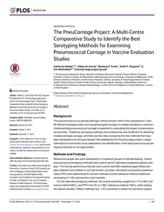
![serotype, whilst only eight methods had 70% sensitivity to detect minor serotypes. For the
field samples, the overall sensitivity ranged from 74.2% to 95.8% (reference method
93.8%), and PPV from 82.2% to 96.4% (reference method 99.6%). The microarray had the
highest sensitivity (95.8%) and high PPV (93.7%). The major limitation of this study is that
not all of the available alternative serotyping methods were included.
Conclusions
Most methods were able to detect the dominant serotype in a sample, but many performed
poorly in detecting the minor serotype populations. Microarray with a culture amplification
step was the top-performing method. Results from this comprehensive evaluation will inform
future vaccine evaluation and impact studies, particularly in low-income settings, where
pneumococcal disease burden remains high.
Introduction
Streptococcus pneumoniae (the pneumococcus) is a dominant cause of childhood illness and
death worldwide. It has been estimated to cause ~800,000 deaths in children aged under 5 y
annually, most due to pneumonia and most in low-income countries [1]. Pneumococci com-
monly colonise the nasopharynx of healthy children. Although carriage is usually asymptom-
atic, it can cause local inflammation and is considered a prerequisite for pneumococcal disease
[2]. Pneumococci are diverse, with 97 serotypes identified to date. Understanding carriage is
important for understanding pneumococcal population biology and transmission, assessing
vaccine impact, and evaluating the performance of new vaccines.
Pneumococcal conjugate vaccines (PCVs) are effective at preventing pneumonia and inva-
sive pneumococcal disease, and are currently being introduced in many resource-poor coun-
tries (e.g., by Gavi, the Vaccine Alliance). PCVs reduce nasopharyngeal carriage of the
pneumococcal serotypes they contain (vaccine types). Although transmission and disease due
to vaccine types is reduced, non-vaccine types fill the ecological niche, becoming more com-
mon in carriage and disease [3–5]. This phenomenon of serotype replacement, which may be
more pronounced in low-income settings (because of higher rates, load, and diversity of pneu-
mococcal carriage [6] and higher rates of disease), represents a significant risk to the global
pneumococcal immunisation strategy. It is difficult to monitor the serotypes causing pneumo-
nia or invasive disease because of the insensitivity of blood culture for detecting pneumococcal
pneumonia, inconsistencies in clinical practice (e.g., performing of lumbar punctures), and
limitations on diagnostic laboratory capacity, particularly in resource-poor settings [7–9]. Fur-
thermore, any improvement in laboratory or clinical diagnostic practices over time in vacci-
nated populations would lead to enhanced detection of non-vaccine types. This increase may
not reflect true replacement, and may cause unnecessary concerns about vaccine effectiveness
and inappropriate changes to vaccine policy.
Carriage studies offer a practical approach for monitoring serotype replacement and can
help in assessing the impact of PCVs and other pneumococcal vaccines [8,10]. Pneumococcal
carriage is an important endpoint for efficacy trials of new pneumococcal vaccines such as
common protein, combination (PCV + common protein), and whole-cell vaccines [11]. Tradi-
tional serotyping methods involve typing a small number of colonies, frequently missing car-
riage of multiple serotypes and providing no quantitative data. The gold-standard serotyping
Serotyping Methods for Pneumococcal Carriage Studies
PLOS Medicine | DOI:10.1371/journal.pmed.1001903 November 17, 2015 2 / 30
org), Grant 52099. This project was also supported
by the Murdoch Childrens Research Institute (www.
mcri.edu.au) and the Victorian Government's
Operational Infrastructure Support Program. The
funders had no role in the study design, data
collection and analysis, decision to publish, or
preparation of the manuscript; except for the specific
roles of DCHB, DH and KPK as described in the
author contributions. All authors have read, and
confirm that they meet, ICMJE criteria for authorship.
Competing Interests: I have read the journal's policy
and the authors of the manuscript have the following
competing interests: DCHB and DH are previous
employees of the Bill and Melinda Gates Foundation,
which provided funding for this project. KPK was
employed by the Bill and Melinda Gates Foundation
towards the completion of the project, but was not
involved in funding the project. KPK is a member of
the Editorial Board of PLOS Medicine. CS and EMD
were awarded the Robert Austrian Award which is
funded by Pfizer. Authors have served on advisory
boards for Merck (CS, KPK, EKM), GSK (KPK,
EKM), and Pfizer (KPK). Research group authors
have a scientific and/or commercial interest in the
performance of their methods, so blinded testing was
conducted.
Abbreviations: CDC, US Centers for Disease
Control and Prevention; CFU, colony-forming units;
ESI-MS, electrospray ionisation mass spectrometry;
HBA, horse blood agar; IQR, interquartile range; MFI,
mean fluorescence intensity; MLST, multilocus
sequence typing; mPCR, multiplex PCR; PBS,
phosphate-buffered saline; PCV, pneumococcal
conjugate vaccine; PPV, positive predictive value;
RFLP, restriction fragment length polymorphism;
RLB, reverse line blot; SSI, Statens Serum Institut;
STGG, skim milk-tryptone-glucose-glycerol; ULT,
ultra-low temperature.](data:image/gif;base64,R0lGODlhAQABAIAAAAAAAP///yH5BAEAAAAALAAAAAABAAEAAAIBRAA7)