- Current estimates of the case fatality rate for H5N1 infection are between 50-80%, but these rates may be skewed high because reporting criteria are too stringent and do not account for mild or asymptomatic cases.
- Molecular studies show that H5N1 NS1 protein contributes to the virus's virulence by triggering apoptosis and hypercytokinemia, helping explain H5N1's associated rapid viral pneumonia and acute respiratory distress syndrome.
- Limited human cell tropism of H5N1, restricted largely to cells in the lower respiratory tract, is thought to currently prevent efficient human-to-human transmission but adaptation remains possible through antigenic drift or shift.
H5N8 virus dutch outbreak (2014) linked to sequences of strains from asiaHarm Kiezebrink
Genetic analysis of influenza A(H5N8) virus from the Netherlands indicates that the virus probably was spread by migratory wild birds from Asia, possibly through overlapping flyways and common breeding sites in Siberia. In addition to the outbreak in the Netherlands, several other outbreaks of HPAI (H5N8) virus infections were reported in Europe at the end of 2014 after exponentially increasing deaths occurred in chicken and turkey flocks.
Genetic sequences submitted to the EpiFlu database indicated that the viruses from Europe showed a strong similarity to viruses isolated earlier in 2014 in South Korea, China, and Japan. An H5N8 virus isolated from a wigeon in Russia in September 2014 is located in the phylogenetic tree near the node of all sequences for H5N8 viruses from Europe.
In regard to time, this location fits the hypothesized route of H5N8 virus introduction into Europe. Furthermore, for several reasons, it is highly likely that the introduction of HPAI (H5N8) virus into the indoor-layer farm in the Netherlands occurred via indirect contact.
First, despite intensive monitoring, H5N8 viruses have never been detected in commercial poultry or wild birds in the Netherlands.
Second, when the virus was detected, the Netherlands had no direct trade contact with other European countries or Asia that might explain a route of introduction.
Third, because of the severity of disease in galliforms, outbreaks of H5N8 in the Netherlands before November 2014 would have been noticed.
Reseach on H9N2: evidence that link outbreaks in Eurasia, China, South Korea,...Harm Kiezebrink
In this study, scientists from the U.S. Geological Survey and U.S. Fish and Wildlife Service harnessed a new type of DNA technology to investigate avian influenza viruses in Alaska. Using a “next generation” sequencing approach, which identifies gene sequences of interest more rapidly and more completely than by traditional techniques, scientists identified low pathogenic avian influenza viruses in Alaska that are nearly identical to viruses found in China and South Korea.
The viruses were found in an area of western Alaska that is known to be a hot spot for both American and Eurasian forms of avian influenza.
“Our past research in western Alaska has shown that 70 percent of avian influenza viruses isolated in this area were found to contain genetic material from Eurasia, providing evidence for high levels of intercontinental viral exchange,” said Andy Ramey, a scientist with the USGS Alaska Science Center and lead author of the study. “This is because Asian and North American migratory flyways overlap in western Alaska.”
The new study, led by the USGS, found low pathogenic H9N2 viruses in an Emperor Goose and a Northern Pintail. Both of the H9N2 viruses were nearly identical genetically to viruses found in wild bird samples from Lake Dongting, China and Cheon-su Bay, South Korea.
“These H9N2 viruses are low pathogenic and not known to infect humans, but similar viruses have been implicated in disease outbreaks in domestic poultry in Asia,” said Ramey.
There is no commercial poultry production in western Alaska and highly similar H9N2 virus strains have not been reported in poultry in East Asia or North America, so it is unlikely that agricultural imports influenced this result.
The finding provides evidence for intercontinental movement of intact avian influenza viruses by migratory birds. The USGS recently released a publication about the detection of a novel highly pathogenic H5N8 virus in the U.S. that is highly similar to the Eurasian H5N8 viruses. This suggests that the novel re-assortment may be adapted to certain waterfowl species, enabling it to survive long migrations. That virus, and associated strains, have now spread from early detections in wild and domestic birds in Pacific states to poultry outbreaks in Minnesota, Missouri and Arkansas.
“The frequency of inter-hemispheric dispersal events of avian influenza viruses by migratory birds may be higher than previously recognized,” said Ramey.
While some of the samples for the project came from bird fecal samples collected from beaches at Izembek National Wildlife Refuge, most of the samples came from sport hunters.
“For the past several years, we’ve worked closely with sport hunters in the fall to obtain swab samples from birds and that has really informed our understanding of wildlife disease in this area,” said Bruce Casler, formerly a biologist with the USFWS Izembek National Wildlife Refuge and a co-author of the study. Non
Outbreak of High Patogen Avian Influenza H5N8 in GermanyHarm Kiezebrink
Germany has reported an outbreak of highly pathogenic avian influenza, H5N8 in fattening turkeys in North East Germany
(Mecklenburg - Western Pomerania). Increased mortality was observed in one of the six sheds of 15 week old birds for fattening (total number of turkeys on the premises ~ 31,000 of which each shed contained 5,000).
Analysis in to the Epidemiology and Pathophysiology of Respiratory Syncytial ...Pırıl Erel
Respiratory Syncytial Virus (RSV) places the heaviest clinical burden on paediatric wards in the UK and the US. It is in fact, a global issue with 3.4 million hospitalisations and approximately 66,000 deaths worldwide per annum (Bush et al., 2007) (Lambert et al., 2014). RSV is the leading cause, especially during the winter months, of severe respiratory infections in infants resulting in a rise in hospital admissions where 0.5-1% of infected babies die from respiratory failure. It is also a significant respiratory concern in the elderly population. (Agoti et al., 2014)
RSV has shown to have a willful ability to enter the host resulting in illness both by viral mechanisms and proteins encoded by RSV, dysregulating the synthesis of systemic immune response of the host. Alongside the infiltration of RSV, the heath status and genotype of the host will be a key factor in predetermining disease susceptibility and severity.
It is important to understand RSV has been implicated with further acute and chronic illnesses therefore by considering the epidemiology and pathophysiology of RSV treatment may be implicated during early stages which can influence possible outcomes in the future.
Human-to-Human transmission of H7H7 in Holland 2003Harm Kiezebrink
The outbreak of highly pathogenic avian influenza A virus subtype H7N7 started at the end of February, 2003, in commercial poultry farms in the Netherlands. In this study, published in The Lancet in 2004, it is noted that an unexpectedly high number of transmissions of avian influenza A virus subtype H7N7 to people directly involved in handling infected poultry, providing evidence for person-to-person transmission.
Although the risk of transmission of these viruses to humans was initially thought to be low, an outbreak investigation was launched to assess the extent of transmission of influenza A virus subtype H7N7 from chickens to humans.
453 people had health complaints—349 reported conjunctivitis, 90 had influenza-like illness, and 67 had other complaints. We detected A/H7 in conjunctival samples from 78 (26·4%) people with conjunctivitis only, in five (9·4%) with influenza-like illness and conjunctivitis, in two (5·4%) with influenza-like illness only, and in four (6%) who reported other symptoms. Most positive samples had been collected within 5 days of symptom onset. A/H7 infection was confirmed in three contacts (of 83 tested), one of whom developed influenza-like illness. Six people had influenza A/H3N2 infection. After 19 people had been diagnosed with the infection, all workers received mandatory influenza virus vaccination and prophylactic treatment with oseltamivir. More than half (56%) of A/H7 infections reported here arose before the vaccination and treatment programme.
Hundred samples viz. urine, blood, wound, pus and sputum collected from different patients were found to harbour Pseudomonas aeruginosa (P. aeruginosa) (27%) with a maximum isolation from wound samples (33.33%) and minimum from blood samples (11.11%). The degree of resistance of P. aeruginosa isolates to different antibiotics like Ceftazidime (30µg), Amikacin (30µg), Imipenem (10µg), Ciprofloxacin (30µg), Tetracycline (30µg), Gentamicin (10µg), Norfloxacin (10µg), Penicillin (30µg), Chloramphenicol (30µg), and Ofloxacin (5µg) varied from 56% to 100%. Antiseptics i.e. Betadine and Dettol were found to be more effective against the MDR strain of P. aeruginosa at the dilutions of 10-1 and 10-2. Duration of the disease and hospitalization duration, evaluated as risk factors for P. aeruginosa colonization were found to be statistically significant while age and gender were found to be statistically non- significant. The incidence of multidrug resistance of P. aeruginosa is increasing fast due to the frequent use of antibiotics and antiseptics, which are used extensively in hospitals and healthcare centers, therefore it is a need to develop alternative antimicrobial agents for the treatment of infectious diseases.
Key-words- Antibiotic, Antiseptic, Betadine and Dettol, Disinfectants, P. aeruginosa
Dossier transmission: Transmission of Avian Influenza Virus to DogsHarm Kiezebrink
Avian influenza was found in a dog on a farm in South Gyeongsang Province amid growing concerns that the disease could spread to other animals, officials the Ministry of Agriculture, Food and Rural Affairs said. The dog ― one of three at a duck farm in Goseong-gun, South Gyeongsang Province ― had antigens for the highly pathogenic H5N8 strain of bird flu, the Ministry of Agriculture, Food and Rural Affairs said. The disease affected the farm on Jan. 23.
Since the first case of a dog being infected with the poultry virus in March 2014, there have been 55 dogs found with antibodies to the bird flu virus. The antibody means the immune system of the dogs eliminated the virus. This is the first time bird flu has been found in a dog in Korea through the detection of antigens.
“None of these dogs had shown symptoms. No antigens or antibodies for the virus were found in the two other dogs, which means that dog-to-dog transmission is unlikely to have happened,” quarantine officials said.
The ministry suspected that the dog may have eaten infected animals at the farm. All poultry and dogs at the concerned farm were slaughtered as part of the preventive measures right after the farm was reported to have been infected with the disease, officials said.
Meanwhile, quarantine officials rejected the possibility of viral transmission to humans. According to the ministry’s report, about 450 workers at infected farms across the country had been given an antigen test, with none showing signs of infection. None of Korea’s 20,000 farm workers have reported any symptoms so far, officials added.
“It is thought that infected dogs do not show symptoms of the disease as they are naturally resistant to bird flu,” the ministry said. Meanwhile, the Agriculture Ministry has toughened the quarantine measures in Goseong-gun. The region is a frequented by migratory birds, which are suspected to have spread the viral disease.
Spatio temporal dynamics of global H5N1 outbreaks match bird migration patternsHarm Kiezebrink
The global spread of highly pathogenic avian influenza H5N1 in poultry, wild birds and humans, poses a significant pandemic threat and a serious public health risk.
An efficient surveillance and disease control system relies on the understanding of the dispersion patterns and spreading mechanisms of the virus. A space-time cluster analysis of H5N1 outbreaks was used to identify spatio-temporal patterns at a global scale and over an extended period of time.
Potential mechanisms explaining the spread of the H5N1 virus, and the role of wild birds, were analyzed. Between December 2003 and December 2006, three global epidemic phases of H5N1 influenza were identified.
These H5N1 outbreaks showed a clear seasonal pattern, with a high density of outbreaks in winter and early spring (i.e., October to March). In phase I and II only the East Asia Australian flyway was affected. During phase III, the H5N1 viruses started to appear in four other flyways: the Central Asian flyway, the Black Sea Mediterranean flyway, the East Atlantic flyway and the East Africa West Asian flyway.
Six disease cluster patterns along these flyways were found to be associated with the seasonal migration of wild birds. The spread of the H5N1 virus, as demonstrated by the space-time clusters, was associated with the patterns of migration of wild birds. Wild birds may therefore play an important role in the spread of H5N1 over long distances.
Disease clusters were also detected at sites where wild birds are known to overwinter and at times when migratory birds were present. This leads to the suggestion that wild birds may also be involved in spreading the H5N1 virus over short distances.
Supplementary information wind mediated transmission HPAIHarm Kiezebrink
A comparison between the transmission risk pattern predicted by the model and the pattern observed during the 2003 epidemic reveals that the wind-borne route alone is insufficient to explain the observations although it could contribute substantially to the spread over short distance ranges, for example, explaining 24% of the transmission over distances up to 25 km.
In this generic overview, you will find the date used in the publication “Modelling the Wind-Borne Spread of Highly Pathogenic Avian Influenza Virus between Farms”, published February 2012 (http://n2gf.com/?p=2377). For the outbreak of avian influenza A(H7N7) in the Netherlands in 2003, much data are available. The overview gives a description of the data used in the analyses of the mentioned publication:
Epidemiological data
There were 5360 poultry farms in the Netherlands in 2003, for all of which geographical information x is available. For 1531 farms the flocks were culled, for all of these the date of culling Tcull is known. For 227 of the 241 infected farms the date of infection tinf has been estimated, based on mortality data. The remaining 14 farms are hobby farms, defined as farms with less than 300 animals, for which no mortality data are available.
The geographic and temporal data together have previously been used to estimate the critical farm density, i.e. above what density of farms outbreaks are can occur.
Genetic data
The HA, NA and PB2 genes of viral samples from 231 farms have previously been sequenced. Sequence data RNA can be found in the GISAID database under accession numbers EPI ISL 68268-68352, EPI ISL 82373-82472 and EPI ISL 83984-84031. These data have previously been used to give general characteristics of the outbreak, to reconstruct the transmission tree and to assess the public health threat due to mutations of the virus in the animal host.
Meteorological data
Available meteorological data include wind speed wv and direction wdir (with a ten degree precision) and the fraction of time r without precipitation for every hour of every day of the outbreak, measured at five weather stations close to the infected farms. These data are available from the Royal Dutch Meteorological Institute at www.knmi.nl.
Human Coronaviruses (HCoV) exhibit positive single stranded RNA genome with enveloped nucleocapsid. Coronavirus belongs to the family Coronaviridae, originated from avian and mammalian species causes upper respiratory tract infection in humans by novel HCoVs viruses named as HCoV-HKU1, HCoV-NL63 but predominant species is Middle East respiratory syndrome (MERS-CoV) across the world. HCoV-HKU1 sp. is associated with chronic pulmonary disease, while HCoV-NL63 causes upper and lower respiratory tract disease in both children and adults, but most recent one was MERS-CoV, which caused acute pneumonia and occasional renal failure. The novel coronavirus SARS-CoV-2 is a new strain that causes the Coronavirus Disease 2019 (COVID-19) as named by the World Health Organization. According to the recent world statistics report about the COVID-19 cases approx. 101,500 confirmed cases and 3,500 death cases appeared. And mostly, a case of infection with CoV was identified in Wuhan, China. Structurally viral genome constitutes of 2/3rd of replicase gene encoding ORFs regions and rest of the 1/3rd region of genome form the structural proteins. The aim of the study was to understand the viral genetic systems in order to facilitate the genetic manipulation of the viral genome and to know the fundamental mechanism during the viral replication, facilitating the development of antidotes against the virus.
This paper reviews the evolution of the definition of sepsis and the controversy surrounding the sepsis-3 definition and the sepsis screening tool, qSOFA.
I returned to Dubai to begin my Entrepreneur journey and started various entities including Avigo Mauritius, which was commercially sold to Siguler Guff. For more details : https://www.facebook.com/achalghai
The Significance of Bacterial and Fungal Coinfection in the Setting of Viral ...Texas Children's Hospital
Keystone ECMO meeting 2018: To better characterize the frequency of bacterial and/or fungal coinfections in patients with viral pneumonias placed on ECMO and to understand their impact on mortality.
potassium, chloride, bicarbonate, blood urea nitrogen (BUN), magnesium, creatinine, glucose, and sometimes calcium. Tests that focus on cholesterol levels can determine LDL and HDL cholesterol levels, as well as triglyceride levels.[6]
There are nearly 100 viruses of the herpes group that infect many different animal species.
Official name of herpesviruses that commonly infect human is Humans herpesvirus (HHV)
herpes simplex virus types 1 (HHV 1)
Herpes simplex virus type 2 (HHV 2)
Varicella-zoster virus (HHV 3)
Epstein-Barr virus, (HHV 4)
Cytomegalovirus (HHV 5)
Human herpesvirus 6 (HHV 6)
Human herpesvirus 7 (HHV 7)
Human herpesvirus 8 (HHV 8) (Kaposi's sarcoma-associated herpesvirus).
Herpes B virus of monkeys can also infect humans
hELMINTHS#corona virus#Aspergillosis#BUGANDO#CUHAS#CUHAS#CUHAS
1. 2012
Abstract:
Current World Health Organization (WHO) estimates give a case fatality rate of 50-80% for
people infected with H5N1. Molecular studies into the virulence displayed by H5N1 provide
some basis for an increased case fatality rate, however they cannot explain it completely.
Criteria for reporting H5N1 infections are currently too stringent to allow accurate and
representative reporting of true H5N1 case fatality rates. Molecular studies can, however,
explain the abnormal symptoms of primary viral pneumonia and Acute Respiratory Distress
Syndrome (ARDS) displayed by patients infected with H5N1. Limited human cell tropism is
currently thought to be responsible for preventing human-human H5N spread, preventing
further fatalities beyond the 603 witnessed as of May, 2012. Intense interest in producing an
adaptable or universal H5N1 vaccine has lead to an ever improving field of H5N1 vaccine
production.
Introduction:
A highly virulent pathogen, H5N1 is yet to display a high degree of pathogenicity in humans,
the few infections observed thus far often resulting in fatal acute respiratory distress
syndrome (ARDS) (Lee et al, 2009). Current epidemiology suggests an extremely high
mortality rate amongst humans, though estimates are skewed by monitoring methods. There
is sufficient molecular evidence to raise concerns on the potential for H5N1 to achieve a
human-human transmissible form and cause a pandemic. As a result there have been a
number of recent varied studies undertaken to determine the molecular basis of H5N1
virulence. Due to the current public concern surrounding H5N1 there have been a number of
efforts targeted at producing an H5N1 vaccine and prospects for an H5N1 vaccine produced
in time to immunise against a pandemic strain are constantly improving.
Reasons for the absence of a human H5N1 pandemic
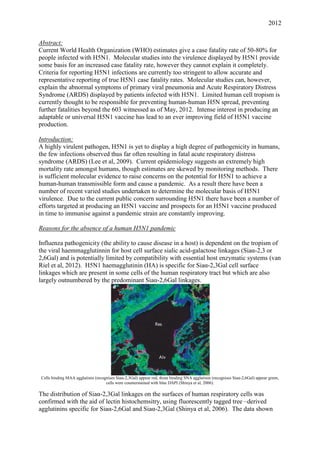
Influenza pathogenicity (the ability to cause disease in a host) is dependent on the tropism of
the viral haemmagglutinnin for host cell surface sialic acid-galactose linkages (Siaα-2,3 or
2,6Gal) and is potentially limited by compatibility with essential host enzymatic systems (van
Riel et al, 2012). H5N1 haemagglutinin (HA) is specific for Siaα-2,3Gal cell surface
linkages which are present in some cells of the human respiratory tract but which are also
largely outnumbered by the predominant Siaα-2,6Gal linkages.
Cells binding MAA agglutinin (recognises Siaα-2,3Gal) appear red, those binding SNA agglutinin (recognises Siaα-2,6Gal) appear green,
cells were counterstained with blue DAPI (Shinya et al, 2006).
The distribution of Siaα-2,3Gal linkages on the surfaces of human respiratory cells was
confirmed with the aid of lectin histochemsitry, using fluorescently tagged tree –derived
agglutinins specific for Siaα-2,6Gal and Siaα-2,3Gal (Shinya et al, 2006). The data shown
2. 2012
above displays the relative paucity of cells displaying Siaα-2,3Gal linkages on their cell
surfaces in the lower respiratory tract relative to those which display Siaα-2,6Gal.
Considering Siaα-2,3Gal linkages are generally restricted to the lower respiratory tract, this
shows the overall lack of Siaα-2,3Gal in the human respiratory system. Viral histochemistry
has since confirmed that host cell range in the lower respiratory tract is predominantly limited
to type II pneumocytes and alveolar macrophages as well as Clara cells and non-ciliated
epithelial cells in the terminal bronchioles (van Riel et al, 2007), binding to the upper
respiratory tract cells occurring as well but only relatively rarely (Nicholls, 2007 and van Riel
et al, 2012).
Attachment of human and avian viruses to ciliated epithelial cells, goblet cells, and submucosal glands in the human URT, Upper respiratory
tract cells to which the virus bound display a red border which is indicative of virus-cell binding and thus virus-cell recognition. (van Riel
et al, 2010)
Virus histochemistry (use of labelled virus) has confirmed that tropism is associated with
sialic acid distribution and that tropism is associated with ability to spread (van Riel et al.,
2010). H5N1 has a different cell binding profile relative to seasonal (H1N1 and H3N2) and
pandemic (H1N1) forms of human influenza. As can be seen, H5N1 does not display a clear
binding to upper respiratory tract cells, with the exception of cells of the submucosal glands.
The human influenza strains tested are much more widespread amongst humans than H5N1
and so it is currently thought that tropism to upper respiratory tract epithelia may allow an
increased ability to undergo human-human spread (van Riel et al, 2010).
H5N1 human-human transmission may require compatible enzyme systems in human cells
allowing viral function and replication to occur (Webster et al,2006). This is not true in the
case of limited proteolysis of HA during the maturation of H5N1 (an extremely important
step in influenza replication) because the proteases responsible are virtually ubiquitous in our
cell membranes, more so than those responsible for seasonal and pandemic influenza HA
cleavage (Kido et al, 2012) suggesting that host proteases do not limit H5N1 as much as was
previously thought.
Thus, H5N1 has not caused more deaths in the human population probably because it is not
currently adapted to our upper respiratory tract, although adaptation is possible through
antigenic drift, as was seen in a reassorted H5N1 virus in ferrets (Kawaoka et al, 2012), or
antigenic shift through reassortment with human-adapted strains in intermediate reservoirs
3. 2012
such as pigs (Cong et al, 2010). Both processes are currently occurring in native H5N1
populations (Kawaoka et al 2012 and Ibid.) and so monitoring of the situation is a key factor
in our preparedness for a potential H5N1 pandemic.
General Molecular mechanisms of H5N1 virulence:
Rapidly progressive primary viral pneumonia is a major feature of most reported H5N1-
related fatalities, a relatively rare event in other forms of influenza (Abdel-Ghafar,2008).
Resulting acute respiratory distress syndrome (ARDS) is associated with damage to the
alveoli and is the main source of mortality amongst H5N1 patients (Peiris et al, 2009).
Molecular studies have sought to find the answers behind these abnormal symptoms, an
important and much-studied virulence factor being the H5N1 NS1 protein. H5N1 NS1 is
implicated in caspase-dependent apoptosis (Zhang et al, 2010) and hypercytokinemia (Lam et
al, 2011), allowing the symptoms of viral infection to extend far beyond a localised area in
the lungs (deJong et al, 2006). Systems-level comparisons between the H5N1 and the
seasonal form of H1N1 have found that H5N1 induces a similar pro-inflammatory cytokine
response to human-adapted influenza in vitro but that this response is quantitatively stronger
(Lee et al, 2009). Further study has also implicated NS1 in this response in vitro (Lam et al,
2011).
Transmission electron microscopy (TEM) on normal (left) and plasmid transfected cells (right) X5,000 magnification (Zhang et al, 2010).
NS1 is a non-structural protein which normally has the role of inhibiting the host
interferon(IFN)-mediated innate immune response by binding double-stranded RNA, thereby
preventing the activation of the downstream cytosolic viral-detection machinery (Peiris et al,
2009). The role of H5N1 NS1 in triggering apoptosis was discovered with the use of
plasmid transfection on carcinomic alveolar epithelial cells. Use of recombinant techniques
and generation of an Escherichia coli (E. coli) library lead to the production of plasmids
encoding H5NS1. The cells were transfected with these plasmids as a surrogate for viral
infection and incubated for 24 hours (Zhang et al, 2010). Shown above are two transmission
electron micrographs on non-transfected and H5N1 NS1-transfected cells. Note how the
transfected cells are characteristic of post-apoptotic cells with condensed chromatin
aggregated along the nuclear membrane (red arrow) and apoptotic bodies (orange arrow).
4. 2012
Enzyme activities of caspase-3 and caspase-9 in NS1-transfected alveolar epithelial cell line. Enzyme activities based on transformed y-axis
indicating levels of caspase activity based on absorbance according to Caspase detection kits(Zhang et al, 2010)
Western blotting showed that Caspases 3 and 9 (two downstream effectors of apoptosis in
mammalian cells) were present in transfected cells. Subsequent colorimetric assays for
activities indicated that both were activated during the infection of the alveolar epithelial cells
(Zhang et al, 2010). This study was done on an immortal cell line rather than primary ex vivo
alveolar epithelium in addition to only the NS1 gene in a modified form being substituted for
a native H5N1 particle with its associated myriad of virulence factors, limiting how
applicable these findings would be to the role of NS1 in a whole virus infection. However a
causative link between the presence of NS1 and caspase-dependent apoptosis has been
established (Zhang et al, 2010), providing a further clue into the molecular basis of H5N1
virulence as apoptosis on other forms of cell lines has also been observed and found to be an
essential mechanism for viral propagation (Wurzer et al, 2003) as well as being a contributing
factor to the viral pneumonia in fatal cases of H5N1 infection (Peiris, 2009).
Heatmap for averaged microarray gene expression profiles of H1N1 and H5N1 infected primary human macrophages. Results are averaged
across macrophages from three different individuals (Lee et al, 2009).
A systems-level approach showed that an infection of primary alveolar macrophages from 3
different individuals by a native H5N1 virus strain in vitro led to a much more pronounced
pro-inflammatory cytokine response than for a low-pathogenicity seasonal H1N1 subtype
5. 2012
inoculated at an equivalent multiplicity of infection (Lee et al, 2009). This study came in lieu
of a number of studies which collectively showed that H5N1 infection caused a hyper-
induction of the cytokine response by alveolar macrophages and alveolar epithelial cells (Lee
et al, 2009). Global gene expression of macrophages infected with either H5N1 or H1N1 was
recorded using GeneChip arrays. A heat map of the relative gene expression between two
sets of alveolar macrophages, those infected with H1N1 and those with H5N1, shows that
similar pathways were upregulated at similar time points but that H5N1 infection caused a
much more intense upregulation. Overall it was found that components of cytokine pathways
for IFN-β and TNF-α were hyper-expressed in vitro (Lee et al, 2009) indicating the H5N1
viral particle was responsible for the hypercytokinemia seen in H5N1 patients (deJong et al,
2006).
Proportion overall cells displaying signs of early apoptosis upon transfection with influenza NS1 mRNA. (CC= non transfected controls).
(Lam et al, 2011)
Transfection of a lung epithelial cell line with mRNA encoding viral NS1 confirmed that
H5N1 NS1 is involved in the induction of apoptosis and inducing the abnormal pro-
inflammatory cytokine response behind ARDS (Lam et al, 2011). The proportion of early
apoptotic cells sampled at irregular intervals was determined flow cytometrically with the aid
of a fluorescent dye specific for early apoptotic cells. Note how large the difference between
H5 and other influenza subtype NS1 is in terms of both biological and statistical significance,
especially at 6 hours. H5 subtype NS1 mRNA transfection resulted in a maximum proportion
of early apoptotic cells double any other influenza NS1 in addition to having a 95%
confidence interval which was not overlapping any of the other ones, indicating a statistically
as well as biologically significant difference (Lam et al, 2011). This increase in apoptosis
could explain why primary viral pneumonia with diffuse alveolar damage is so common in
H5N1 infections, especially considering that the virus is generally limited to the lower
respiratory tract.
6. 2012
Cytometric bead array was used to quantify the levels of relevant cytokines and chemokines.
As shown below, all influenza subtype NS1 mRNA transfections lead to levels of cytokine
both biologically and statistically significantly different from non-transfected cells, however
H5 subtype NS1 mRNA clearly caused a greater amount of expression of the pro-
inflammatory cytokines measured, indicating that it the hypercytokinemia displayed in
ARDS is at least partially due to the effect of H5N1 NS1(Lam et al, 2011).
Cytokines/chemokines induced by transfection of NS1 mRNA of different influenza subtypes. H5 subtype NS1 mRNA transfected cells are
shown in purple. Cytokine/chemokine levels are expressed as fold-changes compared to the non-transfected cell controls measured at the
same time points (Lam et al, 2011).
High case fatality rate
The current case fatality rate for reported H5N1 infections is between 50-80% (“WHO:
Cumulative number of H5N1 cases”, accessed 18.5.2012), extremely high compared to the
1918 H1N1-attributed case fatality rate of 2.5% (Marks et al, 1976). The difference between
case fatality rates is highly unlikely to be due to differences in virulence at the molecular
level alone. A good point has been made that the case fatality rate may be falsely high
because the WHO requires a very stringent number of conditions to be met before confirming
a case of H5N1 (Palesea,2012). A patient must report to a hospital with a laboratory
advanced enough to allow for serological or genetic testing with advanced symptoms of
influenza infection (“WHO: H5N1 Case definition”, accessed 14.05.12). These selection
criteria may provide a skewness towards patients who are severely ill (Peiris et al, 2009),
leaving little room for those who may have an asymptomatic H5N1 infection (if this is
possible) or people who cannot reach a hospital with the necessary facilities. Rural areas
where waterfowl and other reservoir species are abundant are also under-represented,
meaning that this stringent method is very likely giving a non-representative estimate of true
fatality rates amongst those infected with H5N1.
7. 2012
Cumulative number of confirmed human cases for avian influenza A(H5N1) reported to WHO, 2003-2012 (“WHO: Cumulative
number of H5N1 cases”, accessed 18.5.2012)
Presence of unreported H5N1 infection with a successful outcome is possible as shown by the
discovery of anti-H5N1 HA antibodies in a sample of the poultry workers in the Chinese
province of Jiangsu where previous H5N1 infections and a death attributed to H5N1 had
occurred (Huo et al, 2012). Amongst the sample taken there were no people with prior severe
respiratory illness and the poultry in the villages sampled were not vaccinated against H5-
subtype avian influenza. Hemagglutination inhibition assays on blood samples for anti-H5N1
HA antibodies showed that seropositivity was present in two of the three villages and that
that the true population seropositivity rate for these two villages concerned was significantly
higher than zero (as high as between 2.19% and 10.78% in Gaoyou (95% confidence)). If
we take the number of H5N1 seropositive individuals just from Gaoyou (n=7) and added it to
all reported cases in China at the time of the study (n=46 from the above table) then we can
see that the case fatality rate for China shifts immediately from 66.6% (100*[28/42]) down
roughly 10% to 57.1% (100*[28/49]) if we assume that the anti-H5N1 antibodies came from
H5N1 infection with a successful outcome. This crude calculation serves to show that
making case fatality rate calculations with such stringent selection criteria leads to the
generation of inaccurate and error-prone statistics. This study was limited by a number of
factors, not the least of which being the restricted geography of sampling and a small sample
size, but it showed that it is possible to have H5N1 infections which do not result in
extremely adverse effects and that go unreported (Huo et al, 2012). Therefore WHO
estimates of case fatality rate may be skewed and more research into the true rate of H5N1
infection worldwide must be done before any reliable estimate of the true worldwide case
fatality rate of H5N1 can be determined.
Prospects for H5N1 vaccine:
H5N1 is constantly and quickly evolving, meaning that genetic diversification may occur so
quickly that even geographically close isolates of H5N1 may differ sufficiently to prevent
immunological cross-recognition of these strains should a vaccine based on either virus be
produced (Balish et al, 2010). Thus a vaccine must be adaptable with an ability to be
produced in large quantitites in a small amount of time (Zhou et al, 2012). Widespread, rapid
deployment and mass production of such a vaccine also pose major challenges to the
production of an H5N1 vaccine (McKenna, 2007).
As H5N1 is constantly evolving, it does not make financial or practical sense to mass produce
a standard vaccine based on any particular strain of H5N1 as such a vaccine may be made
irrelevant in the space of a few years or may not be relevant to a pandemic strain. Biannnual
WHO reports on global monitoring of H5N1 clades and their antigenic and epidemiological
properties allows for constant adjustment, depending on the storage of many different
8. 2012
candidate viruses so that in the case of a pandemic it might be possible for the mass
production of any of these viruses, one of which would hopefully be close or identical to the
pandemic strain. As of February 2012 there were 20 known and available candidate viruses
for vaccines with 3 pending full development (“WHO: Antigenic and genetic properties of
zoonotic viruses”, 2012).
Production of a vaccine in large enough amounts to matter and whether this vaccine would
generate sufficient immunogenicity is another matter. Cell culture in bioreactors would allow
for a great increase in production capacity of an H5N1 vaccine without having to rely on the
low-yield and potentially at-risk embryonated hen egg techniques, a high-yield production
method based on the use of the continuous Vero cell line (kidney epithelium isolated from
Chlorocebus spp.) being recently developed (Zhou et al, 2012). The strength behind pairing
this production technique with an existing candidate virus and reverse genetics technology is
that this would allow high-level production of virtually tailor-made live attenuated viruses for
use in an inactivated or split virus vaccine based on relevant pandemic H5N1 antigens
(Watanabe, 2012).
H5N1 vaccines cause poor immunogenicity in patients and animal models inoculated and so
novel adjuvants increasing immunogenicity are sought after (McKenna, 2007). Multiple
different adjuvants are currently being trialled and developed. An alum-based adjuvant
showed a good immunogenic response in Japanese adults but induced febrile reactions in
minors (Nakayama, 2012) whereas a novel silver nanoparticle-based adjuvant has also been
developed (Jazayeri et al, 2012). The latter inducing pro-inflammatory cytokine release and
enhanced antibody and cell-mediated immune responses in chicks, however this adjuvant has
not yet been trialled in humans (Ibid., 2012).
There are several candidate H5N1vaccines which have been developed, however they are not
yet ready for widespread use (WHO: FAQs, accessed 18.5.12). Much work remains to be
done on an H5N1 vaccine. With constant monitoring and improved production capacities
coupled to reverse genetics, in addition to continuing development of improved novel
adjuvants, the outlook for an H5N1 vaccine and our ability to respond quickly to a pandemic
is looking better as time progresses.
Conclusion:
It is apparent that H5N1 could be the basis for a future pandemic, albeit at a true case fatality
rate likely to be less than 50-80%. This paper has only captured very few of the many
molecular studies on H5N1 virulence, focusing on the role of the multifunctional NS1. It
should be remembered that H5N1 virulence is a polygenic trait (Lam et al, 2011) not
dependent on NS1 function or cellular tropism alone. Epidemiology on H5N1 is currently
error-prone and should not be trusted as the sole measure on which predictions of an H5N1
pandemic should be based. However, the clearly increased molecular virulence has caused
enough concern and sparked enough fear and interest that the outlook for production of a
successful H5N1 vaccine in a short amount of time is constantly improving.
Word count (not counting abstract, figure legends and titles): 2,740 words.
9. 2012
Bibliography:
Reasons for the absence of a human H5N1 pandemic
Cong Y., Wang G., GuanZ., Chang S., Zhang Q, Yang G., Wang W., Meng Q., Ren W.,
Wang C., Ding Z. (2010) Reassortant between Human-Like H3N2 and Avian H5
Subtype Influenza A Viruses in Pigs: A Potential Public Health Risk PLoS ONE 5(9); e12591
Kawaoka Y., Imai M., Watanabe T., Hatta M., Das S.C, Ozawa M., Shinya K., Zhong
Hanson A., Katsura H., Watanabe S., Li S., Kawakami E., Yamada S., Kiso M.,
Suzuki Y., Maher E.A. and Neumann G. Experimental adaptation of an influenza H5 HA
confers respiratory droplet transmission to a reassortant H5 HA/H1N1 virus in ferrets. Nature
(doi:10.1038/nature10831)
Kido H., Okumura Y., Takahashi E., Pan H.Y., Wang S., Yao D., Yao M., Chida J., YanoM.
(2012) Role of host cellular proteases in the pathogenesis of influenza and influenza-induced
multiple organ failure Biochimica et Biophysica Acta 1824: 186–194
Nicholls J.M., Chan M.C.W., Chan W.Y., Wong H.K., Cheung C.Y., Kwong D.L.W., Wong
M.P., Chui W.H., Poon L.L.M., Tsao S.W., GuanY. & Peiris J.S.M. Tropism of avian
influenza A (H5N1) in the upper and lower respiratory tract. Nature medicine 13(2);147-149
Shinya K., Ebina M., Yamada S., Ono M., Kasai N., Kawaoka Y. (2006) Influenza virus
receptors in the human airway. Nature 440 (doi:10.1038/440435a)
van Riel D., Munster V.J., de Wit E., Rimmelzwaan G.F., Fouchier R.A.M., Osterhaus
A.D.M.E., Kuiken T. (2007) Human and avian influenza viruses target different cells in the
lower respiratory tract of humans and other mammals. American Journal of Pathology
171( 4); 1215-1223
van Riel D., den Bakker M.A., Leijten L.M., Chutinimitkul S., Munster V.J., de Wit E.,
Rimmelzwaan G.F., Fouchier R.A.M, Osterhaus A.D.M.E. and Kuiken T. (2010) Seasonal
and pandemic human influenza viruses attach better to human upper respiratory tract
epithelium than avian influenza viruses. Am. J. Pathol. 176; 1614–1618.
van Riel D. and Kuiken T. (2012) The role of cell tropism for the pathogenesis of influenza in
humans Future Virol. 7(3)
Webster R.G and Govorkova E.A. (2006) H5N1:Continuing Evolution and Spread New
England Journal of Medicine 355(21):2174-2177
General Molecular mechanisms of H5N1 virulence:
Abdel-Ghafar A.N., Chotpitayasunondh T., Gao Z., Hayden F.G., Hien N.D., De Jong M.D.,
Naghdaliyev A., Peiris J.S.M., Shindo N., Soeroso S., Uyeki T.M. Update on avian influenza
A (H5N1) virus infection in humans. New England Journal of Medicine 358(3): 261-273
de Jong M.D., Simmons C.P., Thanh T.T., Hien V.M., Smith G.J.D., Chau T.N.B., Hoang
D.M., Chau N.V.V., Khanh T.H., Dong V.C., Qui P.T., Cam B.V., Ha D.Q., Guan Y., Peiris
10. 2012
J.S.M., Chinh N.T., Hien T.T., Farrar J. (2006) Fatal outcome of human influenza A (H5N1)
is associated with high viral load and hypercytokinemia. Nature Medicine 12(10); 1203-1207
Lam W.Y., Yeung A.C.M., Chan P.K.S. (2011) Apoptosis, cytokine and chemokine
induction by non-structural 1 (NS1) proteins encoded by different influenza subtypes
Virology Journal 8:554
Lee S.M., Gardy J.L., Cheung C.Y., Cheung T.K., Hui K.P., Ip N.Y., Guan Y., Hancock
R.E., Peiris J.S. Systems-level comparison of host-responses elicited by avian H5N1 and
seasonal H1N1 influenza viruses in primary human macrophages. PloS ONE 4(12); e8072
Peiris J.S.M., Cheung C.Y., Leung C.Y.H., Nicholls J.M. (2009) Innate immune responses to
influenza A H5N1: friend or foe? Trends in Immunology 30(12); 574-584
Wurzer W.J., Planz O., Ehrhardt C., Giner M., Silberzahn T., Pleschka S., Ludwig S. (2003)
Caspase 3 activation is essential for efficient influenza virus propagation. The EMBO Journal
22:2717-2728.
Zhang C., Yang Y., Zhou X., Liu X., Song H., He Y., Huang P. (2010) Highly pathogenic
avian influenza A virus H5N1 NS1 protein induces caspase-dependent apoptosis in human
alveolar basal epithelial cells. Virology Journal 7:51
High case fatality rate
Huo, X.a
, Zu, R.a
, Qi, X.a
, Qin, Y.a
, Li, L.a
, Tang, F.a
, Hu, Z.b
, Zhu, F.a
(2012) Seroprevalence of avian influenza A (H5N1) virus among poultry workers in Jiangsu
Province, China: An observational study BMC Infectious Diseases 12; 93
Marks G., Beatty W.K. Epidemics. New York: Scribners; 1976.
Palesea P., Wanga T.T. (2012) H5N1 influenza viruses: Facts, not fear. Proceedings of the
National Academy of Sciences of the United States of America 109(7); 2211-2213
“WHO: H5N1 Case definition”:
(http://www.who.int/influenza/resources/documents/case_definition2006_08_29/en/index.ht
ml, accessed 14.05.12)
“WHO: Cumulative number of H5N1 cases”:
(http://www.who.int/influenza/human_animal_interface/EN_GIP_20120502CumulativeNum
berH5N1cases.pdf, accessed 18.5.2012)
Prospects for H5N1 vaccine:
Balish, A.L., Davis C.T., Saad M.D., El-Sayed N., Esmat H., Tjaden J.A., Earhart K.C.,
Ahmed L.E., El-Halem M.A., Ali A.H.M., Nassif S.A., El-Ebiary E.A., Taha M., Mona
M.A., Arafa A., O'Neill E., Xiyan X., Cox N.J., Donis R.O., Klimov A.I. (2010) Antigenic
and genetic diversity of highly pathogenic avian influenza A (H5N1) viruses isolated in
Egypt. Avian Diseases 54; 329–334
Jazayeri S.D., Ideris A., Zakaria Z., Shameli K., Moeini H., Omar A.R. (2012) Cytotoxicity
and immunological responses following oral vaccination of nanoencapsulated avian influenza
11. 2012
virus H5 DNA vaccine with green synthesis silver nanoparticles Journal of Controlled
Release (Article in Press)
McKenna M. (2007), The Pandemic Vaccine Puzzle, Centre for Infectious Disease Research
and Policy.
(http://www.cidrap.umn.edu/cidrap/content/influenza/panflu/news/oct2507panvax1.html
,accessed 18.5.12)
Nakayama T., Kashiwagi Y., Kawashima H., Kumagai T., Ishii K.J., Ihara T.(2012) Alum-
adjuvanted H5N1 whole virion inactivated vaccine (WIV) enhanced inflammatory cytokine
productions. Vaccine 30; 3885– 3890
Steel, J. (2011) New strategies for the development of H5N1 subtype influenza vaccines:
Progress and challenges. BioDrugs 25(5); 285-298
Watanabe Y., Ibrahim M.S., Suzuki, Y., Ikuta, K. (2012) The changing nature of avian
influenza A virus (H5N1) Trends in Microbiology 20(1); 11-20
“WHO: Antigenic and genetic properties of zoonotic viruses”
(http://www.who.int/influenza/vaccines/virus/characteristics_virus_vaccines/en/index.html,
accessed 19.5.12)
“WHO: FAQs”
(http://www.who.int/influenza/human_animal_interface/avian_influenza/h5n1_research/faqs/
en/index.html, accessed 18.5.2012)
Zhou F., Zhou J., Ma L., Song S., Zhang X., Li W., Jiang S., Wang Y., Liao G. (2012) High-
yield production of a stable Vero cell-based vaccine candidate against the highly pathogenic
avian influenza virus H5N1 Biochemical and Biophysical Research Communications (Article
in Press)