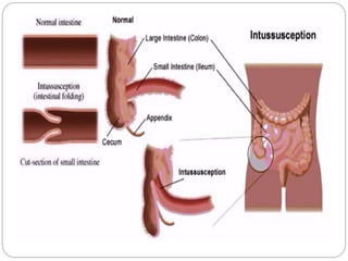
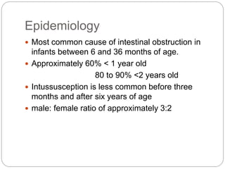
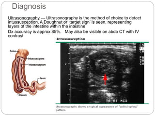
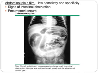
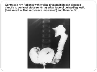
RAJIV LAL
s110677
Intussusception
 Intussusception – telescoping of a proximal
segment of the intestine(intussusceptum) into a
distal segment (intussuscipien).
 It is the most common abdominal emergency in early
childhood, particularly in children younger than two
years of age.
 The majority of cases in children are idiopathic.
Epidemiology
 Most common cause of intestinal obstruction in
infants between 6 and 36 months of age.
 Approximately 60% < 1 year old
80 to 90% <2 years old
 Intussusception is less common before three
months and after six years of age
 male: female ratio of approximately 3:2
INTUSSUSCEPTION
ANATOMIC LOCATIONS
 ILEOCOLIC
MOST COMMON IN CHILDREN
 ILEO-ILEOCOLIC
SECOND MOST COMMON
 ENTEROENTERIC
ILEO-ILEAL, JEJUNO-JEJUNAL
MORE COMMON IN ADULTS
 CAECOCOLIC, COLOCOLIC
Pathogenesis
The intussusceptum, telescopes into the intussuscipien →
dragging the associated mesentery with it.
 Venous and lymphatic congestion
 Edema
 Strangulated obstruction
 Ischemia….necrosis….perforation….peritonitis
sepsis….shock…death
Lead point
A lead point is a lesion or variation in the intestine
that is trapped by peristalsis and dragged into a
distal segment of the intestine, causing
intussusception.
A Meckel diverticulum, intestinal polyp,
intestinal duplication, hemangioma, tumor
(lymphoma), appendix, ectopic pancreas can
act as a lead point for intussusception.
25% of cases have pathological lead point.
Aetiology
 Approximately 75% of cases are idiopathic
because there is no clear disease trigger or
pathological lead point.
 Viral infections can stimulate lymphatic tissue in
the intestinal tract, resulting in hypertrophy of
Peyer patches in the lymphoid rich terminal ileum,
which may act as a lead point for ileocolic
intussusception
 Postoperative- The intussusception is thought to
be caused by uncoordinated peristaltic activity
and/or traction from sutures or devices such as a
gastrojejunal feeding tube.
CLINICAL MANIFESTATIONS
History
Early
 Patients with intussusception typically develop the sudden onset of intermittent,
severe, crampy, progressive abdominal pain, accompanied by inconsolable
crying and drawing up of the legs toward the abdomen.
 Between symptoms child will be playing and doing normal activity.
 Vomiting
Later
 Continuous abdominal pain
 The stool may contains gross or occult blood or be a mixture of blood and
mucous and sloughing mucosa, giving it the appearance of currant jelly.
 Lethargy
 Palpable abdominal mass.
Physical
 A sausage shaped abdominal mass.
 Abdominal distension
 Dehydration
 Classic triad ( <15% of cases)
 Intermittent colicky abdominal pain
 RLQ sausage shaped abdominal mass
 currant jelly stool is seen in less than 15% of patients at
the time of presentation.
 Occasionally, the initial presenting sign is
lethargy or altered consciousness alone, without
pain, rectal bleeding, or other symptoms that
suggest an intra-abdominal process and is often
confused with sepsis.
Diagnosis
Ultrasonography — Ultrasonography is the method of choice to detect
intussusception. A Doughnut or ‘target sign’ is seen, representing
layers of the intestine within the intestine
Dx accuracy is approx 85%. May also be visible on abdo CT with IV
contrast.
Abdominal plain film – low sensitivity and specificity
 Signs of intestinal obstruction
 Pneumoperitoneum
Contrast x-ray Patients with typical presentation can proceed
directly to contrast study (enema) advantage of being diagnostic
(barium will outline a concave ‘meniscus’) and therapeutic.
Treatment
 Stabilize and resuscitate with intravenous fluids
 Hydration, electrolyte, acid-base balance.
 NBM and Stomach should be decompressed with a
nasogastric tube
 Antibiotics- if signs of infection (fever, peritonitis)
 Ampicillin 50mg/kg IM/IV 4x per day
 Gentamycin 5mg/kg IM/IV OD
 Metronidazole 7.5mg/kg IV TDS
Duration – uncomplicated reduced with air enema 24-48 hrs
- perforated bowel with resection 1 week post-op
Non-operative treatment
 Stable patients and no evidence of bowel perforation
should be treated with Non-operative reduction
 Non-operative reduction using hydrostatic or pneumatic
pressure by enema
 Risk of perforation – adv in pneumatic technique.
Surgical treatment
 Indicated for patients with peritonitis or evidence of
perforation or in whom non-operative reduction is
unsuccessful.
 Manual reduction at operation is attempted
 If manual reduction fails or in case of perforation,
necrosis, pathological lead point – bowel resection and
primary anastomosis is performed.
 The risk of recurrence is approximately 1 percent after
manual reduction and virtually nonexistent after surgical
resection
Complications - Rarely occur when diagnosis is prompt.
 Necrosis and bowel perforation from strangulated
intussusception
 Peritonitis and Sepsis
 Hypovolaemia and circulatory shock
 Electrolyte imbalance
 Perforation during non operative reduction.
 Wound infection.
 Adhesions causing bowel obstruction.
 Recurrence.
 10% after successful non-operative reduction
With early diagnosis, appropriate fluid resuscitation and
therapy, the mortality rate from intussusception in children is <
1%. If left untreated, this condition is uniformly fatal in 2-5
days.
Reference
 Lloyd DA, Kenny SE. The surgical abdomen. In:
Pediatric Gastrointestinal Disease: Pathopsychology,
Diagnosis, Management, 4th, Walker WA, Goulet O,
Kleinman RE, et al (Eds), BC Decker, Ontario 2004.
p.604.
 Rudolph’s peadiatrics 22nd edition
 Mandeville K, Chien M, Willyerd FA, et al.
Intussusception: clinical presentations and imaging
characteristics. Pediatr Emerg Care 2012; 28:842.
 Buettcher M, Baer G, Bonhoeffer J, et al. Three-year
surveillance of intussusception in children in
Switzerland. Pediatrics 2007; 120:473.

Intussusception (2)

  • 1.
  • 3.
     Intussusception –telescoping of a proximal segment of the intestine(intussusceptum) into a distal segment (intussuscipien).  It is the most common abdominal emergency in early childhood, particularly in children younger than two years of age.  The majority of cases in children are idiopathic.
  • 4.
    Epidemiology  Most commoncause of intestinal obstruction in infants between 6 and 36 months of age.  Approximately 60% < 1 year old 80 to 90% <2 years old  Intussusception is less common before three months and after six years of age  male: female ratio of approximately 3:2
  • 5.
    INTUSSUSCEPTION ANATOMIC LOCATIONS  ILEOCOLIC MOSTCOMMON IN CHILDREN  ILEO-ILEOCOLIC SECOND MOST COMMON  ENTEROENTERIC ILEO-ILEAL, JEJUNO-JEJUNAL MORE COMMON IN ADULTS  CAECOCOLIC, COLOCOLIC
  • 6.
    Pathogenesis The intussusceptum, telescopesinto the intussuscipien → dragging the associated mesentery with it.  Venous and lymphatic congestion  Edema  Strangulated obstruction  Ischemia….necrosis….perforation….peritonitis sepsis….shock…death
  • 7.
    Lead point A leadpoint is a lesion or variation in the intestine that is trapped by peristalsis and dragged into a distal segment of the intestine, causing intussusception. A Meckel diverticulum, intestinal polyp, intestinal duplication, hemangioma, tumor (lymphoma), appendix, ectopic pancreas can act as a lead point for intussusception. 25% of cases have pathological lead point.
  • 8.
    Aetiology  Approximately 75%of cases are idiopathic because there is no clear disease trigger or pathological lead point.  Viral infections can stimulate lymphatic tissue in the intestinal tract, resulting in hypertrophy of Peyer patches in the lymphoid rich terminal ileum, which may act as a lead point for ileocolic intussusception  Postoperative- The intussusception is thought to be caused by uncoordinated peristaltic activity and/or traction from sutures or devices such as a gastrojejunal feeding tube.
  • 9.
    CLINICAL MANIFESTATIONS History Early  Patientswith intussusception typically develop the sudden onset of intermittent, severe, crampy, progressive abdominal pain, accompanied by inconsolable crying and drawing up of the legs toward the abdomen.  Between symptoms child will be playing and doing normal activity.  Vomiting Later  Continuous abdominal pain  The stool may contains gross or occult blood or be a mixture of blood and mucous and sloughing mucosa, giving it the appearance of currant jelly.  Lethargy  Palpable abdominal mass. Physical  A sausage shaped abdominal mass.  Abdominal distension  Dehydration
  • 10.
     Classic triad( <15% of cases)  Intermittent colicky abdominal pain  RLQ sausage shaped abdominal mass  currant jelly stool is seen in less than 15% of patients at the time of presentation.  Occasionally, the initial presenting sign is lethargy or altered consciousness alone, without pain, rectal bleeding, or other symptoms that suggest an intra-abdominal process and is often confused with sepsis.
  • 11.
    Diagnosis Ultrasonography — Ultrasonographyis the method of choice to detect intussusception. A Doughnut or ‘target sign’ is seen, representing layers of the intestine within the intestine Dx accuracy is approx 85%. May also be visible on abdo CT with IV contrast.
  • 12.
    Abdominal plain film– low sensitivity and specificity  Signs of intestinal obstruction  Pneumoperitoneum
  • 13.
    Contrast x-ray Patientswith typical presentation can proceed directly to contrast study (enema) advantage of being diagnostic (barium will outline a concave ‘meniscus’) and therapeutic.
  • 14.
    Treatment  Stabilize andresuscitate with intravenous fluids  Hydration, electrolyte, acid-base balance.  NBM and Stomach should be decompressed with a nasogastric tube  Antibiotics- if signs of infection (fever, peritonitis)  Ampicillin 50mg/kg IM/IV 4x per day  Gentamycin 5mg/kg IM/IV OD  Metronidazole 7.5mg/kg IV TDS Duration – uncomplicated reduced with air enema 24-48 hrs - perforated bowel with resection 1 week post-op
  • 15.
    Non-operative treatment  Stablepatients and no evidence of bowel perforation should be treated with Non-operative reduction  Non-operative reduction using hydrostatic or pneumatic pressure by enema  Risk of perforation – adv in pneumatic technique. Surgical treatment  Indicated for patients with peritonitis or evidence of perforation or in whom non-operative reduction is unsuccessful.  Manual reduction at operation is attempted  If manual reduction fails or in case of perforation, necrosis, pathological lead point – bowel resection and primary anastomosis is performed.  The risk of recurrence is approximately 1 percent after manual reduction and virtually nonexistent after surgical resection
  • 16.
    Complications - Rarelyoccur when diagnosis is prompt.  Necrosis and bowel perforation from strangulated intussusception  Peritonitis and Sepsis  Hypovolaemia and circulatory shock  Electrolyte imbalance  Perforation during non operative reduction.  Wound infection.  Adhesions causing bowel obstruction.  Recurrence.  10% after successful non-operative reduction With early diagnosis, appropriate fluid resuscitation and therapy, the mortality rate from intussusception in children is < 1%. If left untreated, this condition is uniformly fatal in 2-5 days.
  • 17.
    Reference  Lloyd DA,Kenny SE. The surgical abdomen. In: Pediatric Gastrointestinal Disease: Pathopsychology, Diagnosis, Management, 4th, Walker WA, Goulet O, Kleinman RE, et al (Eds), BC Decker, Ontario 2004. p.604.  Rudolph’s peadiatrics 22nd edition  Mandeville K, Chien M, Willyerd FA, et al. Intussusception: clinical presentations and imaging characteristics. Pediatr Emerg Care 2012; 28:842.  Buettcher M, Baer G, Bonhoeffer J, et al. Three-year surveillance of intussusception in children in Switzerland. Pediatrics 2007; 120:473.