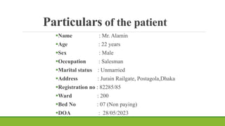
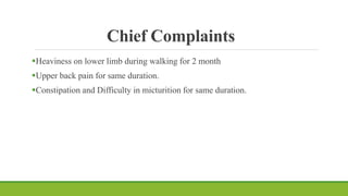
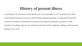
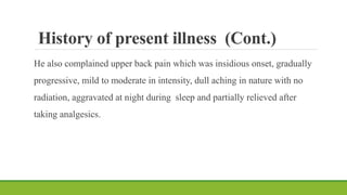
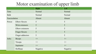
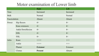
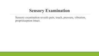
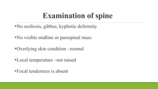
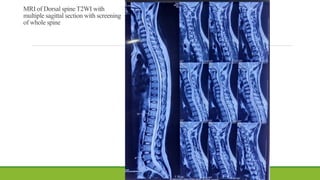
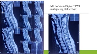
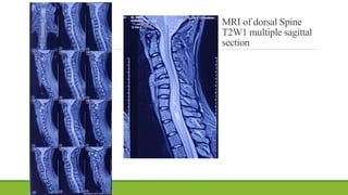
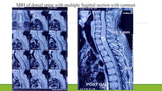
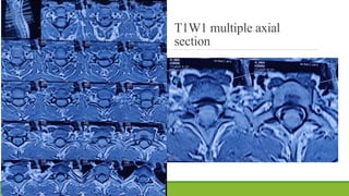
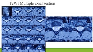
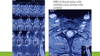
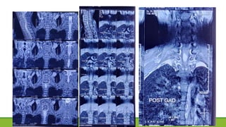
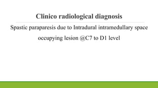
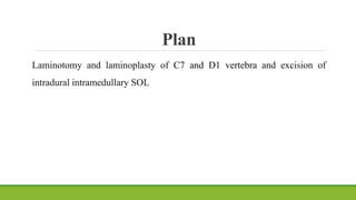
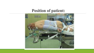
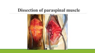
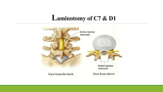
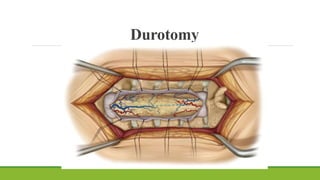
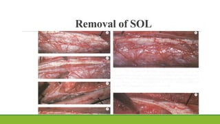
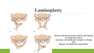
Preoperative case presentation
Dr. Md. Toufiq Hasan
Resident, Phase-A(R-9)
Department Of Neurosurgery
Dhaka Medical College Hospital
On behalf of NSU-Green Unit
Particulars of the patient
Name : Mr. Alamin
Age : 22 years
Sex : Male
Occupation : Salesman
Marital status : Unmarried
Address : Jurain Railgate, Postagola,Dhaka
Registration no : 82285/85
Ward : 200
Bed No : 07 (Non paying)
DOA : 28/05/2023
Chief Complaints
Heaviness on lower limb during walking for 2 month
Upper back pain for same duration.
Constipation and Difficulty in micturition for same duration.
History of present illness
According to the statement of the patient, he was reasonably well 2 months back. Then,
he developed heaviness on lower limb during walking primarily on right then both limb
which was insidious, intermittent, intensity of weakness gradually progressive with
duration of walking ,day by day activities and relieved by stopping walking, activities and
taking rest on bed.
History of present illness (Cont.)
He also complained upper back pain which was insidious onset, gradually
progressive, mild to moderate in intensity, dull aching in nature with no
radiation, aggravated at night during sleep and partially relieved after
taking analgesics.
History of present illness (Cont.)
He gave history of bowel and bladder dysfunction in form of constipation and
difficulty in micturition followed by hesitancy and urgency.
During the course of his illness, he gave no history of headache, neck pain,
trauma, prolonged fever, unexplained weight loss, anorexia, cough, hemoptysis,
prolong use of steroid or close contact with known TB patient.
History of present illness (Cont.)
With the above complaints, he visited Mugda Medical college hospital and then
referred to Neurosurgery department ,DMCH for better management.
Associated illness
He has no history of Diabetes, Hypertension,any heart, kidney ,lung
disease
Past medical history
He has no H/O major childhood illness, previous surgery,
accident or injury.
Immunization History
Vaccinated as per EPI schedule and 1 dose of covid vaccine
Family history
Nothing significant. Boy of a non consanguineous married parents.
Other family members are leading a healthy life.
Personal history
He is non smoker, non alcoholic, non betel nut chewer
Socio economic history
He belongs to a family with low socio-economic condition
Drug history
Nothing significant.
General Physical Examination
Appearance: Anxious
Body built: Average
 Co-operative
Decubitus: On choice
Nutritional status: Average
Pulse: 84 b/m
BP: 130/80 mmHg
Temp: Not raised
RR: 18 /m
 Jaundice
 Clubbing
 Cyanosis
 Anemia
 Leukonychia
 Edema
 Dehydration
 Koilonychia
 Lymphadenopathy
 Catheter in situ
No abnormality
detected
Systemic Examination
Cardiovascular System-No abnormality detected
Respiratory System-No abnormality detected
Alimentary System-No abnormality detected
Nervous System Examination
Higher psychic function: Intact
Memory: Intact
GCS: E₄V₅M₆
Gait: Spastic
Right handed person
Cranial nerves Intact
Speech : Normal
No sign of cerebellar dysfunction
No sign of meningeal irritation
Motor examination of upper limb
Right Left
Tone Normal Normal
Bulk Normal Normal
Fasciculation Absent Absent
Power Elbow flexors 5 5
Wrist extensors 5 5
Elbow extensors 5 5
Finger flexors 5 5
Finger adductors 5 5
Jerks Biceps ++ ++
Triceps ++ ++
Supinator ++ ++
Hoffman Negative Negative
Motor examination of Lower limb
Right Left
Tone Increased Increased
Bulk Normal Normal
Fasciculation Absent Absent
Power Hip flexors 4+ 4+
Knee extensors 4+ 4+
Ankle Dorsiflexion 4+ 4+
EHL 4+ 4+
FHL 4+ 4+
Jerks Knee ++++ +++
Ankle ++++ +++
Planter Extensor Extensor
Clonus Present Absent
Sensory Examination
Sensory examination reveals pain, touch, pressure, vibration,
proprioception intact.
Examination of spine
No scoliosis, gibbus, kyphotic deformity
No visible midline or paraspinal mass
Overlying skin condition –normal
Local temperature –not raised
Focal tenderness is absent
Provisional diagnosis
A case of spastic paraparesis due to dorsal spinal Space
occupying lesion
INVESTIGATIONS
MRI of Dorsal Spine with screening of
whole spine
MRI of Dorsal spine T2WI with
multiple sagittal section with screening
of whole spine
MRI of dorsal Spine T1W1
multiple sagittal section
MRI of dorsal Spine
T2W1 multiple sagittal
section
MRI of dorsal spine with multiple Sagittal section with contrast
T1W1 multiple axial
section
T2WI Multiple axial section
MRI of dorsal spine with
multiple axial section with
contrast
Clinico radiological diagnosis
Spastic paraparesis due to Intradural intramedullary space
occupying lesion @C7 to D1 level
Differential Diagnosis
Plan
Laminotomy and laminoplasty of C7 and D1 vertebra and excision of
intradural intramedullary SOL
Position of patient:
Dissection of paraspinal muscle
Laminotomy of C7 & D1
Durotomy
Removal of SOL
Laminoplasty
Various methods commonly used for laminoplasty.
(a) Single-door suture
technique. (b) Double-door allograft. (c) Single
door
allograft. (d) Single-door plate fixation.
Complication
1. Risks of spinal cord or nerve root injury
2. CSF leak
3. Infection
4. Dural scarring
5. Postoperative hematoma
THANK YOU
Points in favour Points against
Schwannoma Most common nerve sheath mass
•
"dumbbell-shaped' transforaminal mass
• Hemorrhage, cystic, or fatty degeneration
• Solitary spinal lesion more likely schwannoma than NF
• Bony remodeling– Thinned pedicle
.
• No uniform enhancement pattern
Neurofibroma • Target sign
• Hemorrhage, cystic, or fatty degeneration
Bulky multilevel spinal nerve root tumors
in patient with
stigmata of neurofibromatosis type 1
extramedullary
ependymoma
Ependymoma: Unusual
differential for a totally
No Cap sign: Hemosiderin at cranial or
caudal margin

Intramedullary SOL

  • 1.
    Preoperative case presentation Dr.Md. Toufiq Hasan Resident, Phase-A(R-9) Department Of Neurosurgery Dhaka Medical College Hospital On behalf of NSU-Green Unit
  • 2.
    Particulars of thepatient Name : Mr. Alamin Age : 22 years Sex : Male Occupation : Salesman Marital status : Unmarried Address : Jurain Railgate, Postagola,Dhaka Registration no : 82285/85 Ward : 200 Bed No : 07 (Non paying) DOA : 28/05/2023
  • 3.
    Chief Complaints Heaviness onlower limb during walking for 2 month Upper back pain for same duration. Constipation and Difficulty in micturition for same duration.
  • 4.
    History of presentillness According to the statement of the patient, he was reasonably well 2 months back. Then, he developed heaviness on lower limb during walking primarily on right then both limb which was insidious, intermittent, intensity of weakness gradually progressive with duration of walking ,day by day activities and relieved by stopping walking, activities and taking rest on bed.
  • 5.
    History of presentillness (Cont.) He also complained upper back pain which was insidious onset, gradually progressive, mild to moderate in intensity, dull aching in nature with no radiation, aggravated at night during sleep and partially relieved after taking analgesics.
  • 6.
    History of presentillness (Cont.) He gave history of bowel and bladder dysfunction in form of constipation and difficulty in micturition followed by hesitancy and urgency. During the course of his illness, he gave no history of headache, neck pain, trauma, prolonged fever, unexplained weight loss, anorexia, cough, hemoptysis, prolong use of steroid or close contact with known TB patient.
  • 7.
    History of presentillness (Cont.) With the above complaints, he visited Mugda Medical college hospital and then referred to Neurosurgery department ,DMCH for better management.
  • 8.
    Associated illness He hasno history of Diabetes, Hypertension,any heart, kidney ,lung disease
  • 9.
    Past medical history Hehas no H/O major childhood illness, previous surgery, accident or injury.
  • 10.
    Immunization History Vaccinated asper EPI schedule and 1 dose of covid vaccine
  • 11.
    Family history Nothing significant.Boy of a non consanguineous married parents. Other family members are leading a healthy life.
  • 12.
    Personal history He isnon smoker, non alcoholic, non betel nut chewer
  • 13.
    Socio economic history Hebelongs to a family with low socio-economic condition
  • 14.
  • 15.
    General Physical Examination Appearance:Anxious Body built: Average  Co-operative Decubitus: On choice Nutritional status: Average Pulse: 84 b/m BP: 130/80 mmHg Temp: Not raised RR: 18 /m  Jaundice  Clubbing  Cyanosis  Anemia  Leukonychia  Edema  Dehydration  Koilonychia  Lymphadenopathy  Catheter in situ No abnormality detected
  • 16.
    Systemic Examination Cardiovascular System-Noabnormality detected Respiratory System-No abnormality detected Alimentary System-No abnormality detected
  • 17.
    Nervous System Examination Higherpsychic function: Intact Memory: Intact GCS: E₄V₅M₆ Gait: Spastic Right handed person Cranial nerves Intact Speech : Normal No sign of cerebellar dysfunction No sign of meningeal irritation
  • 18.
    Motor examination ofupper limb Right Left Tone Normal Normal Bulk Normal Normal Fasciculation Absent Absent Power Elbow flexors 5 5 Wrist extensors 5 5 Elbow extensors 5 5 Finger flexors 5 5 Finger adductors 5 5 Jerks Biceps ++ ++ Triceps ++ ++ Supinator ++ ++ Hoffman Negative Negative
  • 19.
    Motor examination ofLower limb Right Left Tone Increased Increased Bulk Normal Normal Fasciculation Absent Absent Power Hip flexors 4+ 4+ Knee extensors 4+ 4+ Ankle Dorsiflexion 4+ 4+ EHL 4+ 4+ FHL 4+ 4+ Jerks Knee ++++ +++ Ankle ++++ +++ Planter Extensor Extensor Clonus Present Absent
  • 20.
    Sensory Examination Sensory examinationreveals pain, touch, pressure, vibration, proprioception intact.
  • 21.
    Examination of spine Noscoliosis, gibbus, kyphotic deformity No visible midline or paraspinal mass Overlying skin condition –normal Local temperature –not raised Focal tenderness is absent
  • 22.
  • 23.
    Provisional diagnosis A caseof spastic paraparesis due to dorsal spinal Space occupying lesion
  • 24.
  • 25.
    MRI of DorsalSpine with screening of whole spine
  • 26.
    MRI of Dorsalspine T2WI with multiple sagittal section with screening of whole spine
  • 27.
    MRI of dorsalSpine T1W1 multiple sagittal section
  • 28.
    MRI of dorsalSpine T2W1 multiple sagittal section
  • 29.
    MRI of dorsalspine with multiple Sagittal section with contrast
  • 30.
  • 31.
  • 32.
    MRI of dorsalspine with multiple axial section with contrast
  • 34.
    Clinico radiological diagnosis Spasticparaparesis due to Intradural intramedullary space occupying lesion @C7 to D1 level
  • 35.
  • 36.
    Plan Laminotomy and laminoplastyof C7 and D1 vertebra and excision of intradural intramedullary SOL
  • 37.
  • 38.
  • 39.
  • 40.
  • 41.
  • 42.
    Laminoplasty Various methods commonlyused for laminoplasty. (a) Single-door suture technique. (b) Double-door allograft. (c) Single door allograft. (d) Single-door plate fixation.
  • 43.
    Complication 1. Risks ofspinal cord or nerve root injury 2. CSF leak 3. Infection 4. Dural scarring 5. Postoperative hematoma
  • 44.
  • 46.
    Points in favourPoints against Schwannoma Most common nerve sheath mass • "dumbbell-shaped' transforaminal mass • Hemorrhage, cystic, or fatty degeneration • Solitary spinal lesion more likely schwannoma than NF • Bony remodeling– Thinned pedicle . • No uniform enhancement pattern Neurofibroma • Target sign • Hemorrhage, cystic, or fatty degeneration Bulky multilevel spinal nerve root tumors in patient with stigmata of neurofibromatosis type 1 extramedullary ependymoma Ependymoma: Unusual differential for a totally No Cap sign: Hemosiderin at cranial or caudal margin

Editor's Notes

  • #20 Reflexes can be graded 0= absent +-= present only with reinforcement 1+=present but depressed 2+= Normal 3+= increased 4+= clonus
  • #27 Showing iso to hypointense lesion at the level of C7 and D1 vertebra with fusiform dilatation of spinal cord at this level. Hyperintense signal change above the lesion up to C6 and below the lesion up to D2 vertebra Cervical lordotic curvature is straightening with normal disc space . Anterior and posterior CSF column is maintained. Rest of the spine screening is normal.
  • #28 Showing iso to hyperintense lesion at the level of C7 to D1 vertebra with fusiform dilatation of spinal cord at this level.
  • #29 Showing iso to hypointense oval shaped lesion at the level of C7 to D1 with peripheral hyperintense signal change extending superiorly up to C6 and inferiorly up to D2 vertebra
  • #30 Homogenous contrast enhanced ovoid shaped lesion at the level of C7 to D1 vertebra.
  • #31 Iso intense lesion in the central portion of the spinal cord with poorly demarcated outline
  • #32 Iso to hyperintense lesion in the central portion of the spinal cord with poorly demarcated outline
  • #33 Homogenous contrast enhanced circular lesion on central portion of the cord
  • #35  • Target sign more common with NF than schwannoma • Hemorrhage, cystic, or fatty degeneration more common with schwannoma Solitary spinal lesion more likely schwannoma than NF Bony remodeling due to large intraspinal or transforaminal mass – Thinned pedicle, enlarged neural foramen, vertebral body scalloping, widened interpedicular distance Spinal Meningioma • Thoracic tumor in female patient is more likely meningioma • dural "tail" • calcification Broad dural attachment • Strong homogeneous enhancement Neurofibroma Bulky multilevel spinal nerve root tumors in patient with stigmata of neurofibromatosis type 1 Target sign on T2WI Ependymoma Cap sign: Hemosiderin at cranial or caudal margin
  • #36 Ependymoma Astrocytoma Ganglioglioma