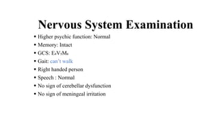
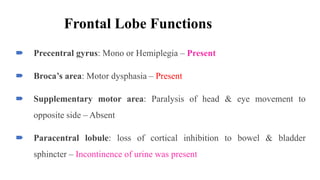
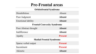
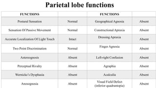
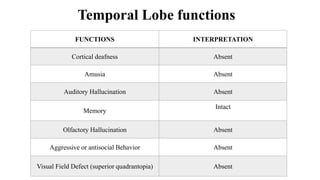
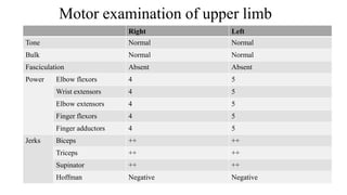
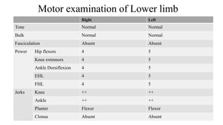
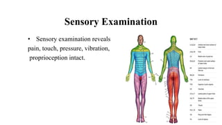
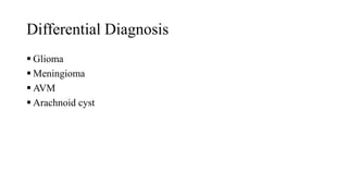
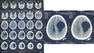
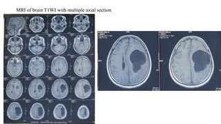
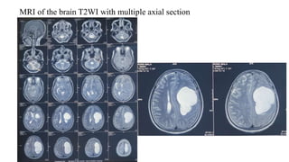
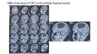
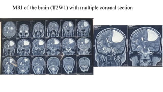
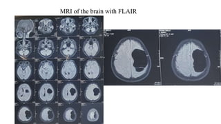
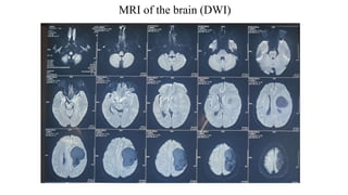
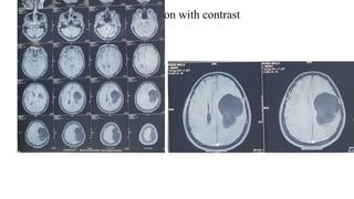
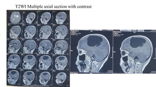
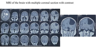
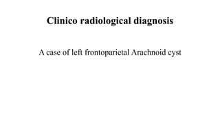
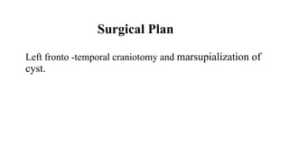
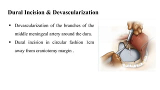
Preoperative case presentation
Dr. Md. Toufiq Hasan
Resident, Phase-A(R-9)
Department Of Neurosurgery
Dhaka Medical College Hospital
On behalf of NSU-Pink Unit
Particulars of the patient
 Name : Mrs Rangmala
 Age : 52 years
 Sex : Female
 Occupation : Housewife
 Marital status : Married
 Address : Zazira, Shariatpur
 Registration no : 71182/242
 Ward : 204
 Bed No : 47 (Non paying)
 DOA : 17/08/2023
Chief Complaints
 Weakness on the right side of the body for 11 months
 Headache for same duration.
 Difficulty in speech for 7 days.
 Continuous dribbling of urine for 7 days
History of present illness
According to the statement of the patient and her sons, she was reasonably well 11
months back. Then, she developed weakness on the right side of the body which was
painless, progressive, initially at right upper limb and then progressed to right lower
limb, could walk freely with hemiplegic gait pattern but from last10 days back, she
experienced a accidental fall and from then she can’t walk and her weakness is
increasing.
History of present illness (Cont.)
She also complained occasional headache for same duration which was
insidious in onset, mild in intensity, dull aching in nature, intermittent in
course, occurring anytime of the day, localized over vertex, no
aggravating factor and relieved by rest without taking any medication
and is not associated with vomiting or any other visual disturbances.
History of present illness (Cont.)
She also gave history of difficulty in speech from 7 days back , initially
she can’t talk but now difficulty in talking with confusion and poor
comprehension.
She also gave history of bladder dysfunction in form of continuous
dribbling of urine for last 7 days.
History of present illness (Cont.)
During the course of his illness, she gave no history of neck pain,
prolonged fever, unexplained weight loss, anorexia, cough, hemoptysis,
prolong use of steroid or close contact with known TB patient.
History of present illness (Cont.)
With the above complaints, she visited Sir Salimullah Medical college
hospital and then referred to Neurosurgery department ,DMCH for
better management.
Associated illness
 She has no history of Diabetes, Hypertension, any heart, kidney,
lung disease
Past medical history
She has no H/O major childhood illness, previous surgery,
accident or injury.
Immunization History
 Vaccinated as per EPI schedule and 3 doses of covid vaccine
Family history
Nothing significant. Girl of a non consanguineous married parents. She
have 4 sons and 1 daughter . Other family members are leading a
healthy life.
Personal history
 She is non smoker, non alcoholic, but betel nut chewer
Socio economic history
 She belongs to a family with low socio-economic condition
Drug history
 Nothing significant.
General Physical Examination
 Appearance: Vacant
 Body built: Below Average
 Less Co-operative
 Decubitus: On choice
 Nutritional status: Below
Average
 Pulse: 72 b/m
 BP: 110/80 mmHg
 Temp: Not raised
 RR: 16/m
 Jaundice
 Clubbing
 Cyanosis
 Anemia
 Leukonychia
 Edema
 Dehydration
 Koilonychia
 Lymphadenopathy
 Catheter in situ
No abnormality
detected
Systemic Examination
• Cardiovascular System-No abnormality detected
• Respiratory System-No abnormality detected
• Alimentary System-No abnormality detected
Nervous System Examination
 Higher psychic function: Normal
 Memory: Intact
 GCS: E₄V₅M₆
 Gait: can’t walk
 Right handed person
 Speech : Normal
 No sign of cerebellar dysfunction
 No sign of meningeal irritation
Cranial Nerve Examination
• Olfactory nerve - Normal
• Optic nerve - Right Left
Visual acuity 6/6 6/6
Visual field No restriction No restriction
Colour vision Intact Intact
Fundoscopy Normal Normal
Oculomotor, Trochlear & Abducens nerve
• Diplopia – Absent
• Nystagmus - Absent
Trigeminal nerve (V)
 Sensory: Intact to all modalities in all the three
divisions.
 Corneal Reflex: Normal (B/E)
 Motor (Muscles of Mastication): Intact Bilaterally
 Jaw Jerk: Absent
Facial nerve (Vll)
• Motor function –
1. Frontalis / Wrinkling of fore head – Intact
2. Orbicularis occuli / Closing of eye – Intact
3. Buccinator / Whistle blowing – Intact
4. Risorius/ Showing of teeth - Normal
5. Platysma / Clenching of neck - Normal
• Taste sensation – Intact
Vestibulocochlear nerve (Vlll)
Vestibulocochlear nerve -
Glossopharyngeal and Vagus nerve
Accessory Nerve (XI)
Deviation of Tongue Absent
Atrophy of tongue muscle Absent
Fasciculation Absent
Frontal Lobe Functions
 Precentral gyrus: Mono or Hemiplegia – Present
 Broca’s area: Motor dysphasia – Present
 Supplementary motor area: Paralysis of head & eye movement to
opposite side – Absent
 Paracentral lobule: loss of cortical inhibition to bowel & bladder
sphincter – Incontinence of urine was present
Pre-Frontal areas
Orbitofrontal Syndrome
Disinhibition Absent
Poor Judgment Absent
Emotional lability Absent
Frontal Convexity Syndrome
Poor Abstract thought Absent
Indifference Absent
Apathy Present
Medial Frontal Syndrome
Sparse verbal output Present
Incontinent Present
Akinetic Absent
Parietal lobe functions
FUNCTIONS FUNCTIONS
Postural Sensation Normal Geographical Agnosia Absent
Sensation Of Passive Movement Normal Constructional Apraxia Absent
Accurate Localization Of Light Touch Intact
Dressing Apraxia
Absent
Two Point Discrimination Normal
Finger Agnosia
Absent
Astereognosis Absent Left-right Confusion Absent
Perceptual Rivalry Absent Agraphia Absent
Wernicke’s Dysphasia Absent Acalculia Absent
Anosognosia Absent
Visual Field Defect
(inferior quadrantopia)
Absent
FUNCTIONS INTERPRETATION
Cortical deafness Absent
Amusia Absent
Auditory Hallucination Absent
Memory
Intact
Olfactory Hallucination Absent
Aggressive or antisocial Behavior Absent
Visual Field Defect (superior quadrantopia) Absent
Temporal Lobe functions
Motor examination of upper limb
Right Left
Tone Normal Normal
Bulk Normal Normal
Fasciculation Absent Absent
Power Elbow flexors 4 5
Wrist extensors 4 5
Elbow extensors 4 5
Finger flexors 4 5
Finger adductors 4 5
Jerks Biceps ++ ++
Triceps ++ ++
Supinator ++ ++
Hoffman Negative Negative
Motor examination of Lower limb
Right Left
Tone Normal Normal
Bulk Normal Normal
Fasciculation Absent Absent
Power Hip flexors 4 5
Knee extensors 4 5
Ankle Dorsiflexion 4 5
EHL 4 5
FHL 4 5
Jerks Knee ++ ++
Ankle ++ ++
Planter Flexor Flexor
Clonus Absent Absent
Sensory Examination
• Sensory examination reveals
pain, touch, pressure, vibration,
proprioception intact.
Examination of spine
No scoliosis, gibbus, kyphotic deformity
No visible midline or paraspinal mass
Overlying skin condition –normal
Local temperature –not raised
Focal tenderness is absent
Provisional diagnosis
• A case of left Supratentorial Space occupying lesion
Differential Diagnosis
 Glioma
 Meningioma
 AVM
 Arachnoid cyst
INVESTIGATIONS
CT Scan of the head
MRI of the brain with contrast
MRI of brain T1WI with multiple axial section
MRI of the brain T2WI with multiple axial section
MRI of the brain (T1W1) with multiple Sagittal section
MRI of the brain (T2W1) with multiple coronal section
MRI of the brain with FLAIR
MRI of the brain (DWI)
T1W1 multiple axial section with contrast
T2WI Multiple axial section with contrast
MRI of the brain with multiple coronal section with contrast
Clinico radiological diagnosis
A case of left frontoparietal Arachnoid cyst
Surgical Plan
Left fronto -temporal craniotomy and marsupialization of
cyst.
Positioning
•Supine position
•The neck rotated towards left.
•Head should be placed above heart level.
•Padding should be placed at bony
prominence
Incision
Horse shoe incision
Craniotomy
Making 2 or 3 burr hole centering the cyst & each burr hole will be
connected.
The dura will be dissected off from inner table with penfield - 3
dissector.
Dural Incision & Devascularization
 Devascularization of the branches of the
middle meningeal artery around the dura.
 Dural incision in circular fashion 1cm
away from craniotomy margin .
Cyst Removal
 Microsurgical marsupialization of cyst.
Hemostasis
 Bipolar
 Gelatin sponge
 Fibrillar / Surgicel
Closure
 Closure of dura mater necessarily implies application of graft.
 For this purpose, local tissue can be used such as galea
aponeurotica, pericranium, fascia lata or synthetic dural
substitute.
 Water tight fashion dural closure will be ensured.
Complications
Per-operative:
 Excessive hemorrhage during craniotomy & cyst removal
 Injury to underlying cortex
 Cerebral contusion, swelling or infarction
 Air embolism
Post-operative:
 Onset of new neurological deficit.
 Seizure
 CSF leakage
 Pseudomeningocele
 Meningitis
 Wound infection
Case 22.8.23.pptx

Case 22.8.23.pptx

  • 1.
    Preoperative case presentation Dr.Md. Toufiq Hasan Resident, Phase-A(R-9) Department Of Neurosurgery Dhaka Medical College Hospital On behalf of NSU-Pink Unit
  • 2.
    Particulars of thepatient  Name : Mrs Rangmala  Age : 52 years  Sex : Female  Occupation : Housewife  Marital status : Married  Address : Zazira, Shariatpur  Registration no : 71182/242  Ward : 204  Bed No : 47 (Non paying)  DOA : 17/08/2023
  • 3.
    Chief Complaints  Weaknesson the right side of the body for 11 months  Headache for same duration.  Difficulty in speech for 7 days.  Continuous dribbling of urine for 7 days
  • 4.
    History of presentillness According to the statement of the patient and her sons, she was reasonably well 11 months back. Then, she developed weakness on the right side of the body which was painless, progressive, initially at right upper limb and then progressed to right lower limb, could walk freely with hemiplegic gait pattern but from last10 days back, she experienced a accidental fall and from then she can’t walk and her weakness is increasing.
  • 5.
    History of presentillness (Cont.) She also complained occasional headache for same duration which was insidious in onset, mild in intensity, dull aching in nature, intermittent in course, occurring anytime of the day, localized over vertex, no aggravating factor and relieved by rest without taking any medication and is not associated with vomiting or any other visual disturbances.
  • 6.
    History of presentillness (Cont.) She also gave history of difficulty in speech from 7 days back , initially she can’t talk but now difficulty in talking with confusion and poor comprehension. She also gave history of bladder dysfunction in form of continuous dribbling of urine for last 7 days.
  • 7.
    History of presentillness (Cont.) During the course of his illness, she gave no history of neck pain, prolonged fever, unexplained weight loss, anorexia, cough, hemoptysis, prolong use of steroid or close contact with known TB patient.
  • 8.
    History of presentillness (Cont.) With the above complaints, she visited Sir Salimullah Medical college hospital and then referred to Neurosurgery department ,DMCH for better management.
  • 9.
    Associated illness  Shehas no history of Diabetes, Hypertension, any heart, kidney, lung disease
  • 10.
    Past medical history Shehas no H/O major childhood illness, previous surgery, accident or injury.
  • 11.
    Immunization History  Vaccinatedas per EPI schedule and 3 doses of covid vaccine
  • 12.
    Family history Nothing significant.Girl of a non consanguineous married parents. She have 4 sons and 1 daughter . Other family members are leading a healthy life.
  • 13.
    Personal history  Sheis non smoker, non alcoholic, but betel nut chewer
  • 14.
    Socio economic history She belongs to a family with low socio-economic condition
  • 15.
  • 16.
    General Physical Examination Appearance: Vacant  Body built: Below Average  Less Co-operative  Decubitus: On choice  Nutritional status: Below Average  Pulse: 72 b/m  BP: 110/80 mmHg  Temp: Not raised  RR: 16/m  Jaundice  Clubbing  Cyanosis  Anemia  Leukonychia  Edema  Dehydration  Koilonychia  Lymphadenopathy  Catheter in situ No abnormality detected
  • 17.
    Systemic Examination • CardiovascularSystem-No abnormality detected • Respiratory System-No abnormality detected • Alimentary System-No abnormality detected
  • 18.
    Nervous System Examination Higher psychic function: Normal  Memory: Intact  GCS: E₄V₅M₆  Gait: can’t walk  Right handed person  Speech : Normal  No sign of cerebellar dysfunction  No sign of meningeal irritation
  • 19.
    Cranial Nerve Examination •Olfactory nerve - Normal • Optic nerve - Right Left Visual acuity 6/6 6/6 Visual field No restriction No restriction Colour vision Intact Intact Fundoscopy Normal Normal
  • 21.
    Oculomotor, Trochlear &Abducens nerve • Diplopia – Absent • Nystagmus - Absent
  • 22.
    Trigeminal nerve (V) Sensory: Intact to all modalities in all the three divisions.  Corneal Reflex: Normal (B/E)  Motor (Muscles of Mastication): Intact Bilaterally  Jaw Jerk: Absent
  • 23.
    Facial nerve (Vll) •Motor function – 1. Frontalis / Wrinkling of fore head – Intact 2. Orbicularis occuli / Closing of eye – Intact 3. Buccinator / Whistle blowing – Intact 4. Risorius/ Showing of teeth - Normal 5. Platysma / Clenching of neck - Normal • Taste sensation – Intact
  • 24.
  • 25.
  • 26.
    Accessory Nerve (XI) Deviationof Tongue Absent Atrophy of tongue muscle Absent Fasciculation Absent
  • 27.
    Frontal Lobe Functions Precentral gyrus: Mono or Hemiplegia – Present  Broca’s area: Motor dysphasia – Present  Supplementary motor area: Paralysis of head & eye movement to opposite side – Absent  Paracentral lobule: loss of cortical inhibition to bowel & bladder sphincter – Incontinence of urine was present
  • 28.
    Pre-Frontal areas Orbitofrontal Syndrome DisinhibitionAbsent Poor Judgment Absent Emotional lability Absent Frontal Convexity Syndrome Poor Abstract thought Absent Indifference Absent Apathy Present Medial Frontal Syndrome Sparse verbal output Present Incontinent Present Akinetic Absent
  • 29.
    Parietal lobe functions FUNCTIONSFUNCTIONS Postural Sensation Normal Geographical Agnosia Absent Sensation Of Passive Movement Normal Constructional Apraxia Absent Accurate Localization Of Light Touch Intact Dressing Apraxia Absent Two Point Discrimination Normal Finger Agnosia Absent Astereognosis Absent Left-right Confusion Absent Perceptual Rivalry Absent Agraphia Absent Wernicke’s Dysphasia Absent Acalculia Absent Anosognosia Absent Visual Field Defect (inferior quadrantopia) Absent
  • 30.
    FUNCTIONS INTERPRETATION Cortical deafnessAbsent Amusia Absent Auditory Hallucination Absent Memory Intact Olfactory Hallucination Absent Aggressive or antisocial Behavior Absent Visual Field Defect (superior quadrantopia) Absent Temporal Lobe functions
  • 31.
    Motor examination ofupper limb Right Left Tone Normal Normal Bulk Normal Normal Fasciculation Absent Absent Power Elbow flexors 4 5 Wrist extensors 4 5 Elbow extensors 4 5 Finger flexors 4 5 Finger adductors 4 5 Jerks Biceps ++ ++ Triceps ++ ++ Supinator ++ ++ Hoffman Negative Negative
  • 32.
    Motor examination ofLower limb Right Left Tone Normal Normal Bulk Normal Normal Fasciculation Absent Absent Power Hip flexors 4 5 Knee extensors 4 5 Ankle Dorsiflexion 4 5 EHL 4 5 FHL 4 5 Jerks Knee ++ ++ Ankle ++ ++ Planter Flexor Flexor Clonus Absent Absent
  • 33.
    Sensory Examination • Sensoryexamination reveals pain, touch, pressure, vibration, proprioception intact.
  • 34.
    Examination of spine Noscoliosis, gibbus, kyphotic deformity No visible midline or paraspinal mass Overlying skin condition –normal Local temperature –not raised Focal tenderness is absent
  • 35.
  • 36.
    Provisional diagnosis • Acase of left Supratentorial Space occupying lesion
  • 37.
    Differential Diagnosis  Glioma Meningioma  AVM  Arachnoid cyst
  • 38.
  • 39.
    CT Scan ofthe head
  • 41.
    MRI of thebrain with contrast
  • 42.
    MRI of brainT1WI with multiple axial section
  • 43.
    MRI of thebrain T2WI with multiple axial section
  • 44.
    MRI of thebrain (T1W1) with multiple Sagittal section
  • 45.
    MRI of thebrain (T2W1) with multiple coronal section
  • 46.
    MRI of thebrain with FLAIR
  • 47.
    MRI of thebrain (DWI)
  • 48.
    T1W1 multiple axialsection with contrast
  • 49.
    T2WI Multiple axialsection with contrast
  • 50.
    MRI of thebrain with multiple coronal section with contrast
  • 51.
    Clinico radiological diagnosis Acase of left frontoparietal Arachnoid cyst
  • 52.
    Surgical Plan Left fronto-temporal craniotomy and marsupialization of cyst.
  • 53.
    Positioning •Supine position •The neckrotated towards left. •Head should be placed above heart level. •Padding should be placed at bony prominence
  • 54.
  • 55.
    Craniotomy Making 2 or3 burr hole centering the cyst & each burr hole will be connected. The dura will be dissected off from inner table with penfield - 3 dissector.
  • 56.
    Dural Incision &Devascularization  Devascularization of the branches of the middle meningeal artery around the dura.  Dural incision in circular fashion 1cm away from craniotomy margin .
  • 57.
    Cyst Removal  Microsurgicalmarsupialization of cyst.
  • 58.
    Hemostasis  Bipolar  Gelatinsponge  Fibrillar / Surgicel
  • 59.
    Closure  Closure ofdura mater necessarily implies application of graft.  For this purpose, local tissue can be used such as galea aponeurotica, pericranium, fascia lata or synthetic dural substitute.  Water tight fashion dural closure will be ensured.
  • 60.
    Complications Per-operative:  Excessive hemorrhageduring craniotomy & cyst removal  Injury to underlying cortex  Cerebral contusion, swelling or infarction  Air embolism
  • 61.
    Post-operative:  Onset ofnew neurological deficit.  Seizure  CSF leakage  Pseudomeningocele  Meningitis  Wound infection

Editor's Notes

  • #21 Disc colour is normal, margin well defined, vessel count is 8 on both eye, peripheral retina is also normal
  • #29 Akinetic- not having the ability to move body
  • #33 Reflexes can be graded 0= absent +-= present only with reinforcement 1+=present but depressed 2+= Normal 3+= increased 4+= clonus
  • #41 Plain CT scan of Head revealed Smooth bordered non calcified cystic lesion with single septation in left frontoparietal region with density similar to CSF with crowding of adjacent sulcus & gyrus, effacement of adjacent lateral ventricle and slight midline shifting Rest of the brain parenchyma, bony calvaria is normal. No extracalvarial lesion is visible.
  • #43 MRI of brain T1WI multiple axial sections showing a well defined fairly large lesion measuring 7.5cm * 4.5cm in diameter, smooth bordered homogenously hypointense lesion in left frontoparietal region with intensity similar to CSF. There is crowding of adjacent sulcus & gyrus and effacement of adjacent lateral ventricle and slight midline shifting Rest of the brain parenchyma is normal. No extracalvarial lesion is present.
  • #44 T2WI shows the lesion is homogenously hyperintense.
  • #45 Sagittal image shows the lesion compressed and distorted the lateral ventricle.
  • #46 In coronal view the lesion pused the adjacent to left lateral ventricle medially and inferiorly.
  • #47 FLAIR image shows the lesion is homogenously hypointense
  • #48 No restriction; nearly identical to ventricles Diffusion restricted –pheriphery –neoplasm Centrally-radiation necrosis
  • #49 Contrast enhanced axial MRI shows there is no contrast uptake by the lesion and also there is no ring enhancement
  • #50 Contrast is not uptaken by the lesion.
  • #52 D/D- Epidermoid cyst, subdural hygroma, Epidermoid Cyst • Scalloped margins • Insinuating growth pattern ○ Creeps along into CSF cisterns ○ Surrounds, engulfs vessels/nerves – ACs displace, but usually do not engulf vessels, cranial nerves • Does not suppress on FLAIR • Restricted diffusion (bright) on DWI Chronic Subdural Hematoma • Signal not identical to CSF • Often bilateral, lentiform-shaped • ± enhancing membrane • Look for foci of "blooming" on T2* ○ < 5% of ACs hemorrhage Subdural Hygroma • Often bilateral • Crescentic or flat configuration