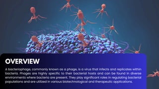
OVERVIEW
A bacteriophage, commonlyknown as a phage, is a virus that infects and replicates within
bacteria. Phages are highly specific to their bacterial hosts and can be found in diverse
environments where bacteria are present. They play significant roles in regulating bacterial
populations and are utilized in various biotechnological and therapeutic applications.
PCR Setup
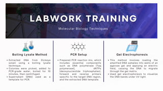
Boiling LysateMethod Gel Electrophoresis
Extracted DNA from Dickeya
solani using a boiling lysate
technique.
Colonies were picked, added to
PCR-grade water, boiled for 10
minutes, then centrifuged.
Supernatant (DNA) used as a
template for PCR.
Prepared PCR reaction mix, which
includes essential components
such as DNA polymerase (Taq
polymerase), dNTPs
(deoxynucleotide triphosphates),
forward and reverse primers
specific to the target DNA region,
and the extracted DNA template.
This method involves loading the
amplified DNA samples into wells of an
agarose gel and applying an electric
field, causing the DNA to migrate
through the gel matrix.
Used gel electrophoresis to visualize
the DNA bands under UV light.
Molecular Biology Techniques
11.
Spot Test Assay
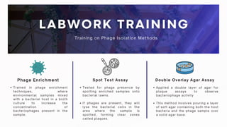
PhageEnrichment Double Overlay Agar Assay
Trained in phage enrichment
techniques, where
environmental samples mixed
with a bacterial host in a broth
culture to increase the
concentration of
bacteriophages present in the
sample.
Tested for phage presence by
spotting enriched samples onto
bacterial lawns.
If phages are present, they will
lyse the bacterial cells in the
area where the sample is
spotted, forming clear zones
called plaques.
Applied a double layer of agar for
plaque assays to observe
bacteriophage activity
This method involves pouring a layer
of soft agar containing both the host
bacteria and the phage sample over
a solid agar base.
Training on Phage Isolation Methods
12.
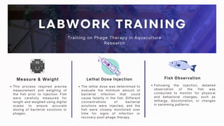
Lethal Dose Injection
Measure& Weight Fish Observation
This process required precise
measurement and weighing of
the fish prior to injection. Fish
were carefully measured for
length and weighed using digital
scales to ensure accurate
dosing of bacterial solutions or
phages.
The lethal dose was determined to
evaluate the minimum amount of
bacterial infection that could
cause fatality in the fish. Different
concentrations of bacterial
solutions were injected, and the
fish were closely monitored over
time for signs of infection or
recovery post-phage therapy.
Following the injection, detailed
observation of the fish was
conducted to monitor for physical
and behavioral changes, such as
lethargy, discoloration, or changes
in swimming patterns.
Training on Phage Therapy in Aquaculture
Research
13.
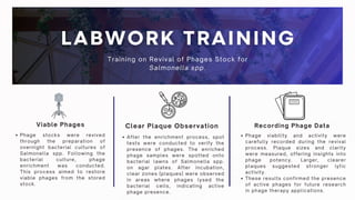
Viable Phages
Phage stockswere revived
through the preparation of
overnight bacterial cultures of
Salmonella spp. Following the
bacterial culture, phage
enrichment was conducted.
This process aimed to restore
viable phages from the stored
stock.
Recording Phage Data
Clear Plaque Observation
After the enrichment process, spot
tests were conducted to verify the
presence of phages. The enriched
phage samples were spotted onto
bacterial lawns of Salmonella spp.
on agar plates. After incubation,
clear zones (plaques) were observed
in areas where phages lysed the
bacterial cells, indicating active
phage presence.
Training on Revival of Phages Stock for
Salmonella spp.
Phage viability and activity were
carefully recorded during the revival
process. Plaque sizes and clarity
were measured, offering insights into
phage potency. Larger, clearer
plaques suggested stronger lytic
activity.
These results confirmed the presence
of active phages for future research
in phage therapy applications.
14.
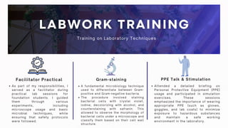
Gram-staining
Facilitator Practical PPETalk & Stimulation
A fundamental microbiology technique
used to differentiate between Gram-
positive and Gram-negative bacteria.
The procedure involved staining
bacterial cells with crystal violet,
iodine, decolorizing with alcohol, and
counterstaining with safranin. This
allowed to observe the morphology of
bacterial cells under a microscope and
classify them based on their cell wall
structure.
As part of my responsibilities, I
served as a facilitator during
practical lab sessions for
foundation students. I guided
them through various
experiments, including
microscope usage and basic
microbial techniques, while
ensuring that safety protocols
were followed.
Attended a detailed briefing on
Personal Protective Equipment (PPE)
usage and participated in simulation
exercises. These sessions
emphasized the importance of wearing
appropriate PPE (such as gloves,
goggles, and lab coats) to minimize
exposure to hazardous substances
and maintain a safe working
environment in the laboratory.
Training on Laboratory Techniques
15.
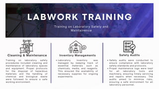
Inventory Managements
Cleaning &Maintenance Safety Audits
Training on laboratory safety
procedures included cleaning and
maintenance of laboratory spaces
and equipment. Proper protocols
for the disposal of hazardous
materials and the handling of
chemical and biological waste
were followed to ensure a safe
working environment.
Laboratory inventory was
managed by keeping track of
essential materials such as
chemicals, media, and reagents.
This ensured the availability of
necessary supplies for ongoing
experiments.
Safety audits were conducted to
ensure compliance with laboratory
safety standards and protocols.
Proper maintenance logs were kept
for all laboratory tools and
machinery, ensuring timely servicing
and repairs when necessary. The
audits aimed to minimize risks,
ensuring a safe environment for all
laboratory personnel.
Training on Laboratory Safety and
Maintainence
16.
Sample Preparation
Lorem ipsumdolor sit amet, consectetur
adipiscing elit. Duis vulputate nulla at ante
rhoncus, vel efficitur felis condimentum. Proin
odio odio.
Prepare Overnight Culture
Lorem ipsum dolor sit amet, consectetur
adipiscing elit. Duis vulputate nulla at ante
rhoncus, vel efficitur felis condimentum. Proin
odio odio.
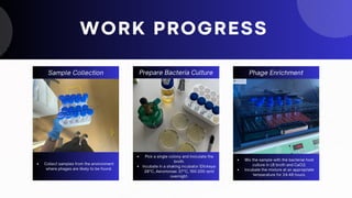
WORK PROGRESS
Phage Enrichment
Lorem ipsum dolor sit amet, consectetur
adipiscing elit. Duis vulputate nulla at ante
rhoncus, vel efficitur felis condimentum. Proin
odio odio.
Sample Collection
Collect samples from the environment
where phages are likely to be found.
Prepare Bacteria Culture
Pick a single colony and inoculate the
broth.
Incubate in a shaking incubator (Dickeya:
28°C, Aeromonas: 37°C, 150-200 rpm)
overnight.
Phage Enrichment
Mix the sample with the bacterial host
culture in LB broth and CaCl2.
Incubate the mixture at an appropriate
temperature for 24-48 hours.
17.
Centridfuge and Filter
Loremipsum dolor sit amet, consectetur
adipiscing elit. Duis vulputate nulla at ante
rhoncus, vel efficitur felis condimentum. Proin
odio odio.
Spot Test Assay
Lorem ipsum dolor sit amet, consectetur
adipiscing elit. Duis vulputate nulla at ante
rhoncus, vel efficitur felis condimentum. Proin
odio odio.
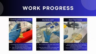
WORK PROGRESS
Bacteria Lawn
Lorem ipsum dolor sit amet, consectetur
adipiscing elit. Duis vulputate nulla at ante
rhoncus, vel efficitur felis condimentum.
Proin odio odio.
Centrifugation & Filtration
Centrifuge the enriched culture to remove
bacterial cells and debris.
Filter the supernatant using a 0.45 and
0.22 µm filter to obtain a phage lysate.
Spot Test Assay
Test the phage against different bacterial
strains by spotting phage lysate onto a lawn
of bacteria on agar plates.
Observe the formation of plaques (clear zone
formation).
Bacteria Lawn
Pick a colony on agar plate using cotton
swab.
Swab the entire surface of the agar plate
uniformly to ensure even coverage.
18.
10-fold serial dilution
Loremipsum dolor sit amet, consectetur
adipiscing elit. Duis vulputate nulla at ante
rhoncus, vel efficitur felis condimentum. Proin
odio odio.
Double Overlay Assay
Lorem ipsum dolor sit amet, consectetur
adipiscing elit. Duis vulputate nulla at ante
rhoncus, vel efficitur felis condimentum. Proin
odio odio.
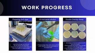
Preparing Soft Agar
Lorem ipsum dolor sit amet, consectetur
adipiscing elit. Duis vulputate nulla at ante
rhoncus, vel efficitur felis condimentum. Proin
odio odio.
WORK PROGRESS
10-fold serial dilution
Prepare phage stock, host culture, and LB
broth. Perform a 10-fold serial dilution of
the phage stock.
Double Overlay Assay
Add phages to the host cultures in a 1:1
ratio and incubate.
Mix the phage-host mixture with soft agar
and pour it onto LB agar plates.
Swirl the plates to spread the agar evenly
and count the plaques after incubation.
Preparing Soft Agar
Use 0.3% soft agar and LB Broth.
Stir the mixture until dissolved.
Aliquot the soft agar into sterile tubes and
pour over plates for plaque assays
20.
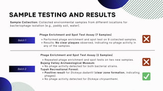
SAMPLE TESTING ANDRESULTS
Sample Collection: Collected environmental samples from different locations for
bacteriophage isolation (e.g., paddy soil, water).
Batch 1
Phage Enrichment and Spot Test Assay (7 Samples)
Performed phage enrichment and spot test on 9 collected samples.
Results: No clear plaques observed, indicating no phage activity in
any of the samples.
Batch 2
Phage Enrichment and Spot Test Assay (2 Samples)
Repeated phage enrichment and spot tests on two new samples.
Bujang Valley Archaeological Museum:
No phage activity detected for both bacterial strains.
Tupah Recreational Forest:
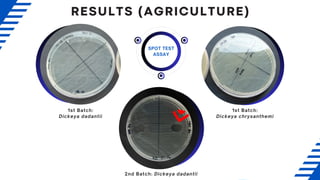
Positive result for Dickeya dadantii (clear zone formation, indicating
phages).
No phage activity detected for Dickeya chrysanthemi.
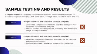
SAMPLE TESTING ANDRESULTS
Sample Collection: Collected environmental samples from different locations for
bacteriophage isolation (e.g., fish pond water, sewage water, fish farm water and etc).
Batch 1
Batch 2
Phage Enrichment and Spot Test Assay (9 Samples)
Conducted sample enrichment and spot test assays on nine
collected environmental samples.
Results showed no clear plaques, indicating null results (no
phage activity detected).
Phage Enrichment and Spot Test Assay (2 Samples)
Repeated phage enrichment and spot tests on the two new
environmental samples.
Again obtained null results (no phage activity detected).
23.
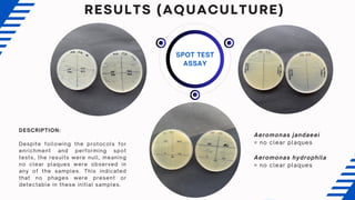
SPOT TEST
ASSAY
DESCRIPTION:
Despite followingthe protocols for
enrichment and performing spot
tests, the results were null, meaning
no clear plaques were observed in
any of the samples. This indicated
that no phages were present or
detectable in these initial samples.
Aeromonas jandaeei
= no clear plaques
Aeromonas hydrophila
= no clear plaques
RESULTS (AQUACULTURE)
25.
DISCUSSION
The phage enrichmentand spot test assays were first conducted on the initial set of collected
samples, followed by a repeat experiment using newly collected additional samples. Despite
meticulous adherence to the enrichment and spot test protocols, no clear plaques were observed
on any of the plates, indicating null results, or the absence of phage activity.
The lack of visible plaques in the assays suggests that no viable phages were present or able to
infect the target bacteria in the tested samples. There are several potential reasons for this
outcome:
Phage absence: The environmental samples might simply lack bacteriophages specific to the
targeted bacterial strains.
1.
Low phage concentration: Phages might be present in the samples but at concentrations too
low to be detected by the spot test assay. Enrichment might not have amplified the phage
numbers sufficiently for visible plaque formation.
2.
Phage-bacteria specificity: It is possible that the collected samples contain phages that do not
target the tested bacterial strains, or the bacterial strains used are resistant to any phages
present.
3.