Inflammation Protocols 1st Edition Paul G Winyard Auth Paul G Winyard
Inflammation Protocols 1st Edition Paul G Winyard Auth Paul G Winyard
Inflammation Protocols 1st Edition Paul G Winyard Auth Paul G Winyard
Inflammation Protocols 1st Edition Paul G Winyard Auth Paul G Winyard
Inflammation Protocols 1st Edition Paul G Winyard Auth Paul G Winyard
1.
Inflammation Protocols 1stEdition Paul G
Winyard Auth Paul G Winyard download
https://ebookbell.com/product/inflammation-protocols-1st-edition-
paul-g-winyard-auth-paul-g-winyard-4327154
Explore and download more ebooks at ebookbell.com
2.
Here are somerecommended products that we believe you will be
interested in. You can click the link to download.
Inflammation Methods And Protocols 1st Edition Bjrn E Clausen
https://ebookbell.com/product/inflammation-methods-and-protocols-1st-
edition-bjrn-e-clausen-5735810
Inflammation And Cancer Methods And Protocols Methods In Molecular
Biology 2691 2nd Edition Brendan J Jenkins Editor
https://ebookbell.com/product/inflammation-and-cancer-methods-and-
protocols-methods-in-molecular-biology-2691-2nd-edition-brendan-j-
jenkins-editor-50819718
Inflammation And Cancer Methods And Protocols Volume 2 Molecular
Analysis And Pathways 1st Edition Yusuke Hiraku
https://ebookbell.com/product/inflammation-and-cancer-methods-and-
protocols-volume-2-molecular-analysis-and-pathways-1st-edition-yusuke-
hiraku-4501792
Inflammation And Cancer Methods And Protocols 1st Edition Brendan J
Jenkins Eds
https://ebookbell.com/product/inflammation-and-cancer-methods-and-
protocols-1st-edition-brendan-j-jenkins-eds-6840712
3.
Inflammation And CancerMethods And Protocols Volume 1 Experimental
Models And Practical Approaches 1st Edition Yelena Golubeva
https://ebookbell.com/product/inflammation-and-cancer-methods-and-
protocols-volume-1-experimental-models-and-practical-approaches-1st-
edition-yelena-golubeva-1301348
Human Airway Inflammation Sampling Techniques And Analytical Protocols
1st Edition Stephen T Holgate Auth
https://ebookbell.com/product/human-airway-inflammation-sampling-
techniques-and-analytical-protocols-1st-edition-stephen-t-holgate-
auth-4327068
Cell Migration In Inflammation And Immunity Methods And Protocols 1st
Edition Silvano Sozzani
https://ebookbell.com/product/cell-migration-in-inflammation-and-
immunity-methods-and-protocols-1st-edition-silvano-sozzani-4327178
Crohns Disease Aip Cookbook Recipes To Reduce Inflammation And
Eliminate Food Triggers On The Autoimmune Protocol Joshua Bradley
https://ebookbell.com/product/crohns-disease-aip-cookbook-recipes-to-
reduce-inflammation-and-eliminate-food-triggers-on-the-autoimmune-
protocol-joshua-bradley-53855946
Inflammation And Obesity A New And Novel Approach To Manage Obesity
And Its Consequences Raman Mehrzad
https://ebookbell.com/product/inflammation-and-obesity-a-new-and-
novel-approach-to-manage-obesity-and-its-consequences-raman-
mehrzad-46450198
M E TH O D S I N M O L E C U L A R B I O L O G YTM
John M. Walker, SERIES EDITOR
240. Mammalian Artificial Chromosomes: Methods and Proto-
cols, edited by Vittorio Sgaramella and Sandro Eridani, 2003
239. Cell Migration in Inflammation and Immunity: Methods
and Protocols, edited by Daniele D’Ambrosio and Francesco
Sinigaglia, 2003
238. Biopolymer Methods in Tissue Engineering, edited by
Anthony P. Hollander and Paul V. Hatton, 2003
237. G Protein Signaling: Methods and Protocols, edited by Alan
V. Smrcka, 2003
236. Plant Functional Genomics: Methods and Protocols, edited
by Erich Grotewold, 2003
235. E. coli Plasmid Vectors: Methods and Applications, edited
by Nicola Casali and Andrew Preston, 2003
234. p53 Protocols, edited by Sumitra Deb and Swati Palit Deb,
2003
233. Protein Kinase C Protocols, edited by Alexandra C. New-
ton, 2003
232. Protein Misfolding and Disease: Principles and Protocols,
edited by Peter Bross and Niels Gregersen, 2003
231. Directed Evolution Library Creation: Methods and Proto-
cols, edited by Frances H. Arnold and George Georgiou, 2003
230. Directed Enzyme Evolution: Screening and Selection Meth-
ods, edited by Frances H. Arnold and George Georgiou, 2003
229. Lentivirus Gene Engineering Protocols, edited by Maurizio
Federico, 2003
228. Membrane Protein Protocols: Expression, Purification, and
Characterization, edited by Barry S. Selinsky, 2003
227. Membrane Transporters: Methods and Protocols, edited by
Qing Yan, 2003
226. PCR Protocols, Second Edition, edited by John M. S. Bartlett
and David Stirling, 2003
225. Inflammation Protocols, edited by Paul G. Winyard and
Derek A. Willoughby, 2003
224. Functional Genomics: Methods and Protocols, edited by
Michael J. Brownstein and Arkady B. Khodursky, 2003
223. Tumor Suppressor Genes: Volume 2: Regulation, Function,
and Medicinal Applications, edited by Wafik S. El-Deiry, 2003
222. Tumor Suppressor Genes: Volume 1: Pathways and Isola-
tion Strategies, edited by Wafik S. El-Deiry, 2003
221. Generation of cDNA Libraries: Methods and Protocols,
edited by Shao-Yao Ying, 2003
220. Cancer Cytogenetics: Methods and Protocols, edited by John
Swansbury, 2003
219. Cardiac Cell and Gene Transfer: Principles, Protocols, and
Applications, edited by Joseph M. Metzger, 2003
218. Cancer Cell Signaling: Methods and Protocols, edited by
David M. Terrian, 2003
217. Neurogenetics: Methods and Protocols, edited by Nicholas
T. Potter, 2003
216. PCR Detection of Microbial Pathogens: Methods and Pro-
tocols, edited by Konrad Sachse and Joachim Frey, 2003
215. Cytokines and Colony Stimulating Factors: Methods and
Protocols, edited by Dieter Körholz and Wieland Kiess, 2003
214. Superantigen Protocols, edited by Teresa Krakauer, 2003
213. Capillary Electrophoresis of Carbohydrates, edited by
Pierre Thibault and Susumu Honda, 2003
212. Single Nucleotide Polymorphisms: Methods and Protocols,
edited by Pui-Yan Kwok, 2003
211. Protein Sequencing Protocols, 2nd ed., edited by Bryan John
Smith, 2003
210. MHC Protocols, edited by Stephen H. Powis and Robert W.
Vaughan, 2003
209. Transgenic Mouse Methods and Protocols, edited by Mar-
ten Hofker and Jan van Deursen, 2003
208. Peptide Nucleic Acids: Methods and Protocols, edited by
Peter E. Nielsen, 2002
207. Recombinant Antibodies for Cancer Therapy: Methods and
Protocols. edited by Martin Welschof and Jürgen Krauss, 2002
206. Endothelin Protocols, edited by Janet J. Maguire and Anthony
P. Davenport, 2002
205. E. coli Gene Expression Protocols, edited by Peter E.
Vaillancourt, 2002
204. Molecular Cytogenetics: Protocols and Applications, edited
by Yao-Shan Fan, 2002
203. In Situ Detection of DNA Damage: Methods and Protocols,
edited by Vladimir V. Didenko, 2002
202. Thyroid Hormone Receptors: Methods and Protocols, ed-
ited by Aria Baniahmad, 2002
201. Combinatorial Library Methods and Protocols, edited by
Lisa B. English, 2002
200. DNA Methylation Protocols, edited by Ken I. Mills and
Bernie H. Ramsahoye, 2002
199. Liposome Methods and Protocols, edited by Subhash C.
Basu and Manju Basu, 2002
198. Neural Stem Cells: Methods and Protocols, edited by Tanja
Zigova, Juan R. Sanchez-Ramos, and Paul R. Sanberg, 2002
197. Mitochondrial DNA: Methods and Protocols, edited by Will-
iam C. Copeland, 2002
196. Oxidants and Antioxidants: Ultrastructure and Molecular
Biology Protocols, edited by Donald Armstrong, 2002
195. Quantitative Trait Loci: Methods and Protocols, edited by
Nicola J. Camp and Angela Cox, 2002
194. Posttranslational Modifications of Proteins: Tools for Func-
tional Proteomics, edited by Christoph Kannicht, 2002
193. RT-PCR Protocols, edited by Joe O’Connell, 2002
192. PCR Cloning Protocols, 2nd ed., edited by Bing-Yuan Chen
and Harry W. Janes, 2002
191. Telomeres and Telomerase: Methods and Protocols, edited
by John A. Double and Michael J. Thompson, 2002
190. High Throughput Screening: Methods and Protocols, edited
by William P. Janzen, 2002
189. GTPase Protocols: The RAS Superfamily, edited by Edward
J. Manser and Thomas Leung, 2002
188. Epithelial Cell Culture Protocols, edited by Clare Wise,
2002
187. PCR Mutation Detection Protocols, edited by Bimal D. M.
Theophilus and Ralph Rapley, 2002
186. Oxidative Stress Biomarkers and Antioxidant Protocols,
edited by Donald Armstrong, 2002
7.
Humana Press Totowa,New Jersey
M E T H O D S I N M O L E C U L A R B I O L O G Y™
Inflammation
Protocols
Edited by
Paul G. Winyard
and
Derek A. Willoughby
William Harvey Research Institute,
London, UK
v
Preface
Inflammation has beendescribed as the basis of many pathologies of
human disease. When one considers the updated signs of inflammation, they
would be vasodilation, cell migration, and, in the case of chronic inflamma-
tion, cell proliferation, often with an underlying autoimmune basis. Gener-
ally, inflammation may be divided into acute, chronic, and autoimmune, al-
though the editors believe that most, if not all, chronic states are often the
result of an autoimmune response to an endogenous antigen. Thus, a proper
understanding of the inflammatory basis may provide clues to new therapeu-
tic targets not only in classical inflammatory diseases, but atherosclerosis,
cancer, and ischemic heart disease as well.
The lack of advances in classical inflammatory diseases, such as rheu-
matoid arthritis, may in part arise from a failure to classify the disease into
different forms. That different forms exist is exemplified in patients with dif-
fering responses to existing antiinflammatory drugs, ranging from
nonresponders to very positive responders for a particular nonsteroidal anti-
inflammatory drug (NSAID). Though researchers have progressively unrav-
eled the mechanisms, the story is far from complete. It should also be noted
that the inflammatory response is part of the innate immune response, or to
use John Hunter’s words in 1795, “inflammation is a salutary response.” That
may be applied in particular to the defensive response to invading microor-
ganisms.
Because of the large multidisciplinary scope of inflammation research,
it is inevitable that a protocols collection such as Inflammation Protocols will
represent a limited selection of the more important tools for studying
inflammation. The editors have therefore focused this volume on those methods
that they believe are most likely to be applicable to investigations of potential
new antiinflammatory drugs in active target areas for R&D such as
transcription factors, adhesion molecules, cyclooxygenase-2 (COX-2)
inhibitors, nitric oxide synthases, and metalloproteinases. Some of the
experimental protocols (especially the in vitro ones) described in this book
are generic, in the sense that they are applicable to the study of many different
inflammatory diseases, whereas others attempt to model particular human
inflammatory diseases or particular aspects of inflammation.
10.
vi Preface
Inflammation Protocolshas been divided into three sections: (1) in
vitro systems for studying aspects of inflammation, (2) in vivo models, and
(3) relevant pharmacodynamic measurements for the assessment of antiin-
flammatory compounds. Each section opens with one or two introductory chap-
ters that attempt to provide an overview, or at least viewpoints, of the signifi-
cance of the methods in that section of the book.
There is no doubt that we will have introduced a particular “flavor” to
the book that might not be to everyone’s taste–inflammation researchers are
of course notorious for having their own favorite cell or inflammatory cas-
cade. Perhaps a second volume will eventually be needed to produce a wider
coverage of inflammation protocols, but the editors must recoup their ener-
gies before contemplating that prospect. Despite this, we hope that the present
volume will provide a unique and contemporary collection of methods that
will be useful to both the established experimenter and newcomers to the
field of inflammation.
Finally, we are very grateful indeed to the contributing authors, all of
them leading researchers within their respective fields. We would also like to
thank the series editor, John M. Walker, for his efficient help in reviewing the
manuscripts. Most of all, we would like to express our appreciation to Lin
Wells, who has somehow managed to maintain a high level of administrative
organization within the project, despite the disruptive behavior of the Editors
and some of the Contributors!
Paul G. Winyard
Derek A. Willoughby
11.
vii
Contents
Preface .............................................................................................................v
Contributors .....................................................................................................xi
PART I. IN VITRO SYSTEMS FOR STUDYING ASPECTS OF THE INFLAMMATORY RESPONSE
AND TESTING ANTIINFLAMMATORY DRUGS
1. Key Stages in the Acute Inflammatory Response
and Their Relevance as Therapeutic Targets: Introduction to Part 1
Paul G. Winyard ..................................................................................... 3
2. IgB Kinase and NF-gB Signaling
in Response to Pro-Inflammatory Cytokines
Mireille Delhase...................................................................................... 7
3. Screening for Inhibitors of Transcription Factors
Using Luciferase Reporter Gene Expression in Transfected Cells
Deborah Phippard and Anthony M. Manning ................................... 19
4. Adhesion Molecule Expression
on Cytokine-Stimulated Human Endothelial Cells
Susan L. Cuvelier and Kamala D. Patel ............................................ 25
5. Phagocytosis by Inflammatory Phagocytes:
Experimental Strategies for Stimulation and Quantification
M. Rachel Morris, Sharon Dewitt, Iraj Laffafian,
and Maurice B. Hallett..................................................................... 35
6. Cytosolic Ca2+
Measurement and Imaging in Inflammatory Cells
Sharon Dewitt, Iraj Laffafian, M. Rachel Morris,
and Maurice B. Hallett..................................................................... 47
7. Detection and Visualization of Oxidase Activity in Phagocytes
Maurice B. Hallett, Caroline Cole, and Sharon Dewitt .................... 61
8. Measurement of Complement Activation
Tom Eirik Mollnes ................................................................................ 69
9. Measurement of Matrix Metalloproteinase Activities
in the Medium of Cultured Synoviocytes Using Zymography
Linda Troeberg and Hideaki Nagase ................................................. 77
12.
viii Contents
10. Measurementof Aggrecanase-Generated Interglobular Domain
Catabolites in the Medium and Extracts of Cartilage Explants
Using Western Blot Analysis
Clare E. Hughes, Christopher B. Little, and Bruce Caterson ........ 89
11. In Vitro Model of Human Articular Cartilage Degradation
William D. Shingleton .......................................................................... 99
PART II. IN VIVO MODELS OF INFLAMMATION
12. In Vivo Models of Inflammation: Introduction to Part 2
Derek A. Willoughby.......................................................................... 109
13. Carrageenan-Induced Paw Edema in the Rat and Mouse
Christopher J. Morris ........................................................................ 115
14. Pleural Models of Inflammation: Immune and Nonimmune
Adrian R. Moore ................................................................................. 123
15. Models of Acute Inflammation in the Ear
Miklós Gábor ...................................................................................... 129
16. Migration of Specific Leukocyte Subsets in Response
to Cytokine or Chemokine Application In Vivo
Mauro Perretti and Stephen J. Getting ........................................... 139
17. Inflammatory Joint Disease: Clinical, Histological,
and Molecular Parameters of Acute and Chronic Inflammation
and Tissue Destruction
Nancy L. McCartney-Francis, James Chan,
and Sharon M. Wahl ...................................................................... 147
18. The Assessment of Inflammation, Cartilage Matrix, and Bone Loss
in Experimental Monoarticular Arthritis of the Rat
Michael P. Seed.................................................................................. 161
19. Collagen-Induced Arthritis
Adrian R. Moore ................................................................................. 175
20. Air-Pouch Models of Inflammation and Modifications for the Study
of Granuloma-Mediated Cartilage Degradation
Paul Colville-Nash and Toby Lawrence .......................................... 181
21. Quantitative Analysis of Angiogenesis
Using the Murine Chronic Granulomatous Air Pouch
Chandan A. S. Alam........................................................................... 191
13.
Contents ix
22. Modelsof Coronary Artery Occlusion and Reperfusion
for the Discovery of Novel Antiischemic
and Antiinflammatory Drugs for the Heart
Nicole S. Wayman, Michelle C. McDonald,
Prabal K. Chatterjee, and Christoph Thiemermann .................. 199
23. Assessment of Anticolitic Drugs in the Trinitrobenzene
Sulfonic Acid (TNBS) Rat Model of Inflammatory Bowel Disease
Brendan J. R. Whittle, Maryan Cavicchi,
and Dominique Lamarque ............................................................ 209
24. An In Vivo Model of Ischemia/Reperfusion and Inflammation
of the Kidneys of the Rat
Prabal K. Chatterjee and Christoph Thiemermann ....................... 223
25. In Vivo Models of Inflammation:
Immune Rejection and Skin Transplantation In Vivo
Isabelle Binet and Kathryn J. Wood ................................................ 239
26. Wound Healing: A Model of Dermal Wound Repair
Annette Tomlinson and Mark W. J. Ferguson ............................... 249
PART III. PHARMACODYNAMIC ENDPOINTS IN EXPERIMENTAL MODELS
AND IN CLINICAL STUDIES IN HUMANS
27. An Iconoclastic Approach to Pharmacodynamics in Model Systems:
Their Relevance to Humans: Introduction to Part 3
David R. Blake and Gordon J. Taylor.............................................. 263
28. A Reply to “An Iconoclastic Approach to Pharmacodynamics
in Model Systems: Their Relevance to Humans”
Derek A. Willoughby.......................................................................... 269
29. Quantifying Inflammation In Vivo
Using Radiolabeled Antibodies and Leukocytes
Diane Marshall and Dorian O. Haskard........................................... 273
30. Immunoperoxidase Histochemistry
for the Detection of Cellular Adhesion Molecule,
Cytokine, and Chemokine Expression in the Arthritic Synovium
Zoltan Szekanecz and Alisa E. Koch............................................... 283
31. Roles of Nitric Oxide and Superoxide in Inflammation
Daniela Salvemini, Harry Ischiropoulos,
and Salvatore Cuzzocrea.............................................................. 291
32. Analysis of Nitrite and Nitrate in the Study of Inflammation
Claire A. Davies, Sophie A. Rocks, Meg C. O’Shaughnessy,
David Perrett, and Paul G. Winyard ............................................ 305
14.
33. In VivoAssays for COX-2
Chi-Chung Chan................................................................................. 321
34. Measurement of 8-epi-PGF2_
as a Marker of Lipid Peroxidation
In Vivo by Immunoaffinity Extraction
and Gas Chromatography-Mass Spectrometry
Nitin K. Gopaul and Erik E. Änggård .............................................. 329
35. Laboratory Assessment of the Acute Phase Response:
Using CRP as a Model
Robert F. Ritchie and Thomas B. Ledue......................................... 343
36. Assays of Matrix Metalloproteinases (MMPs) and MMP Inhibitors:
Bioassays and Immunoassays
Applicable to Cell Culture Medium, Serum, and Synovial Fluid
Jon B. Catterall and Tim E. Cawston .............................................. 353
Index ............................................................................................................ 365
x Contents
15.
Contributors
xi
CHANDAN A. S.ALAM • The William Harvey Research Institute, London, UK
ERIK E. ÄNGGÅRD • The William Harvey Research Institute, London, UK
ISABELLE BINET • University Hospital of Geneva, Geneva, Switzerland
DAVID R. BLAKE • University of Bath, Bath, UK
BRUCE CATERSON • University of Cardiff, Cardiff, UK
JON B. CATTERALL • University of Newcastle, UK
MARYAN CAVICCHI • Hospitalier Universitaire Henri Mondor, Paris, France
TIM E. CAWSTON • University of Newcastle, Newcastle, UK
CHI-CHUNG CHAN • Merck Frosst Centre for Therapeutic Research, Quebec,
Canada
JAMES CHAN • NIDCR, NIH, Bethesda, MD
PRABAL K. CHATTERJEE • The William Harvey Research Institute, London, UK
CAROLINE COLE • University of Wales College of Medicine, Cardiff, UK
PAUL COLVILLE-NASH • The William Harvey Research Institute, London, UK
SUSAN L. CUVELIER • University of Calgary, Calgary, Alberta, Canada
SALVATORE CUZZOCREA • University of Messina, Italy
CLAIRE A. DAVIES • The William Harvey Research Institute, London, UK
MIREILLE DELHASE • University of California at San Diego, La Jolla, CA
SHARON DEWITT • University of Wales College of Medicine, Cardiff, UK
MARK W. J. FERGUSON • University of Manchester, Manchester, UK
MIKLÓS GÁBOR • University of Szeged, Szeged, Hungary
STEPHEN J. GETTING • The William Harvey Research Institute, London, UK
NITIN K. GOPAUL • The William Harvey Research Institute, London, UK
MAURICE B. HALLETT • University of Wales College of Medicine, Cardiff, UK
DORIAN O. HASKARD • Imperial College School of Medicine, London, UK
CLARE E. HUGHES • University of Cardiff, Cardiff, UK
HARRY ISCHIROPOULOS • University of Pennsylvania, Philadelphia, PA
ALISA E. KOCH • Northwestern University Medical School and Department
of Veterans Affairs, Chicago, IL
IRAJ LAFFAFIAN • University of Wales College of Medicine, Cardiff, UK
DOMINIQUE LAMARQUE • Hospitalier Universitaire Henri Mondor, Paris,
France
TOBY LAWRENCE • The William Harvey Research Institute, London, UK
THOMAS B. LEDUE • Foundation for Blood Research, Scarborough, ME
16.
xii Contributors
CHRISTOPHER B.LITTLE • Cell and Matrix Biology Research Unit, University
of Melbourne, Melbourne, Australia
NANCY L. MCCARTNEY-FRANCIS • NIDCR, NIH, Bethesda, MD
MICHELLE C. MCDONALD • The William Harvey Research Institute, London, UK
ANTHONY M. MANNING • Roche Pharmaceuticals, Palo Alto, CA
DIANE MARSHALL • Celltech, Slough, UK
TOM EIRIK MOLLNES • University of Oslo, Oslo, Norway
ADRIAN R. MOORE • Celltech, Slough, UK
CHRISTOPHER J. MORRIS • University of Bath, Bath, UK
M. RACHEL MORRIS • University of Wales College of Medicine, Cardiff, UK
HIDEAKI NAGASE • Imperial College, London, UK
MEG C. O’SHAUGHNESSY • The William Harvey Research Institute, London, UK
KAMALA D. PATEL • University of Calgary, Calgary, Alberta, Canada
DAVID PERRETT • Barts and The London School of Medicine and Dentistry,
London, UK
MAURO PERRETTI • The William Harvey Research Institute, London, UK
DEBORAH PHIPPARD • Roche Pharmaceuticals, Palo Alto, CA
ROBERT F. RITCHIE • Foundation for Blood Research, Scarborough, ME
SOPHIE A. ROCKS • The William Harvey Research Institute, London, UK
DANIELA SALVEMINI • Metaphore Pharmaceuticals, St. Louis, MO
MICHAEL P. SEED • The William Harvey Research Institute, London, UK
WILLIAM D. SHINGLETON • University of Newcastle, Newcastle, UK
ZOLTAN SZEKANECZ • University of Debrecen, Debrecen, Hungary
GORDON J. TAYLOR • University of Bath, Bath, UK
CHRISTOPH THIEMERMANN • The William Harvey Research Institute, London, UK
ANNETTE TOMLINSON • University of Manchester, Manchester, UK
LINDA TROEBERG • Imperial College, London, UK
SHARON M. WAHL • NIDCR, NIH, Bethesda, MD
NICOLE S. WAYMAN • The William Harvey Research Institute, London, UK
BRENDAN J. R. WHITTLE • The William Harvey Research Institute, London, UK
DEREK A. WILLOUGHBY • The William Harvey Research Institute, London, UK
PAUL G. WINYARD • Peninsula Medical School, St. Luke's Campus,
Universities of Exeter and Plymouth, Exeter, UK
KATHRYN J. WOOD • University of Oxford, Oxford, UK
17.
Key Stages inthe Acute Inflammatory Response 1
I
IN VITRO SYSTEMS FOR STUDYING ASPECTS
OF THE INFLAMMATORY RESPONSE
AND TESTING ANTIINFLAMMATORY DRUGS
4 Winyard
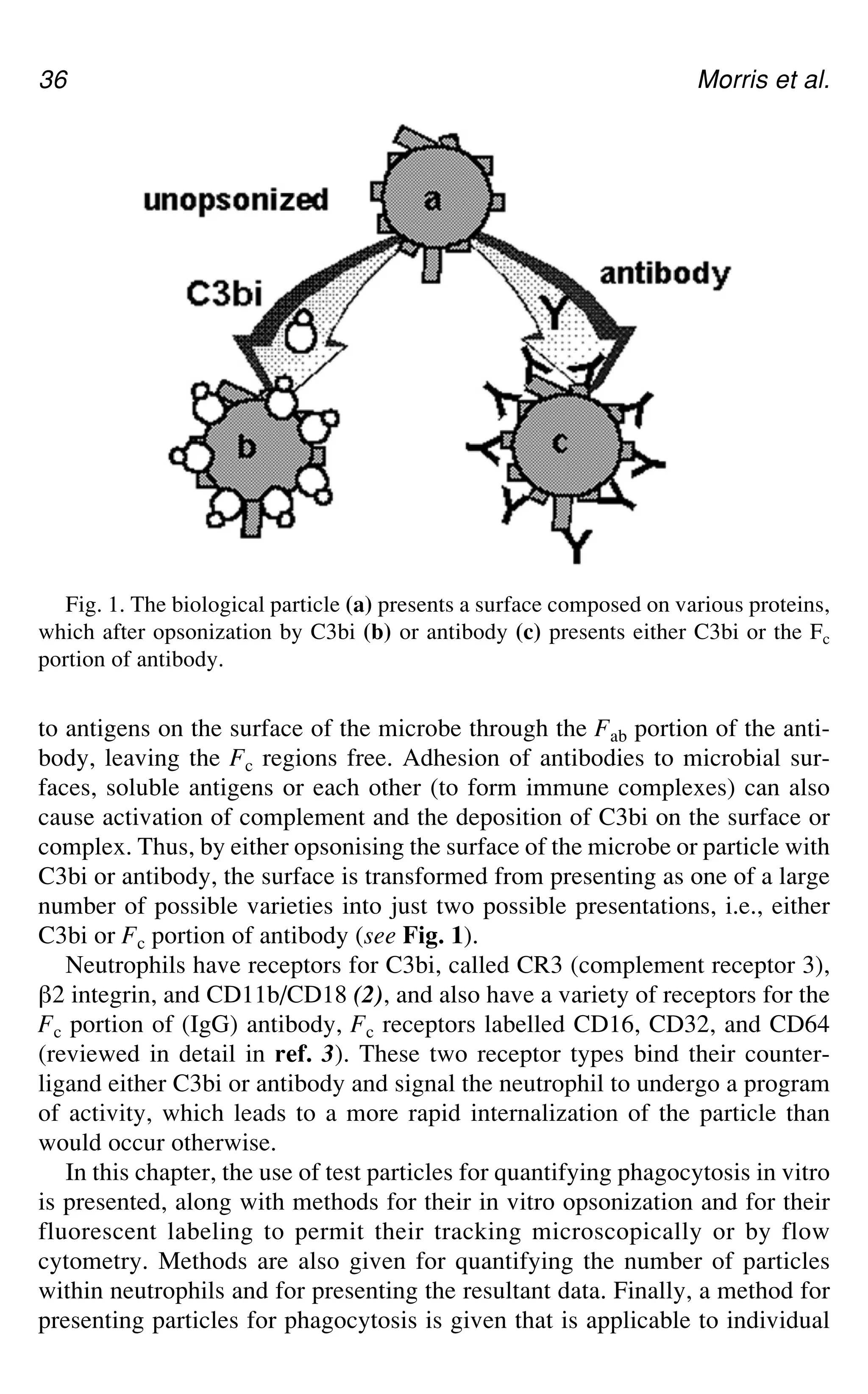
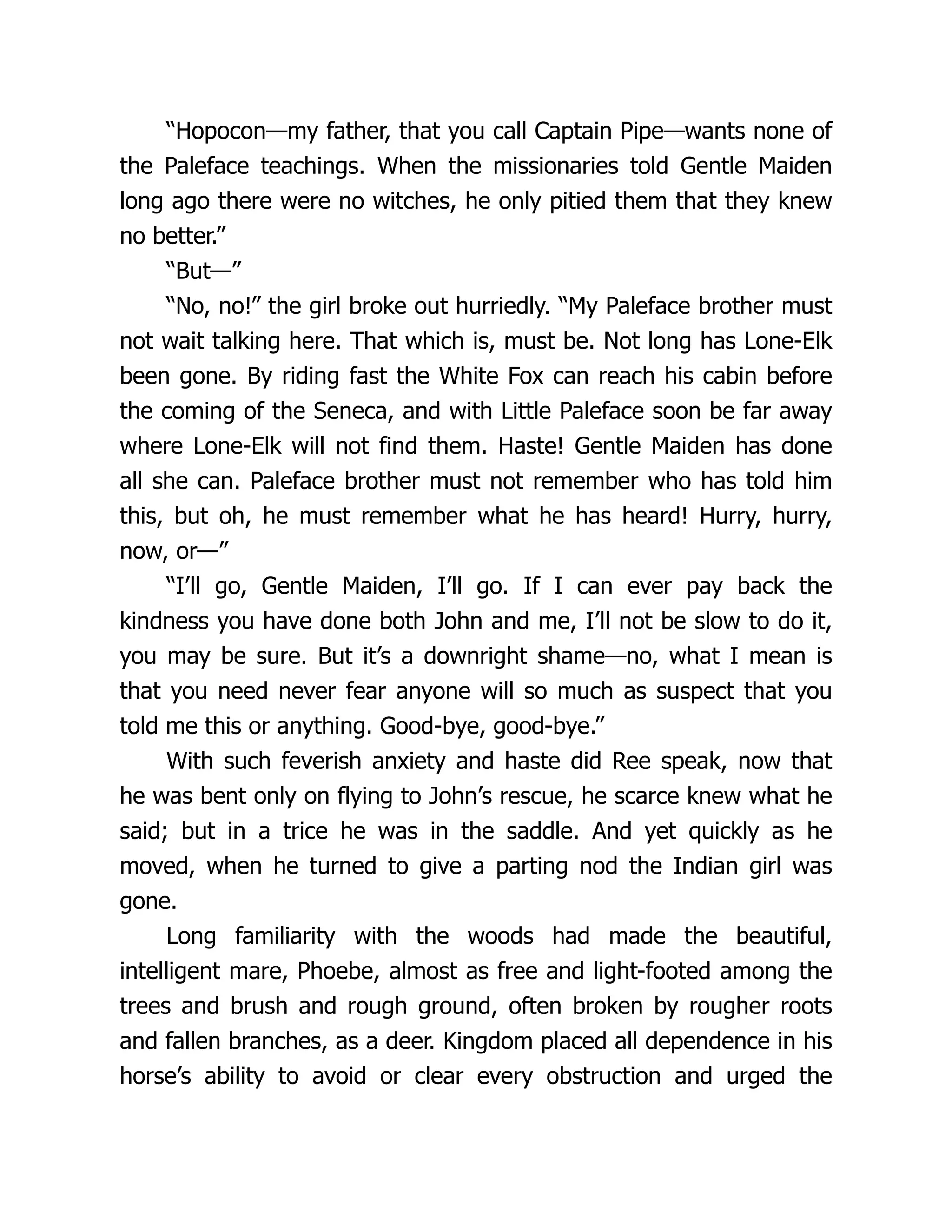
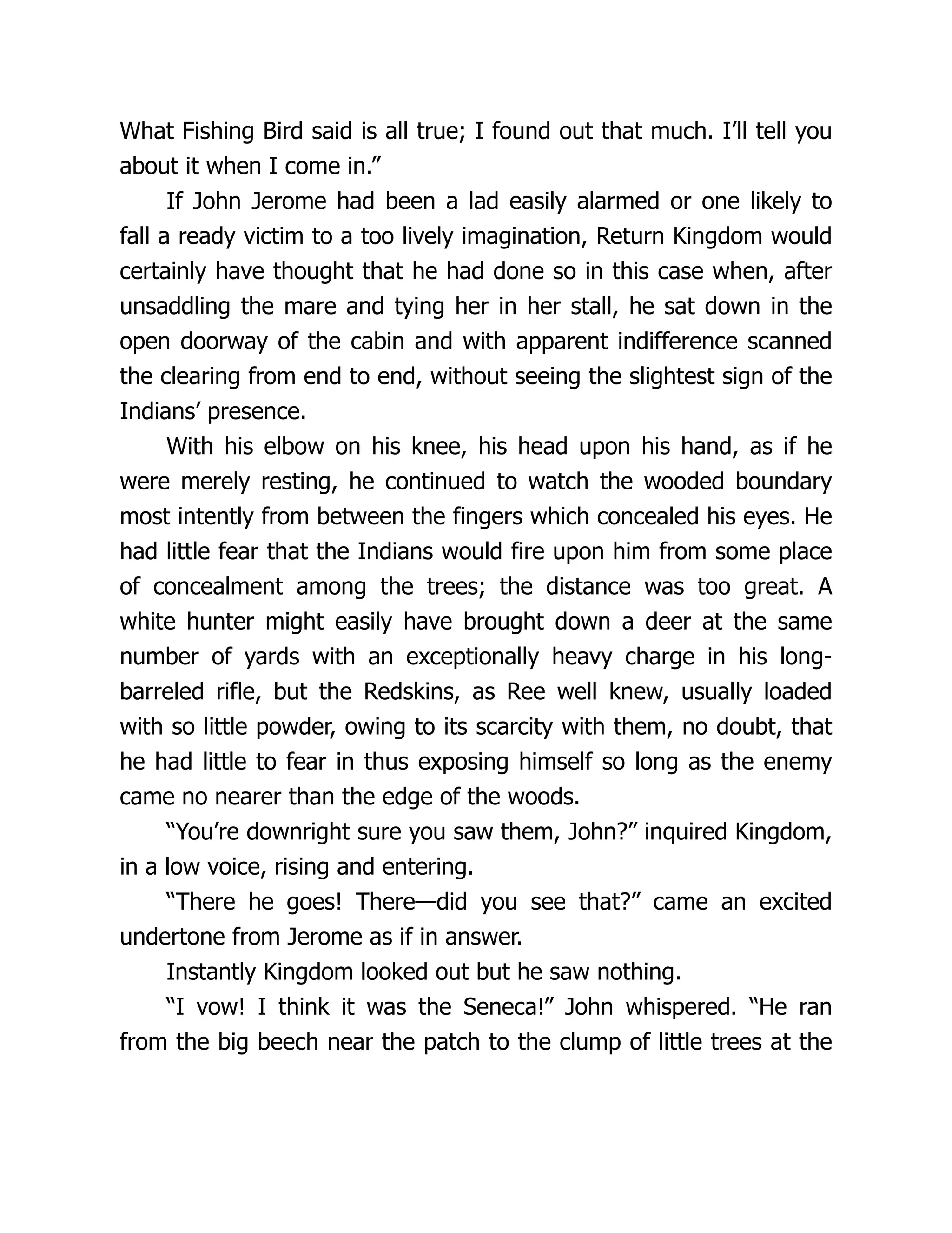
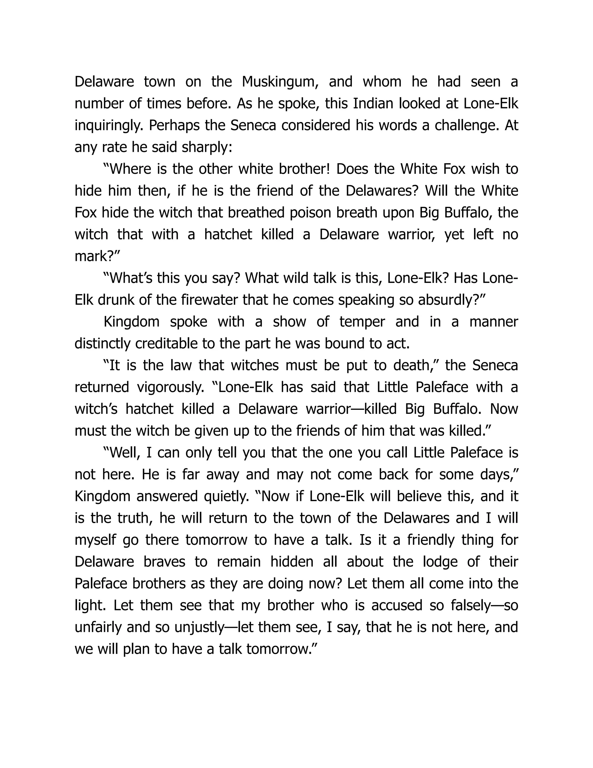
Fig. 1.Grossly simplified schematic diagram representing some of the key events
of the inflammatory response. The letters correspond to some of the processes that can
be investigated using protocols described within this first section of the book (or, if in
brackets, elsewhere in this book). A, intracellular kinase activation by cytokines; B,
activation of NF-gB; C, adhesion molecule expression; D, migration (of specific leu-
kocyte subsets); E, stimulation of inflammatory phagocytes, including changes in
cytosolic Ca2+ and plasma membrane NADPH oxidase; F, opsonisation involving
activation of the complement cascade; G, release and activation of MMPs within the
extracellular matrix; H, breakdown of the extracellular matrix by proteinases—e.g.,
degradation of cartilage by a MMPs and aggrecanase.
that are not discussed here. In many cases, the individual protocols described
in this section will allow the in vitro screening of candidate therapeutic com-
pounds.
Returning to a description of our illustrative “main highway” of inflamma-
tion, the binding of pro-inflammatory cytokines to their cognate plasma mem-
brane receptors results in the activation of intracellular kinases that catalyze a
cascade of phosphorylation events, including the phosphorylation (catalyzed
by the IKK complex) of IgB, an inhibitory protein subunit bound to the tran-
scription factor NF-gB. The control of gene transcription by specific proteins—
20.
Key Stages inthe Acute Inflammatory Response 5
transcription factors—involves the binding of these proteins to short DNA
sequence elements located adjacent to the promotor or in the enhancer regions
of genes. The phosphorylation of IgB targets it for proteolytic degradation by
the 26S proteasome, leaving the active NF-gB complex to be translocated to
the nucleus, where it binds to a specific DNA motif and is thereby involved,
together with other transcription factors, in controlling the transcription of an
array of inflammation-associated genes. Indeed, a key feature of inflammatory
diseases such as rheumatoid arthritis (RA) is the increased expression of cer-
tain genes that encode “inflammatory” proteins. These include a variety of
adhesion molecules, as well as TNF_, IL-1`, and inducible nitric oxide syn-
thase (iNOS) among many others.
Dr. Mireille Delhase describes methods for measuring cytokine-induced
IKK activity and NF-gB activation in cultured cells. By stably transfecting
cells with a construct containing the NF-gB-binding motif upstream of the
luciferase gene, a convenient system for screening potential inhibitors of NF-
gB may be created, as described by Dr. Deborah Phippard and Dr. Tony Man-
ning. Recently, there has been considerable interest in the idea that transcription
factors may be useful targets for novel therapeutic strategies in the treatment of
human diseases, including inflammatory diseases.
As aforementioned, induction of the expression of several adhesion mol-
ecules, such as intercellular adhesion molecule-1 (ICAM-1), and E-selectin
vascular cell adhesion molecule-1 (VCAM-1), represents a significant inflam-
matory event and a potential therapeutic target. The increased transcription of
these proteins ultimately results in their upregulation on the plasma membrane
surface of endothelial cells, and the levels of adhesion molecules may be mea-
sured using the techniques described by Dr. Susan Cuvelier and Dr. Kamala
Patel. The increased expression of adhesion molecules, in turn, facilitates the
“rolling” and adhesion to the vascular endothelium of inflammatory phago-
cytic cells at vessel sites adjacent to the site of inflammation. The phagocytic
cells, such as neutrophils, then migrate through the vessel wall, via the process
of diapedesis, and arrive at the focus of “acute” inflammation. The reader is
referred to the second section of this book for a description of an in vivo tech-
nique for the study of this process, as provided by Professor Mauro Perretti and
Dr. Stephen Getting. Later, in the “chronic” phase of inflammation, other cell
types (e.g., macrophages and lymphocytes) will be recruited to the site of inflam-
mation by an analogous process. As part of the classic, acute inflammatory
response, neutrophils are activated by immune complexes, the complement
system and/or pathogens to produce both free radicals (such as superoxide)
and proteinases (such as elastase and cathepsin G) which act in concert to kill
invading fungi or bacteria, but which may also cause tissue damage. This stimu-
lation of neutrophils can be reproduced in vitro using isolated cells and the
extent of the response in the presence of putative antiinflammatory compounds
21.
6 Winyard
may betested using the methods described by Dr. Maurice Hallett et al. This
group also describes specific methods for the measurement of neutrophil intra-
cellular Ca2+ fluxes and O2
•– production by a plasma membrane NADPH oxi-
dase. The killing of bacteria by neutrophils involves their opsonization by plasma
proteins, including various components of the complement cascade. Furthermore,
certain components of the complement cascade are chemotactic for neutrophils.
A protocol by which the extent of complement activation may be determined is
described by Professor Tom Eirik Mollnes.
Phagocytic cells, and sometimes resident cells at the site of inflammation,
are also stimulated by cytokines such as TNF and IL-1 to release matrix
metalloproteinases (MMPs). In respect of the inflamed joint, the resident cells
of the synovial membrane—the so-called type B synoviocytes—are activated
by cytokines to release MMPs. The MMPs involved in inflammatory tissue
destruction include collagenases, gelatinases, and stromelysins. The activities
of these proteinases may be determined, again in an in vitro system, according
to the zymographic technique detailed by Dr. Linda Troeberg and Professor
Hideaki Nagase. Among the expressed metalloproteinases is aggrecanase,
which plays an important role in the degradation of cartilage within the rheu-
matoid joint. Professor Bruce Caterson et al. describe a protocol for the mea-
surement of aggrecanase activity, whereas Dr. Bill Shingleton describes an in
vitro model of articular cartilage degradation.
In finishing this introduction to in vitro protocols for the study of inflamma-
tion it should be stressed again that, for the sake of simplicity, the description
above refers to one of many pathways of acute inflammation. Many of the exper-
imental systems described in this section of the book are relevant to more than
one stage or type of inflammation, e.g., acute versus chronic, immune vs
nonimmune, and so on. Although, for convenience, the inflammation protocols
in this book have been divided between in vitro and in vivo methods, it is vital
to have both. It is, of course, impossible to reproduce inflammation in vitro. In
vivo, the environment at the site of inflammation changes millisecond by milli-
second—this can never be reproduced in a test tube. However, the complemen-
tary use of both in vitro and in vivo techniques is a powerful strategy in the
study of inflammation and antiinflammatory drug development: For example,
the cytokines can be identified in vivo, whereas the signal transduction path-
ways leading to the production of such cytokines can be characterized in vitro.
The combination of the outputs from these two approaches may then allow the
demonstration of the importance of a particular pathway by the in vivo testing
of selective inhibitors.
8 Delhase
catalytic subunits,IKK_ and IKK` and a regulatory subunit, IKKa (or NEMO)
(4). Biochemical studies and analysis of knockout mice indicate that the cata-
lytic subunit IKK` and the regulatory subunit IKKa but not the catalytic sub-
unit IKK_, are absolutely required for IgB phosphorylation and NF-gB
activation in response to pro-inflammatory stimuli (7–14). IKK activation by
such stimuli depends on phosphorylation of the IKK` subunit at two serine
residues (Ser-177 and Ser-181) within its activation loop (7). Despite the con-
servation of these serines in the IKK_ subunit, IKK_ does not play an essential
role in IKK and NF-gB activation by all major stimuli (7). Instead, IKK_ plays
a critical role in developmental processes, in particular terminal differentiation
of keratinocytes during formation of the epidermis (13,14). Recently, IKK_
has been found to be involved in activation of a second NF-gB pathway involv-
ing processing of NF-gB2 (15). This pathway is particularly important for B-cell
maturation and secondary lymphoid organ formation, key events in the devel-
opment of adaptive immunity.
This chapter focuses on the basic techniques allowing the study of IKK and
NF-gB activation in response to cell stimulation by pro-inflammatory
cytokines.
2. Materials
2.1. Stock Solutions
1. Stocks of the reagents are prepared and stored as follows and are stable for at
least 6 mo.
a. Dithiothreitol (DTT) (1 M) in 10 mM sodium acetate, stored at –80°C.
b. Phenylmethylsulfonide fluoride (PMSF) (100 mM) in ethanol, stored at room
temperature.
c. p-nitrophenyl phosphate (pNPP) (1 M) in H2O, stored at –20°C.
d. Na3VO4 (100 mM) in H2O, stored at 4°C.
e. Aprotinin (10 mg/mL) in 10 mM HEPES, pH 8.0, stored at –80°C.
f. Bestatin (10 mg/mL) in H2O, stored at –80°C.
g. Leupeptin (10 mg/mL) in H2O, stored at –80°C.
h. Pepstatin (10 mg/mL) in ethanol, stored at –80°C.
i. All protease inhibitors are available from Sigma and Calbiochem (San Diego,
CA).
2. Sterile penicillin/streptomycin stock solution and 200 mM L-glutamine stock
solution are kept at –20°C in 10-mL aliquots. These reagents, as well as
Dulbecco’s modified Eagle’s medium (DMEM), may be obtained from Gibco
BRL, Life Technologies, Rockville, MD.
3. Recombinant mouse and human TNF_ and IL-1 are available from several com-
mercial sources (BioSource International, Camarillo, CA or R&D Systems, Min-
neapolis, MN). The lyophilized cytokines are reconstituted at 10 μg/mL in PBS
24.
IgB Kinase andNF-gB Signaling 9
plus 0.1% bovine serum albumin (BSA, tissue culture grade) and stored at –80°C
in 100-μL aliquots.
4. ATP stock solution: 100 mM in H2O, stored at –80°C in 50-μL aliquots.
5. 32P-orthophosphate (cat. no. Nex-53C) is from NEN Life Science Products, Bos-
ton, MA).
6. [a-32P]ATP is from Amersham Pharmacia Biotech, Piscataway, NJ.
2.2. Cell Lysis Buffers
1. Lysis buffer: 50 mM Tris-HCl, pH 7.6, 250 mM sodium chloride (NaCl), 3 mM
ethylenediamine tetraacetic acid (EDTA), 3 mM ethyleneglycol tetraacetic acid
(EGTA), 1% (v/v) Triton X-100, 0.5% (v/v) Nonidet (P-40) (NP40), 10% glyc-
erol, 20 mM sodium fluoride (NaF), 40 mM `-glycero-3-phosphate. This buffer
is prepared as a stock solution, filtered through a 0.45-μm filter unit, and stored
at 4°C. Before use, the following should be added: 2 mM DTT, 1 mM PMSF,
2 mM pNPP, 1 mM sodium orthovanadate (Na3VO4), and 10 μg/mL each of
aprotinin, bestatin, leupeptin, and pepstatin (see Subheading 2.1.).
2. RIPA buffer: 20 mM Tris-HCl, pH 7.6, 150 mM NaCl, 1 mM EDTA, 1 mM
EGTA, 1% NP40, 0.5% (w/v) sodium deoxycholate (DOC), 0.05% sodium
dodecyl sulfate (SDS), 10% glycerol, 20 mM NaF, 40 mM `-glycero-3-phos-
phate, 2.5 mM sodium metabisulfite, 5 mM benzamidine, 2 mM DTT, 1 mM
PMSF, 20 mM pNPP, 1 mM Na3VO4, and 10 μg/mL each of aprotinin, bestatin,
leupeptin, and pepstatin (see Subheading 2.1.).
2.3. Bacterial Culture Medium and Bacteria Lysis Buffers
1. Lbroth: bacto-tryptone 10 g/L, bacto-yeast extract 5 g/L, NaCl 5 g/L, adjusted to
pH 7.5 with NaOH. 100 μg/mL ampicillin and 2% glucose are added.
2. Base buffer: PBS, pH 8.0, 1 mM EDTA, 0.5% (v/v) Triton X-100, 5 mM DTT,
store at 4°C. This buffer is stable for several months if prepared without DTT.
DTT can be added just before use (see Subheading 2.1.).
3. Buffer I : Base buffer containing 0.3 M ammonium sulfate [(NH4)2SO4].
4. Lysis buffer: Buffer I supplemented with 1 mM PMSF and 10 μg/mL of protease
inhibitors (aprotinin, bestatin, leupeptin, and pepstatin) (see Subheading 2.1.).
5. Elution buffer: 100 mM Tris-HCl, pH 8.0, 20 mM glutathione.
6. Dialysis buffer: 20 mM Tris-HCl, pH 8.0, 100 mM NaCl, 0.2 mM EDTA, 10 mM
`-glycero-3-phosphate and 10% glycerol.
2.4. Kinase Assay
1. Kinase buffer (10X): 200 mM HEPES, pH 7.5, 100 mM magnesium chloride
(MgCl2).
2. Reaction buffer: kinase buffer [1X] supplemented with 20 mM `-glycero-3-phos-
phate, 10 mM pNPP, 1 mM DTT, and 20 μM “unlabeled” ATP. This buffer should
be prepared just before use.
25.
10 Delhase
2.5. Reagentsfor Electrophoretic Mobility Shift Assay
1. Oligonucleotide probes:
NF-gB:5'-GGATCCTCAACAGAGGGGACTTTCCGAGGCCA-3'
3'- AGTTGTCTCCCCTGAAAGGCTCCGGTCCTAGG-5'
NF-Y:5'-GTAGGAACCAATGAAATGCGAGG-3'
3'- TTGGTTACTTTACGCTCCGGATG-5'
Consensus oligonucleotide sequences for binding of transcription factors are also
available from Promega (Madison, WI). The oligonucleotides are diluted at 1 μg/μL
in TE buffer (10 mM Tris-HCl, pH 8.0, 1 mM EDTA) and stored at –20°C.
2. Klenow buffer (1X): 10 mM Tris-HCl, pH 7.9, 50 mM NaCl, 10 mM MgCl2, and
1 mM DTT).
3. EMSA buffer (10X): 100 mM Tris, pH 7.6, 500 mM potassium chloride (KCl),
10 mM EDTA, and 50% glycerol, stored at –20°C.
4. Nu-Clean D25 spin columns for probe purification are available from Eastman
Kodak Company, Rochester, NY.
5. Poly(dI.dC) is from Amersham Pharmacia Biotech.
6. Gel loading buffer (10X): 250 mM Tris-HCl, pH 7.6, 40% glycerol, and 0.2%
(w/v) bromophenol blue, stored at 4°C.
7. Acrylamide stock solution (40%): Dissolve 38.71 g of acrylamide and 1.29 g of
bis-acrylamide in 100 mL H2O. Solution is filtered and kept at 4°C.
8. TBE (10X): Dissolve 121.1 g of Tris base, 55 g of boric acid, and 7.4 g of EDTA
into 1 L of H2O. Adjust the pH to 8.3 with solid boric acid.
9. Nondenaturing gel: Mix 36 mL of H2O, 4.5 mL of acrylamide stock solution,
4.5 mL of TBE 10X, 450 μL of a 10% (w/v) solution of ammonium persulfate
(APS) in H2O, and 45 μL of TEMED.
3. Methods
3.1. Cell Culture and Cytokine Stimulation
1. Adherent cells (HeLa, HEK295, 3T3 fibroblasts) are cultured in DMEM high
glucose supplemented with 10% fetal bovine serum (FBS), 2 mM L-glutamine,
and 1% (v/v) penicillin/streptomycin stock solution at 37°C in a 5% CO2 atmo-
sphere.
2. Tumor necrosis factor _ (TNF_) and interleukin-1 (IL-1) stocks solutions are
diluted, respectively, at 20 ng/mL and 10 ng/mL final concentration in pre-
warmed serum-free medium, then added to the cells. Stimulation is performed at
37°C for various periods of time.
3. After stimulation, the cells are placed on ice, medium is removed and the cells
are washed twice with ice-cold phosphate-buffered saline (PBS). Cells are har-
vested in 1 mL PBS/100-mm dish using a cell lifter and pelleted by centrifuga-
tion for 5 min at 4°C at 2000 g.
4. Cell pellets are kept frozen at –80°C or directly lysed by resuspension in lysis
buffer (400 μL per cell pellet from a 100-mm dish) with incubation on ice for
10–15 min. The lysate is centrifuged at 13,000g for 10 min at 4°C.
26.
IgB Kinase andNF-gB Signaling 11
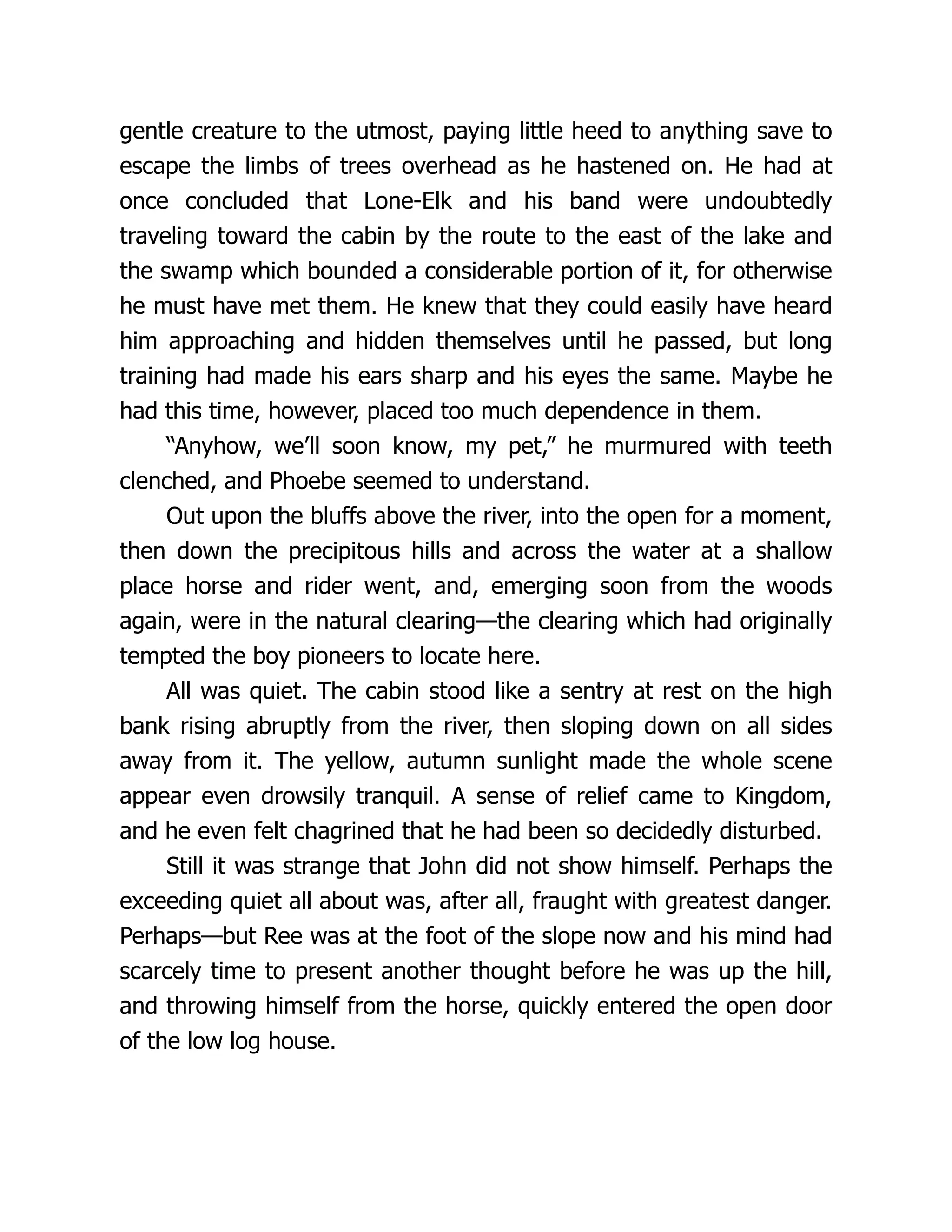
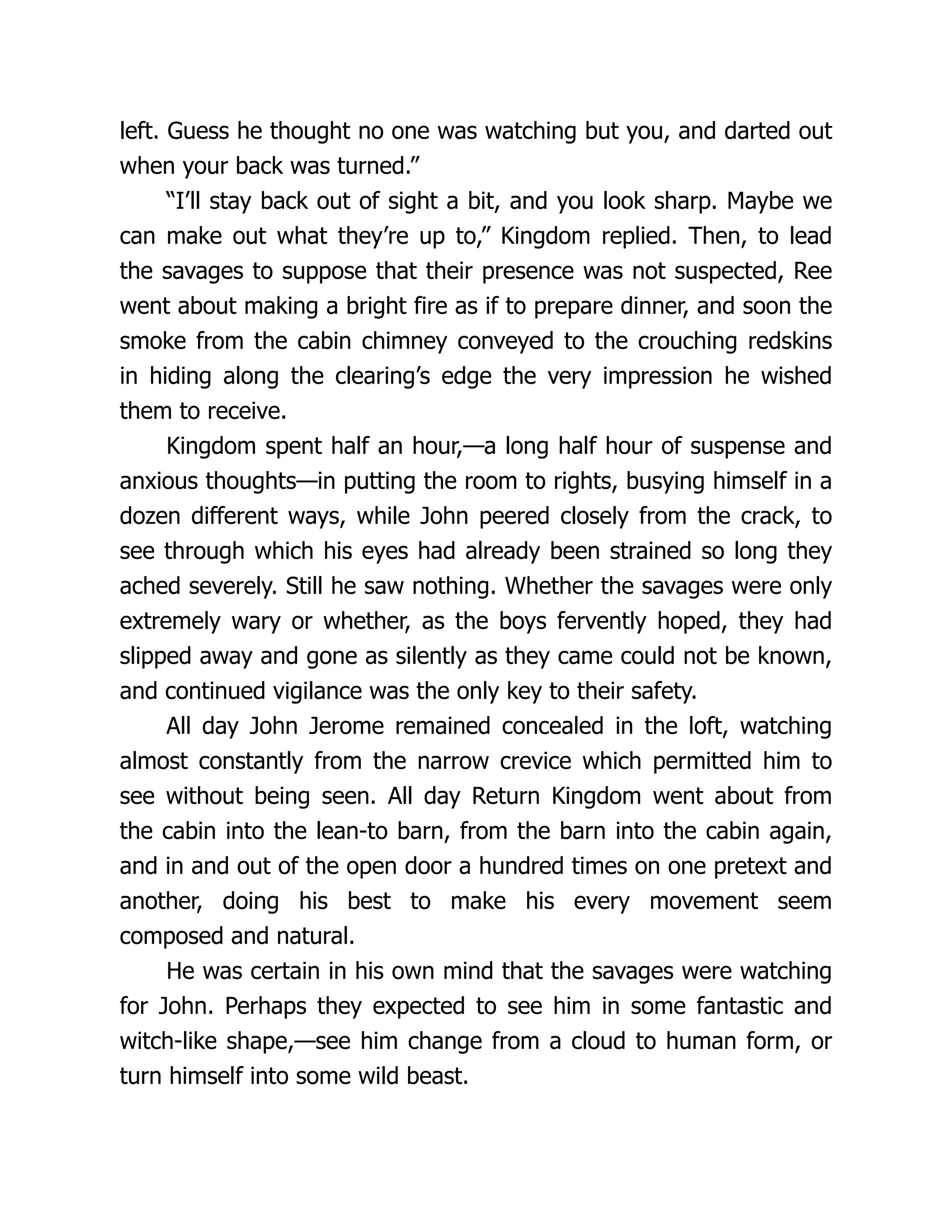
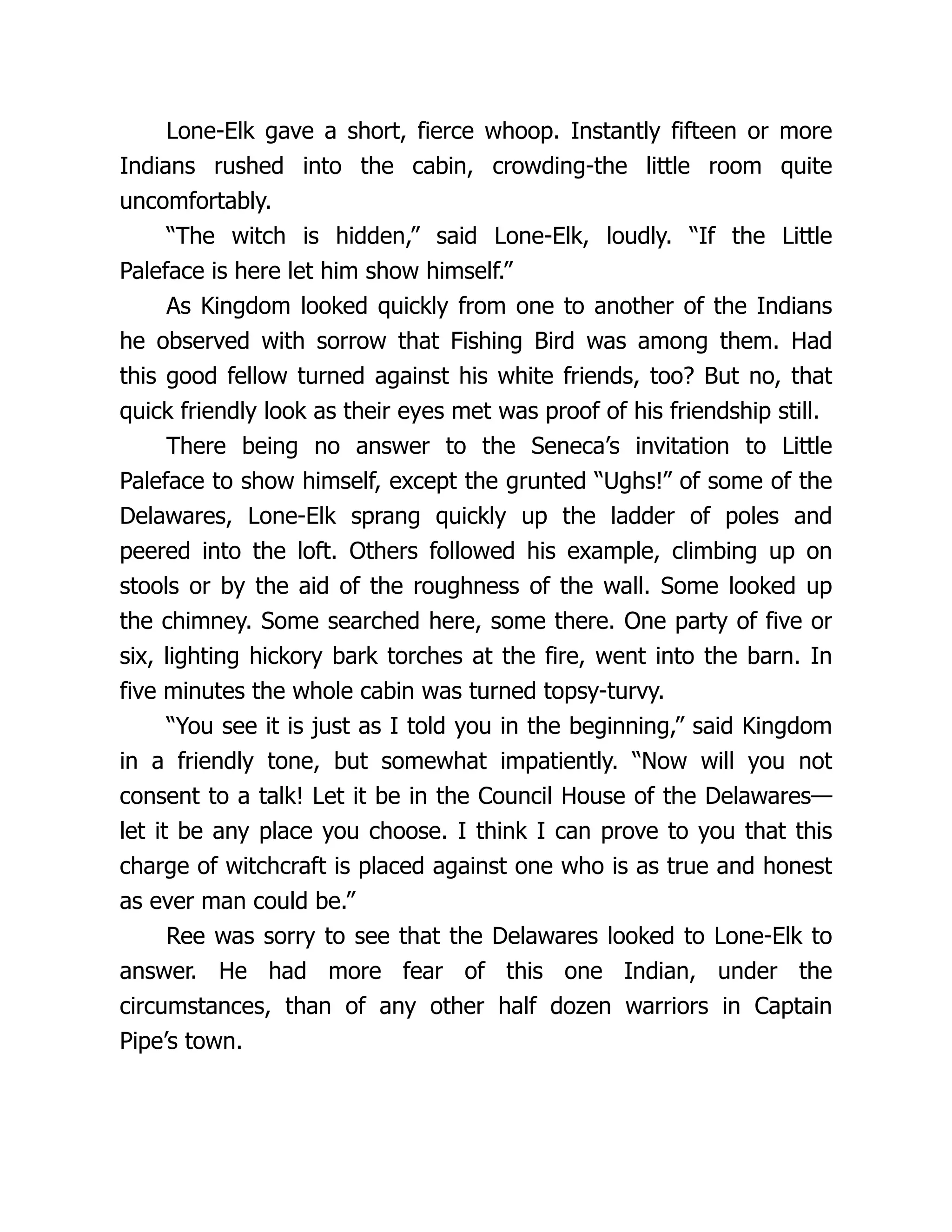
Fig. 1. Cytokine-induced IKK phosphorylation. HeLa cells incubated for 5 h with
[32P]orthophosphate were stimulated with TNF_ (20 ng/mL) for the indicated times,
then lysed. The IKK complex was immunoprecipitated (IP) with an antibody to IKK_
(clone # B78-1 from PharMingen), resolved by gel electrophoresis and transferred onto
a PVDF membrane. Phosphoproteins were detected by autoradiography. IKK_ and
IKK` were identified by immunoblotting (IB) using specific antibodies (anti-IKK_,
clone # B78-1 from PharMingen and anti-IKK` H470 from Santa Cruz Biotechnology).
5. The supernatant is transferred to a clean tube and the protein concentration is
determined by Bradford assay (17).
6. The lysates (WCE, whole cell extracts) are stored at –80°C until use. WCEs can
be used for analysis of protein composition by western blotting, immune-com-
plex kinase assay and electrophoretic mobility shift assay (EMSA).
3.2. Detection of IKK Phosphorylation by 32P Metabolic Labeling
(see Note 1)
1. Cells at subconfluence (70–80%) (one 60-mm dish per assay) are washed twice
in phosphate-free DMEM and incubated for 1 h in the same medium.
2. The medium is then replaced by labeling medium (4 mL per dish) consisting of
phosphate-free DMEM supplemented with 10% dialyzed FBS and 1–2 mCi/mL
32P-orthophosphate. Cells are labeled for 4–5 h at 37°C.
3. TNF_ or IL-1 stimulation of the cells is performed as described in Subheading
27.
12 Delhase
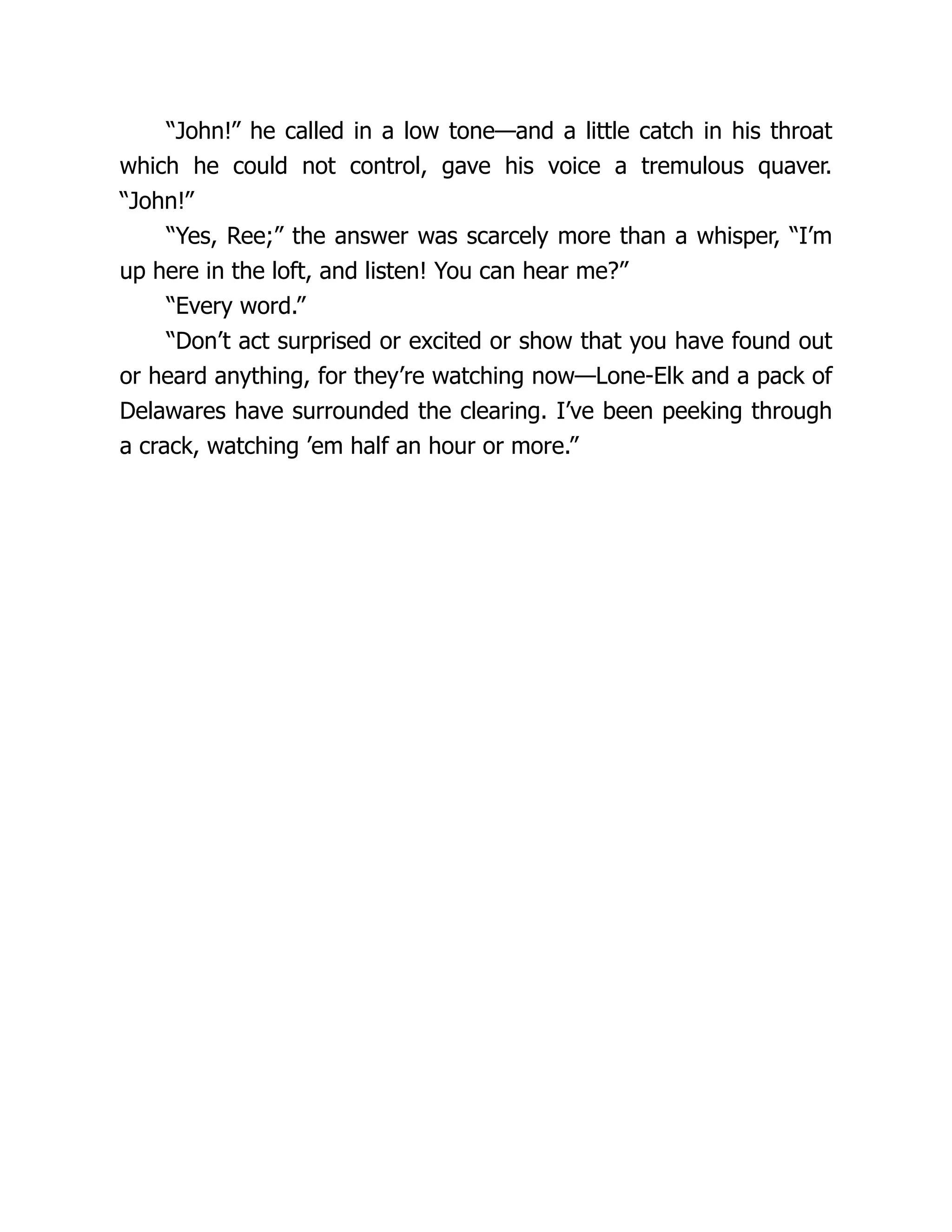
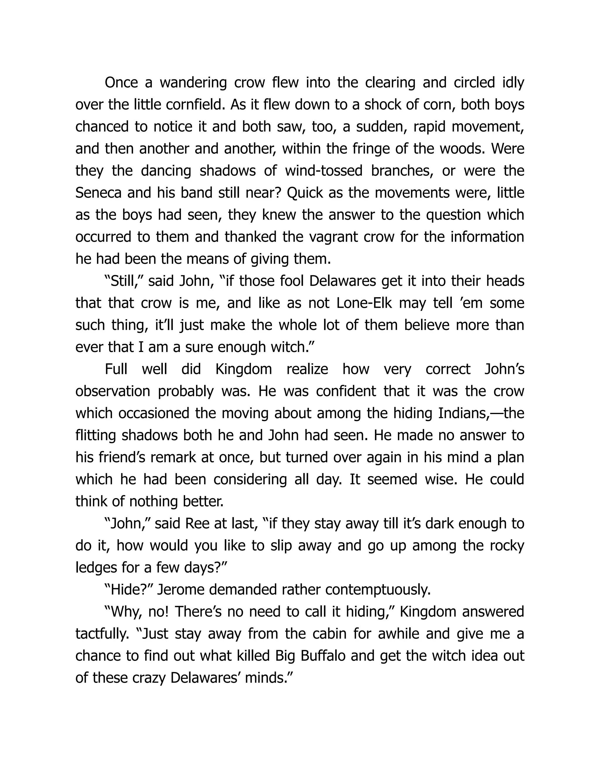
Fig. 2.(opposite page) IKK and NF-gB activation by TNF_ and IL-1. (A) HeLa
cells (left panel) and LL-2 cells (lung carcinoma cell line, right panel) were stimulated
with TNF_ and IL-1 for the indicated times. IKK was isolated by immunoprecipitation
with anti-IKK_ (clone # B78-1, PharMingen) or anti-IKKa(clone 73-764, PharMingen)
and its activity was measured by phosphorylation of GST-IgB_ (1-54) as described in
Subheading 3.3. (B) IgB_ degradation was analyzed by immunoblotting. IgB_ phos-
phorylation (middle panel) was detected using an antibody directed against P-Ser-32 of
IgB_ (#9241, New England Biolabs, Beverly, MA). IgB_ (bottom panel) was detected
by immunoblotting using an anti-IgB_ antibody (#IMG-127, Imgenex or # C-21, Santa
3.1. Cytokines are diluted directly into the labeling medium (20 ng/mL for TNF_
and 10 ng/mL for IL-1) and stimulation is performed for 5 to 15 min.
4. Bulk IKK phosphorylation is observed after 5 min following cytokine stimula-
tion (see ref. 7 and Fig. 1). Labeling medium is removed, cells are placed on ice,
and washed twice with ice-cold PBS.
5. Cells are lysed by addition of 1 mL of lysis buffer directly to the culture dish.
Cells are scraped using a cell lifter, transferred to a microcentrifuge tube and
lysed on ice for 10 min. Cellular debris are pelleted by centrifugation at 13,000g
for 10 min at 4°C.
6. Supernatants are transferred to clean tubes. Cell lysates are clarified by incuba-
tion for 1–2 h at 4°C on a rotator in the presence of 10 μL of preimmune serum (if
available) or 10 μg of nonspecific control IgG and 50 μL of 50% slurry of protein
A/G-Sepharose beads. Beads are pelleted by centrifugation at 13,000g for 5 min
at 4°C and supernatants are transferred to clean tubes.
7. Immunoprecipitation of the IKK complex is performed by rotating the cell lysate
overnight at 4°C after addition of 1–5 μg of anti-IKK antibody (see Note 2) and
25 μL of 50% of slurry of protein A/G-Sepharose beads. The immune-complexes
are collected by centrifugation for 1 min at 13,000g, then washed once in lysis
buffer containing 300 mM NaCl, once in the same buffer supplemented with 2 M
urea and twice in RIPA buffer. The supernatants are discarded and the immune
complexes are resuspended in SDS-loading buffer.
8. The IKK subunits are resolved on a 7.5% SDS-PAGE gel (12X 16 cm) according
to a standard procedure (17). The proteins are transferred onto a nitrocellulose or
Immobilon-P polyvinylidene fluoride membrane. The phosphoproteins are
detected by exposure of the membrane to Kodak, Biomax-MR film for 2–12 h at
–80°C. After exposure, the membrane can be used to detect protein levels by
immunoblotting.
3.3. Measurement of IKK Activity by An Immune-Complex
Kinase Assay
Rapid and transient activation of the IKK complex in response to cell stimu-
lation by pro-inflammatory cytokines (TNF_ and IL-1) can be best measured
and quantified by an immune-complex kinase assay using GST-IgB_ (1–54)
as a substrate (see Fig. 2A).
28.
IgB Kinase andNF-gB Signaling 13
Fig. 2. (continued) Cruz Biotechnology). IKK_ expression (top panel), used as control
for equal protein loading, was determined by immunoblotting using an anti-IKK_ anti-
body (#IMG-136, Imgenex). (C) NF-gB DNA-binding activity was measured by elec-
trophoretic mobility shift assay (EMSA). Cells were left untreated or were stimulated
with TNF_ (left panel) or IL-1 (right panel) for the indicated times, after which WCEs
were prepared and NF-gB and NF-Y DNA-binding activities were measured by EMSA
as described in Subheading 3.4. ns = nonspecific.
29.
14 Delhase
3.3.1. Expressionand Purification of GST-IkBa
(1–54) Fusion Protein in Escherichia coli
1. A cDNA fragment encoding the first 54 N-terminal amino acids (including Ser 32
and Ser 36) of human IgB_ was inserted in frame at the 3' end of the GST sequence
of a pGEX expression vector (Amersham Pharmacia Biotech) (see Note 3).
2. BL21 bacteria transformed with this recombinant plasmid are grown at 37°C in
500 mL Lbroth containing 100 μg/mL ampicillin and 2% glucose until the absor-
bance at 600 nm measured in a 1-cm pathlength cuvet is between 0.4 and 0.5.
3. Protein expression is induced by addition of 2 mM isopropyl-`-thioga-
lactopyranoside (IPTG) for 3 h at 37°C.
4. Bacteria are collected by centrifugation and resuspended on ice in 30 mL of ice-
cold bacteria lysis buffer. Lysozyme is added to a final concentration of 1 mg/mL
and bacteria are lysed by sonication (Virsonic 600 sonicator). Bacterial debris
are removed by centrifugation at 20,000g for 20 min at 4°C.
5. The supernatant is transferred to a clean tube and incubated for 1 h at 4°C with
500 μL of 50% slurry of glutathione Sepharose 4B equilibrated with PBS.
6. The beads are collected by centrifugation at 500g for 5 min and washed twice
with buffer I containing 1 mM PMSF, twice with base buffer containing 1 mM
PMSF and once with PBS (10 bed volumes for each wash). The beads are then
transferred to a microcentrifuge tube.
7. The GST fusion protein is eluted by incubation of the beads with two bed vol-
umes of elution buffer for 1 h at 4°C on a rotator. After centrifugation, the super-
natant is collected and the beads are subjected to a second 1-h elution step. The
supernatants from the two elution steps are combined and dialyzed at 4°C against
2 L of dialysis buffer.
8. The quality and quantity of fusion protein is evaluated by SDS-PAGE followed
by Coomassie blue staining (17) using known amounts of BSA as standard. GST-
IgB_ (1–54) protein solution is stored at –80°C in 100 μL aliquots.
3.3.2. IKK Immune-Complex Kinase Assay
1. To determine whether IKK activity is induced by a given stimulus, cells are left
untreated or are treated for various periods of times with a specific activator (for
example TNF_ or IL-1) as described in Subheading 3.1. WCEs are prepared as
described in Subheading 3.1.
2. WCEs containing equal amounts of proteins (typically 20–100 μg) from
nonstimulated and stimulated cells are aliquoted into prechilled microcentrifuge
tubes. The volume is adjusted to 300 μL with cell lysis buffer.
3. Anti-IKK antibody (1 to 2 μg per reaction) (see Note 2) or control serum (pre-
immune serum or control IgG) and 20 μL of 50% slurry of protein A/G-Sepharose
are added to the lysates. Immunoprecipitation is performed for a minimum of 2 h
at 4°C on a rotator.
4. The immune-complexes bound to the beads are collected by centrifugation at
2000g for 1 min at 4°C. The supernatants are discarded and the beads are washed
once with lysis buffer containing 400 mM NaCl, once with the same buffer con-
taining 2 M urea and twice with 1X kinase buffer.
30.
IgB Kinase andNF-gB Signaling 15
5. The immune-complexes are resuspended in a 30-μL reaction buffer as described
in Subheading 2.4. containing 5μCi of [a-32P]ATP (6000 Ci/mmole) and 1 μg of
GST-IgB_(1–54) substrate (see Subheading 3.3.1). The kinase reaction is
allowed to proceed for 30 min at 30°C.
6. The reactions are stopped by addition of SDS sample loading buffer (16). The
samples are heated at 100ºC for 5 min to denature the proteins and then analyzed
by SDS-PAGE (10% gel). Electrophoresis is stopped before the free isotope
(which migrates on the gel together with the yellow pNPP band) runs off the gel.
7. The bottom of the gel containing the free isotope is cut off and discarded. The
gel is stained with Coomassie blue (17), destained in 10% acetic acid, 10%
methanol in H2O and dried. This step allows visualization of the substrate to
verify equal loading.
8. The dried gel is exposed to Kodak X-OMAT-AR films. Alternatively, the kinase
assay gel can be directly transferred to a PVDF membrane, which is then exposed
to film. This allows further detection of IKK in the immune-complexes by
immunoblotting.
9. The IKK activity is determined by the amount of radioactive 32P incorporated
into the GST-IgB_(1-54) substrate. Quantification can be performed on a
phosphoimager (preferred) or by densitometric scanning of the films.
3.4. Measurement of NF-g
g
g
g
gB Activation
by Electrophoretic Mobility Shift Assay
3.4.1. Oligonucleotide Probe Labeling
1. Complementary oligonucleotides are synthesized according to consensus seq-
uences for binding of transcription factors (see Subheading 2.5.).
2. 10 μL of both oligonucleotide stock solutions (at 1 μg/μL) are diluted into 100 μL
of TE in a microcentrifuge tube. The tube is heated at 80°C for 10 min in a beaker
of water, then slowly cooled down to room temperature (about 1 h) to allow
annealing of the oligonucleotides. The double-stranded (ds) oligonucleotide stock
solution is stored at –20°C.
3. The EMSA probe is prepared by incubation for 20 min at room temperature of
1 μL of ds oligonucleotide stock solution in a 20-μL reaction mixture containing
1X Klenow buffer (see Subheading 2.5.). 25 μM of each dATP, dTTP, and
dGTP, 50 μCi [_-32P]dCTP and 5 units of Klenow enzyme (see Note 4).
4. The labeling reaction is stopped by addition of 25 μM of unlabelled dCTP and
incubation is continued for 5 min at room temperature.
5. The probe is purified on a Nu-Clean D25 spin column (see Note 5) and stored at
–20°C.
3.4.2. Electrophoretic Mobility Shift Assay
1. Aliquots of WCEs containing 10 μg of protein (adjusted to 10 μL with cell lysis
buffer) are incubated for 30 min at room temperature in a 20-μL volume reaction
containing 2 μL of 10X EMSA buffer (see Subheading 2.5.), 1 μL of 20 mM
DTT, 2 μg of poly(dI.dC) and 15,000–30,000 cpm of probe. H2O is added to
bring the volume to 20 μL.
31.
16 Delhase
2. Thereactions are stopped by addition of 2 μL of 10X gel loading buffer (see
Subheading 2.5.) and the samples are run on a 4% nondenaturing polyacryla-
mide gel (20 × 20 cm).
3. Electrophoresis is stopped when the free probe (which migrates together with the
bromophenol blue band) is 2.5 cm from the bottom of the gel. The gel is fixed
(optional) for 15 min in a solution of 10% acetic acid, 10% methanol in H2O,
then dried and exposed to Kodak X-OMAT-AR film for autoradiography (see
Fig. 2C).
4. Notes
1. Precautions must be taken when performing metabolic labeling with 32P-ortho-
phosphate. Work must be carried out behind a 2.5-cm-thick acrylic shield. A
thick layer of lead can be attached to the outside of the shield to provide addi-
tional protection against radiation. A 1-cm-thick acrylic cell house should be
used to provide shielding when the cell dishes are in the incubator or when they
are moved in the laboratory. Researchers should wear two layers of gloves dur-
ing all steps of the labeling procedure.
2. Immunoprecipitation of the entire IKK complex can be performed using an anti-
body against any of the IKK subunits. Several commercial antibodies have been
tested and found to quantitatively immunoprecipitate the IKK complex. We cur-
rently use anti-IKK_ [clone # B78-1, PharMingen (San Diego, CA), this mono-
clonal antibody recognizes the human IKK_ but not the mouse IKK_], anti-IKK_
M280 [Santa Cruz Biotechnology (Santa Cruz, CA)], anti-IKKa (clones # C73-764
and C73-1794, PharMingen) and anti-IKK` H470 (Santa Cruz Biotechnology)
(7,13). Intensive washings of the immunoprecipitates in RIPA buffer may disrupt
interaction between IKKa and the IKK_/IKK` dimers. Therefore, to examine IKKa
phosphorylation, we recommend immunoprecipitating the IKK complex using an
antibody against IKKa.
3. This recombinant plasmid as well as a mutant version in which Ser 32 and Ser 36
have been replaced by Ala (GST-IgB_(1-54)AA) are available from the author’s
laboratory upon request.
4. If the annealed oligonucleotides do not contain 5' protruding ends, but instead are
blunt ended, labeling cannot be performed using Klenow enzyme. Instead T4
polynucleotide kinase in the presence of [a-32P] ATP should be used (17).
5. Unincorporated nucleotides can also be removed by centrifugation through a “self
made” 1-mL Sephadex G25 column.
Acknowledgment
The author would like to thank D. Rothwarf for helpful discussions and critical
reading of the manuscript. This work was supported by the Sontag Foundation Fel-
lowship of the Arthritis National Research Foundation and a research grant from the
San Diego Chapter of the Arthritis Foundation.
32.
IgB Kinase andNF-gB Signaling 17
References
1. Barnes, P. J. and Karin, M. (1997) Nuclear factor-gB - A pivotal transcription
factor in chronic inflammatory diseases. N. Engl. J. Med. 15, 1066–1071.
2. Baeuerle, P. A. and Baltimore, D. (1996) NF-kappa B: ten years after. Cell 87,
13–20.
3. Ghosh, S., May, M. J., and Kopp, E. B. (1998) NF-kappa B and Rel proteins: evolution-
arily conserved mediators of immune responses. Ann. Rev. Immunol. 16, 225–260.
4. Rothwarf, D. M. and Karin, M. (1999) The NF-gB activation pathway: a para-
digm in information transfer from the membrane to the nucleus. Science’s STKE.
www.stke.org/cgi/content/full/OC_sigtrans;1999/5/re1.
5. Karin, M. and Delhase, M. (2000) The IgB kinase (IKK) and NF-gB: key ele-
ments of proinflammatory signalling. Sem. Immunol. 12, 85–98.
6. Karin, M. and Ben-Neriah, Y. (2000) Phosphorylation meets ubiquitination: the
control of NF-gB activity. Ann. Rev. Immunol. 18, 621–663.
7. Delhase, M., Hayakawa, M., Chen, Y., and Karin, M. (1999) Positive and nega-
tive regulation of IgB kinase activity through IKK` subunit phosphorylation. Sci-
ence 284, 309–313.
8. Li, Q., Van Antwerp, D., Mercurio, F., Lee, K.-F., and Verma, I. M. (1999) Severe
liver degeneration in mice lacking the IgB kinase 2 gene. Science 284, 321-325.
9. Li, Z.-W., Chu, W., Hu, Y., Delhase, M., Deerinck, T., Ellisman, M., Johnson, R.
and Karin, M. (1999) The IKK` subunit of IgB kinase (IKK) is essential for
NF-gB activation and prevention of apoptosis. J. Exp. Med. 189, 1839–1845.
10. Yamaoka, S., Courtois, G., Bessia, C., Whiteside, S. T., Weil, R., Agou, F., et al.
(1998) Complementation cloning of NEMO, a component of the IgB kinase com-
plex essential for NF-gB activation. Cell 93, 1231–1240.
11. Rothwarf, D. M., Zandi, E., Natoli, G., and Karin, M. (1998) IKKa is an essential
regulatory subunit of the I_B kinase complex. Nature 395, 297–300.
12. Makris, C., Godfrey, V. L., Krahn-Senftleben, G., Takahashi, T., Roberts, J. L.,
Schwarz, T., et al. (2000) Female mice heterozygous for IKKa/NEMO deficien-
cies develop a dermatopathy similar to the human X-linked disorder incontinentia
pigmenti. Mol. Cell 5, 969–979.
13. Hu, Y., Baud, V., Delhase, M., Zhang, P., Deerinck, T., Ellisman, M., et al. (1999)
Abnormal morphogenesis but intact IKK activation in mice lacking the IKK_
subunit of the IgB kinase. Science 284, 316–320.
14. Takeda, K., Takeuchi, O., Tsujimura, T., Itami, S., Adachi, O., Kawai, T., et al.
(1999) Limb and skin abnormalities in mice lacking IKK_. Science 284, 313–316.
15. Senftleben, U., Cao, Y., Xiao, G., Greten, F. R., Krahn, G., Bonizzi, G., et al.
(2001). Activation by IKK_ of a second evolutionary conserved, NF-gB signal-
ing pathway. Science 293, 1495–1499.
16. Mercurio, F., DiDonato, J., Rosette, C., and Karin, M. (1992) Molecular cloning
and characterization of a novel Rel/NF-gB family member displaying structural
and functional homology to NF-gB p50/p105. DNA Cell Biol. 11, 523–537.
17. Ausubel, F., Brent, R., Kingston, R.E., Moore, D. D., Smith, J. A., Seidman, J. G.,
et al., Eds. (1991) Current Protocols in Molecular Biology, Wiley, New York.
20 Phippard andManning
library that inhibited both AP-1 and NF-gB mediated transcriptional activation
without blocking basal transcription driven by the `-actin promoter (3). The
most potent of these compounds, SP-100030, inhibited Phorbol 12-myristate-
13 acetate (PMA)-induced AP-1 or NF-gB-dependent luciferase activity with
an IC50 of 30 nM (4).
IL-2, IL-8, and TNF-_ production by the PMA-stimulated Jurkat cells was
similarly inhibited. Treatment with SP100030 (10 mg/kg/d.i.p.) decreased
arthritis severity in a mouse model of collagen-induced arthritis (5). Analysis
of arthritic joints from these animals demonstrated that SP100030 decreased
NF-gB binding and IL-2 gene expression. Promoter-reporter assays are there-
fore valuable tools in the drug discovery and research settings. This chapter
provides a detailed methodology for screening for transcription factor inhibi-
tors using promoter-reporter constructs in transfected cell cultures.
2. Materials
2.1. Cells and Culture Conditions
1. Human Jurkat T-cell lymphoma cells obtained from the ATCC (American Type
Culture Collection, Manassas, VA) were expanded to give aliquots stored in liq-
uid nitrogen until use.
2. Cells were cultured under sterile conditions in RPMI-1640 (Gibco Laboratories,
Grand Island, NY) supplemented with 10% fetal calf serum (FCS) in the pres-
ence of 100 μg/mL penicillin and streptomycin and 2 mM L-glutamine.
2.2. Plasmid DNA Constructs
1. Firefly luciferase reporter plasmid; p(gB)4LUC which contains four NF-gB sites
(from the MHC promoter) cloned upstream of the minimal SV40 promoter in the
pGL2 vector which is available from Promega Corp., Madison, WI.
2. The AP-1-LUC plasmid, contains an AP-1 binding site from the collagenase pro-
moter driving luciferase expression in the pGL2 vector.
3. `-Actin-LUC. A reporter construct where luciferase expression is driven by the
`-actin promoter.
4. Positive control plasmid; phRL-CMV which provides constitutive expression of
Renilla luciferase under the control of the cytomegalovirus (CMV) promoter is
available from Promega Corp.
2.3. Transfection Reagents
1. DEAE-dextran is available from Pharmacia Biotech, Piscataway, NJ.
2. Sterile phosphate-buffered saline (PBS) is available from Invitrogen Corp.,
Carlsbad, CA, and contains 1.04 mM KH2PO4, 155.17 mM NaCl, and 2.96 mM
Na2HPO4. Store at room temperature.
3. Sterile STBS; 25 mM Tris-HCl, pH 7.4, 5 mM KCl, 0.7 mM CaCl2, 137 mM
NaCl, 0.6 mM Na2HPO4. 0.5 mM MgCl2 is also available from Invitrogen.
35.
Screening for Inhibitorsof Transcription Factors 21
2.4. Luciferase Detection
1. Luciferase cell culture lysis buffer; 25 mM Tris-phosphate (pH 7.8), 2 mM
dithiothreitol (DTT), 2 mM 1,2-diaminocyclohexane-N,N,N’,N’-tetraacetic acid,
10% glycerol, and 1% Triton X-100. Supplied with the Luciferase Assay System
Kit (Promega Corp.). Stored at –20°C.
2. Lyophilized lysosyme, supplied with Promega’s Luciferase Assay System. Pre-
pare a fresh 5 mg/mL working stock for each use. Add 1 volume of 1 M K2HPO4
(pH 7.8), 20 mM ethylenediamine tetraacetic acid (EDTA) to 9 volumes of sterile
distilled water. Add lysosyme to a final concentration of 5 mg/mL and vortex
until dissolved.
3. Luciferase assay reagent is a component of Promega’s Luciferase Assay System.
Dispense reconstituted reagent into aliquots to avoid freeze/thaw cycles. Stored
at –70°C and equilibrated to room temperature before use.
4. Packard TopCount luminometer (Perkin Elmer Life Sciences, Meriden, CT).
2.5. Compounds
1. SP100030 was obtained from Signal Pharmaceuticals, Inc. (San Diego, CA). The
chemical formula is 2-chloro-4-(trifluoromethyl)pyrimidine-5-N-(3',5'-
bis(trifluoromethyl)phenyl)-carboxamide.
2. PMA and phytohemagglutin (PHA) were obtained from Sigma (St. Louis, MO).
3. Methods
3.1. DEAE-Dextran Transfection
1. Count the cells and resuspend them in fresh medium containing 10% FCS at a
density of 1× 10⁶ cells/mL. Plate in 96-well round-bottom plates (200 μL per
well) 18 h prior to transfection.
2. Resuspend cells in 1 mL of STBS containing 2–10 μg of the appropriate plas-
mid DNA and 100–500 μg/mL of DEAE-dextran in PBS. The optimal amount
of DNA and DEAE-dextran should be determined for each cell line used. Each
experiment should utilize appropriate controls, including cells transfected with
target construct (e.g., NF-gB LUC), with control construct (e.g., `-actin or
Renilla luciferase), and mock-transfected cells (DEAE-dextran but no DNA)
(see Notes 1–3).
3. Incubate for 15 min at 37°C.
4. Wash cells with sterile PBS and resuspend in complete media. Divide each trans-
fection into two equal pools, plate cells in 96-well round bottom plates and culti-
vate for approx 48 h.
3.2. Compound Preparation and Cell Treatment
1. Add compounds (e.g., SP100030 and analogs), dissolved in 0.2% dimethyl sul-
foxide/H2O, at the appropriate concentrations (3.30, 0.33, and 0.03 mg/μL for
initial evaluation of screening libraries) to the microtiter plates containing the cells.
36.
22 Phippard andManning
Incubate the plates at 37°C for 30 min prior to stimulation with PMA and PHA.
2. To induce transcriptional activation, 50 ng/mL of PMA and 1 μg/mL of PHA are
added to each well, and the cells incubated for an additional 5 h at 37°C.
3. Note that for cells transfected with the AP-1-LUC construct, only 5 ng/mL of
PMA should be added with the PHA to induce transcriptional activation. Cells
transfected with the `-actin-LUC are not induced with PMA and PHA.
3.3. Detection of Luciferase Activity
1. Centrifuge the plates at 2200 rpm for 1 min at room temperature followed by
removal of the media. 60 μL of luciferase cell lysis buffer, equilibrated at room
temperature, should be added to each well, and cells lysed for 15 min.
2. For each cell lysate, transfer 40 μL to a black 96-well microtiter plate, and add
50 μL of luciferase substrate buffer. Meausure the luminescence immediately
using a Packard TopCount luminometer (Perkin Elmer Life Sciences, Meriden,
CT) (see Note 4).
3. The results are expressed as IC50 values, where the IC50 value is defined as the
concentration of compound required to reduce cytokine levels to 50% of control
values.
4. Notes
1. Standard transfection protocols concurrently expose cells to DEAE-dextran and
DNA with short (approx 15 min incubation times) as long exposures to DEAE-
dextran may be toxic to some cell lines. For maximal DNA uptake the protocol
can be modified such that the cells are pretreated with DEAE-dextran, followed
by a longer DNA incubation (6). The best protocol for a particular cell line should
be determined experimentally. The addition of 80 μM chloroquine together with
the DNA is an option for both protocols and may dramatically improve transfec-
tion efficiency depending on the cell type.
2. For good transfection efficiency it is crucial that the plasmid DNA is of high
quality, the A260/A280 ratio should be *1.8. Transfection efficiency may also be
low if cells at high passage number are used. Also, lymphocytes, in particular,
will exhibit variability in transfection efficiency if they are left in culture beyond
1–2 wk.
3. The optimal amount of DNA to use for cell transfection will vary with the type of
reporter construct and the cell line used. Generally, 2–6 μg of DNA will be suffi-
cient for a 60-mm plate and 4–10 μg for a 100-mm plate. A dose response curve
should be established to determine the optimal DNA concentration to use.
4. Luciferase reporter activity is detectable immediately upon translation as the pro-
tein does not require posttranslational modification. Additionally, the assay is
rapid and sensitive as fluorescence is produced with a high quantum efficiency
and there is no background luminescence in the host cells or the assay chemistry.
Using current technology such as Promega’s Luciferase Assay System it is pos-
sible to obtain 100-fold greater sensitivity over the chloramphenicol acetyltrans-
ferase (CAT) assay.
37.
Screening for Inhibitorsof Transcription Factors 23
References
1. Tijan, R. and Maniatis, T. (1994) Transcriptional activation: a complex puzzle
with few easy pieces. Cell 77, 5–8.
2. Manning, A. M. (1996) Transcription factors: a new frontier for drug discovery.
Drug Discovery Today 1, 151–160.
3. Palanki, M. S. S., Erdman, P. E., Gayo-Fung, L. J., Shevlin, G. I., Sullivan, R. W.,
Suto, M. J., et al. (2000) Inhibitors of NF-gB and AP-1 gene expression: SAR
studies on the pyrimidine portion of 2-chloro-4-trifluoromethylpyrimidine-5-[N-
(3',5'-bis(trifluoromethyl)phenyl carboxamide. J. Med. Chem. 43(21), 3995–4004.
4. Palanki, M. S. S., Erdman, P. E., Manning, A. M., Ow, A., Ransone, L. J., Spooner,
C. et al. (2000) Novel inhibitors of AP-1 and NF-gB mediated gene expression.
Structure-activity relationship studies of ethyl-[(methyl-2,5-dioxo(3-
pyrrolinyl))amino]-2-(trifluoromethyl)pyrimidine-5-carboxylate. Bioorg. Med.
Chem. Lett. 10, 1645–1648.
5. Gerlag, D. M., Ransone-Fong, L., Palanki, M., Han, Z., Tak, P. P., Manning, A.
M., et al. (2000) The effect of a T cell-specific NF-gB inhibitor on cytokine
production in vitro and on murine collagen induced arthritis. J. Immunol. 165,
1652–1658.
6. Al-Moslih, M. I. and Dubes, G. R. (1973) The kinetics of DEAE-dextran-induced
cell sensitization to transfection. J. Gen. Virol. 18, 189.
26 Cuvelier andPatel
2. Materials
2.1. Endothelial Cell Stimulation
1. Cultured human endothelial cells.
2. Appropriate serum-free medium. Stored at 4°C. Heated to 37°C before use.
3. Hanks Balanced Salt Solution (HBSS) with calcium chloride, magnesium chlo-
ride, and magnesium sulfate; without sodium bicarbonate and phenol red (Gibco
BRL, Grand Island, NY), stored at 4°C. Heated to 37°C before use.
4. 25% human serum albumin (HSA) (Bayer Corporation, Elkhart, IN), stored at 4°C.
5. Recombinant human cytokines: IL-1`, TNF, IL-4, and Oncostatin M (R&D Sys-
tems, Minneapolis, MN). Stored in aliquots at –20°C and transferred to storage at
4°C as needed.
2.2. RNA Isolation
1. TRIzol Reagent (Gibco BRL), stored at 4°C.
2. Chloroform and isopropanol. Nucleic acid grade, stored at room temperature.
3. 75% ethanol made from three parts 100% ethanol and one part distilled water
(Gibco BRL), stored at room temperature.
4. Distilled pathogen-free water (Gibco BRL), stored at room temperature.
2.3. Reverse Transcriptase Polymerase Chain Reaction (RT-PCR)
1. RNA samples, stored at –70°C.
2. Oligo (dT) 12-18 primer (Gibco BRL), stored at –20°C.
3. Ultrapure dNTP set 2'-deoxynucleoside 5'-triphosphate (Amersham Pharmacia
Biotech, Little Chalfont, UK), stored at –20°C.
4. 0.1 M dithiothreitol (DTT) (Gibco BRL), stored at –20°C.
5. 5X first strand buffer (Gibco BRL), stored at –20°C.
6. SuperScript II RNase H– Reverse Transcriptase (Gibco BRL), stored at –20°C.
7. Taq PCR Master Mix Kit (Qiagen, Valencia, CA), stored at –20°C.
8. Forward and reverse primers.
9. 2% agarose gel containing 0.5 μg/mL ethidium bromide.
10. 1 Kb Plus DNA Ladder (Gibco BRL).
2.4. Western Blotting
1. 1X phosphate-buffered saline (PBS), pH 7.4 (Gibco BRL), stored at 4°C.
2. Lysis buffer: 1% Triton X-100, 5 mM sodium vanadate, 5 mM sodium ethylene-
diamine tetraacetic acid (EDTA), 1 mM phenylmethyl sulfonyl fluoride (PMSF),
10 μM pepstatin A, 50 mU/mL aprotinin, 10 μg/mL leupeptin, 1X MES-buffered
saline (MBS), pH 7.4. Prepared immediately before use and kept on ice.
3. 5X Laemmli’s buffer: 0.28 M Tris-HCl, pH 6.8, 44% glycerol, 0.09 g/mL sodium
dodecyl sulfate (SDS), 0.3 mg/mL bromophenol blue, stored at room tempera-
ture, heated to 37°C before use. One part 1.0 M DTT is added to 10 parts 5X
Laemmli’s buffer before use.
4. 7.5% SDS-polyacrylamide gel electrophoresis (PAGE) gel.
40.
Adhesion Molecule Expression27
5. Prestained Protein Marker, Broad Range (New England Biolabs, Beverly, MA),
stored at –20°C, heated to 95–100°C for 3–5 min before use.
6. Immobilon-P Transfer membrane (PVDF) (Millipore Corporation, Bedford, MA).
7. Human adhesion molecule-specific antibodies (see Note 1), stored in aliquots at
–20°C and transferred to storage at 4°C as needed.
8. Horseradish peroxidase (HRP)-conjugated sheep anti-mouse antibody
(Amersham Pharmacia Biotech), stored at 4°C.
9. SuperSignal West Pico Chemiluminescent Substrate (Pierce, Rockford, IL),
stored at room temperature or 4°C for up to 1 yr.
10. Hyperfilm (Amersham Pharmacia Biotech).
2.5. Cell Surface ELISA
1. HBSS, HSA, human adhesion molecule-specific antibodies, and HRP-conjugated
sheep anti-mouse antibody (as described in Subheadings 2.1. and 2.4.).
2. Mouse IgG isotype-matched control (non-immune control) (R & D Systems,
Minneapolis, MN), stored at 4°C.
3. TMB-ELISA (Gibco BRL), stored at 4°C in light-proof container for up to 1 yr,
warmed to room temperature before use.
4. 1.0 M phosphoric acid, stored at room temperature.
2.6. Flow Cytometry
1. 1X Trypsin-EDTA in HBSS (Gibco BRL), stored at 4°C, warmed to 37°C before use.
2. 1X PBS (Gibco BRL), stored at 4°C, warmed to 37°C before use.
3. Human adhesion molecule-specific antibodies and mouse IgG isotype-matched
control (as described in Subheadings 2.4. and 2.5.).
4. FITC- or PE-conjugated sheep anti-mouse antibody (Amersham Pharmacia
Biotech), stored at 4°C.
3. Methods
3.1. Endothelial Cell Stimulation
1. Culture endothelial cells as described for HUVEC (3,4), human pulmonary micro-
vascular endothelial cells (HPMEC) (9), and human dermal microvascular endo-
thelial cells (HDMEC) (10–12) (see Note 2). Endothelial cells are also available
commercially from companies such as Cambrex (East Rutherford, NJ). Grow to
tight confluence.
2. Remove the medium and wash cells once with fresh serum-free medium.
3. Stimulate cells in serum-free medium with 0.5% HSA containing cytokine(s) of
interest (see Table 1) at 37°C and 5% CO2. If the stimulation time is 4 h or less,
cells may be stimulated in HBSS with 0.5% HSA instead of medium.
3.2. RNA Isolation
1. Stimulate 35-mm diameter culture dish of endothelial cells as described. Remove
the medium (see Note 3).
41.
28 Cuvelier andPatel
2. Homogenize the cells in TRIzol reagent (1.0 mL per dish) by pipetting the solu-
tion up and down 2–3 times. Transfer the homogenate to a clean 1.7 mL
Eppendorf tube. Incubate at room temperature for 5 min (see Note 4).
3. Add 200 μL of chloroform and mix by shaking vigorously. Incubate at room
temperature for 3 min.
4. Centrifuge the samples at 12,000g for 15 min at 4°C. Carefully remove the upper
aqueous phase and transfer to a clean 1.7 mL Eppendorf tube.
5. Add 500 μL of isopropanol to each tube and mix by inverting. Incubate at room
temperature for 3 min.
6. Pellet the RNA by centrifugation at 12,000g for 15 min at 4°C.
7. Wash the pellet in 1 mL of 75% ethanol and centrifuge at 7500g for 5 min at 4°C.
Air-dry the pellet and dissolve in 10 μL of distilled water.
8. Quantitate the RNA by reading the absorbance at 280 nm and store at –70°C.
3.3. RT-PCR
1. Reverse transcription is performed in a volume of 20 μL containing 5 μg total
RNA, 1 μL of 500 μg/mL Oligo (dT) 12–18 primer, 1 μL 10 mM Ultrapure dNTP
set (10 mM each of dATP, dGTP, dCTP, and dTTP), 2 μL 0.1 M DTT, 4 μL 5X
first strand buffer, and 1 μL SuperScript II reverse transcriptase.
2. Incubate the RT mixture at 21°C for 10 min and then at 42°C for 50 min. Stop the
reaction by heating the RT mixture to 70°C and holding for 15 min. Place the RT
mixture on ice for PCR, or store at –20°C for later use.
3. Prepare the mixture for PCR by adding, in order, 19 μL Taq PCR Master Mix Kit
distilled water, 2 μL 10 μM forward primer (see Table 2), 2 μL 10 μM reverse
primer, 2 μL cDNA (from RT reaction) and 25 μL 2X Taq PCR Master Mix.
Perform PCR for 35 cycles as follows: 1 min at 94°C for denaturing, 1 min at
55°C for annealing and 1 min at 72°C for elongation. Finish with one cycle of 5
min at 72°C for elongation. Hold at 4°C.
4. Separate the PCR products on a 2% agarose gel containing 0.5 μg/mL ethidium
Table 1
Upregulation of Adhesion Molecule Expression in HUVEC
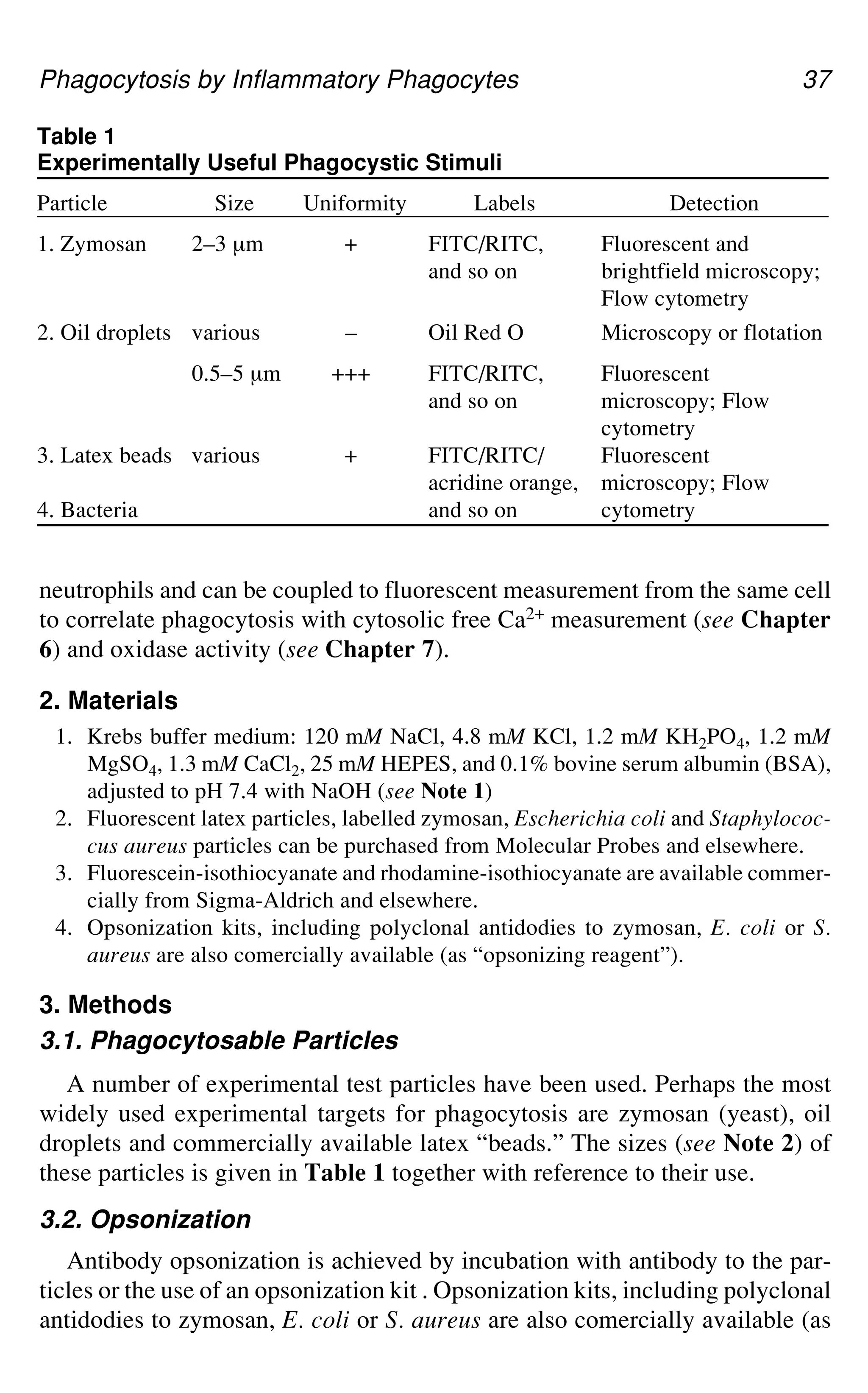
by Various Stimuli
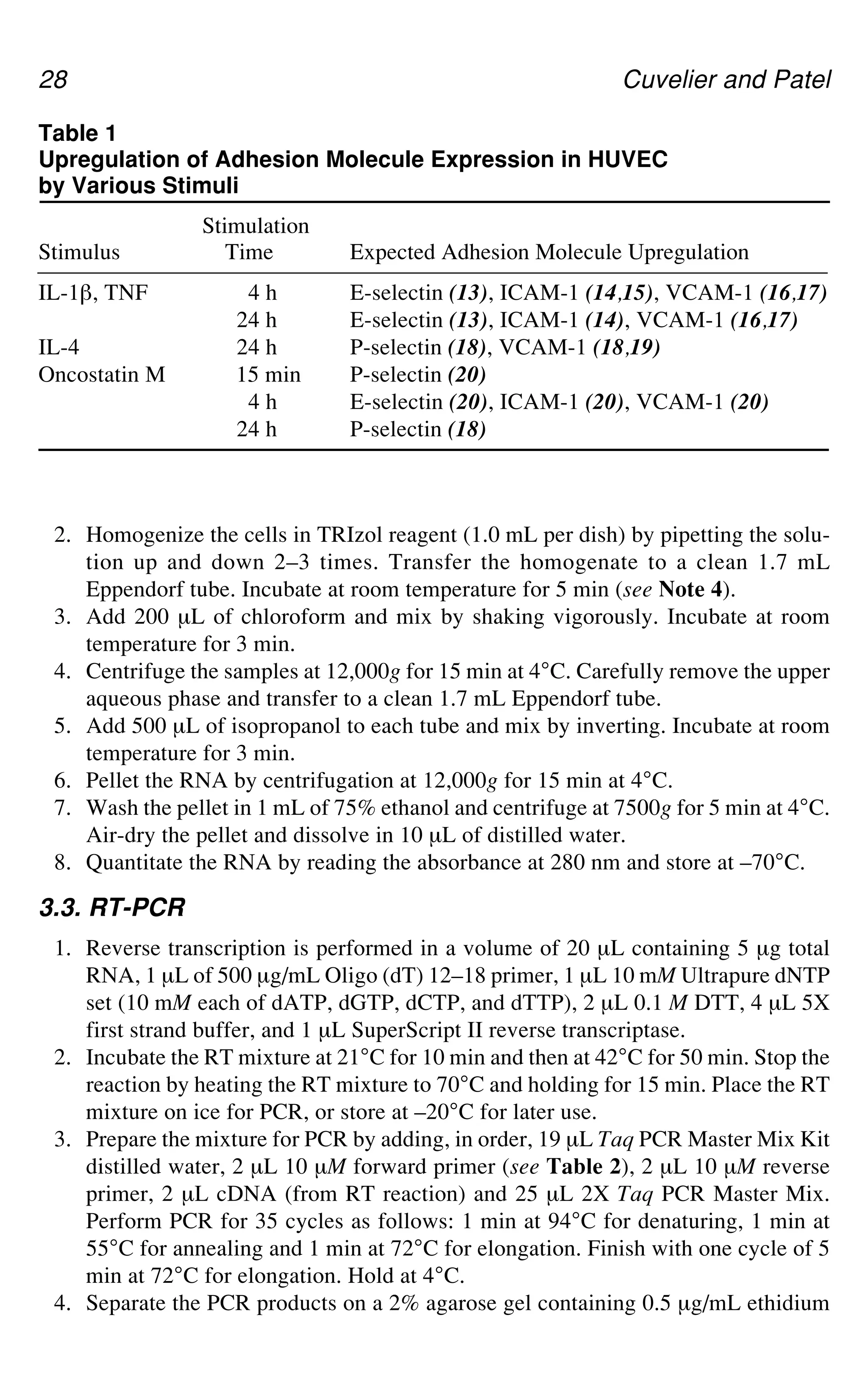
Stimulation
Stimulus Time Expected Adhesion Molecule Upregulation
IL-1`, TNF 4 h E-selectin (13), ICAM-1 (14,15), VCAM-1 (16,17)
24 h E-selectin (13), ICAM-1 (14), VCAM-1 (16,17)
IL-4 24 h P-selectin (18), VCAM-1 (18,19)
Oncostatin M 15 min P-selectin (20)
4 h E-selectin (20), ICAM-1 (20), VCAM-1 (20)
24 h P-selectin (18)
42.
Adhesion Molecule Expression29
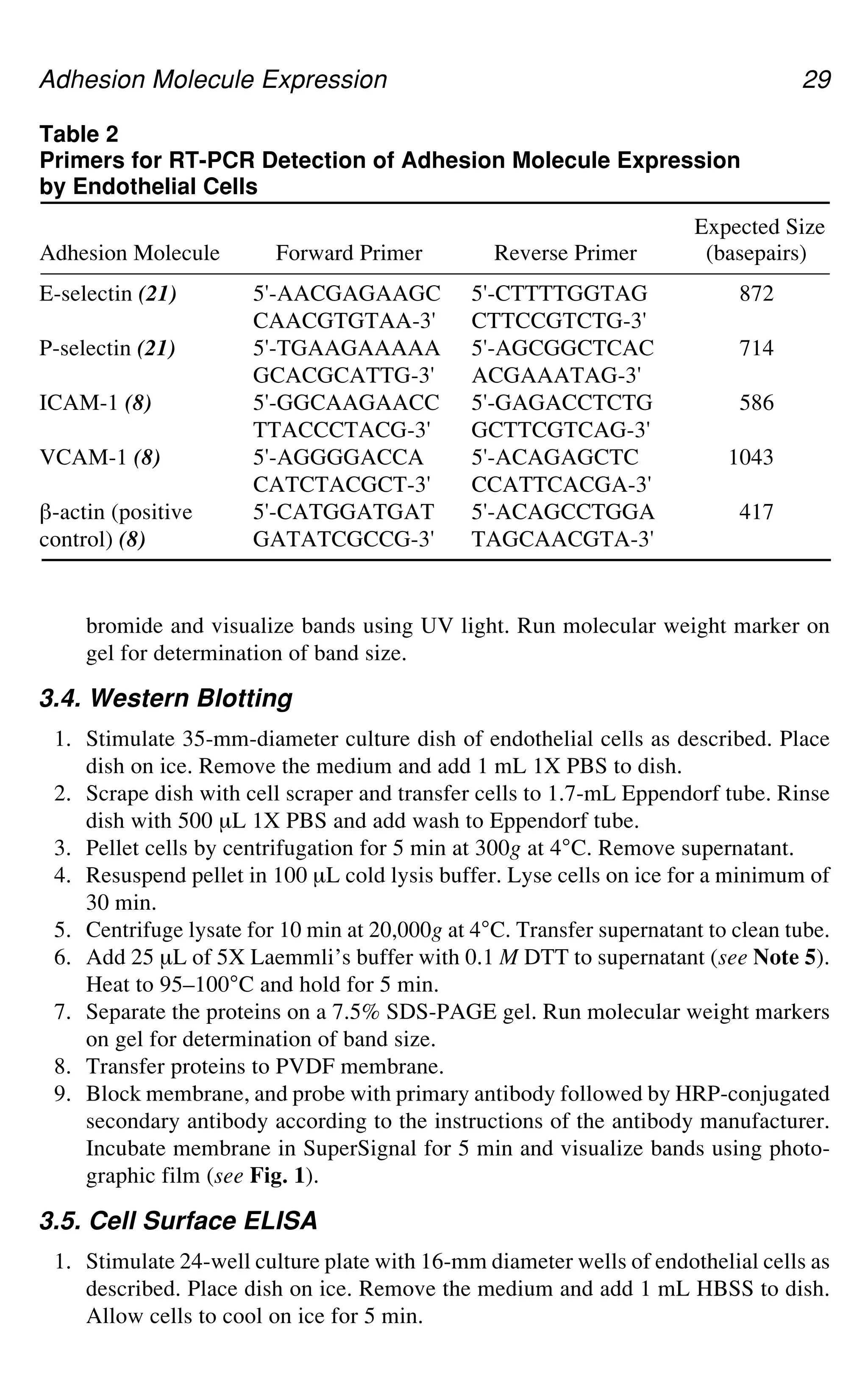
bromide and visualize bands using UV light. Run molecular weight marker on
gel for determination of band size.
3.4. Western Blotting
1. Stimulate 35-mm-diameter culture dish of endothelial cells as described. Place
dish on ice. Remove the medium and add 1 mL 1X PBS to dish.
2. Scrape dish with cell scraper and transfer cells to 1.7-mL Eppendorf tube. Rinse
dish with 500 μL 1X PBS and add wash to Eppendorf tube.
3. Pellet cells by centrifugation for 5 min at 300g at 4°C. Remove supernatant.
4. Resuspend pellet in 100 μL cold lysis buffer. Lyse cells on ice for a minimum of
30 min.
5. Centrifuge lysate for 10 min at 20,000g at 4°C. Transfer supernatant to clean tube.
6. Add 25 μL of 5X Laemmli’s buffer with 0.1 M DTT to supernatant (see Note 5).
Heat to 95–100°C and hold for 5 min.
7. Separate the proteins on a 7.5% SDS-PAGE gel. Run molecular weight markers
on gel for determination of band size.
8. Transfer proteins to PVDF membrane.
9. Block membrane, and probe with primary antibody followed by HRP-conjugated
secondary antibody according to the instructions of the antibody manufacturer.
Incubate membrane in SuperSignal for 5 min and visualize bands using photo-
graphic film (see Fig. 1).
3.5. Cell Surface ELISA
1. Stimulate 24-well culture plate with 16-mm diameter wells of endothelial cells as
described. Place dish on ice. Remove the medium and add 1 mL HBSS to dish.
Allow cells to cool on ice for 5 min.
Table 2
Primers for RT-PCR Detection of Adhesion Molecule Expression
by Endothelial Cells
Expected Size
Adhesion Molecule Forward Primer Reverse Primer (basepairs)
E-selectin (21) 5'-AACGAGAAGC 5'-CTTTTGGTAG 872
CAACGTGTAA-3' CTTCCGTCTG-3'
P-selectin (21) 5'-TGAAGAAAAA 5'-AGCGGCTCAC 714
GCACGCATTG-3' ACGAAATAG-3'
ICAM-1 (8) 5'-GGCAAGAACC 5'-GAGACCTCTG 586
TTACCCTACG-3' GCTTCGTCAG-3'
VCAM-1 (8) 5'-AGGGGACCA 5'-ACAGAGCTC 1043
CATCTACGCT-3' CCATTCACGA-3'
`-actin (positive 5'-CATGGATGAT 5'-ACAGCCTGGA 417
control) (8) GATATCGCCG-3' TAGCAACGTA-3'
43.
30 Cuvelier andPatel
2. Remove supernatant and add 250 μL of primary antibody (2 μg/mL) in HBSS
with 1% HSA (see Notes 6 and 7). Incubate on ice for 1 h.
3. Wash wells three times with 500 μL of 4°C HBSS (see Note 8).
4. Add 250 μL of appropriate HRP-conjugated secondary antibody (1:1000 dilu-
tion) in HBSS with 1% HSA (see Note 9). Incubate on ice for 1 h.
5. Wash wells three times with 500 μL of 4°C HBSS.
6. Add 200 μL of TMB-ELISA. Incubate at room temperature until blue color has
developed (see Note 10). Add 200 μL of 1 M phosphoric acid to wells to stop
color development.
7. Read absorbance of duplicate points at 450 nm (see Fig. 2).
3.6. Flow Cytometry
1. Stimulate T75 flask of endothelial cells as described. Remove the medium and
add 5 mL of trypsin-EDTA (see Note 11). Incubate at room temperature for
approx 1 min. Harvest cells by pipetting the suspension up and down 2–3 times
with an automatic pipettor. Transfer suspension to a 50-mL Falcon tube contain-
ing 5 mL of medium.
2. Rinse flask two times with 1X PBS. Transfer rinses to Falcon tube.
3. Pellet cells by centrifugation for 5 min at 300g. Wash cells by resuspending pel-
let in 20 mL 1X PBS and centrifuging for 5 min at 300g.
4. Discard supernatant and resuspend pellet in 600 μL 1X PBS. Divide into six equal
aliquots. Each aliquot can be stained with a different antibody (see Note 12).
5. Add 1 μg of primary antibody to each tube. Incubate at room temperature for
30 min.
6. Pellet cells by centrifugation for 5 min at 300g. Wash cells by resuspending pel-
let in 3 mL 1X PBS and centrifuging for 5 min at 300g.
7. Aspirate off the supernatant. Resuspend cells in 90 μL of 1X PBS and add 10 μL
of FITC- or PE-conjugated secondary antibody. Incubate at room temperature,
shielded from light, for 30 min.
Fig. 1. Detection of VCAM-1 protein by Western Blotting. HUVEC were stimu-
lated for 24 h at 37°C and 5% CO2 in M199/A alone (Con) or M199/A with 20 ng/mL
of TNF (TNF) or IL-4 (IL-4). Cells were lysed in a Triton X-100-containing buffer
and separated on a 7.5% SDS-PAGE gel. Proteins were transferred to PVDF and the
membrane was probed with an anti-VCAM-1 antibody. The figure shown is represen-
tative of at least three experiments.
44.
Adhesion Molecule Expression31
8. Wash cells by resuspending pellet in 3 mL 1X PBS and centrifuging for 5 min
at 300g.
9. Resuspend cells by adding 0.2–0.3 mL of 1X PBS and vortexing gently. Measure
FITC- or PE-labeled cells using flow cytometry.
4. Notes
1. We frequently obtain adhesion molecule-specific antibodies directly from prin-
ciple investigators. However, antibodies against most adhesion molecules are com-
mercially available from companies such as R & D Systems (Minneapolis, MN),
Coulter-Immunotech (Miami, FL), and Cedarlane Laboratories (Hornby, ON).
2. Many protocols for culturing endothelial cells recommend using fetal bovine
serum (FBS) in the culture medium. Our laboratory, however, has found that
endothelial cells grow better in medium containing human serum than in medium
containing fetal bovine serum. Human serum can be isolated from normal donors
and pooled as described (4).
3. RNA is extremely sensitive to digestion by RNases, which are present on skin
and most workspaces. To prevent loss of RNA samples due to RNase digestion,
use extreme care when working with RNA. Wear gloves, use only RNase- and
DNase-free Eppendorf tubes and pipet tips and keep the work area clean.
Fig. 2. Detection of surface-associated VCAM-1 protein by ELISA. HUVEC were
stimulated for 24 h at 37°C and 5% CO2 in M199/A alone (Control) or M199/A
with 20 ng/mL of IL-4 (IL-4). Cells were labeled with nonimmune mouse IgG1 or an
_-VCAM-1 antibody. After 1 h at 4°C, the monolayers were washed and HRP-conju-
gated sheep _-mouse secondary antibody was added. After 1 h at 4°C, the monolayers
were washed and TMB-ELISA substrate was added. The reaction was stopped after
3 min by the addition of 1.0 M phosphoric acid. Antibody binding was determined by
measuring absorbance at 450 nm. Data shown are mean ± deviation and are from a
representative experiment.
45.
32 Cuvelier andPatel
4. During the RNA isolation, there are a number of steps at which the procedure can
be stopped and samples can be stored. Samples in TRIzol may be stored at –70°C
for up to 1 wk and defrosted prior to continuing. Samples in 75% ethanol may be
stored at –70°C for up to 1 wk and used immediately upon removal from the
freezer.
5. Cell lysates may be frozen after the addition of 5X Laemmli’s buffer and sepa-
rated by SDS-PAGE at a later date.
6. To measure background binding, label a well of cells with an isotype-matched,
non-immune primary antibody as a negative control.
7. P-selectin levels on endothelial cells are generally very low, making detection
difficult. Multiple primary antibodies that recognize different epitopes on
P-selectin can be used in the cell surface enzyme-linked immunoabsorbent assay
(ELISA) to increase the sensitivity of detection.
8. Endothelial cell monolayers tend to be very delicate. Add HBSS washes slowly
to the centre of the wells using an automatic pipettor to prevent lifting of the
cells. Examine the cells using a microscope following the surface ELISA proce-
dure to ensure that cells have not been lost.
9. The amount of secondary antibody used may need to be titrated to achieve an
optimal signal to background ratio.
10. For comparison, all wells in a single experiment should be incubated in TMB-
ELISA for the same amount of time. Color development should be allowed to
occur until the absorbance of all samples falls within the linear range of the spec-
trophotometer used to measure absorbance. Data from samples whose absorbance
falls outside of the linear range of the spectrophotometer should not be used.
11. Surface expression of P-selectin is often difficult to detect and the trypsin-EDTA
digestion required to detach endothelial cells from culture dishes may cleave
adhesion molecules making detection more difficult. Surface ELISA is generally
a preferable method to flow cytometry, especially for detecting P-selectin.
12. To measure background binding, label a tube of cells with an isotype-matched,
non-immune primary antibody as a negative control.
References
1. Silverstein, E. A. (1999) The vascular endothelium, in: Inflammation: Basic Prin-
ciples and Clinical Correlates. (Gallin, J. I., Snyderman, R., Fearon, D. T.,
Haynes, B. F., and Nathan, C., eds.), Lippincott Williams and Wilkins, Philadel-
phia, PA, pp. 207–226.
2. Cohnheim, J. (1889) Lectures on General Pathology. A Handbook for Practitio-
ners and Students. The New Syndenham Society, London, UK.
3. Jaffe, E. A., Nachman, R. L., Becker, C. G., and Minick, C. R. (1973) Culture of
human endothelial cells derived from umbilical veins: Identification by morpho-
logic and immunologic criteria. J. Clin. Invest. 52, 2745–2756.
4. Zimmerman, G. A., McIntyre, T. M., and Prescott, S. M. (1985) Thrombin stimu-
lates the adherence of neutrophils to human endothelial cells in vitro. J. Clin.
Invest. 76, 2235–2246.
46.
Adhesion Molecule Expression33
5. McIntyre, T. M., Zimmerman, G. A., and Prescott, S. M. (1986) Leukotrienes C4
and D4 stimulate human endothelial cells to synthesize platelet-activating factor
and bind neutrophils. Proc. Natl. Acad. Sci. USA 83, 2204–2208.
6. Bevilacqua, M. P., Pober, J. S., Mendrick, D. L., Cotran, R. S., and Gimbrone, M.
A. Jr. (1987) Identification of an inducible endothelial-leukocyte adhesion mol-
ecule. Proc. Natl. Acad. Sci. USA 84, 9238–9242.
7. Springer, T. A. (1994) Traffic signals for lymphocyte recirculation and leukocyte
emigration: The multistep paradigm. Cell 76, 301–314.
8. Ali, S., Kaur, J., and Patel, K. D. (2000) Intercellular cell adhesion molecule-1,
vascular cell adhesion molecule-1, and regulated on activation normal T cell
expressed and secreted are expressed by human breast carcinoma cells and sup-
port eosinophil adhesion and activation. Am. J. Pathol. 157, 313–321.
9. Hewett, P. W. and Murray, J. C. (1993) Human lung microvessel endothelial cells:
Isolation, culture and characterization. Microvasc. Res. 46, 89–102.
10. Marks, R. M., Czerniecki, M., and Penny, R. (1985) Human dermal microvascu-
lar endothelial cells: An improved method for tissue culture and a description of
some singular properties in culture. In Vitro Cell. Dev. Biol. 21, 627–635.
11. Gupta, K., Ramakrishnan, S., Browne, P. V., Solovey, A., and Hebbel, R. P. (1997)
A novel technique for culture of human dermal microvascular endothelial cells
under either serum-free or serum-supplemented conditions: Isolation by panning and
stimulation with vascular endothelial growth factor. Exp. Cell Res. 230, 244–251.
12. Richard, L., Velasco, P., and Detmar, M. (1998) A simple immunomagnetic pro-
tocol for the selective isolation and long-term culture of human dermal microvas-
cular endothelial cells. Exp. Cell Res. 240, 1–6.
13. Bevilacqua, M. P., Stengelin, S., Gimbrone, M. A., Jr., and Seed, B. (1989)
Endothelial leukocyte adhesion molecule 1: An inducible receptor for neutrophils
related to complement regulatory proteins and lectins. Science 243, 1160–1165.
14. Pober, J. S., Gimbrone, M. A., Jr., Lapierre, L. A., Mendrick, D. L., Fiers, W.,
Rothlein, R., et al. (1986) Overlapping patterns of activation of human endothe-
lial cells by interleukin 1, tumor necrosis factor, and immune interferon.
J. Immunol. 137, 1893–1896.
15. Buzby, J. S., Knoppel, E. M., and Cairo, M. S. (1994) Coordinate regulation of
steel factor, its receptor (kit), and cytoadhesion molecule (ICAM-1 and ELAM-1)
mRNA expression in human vascular endothelial cells of differing origins. Exp.
Hematol. 22, 122–129.
16. Osborn, L., Hession, C., Tizard, R., Vassallo, C., Luhowskyj, S., Chi-Rosso,
G., et al. (1989) Direct expression cloning of vascular cell adhesion molecule 1,
a cytokine-induced endothelial protein that binds to lymphocytes. Cell 59,
1203–1211.
17. Carlos, T. M., Schwartz, B. R., Kovach, N. L., Yee, E., Rosso, M., Osborn, L.,
et al. (1990) Vascular cell adhesion molecule-1 mediates lymphocyte adherence
to cytokine-activated cultured human endothelial cells. Blood 76, 965–970.
18. Yao, L., Pan, J., Setiadi, H., Patel, K. D., and McEver, R. P. (1996) Interleukin 4
or oncostatin M induces a prolonged increase in P-selectin mRNA and protein in
human endothelial cells. J. Exp. Med. 184, 81–92.
47.
34 Cuvelier andPatel
19. Schleimer, R. P., Sterbinsky, S. A., Kaiser, J., Bickel, C. A., Klunk, D. A.,
Tomioka, K., et al. (1992) IL-4 induces adherence of human eosinophils and ba-
sophils but not neutrophils to endothelium: Association with expression of
VCAM-1. J. Immunol. 148, 1086–1092.
20. Modur, V., Feldhaus, M. J., Weyrich, A. S., Jicha, D. L., Prescott, S. M.,
Zimmerman, G. A., et al. (1997) Oncostatin M is a proinflammatory mediator: In
vivo effects correlate with endothelial cell expression of inflammatory cytokines
and adhesion molecules. J. Clin. Invest. 100, 158–168.
21. Ali, S. and Patel, K. D. Unpublished observations.
36 Morris etal.
Fig. 1. The biological particle (a) presents a surface composed on various proteins,
which after opsonization by C3bi (b) or antibody (c) presents either C3bi or the Fc
portion of antibody.
to antigens on the surface of the microbe through the Fab portion of the anti-
body, leaving the Fc regions free. Adhesion of antibodies to microbial sur-
faces, soluble antigens or each other (to form immune complexes) can also
cause activation of complement and the deposition of C3bi on the surface or
complex. Thus, by either opsonising the surface of the microbe or particle with
C3bi or antibody, the surface is transformed from presenting as one of a large
number of possible varieties into just two possible presentations, i.e., either
C3bi or Fc portion of antibody (see Fig. 1).
Neutrophils have receptors for C3bi, called CR3 (complement receptor 3),
`2 integrin, and CD11b/CD18 (2), and also have a variety of receptors for the
Fc portion of (IgG) antibody, Fc receptors labelled CD16, CD32, and CD64
(reviewed in detail in ref. 3). These two receptor types bind their counter-
ligand either C3bi or antibody and signal the neutrophil to undergo a program
of activity, which leads to a more rapid internalization of the particle than
would occur otherwise.
In this chapter, the use of test particles for quantifying phagocytosis in vitro
is presented, along with methods for their in vitro opsonization and for their
fluorescent labeling to permit their tracking microscopically or by flow
cytometry. Methods are also given for quantifying the number of particles
within neutrophils and for presenting the resultant data. Finally, a method for
presenting particles for phagocytosis is given that is applicable to individual
50.
Phagocytosis by InflammatoryPhagocytes 37
Table 1
Experimentally Useful Phagocystic Stimuli
Particle Size Uniformity Labels Detection
1. Zymosan 2–3 μm + FITC/RITC, Fluorescent and
and so on brightfield microscopy;
Flow cytometry
2. Oil droplets various – Oil Red O Microscopy or flotation
0.5–5 μm +++ FITC/RITC, Fluorescent
and so on microscopy; Flow
cytometry
3. Latex beads various + FITC/RITC/ Fluorescent
acridine orange, microscopy; Flow
4. Bacteria and so on cytometry
neutrophils and can be coupled to fluorescent measurement from the same cell
to correlate phagocytosis with cytosolic free Ca2+ measurement (see Chapter
6) and oxidase activity (see Chapter 7).
2. Materials
1. Krebs buffer medium: 120 mM NaCl, 4.8 mM KCl, 1.2 mM KH2PO4, 1.2 mM
MgSO4, 1.3 mM CaCl2, 25 mM HEPES, and 0.1% bovine serum albumin (BSA),
adjusted to pH 7.4 with NaOH (see Note 1)
2. Fluorescent latex particles, labelled zymosan, Escherichia coli and Staphylococ-
cus aureus particles can be purchased from Molecular Probes and elsewhere.
3. Fluorescein-isothiocyanate and rhodamine-isothiocyanate are available commer-
cially from Sigma-Aldrich and elsewhere.
4. Opsonization kits, including polyclonal antidodies to zymosan, E. coli or S.
aureus are also comercially available (as “opsonizing reagent”).
3. Methods
3.1. Phagocytosable Particles
A number of experimental test particles have been used. Perhaps the most
widely used experimental targets for phagocytosis are zymosan (yeast), oil
droplets and commercially available latex “beads.” The sizes (see Note 2) of
these particles is given in Table 1 together with reference to their use.
3.2. Opsonization
Antibody opsonization is achieved by incubation with antibody to the par-
ticles or the use of an opsonization kit . Opsonization kits, including polyclonal
antidodies to zymosan, E. coli or S. aureus are also comercially available (as
51.
38 Morris etal.
“opsonizing reagent”). Zymosan particles can also be readily opsonized with
human C3bi. The procedure has two parts: i) producing human serum with
complement, and ii) the opsonization process.
3.2.1. Production of Human Serum
1. Take 10–20mL of blood (human) into a glass container (universal tube) without
heparin or other anticoagulant.
2. Leave for several hours (preferably overnight) at 4°C to clot and for the clot to
contract. It is often useful to release the clot from the sides of the glass tube as
this allows clot contraction to occur. Centrifuge the tube to sediment free red
blood cells.
3. Remove the serum with a plastic pipete trying to avoid collecting free red blood
cells. Do not attempt to recover more serum than about half the original blood
volume.
4. Centrifuge the serum to sediment any contaminating red blood cells. The serum
should be clear and “straw” colored. If the serum remains red after centrifuga-
tion, it should be discarded as this indicates that haemolysis has occurred (possi-
bly as a result of complement activation).
5. The serum can be aliquotted and stored at –20°C until required.
3.2.2. C3bi Opsonization of Particles
1. Zymosan (10 mg) is suspended in Krebs medium (0.5 mL).
2. This is added to 0.5 mL of fresh or thawed stored human serum.
3. The suspension is incubated at 37°C for 30 min (keeping the zymosan in suspen-
sion if possible).
4. The zymosan is washed in PBS by centrifugation (300g, 2 min) and resuspension
(see Note 3)
5. The suspension can be stored at 4°C for short periods. However, sterile condi-
tions or storage at –20°C is required for longer periods.
3.2.3. Characterization of Opsonized Particles
It is important to confirm that opsonisation has been correctly performed by
i) comparison with nonopsonized particles of their uptake by neutrophils, and
ii) showing that blockade of CR3 (`2-integrin) by pretreatment of the neutro-
phils with antibody to CD18 or CD11b reduces the phagocytic uptake to the
nonopsonized level.
3.3. Fluorescent Labeling
There are a number ways by which a fluor can be covalently coupled to a
protein. Although kits are available for some linkages, one of the simplest and
most effective is to employ the reaction between the protein and isothiocyanate.
The fluorescein and rhodamine isothiocyanate-conjugates are commercially
52.
Phagocytosis by InflammatoryPhagocytes 39
available from a number of sources (FITC and RITC). Also, fluorescently
labeled antibody preparations are commercially available. In order to label
zymosan particles with the probe, there are essentially two steps: i) establish-
ing the reaction, which occurs at high pH, and ii) removal of the reactant, high
pH, and separation of the labeled zymosan from the free fluor.
1. High pH bicarbonate reaction buffer (0.25 M) is made from Na2CO3 (1 M) and
NaHCO3 (1 M) stocks mixed in a ratio of 1:9 and diluted in water 1:4, pH
adjusted to pH 9.0.
2. Zymosan (10 mg) is suspended in 900 μL of reaction buffer.
3. 100 μL of FITC (5 mg/mL dissolved in the NaHCO3 buffer) is added to the
reaction buffer containing zymosan.
4. The reactants are incubated at 4°C overnight (being kept suspended if possible).
5. The zymosan is removed from the reaction mixture by centrifugation at 300g for
5 min (see Note 2).
6. The zymosan is resuspended in PBS and washed again by centrifugation.
7. Steps 5 and 6 are repeated until the washing medium is free of fluorescent mate-
rial (at least three times) and when viewed microscopically, the zymosan par-
ticles are observable against a black background which is no brighter than a
control field with no fluor present.
8. The preparation can be stored at 4°C or opsonized. (see Note 3)
3.4. Presentation of Stimuli
The particulate stimuli can be presented to neutrophils in three ways: i) both
cells and particles in suspension (suspension), ii) neutrophils adherent with
particles in suspension “raining” down on the cells (sedimentation), and ii)
neutrophils adherent and particles presented by micromanipulation. Each
method of presentation is useful for different purposes.
3.4.1. Suspension Presentation
In this approach, a suspension of particles is added to a suspension of neu-
trophils. The method relies on the random contact between particles and cells
in the suspension medium. Obviously, the number of random collisions be-
tween the two will depend upon the concentration of each. A common strategy
is to ensure that the number of particles is in large excess to the number of
cells. Neutrophils can internalise more than nine zymosan particles/cell, so it is
clear that if the ratio of particles to cells is less than this, phagocytosis will be
limited not by the capability of the cells, but by the availability of the particles.
A typical protocol is given.
1. Follow protocol for opsonisation, or retrieve from storage.
2. Resuspend neutrophils in Krebs medium (see Table 1) to give 106 cells/mL, warm
to 37°C
53.
40 Morris etal.
3. Add 10 μL of stock zymosan to give final conc of 1 mg/mL
4. Incubate at 37°C, keeping cells suspended.
5. Sample at 10, 15, 30, and 60 min for microscopic counting or flow cytometry.
3.4.2. Sedimentation Presentation
An important feature of the suspension method of presentation is that all
surfaces of the neutrophil have an equal chance of encountering a particle (by
random collision) and so cells can accumulate large numbers of particles. How-
ever, it is an unusual situation for neutrophils to encounter microbes in this
way. Instead, neutrophils encounter microbes in the extravascular space after
they have changed their morphology from spherical (as in the blood stream or in
the suspension) to flattened and chemotactic. For this reason, another approach
which may be used is to adhere the neutrophils to a surface and allow particles
to contact them by sedimenting onto their exposed surface. With this approach,
although the number of particles internalized may be less, the physiological
process may be closer to that which occurs in vivo. There the surface to which
the neutrophils are allowed to adhere is, however, a consideration. If one
chooses an integrin engaging surface such as may be presented by a protein
coating, this may reduce the availability of these receptors for phagocytosis.
However, if one chooses a nonphysiological (and convenient) surface such as
glass, the CR3 receptors may remain available but as the neutrophils have a
tendency to become increasingly flattened onto the glass, this causes a limitation
to the available plasma membrane to form phagosomes (i.e., the cells become so
stretched that there is no possibility of the further “stretching” required for
phagocytosis). A typical methodology is given in which glass is used as the
substrate, but the time for adhesion is short to minimise this effect.
1. An area is marked on a clean (ethanol washed) glass slide.
2. Neutrophils (106/mL) are added to the marked area and allowed to sediment for
10 min at 37°C.
3. Nonadhered neutrophils are removed by washing the slide with Krebs medium.
4. Zymosan is diluted in Krebs medium (10 mg/mL) and 100 μL added to the
marked area of the side.
5. This is transferred to a humid incubator chamber and maintained at 37°C for the
required time (e.g., 10, 30, 60 min).
6. The excess, nonphagocytosed material is removed by washing and phagocytosis
quantified by counting.
3.4.3. Micromanipulation
Another advantage of the sedimentation approach is that an individual liv-
ing neutrophil can be observed before during and after phagocytosis. This
would enable the time between contact, pseudopod formation, and so on to be
54.
Phagocytosis by InflammatoryPhagocytes 41
determined. Also, if the neutrophil were loaded with an cytosolic free Ca2+
indicator (see Chapter 6), the Ca2+ signaling events accompanying phagocyto-
sis would also be observable (4). However, the sedimentation method provides a
random distribution of particles as they sediment and it cannot be predicted
when a particular cell will make contact with a particle. This can be frustrating
as particles may sediment onto the focal plane but fail to contact the cell under
observation. This problem is also compounded by local heating effects caused
by the ullumination source. This can occur even with heated stages and cause
conventional currents away from the source of observation which carry par-
ticles away from the cell under observation.
These problem are, however, overcome by a micromanipulation technique
whereby the particle is fed to the neutrophil under observation (Fig. 3). Thus,
the timing of contact can be precisely controlled. Also the position on the cell
at which contact is made can be controlled and the same cell can be “fed”
multiple times. The method relies on a micropipet attached to a micro-
maniupulator, through which a slight negative pressure is applied. A zymosan
particle which has sedimented near the cell under observation is “picked up”
by positioning the tip of the micropipete near by. This is then carried to the cell
and presented. Once adherence between the particle and cell has occurred, the
particle can be released and phagocytosis observed. The tip of the micropipet
should be smaller than the particle, ideally 1–2 μm so that pressure control can
be effective. The positioning of the micropipet is best achieved using a motor-
ized microprocessor controlled manipulator. These are commercially available
from Eppendorf, WPI, and others. It is not necessary to have the complicated
electronic control required for microinjection (i.e., z stabbing) and less expen-
sive micromanipulators are adequate for the task.
3.5. Microscopic and Flow Cytometric Detection of Phagocytosis
There are essentially two approaches for detecting phagocytosis: i) micro-
scopic, in which individual neutrophils are observed and the number of par-
ticles phagocytosed per cell counted, and ii) the use of flow cytometry to
quantify the fluorescence associated with each cell as it passes through the
laser beam. Both approaches have advantages and disadvantages. The micro-
scopic approach is not automated and requires observation of a large number
of cells. This is obviously time consuming, but has the advantage i) that adher-
ence of particles to cells is not taken as phagocytosis, and ii) that gross effects
on cell morphology or the health of cells (if subjected to a pharmacological
agent, for example) are apparent during the counting. In contrast, flow
cytometry has the advantage that it is automated and quick, but the disadvan-
tage that it is more difficult to distinguish adherence from internalisation, or
have feedback on the health of the cells.
“Hopocon—my father, thatyou call Captain Pipe—wants none of
the Paleface teachings. When the missionaries told Gentle Maiden
long ago there were no witches, he only pitied them that they knew
no better.”
“But—”
“No, no!” the girl broke out hurriedly. “My Paleface brother must
not wait talking here. That which is, must be. Not long has Lone-Elk
been gone. By riding fast the White Fox can reach his cabin before
the coming of the Seneca, and with Little Paleface soon be far away
where Lone-Elk will not find them. Haste! Gentle Maiden has done
all she can. Paleface brother must not remember who has told him
this, but oh, he must remember what he has heard! Hurry, hurry,
now, or—”
“I’ll go, Gentle Maiden, I’ll go. If I can ever pay back the
kindness you have done both John and me, I’ll not be slow to do it,
you may be sure. But it’s a downright shame—no, what I mean is
that you need never fear anyone will so much as suspect that you
told me this or anything. Good-bye, good-bye.”
With such feverish anxiety and haste did Ree speak, now that
he was bent only on flying to John’s rescue, he scarce knew what he
said; but in a trice he was in the saddle. And yet quickly as he
moved, when he turned to give a parting nod the Indian girl was
gone.
Long familiarity with the woods had made the beautiful,
intelligent mare, Phoebe, almost as free and light-footed among the
trees and brush and rough ground, often broken by rougher roots
and fallen branches, as a deer. Kingdom placed all dependence in his
horse’s ability to avoid or clear every obstruction and urged the
57.
gentle creature tothe utmost, paying little heed to anything save to
escape the limbs of trees overhead as he hastened on. He had at
once concluded that Lone-Elk and his band were undoubtedly
traveling toward the cabin by the route to the east of the lake and
the swamp which bounded a considerable portion of it, for otherwise
he must have met them. He knew that they could easily have heard
him approaching and hidden themselves until he passed, but long
training had made his ears sharp and his eyes the same. Maybe he
had this time, however, placed too much dependence in them.
“Anyhow, we’ll soon know, my pet,” he murmured with teeth
clenched, and Phoebe seemed to understand.
Out upon the bluffs above the river, into the open for a moment,
then down the precipitous hills and across the water at a shallow
place horse and rider went, and, emerging soon from the woods
again, were in the natural clearing—the clearing which had originally
tempted the boy pioneers to locate here.
All was quiet. The cabin stood like a sentry at rest on the high
bank rising abruptly from the river, then sloping down on all sides
away from it. The yellow, autumn sunlight made the whole scene
appear even drowsily tranquil. A sense of relief came to Kingdom,
and he even felt chagrined that he had been so decidedly disturbed.
Still it was strange that John did not show himself. Perhaps the
exceeding quiet all about was, after all, fraught with greatest danger.
Perhaps—but Ree was at the foot of the slope now and his mind had
scarcely time to present another thought before he was up the hill,
and throwing himself from the horse, quickly entered the open door
of the low log house.
58.
“John!” he calledin a low tone—and a little catch in his throat
which he could not control, gave his voice a tremulous quaver.
“John!”
“Yes, Ree;” the answer was scarcely more than a whisper, “I’m
up here in the loft, and listen! You can hear me?”
“Every word.”
“Don’t act surprised or excited or show that you have found out
or heard anything, for they’re watching now—Lone-Elk and a pack of
Delawares have surrounded the clearing. I’ve been peeking through
a crack, watching ’em half an hour or more.”
59.
CHAPTER V—IN DRIPPINGRAIN
AND DARKNESS
With what consternation Kingdom received the startling intelligence
John’s words conveyed would never have been guessed from his
actions. He tossed his rough, squirrel-skin cap on the bunk, which
was a bed by night and a lounge by day, and sat down, wiping the
perspiration from his forehead.
“They’re after me, I s’pose, Ree,—blame ’em!” Jerome went on
in the same half whisper. “I just happened to be up here pawing
over some of the skins stored away so long, and got a glimpse of
the rascals among the trees. So I’ve been watching ever since, and I
don’t want you to think I crawled up here to hide. Just so much as
hint at such a thing and I’ll—”
John did not say what he would do, but seeing how he hated
being found in a position which might be taken as a reflection upon
his courage, Ree was considerably tempted to suggest that maybe
he himself had better get under the bed. But it was no time for joke-
making and the facetious thought had no more than occurred to him
than, unspoken, it was forgotten.
“Stay up there, John, old boy; see everything you can. I’ll stroll
out and put Phoebe in the lean-to and gape around some in a
natural sort of way myself. The whole business looks mighty bad.
60.
What Fishing Birdsaid is all true; I found out that much. I’ll tell you
about it when I come in.”
If John Jerome had been a lad easily alarmed or one likely to
fall a ready victim to a too lively imagination, Return Kingdom would
certainly have thought that he had done so in this case when, after
unsaddling the mare and tying her in her stall, he sat down in the
open doorway of the cabin and with apparent indifference scanned
the clearing from end to end, without seeing the slightest sign of the
Indians’ presence.
With his elbow on his knee, his head upon his hand, as if he
were merely resting, he continued to watch the wooded boundary
most intently from between the fingers which concealed his eyes. He
had little fear that the Indians would fire upon him from some place
of concealment among the trees; the distance was too great. A
white hunter might easily have brought down a deer at the same
number of yards with an exceptionally heavy charge in his long-
barreled rifle, but the Redskins, as Ree well knew, usually loaded
with so little powder, owing to its scarcity with them, no doubt, that
he had little to fear in thus exposing himself so long as the enemy
came no nearer than the edge of the woods.
“You’re downright sure you saw them, John?” inquired Kingdom,
in a low voice, rising and entering.
“There he goes! There—did you see that?” came an excited
undertone from Jerome as if in answer.
Instantly Kingdom looked out but he saw nothing.
“I vow! I think it was the Seneca!” John whispered. “He ran
from the big beech near the patch to the clump of little trees at the
61.
left. Guess hethought no one was watching but you, and darted out
when your back was turned.”
“I’ll stay back out of sight a bit, and you look sharp. Maybe we
can make out what they’re up to,” Kingdom replied. Then, to lead
the savages to suppose that their presence was not suspected, Ree
went about making a bright fire as if to prepare dinner, and soon the
smoke from the cabin chimney conveyed to the crouching redskins
in hiding along the clearing’s edge the very impression he wished
them to receive.
Kingdom spent half an hour,—a long half hour of suspense and
anxious thoughts—in putting the room to rights, busying himself in a
dozen different ways, while John peered closely from the crack, to
see through which his eyes had already been strained so long they
ached severely. Still he saw nothing. Whether the savages were only
extremely wary or whether, as the boys fervently hoped, they had
slipped away and gone as silently as they came could not be known,
and continued vigilance was the only key to their safety.
All day John Jerome remained concealed in the loft, watching
almost constantly from the narrow crevice which permitted him to
see without being seen. All day Return Kingdom went about from
the cabin into the lean-to barn, from the barn into the cabin again,
and in and out of the open door a hundred times on one pretext and
another, doing his best to make his every movement seem
composed and natural.
He was certain in his own mind that the savages were watching
for John. Perhaps they expected to see him in some fantastic and
witch-like shape,—see him change from a cloud to human form, or
turn himself into some wild beast.
62.
Once a wanderingcrow flew into the clearing and circled idly
over the little cornfield. As it flew down to a shock of corn, both boys
chanced to notice it and both saw, too, a sudden, rapid movement,
and then another and another, within the fringe of the woods. Were
they the dancing shadows of wind-tossed branches, or were the
Seneca and his band still near? Quick as the movements were, little
as the boys had seen, they knew the answer to the question which
occurred to them and thanked the vagrant crow for the information
he had been the means of giving them.
“Still,” said John, “if those fool Delawares get it into their heads
that that crow is me, and like as not Lone-Elk may tell ’em some
such thing, it’ll just make the whole lot of them believe more than
ever that I am a sure enough witch.”
Full well did Kingdom realize how very correct John’s
observation probably was. He was confident that it was the crow
which occasioned the moving about among the hiding Indians,—the
flitting shadows both he and John had seen. He made no answer to
his friend’s remark at once, but turned over again in his mind a plan
which he had been considering all day. It seemed wise. He could
think of nothing better.
“John,” said Ree at last, “if they stay away till it’s dark enough to
do it, how would you like to slip away and go up among the rocky
ledges for a few days?”
“Hide?” Jerome demanded rather contemptuously.
“Why, no! There’s no need to call it hiding,” Kingdom answered
tactfully. “Just stay away from the cabin for awhile and give me a
chance to find out what killed Big Buffalo and get the witch idea out
of these crazy Delawares’ minds.”
63.
“But, don’t you—”
“Iknow what you’re going to say. It is, don’t I think that the
fact of your being away will make the Indians all the more certain
about this witchcraft business—make them think you’ve skedaddled!
We can’t help what they think. We do know, though, that they’re
after you and either we’ve got to pack up and light out, or get this
witch idea out of their heads. Now I think I can do it, in spite of
Gentle Maiden’s discouraging talk; if I only have a chance.”
On one point, as the discussion continued, hardly above a
whisper, both boys agreed. It was that some time during the night
the Indians would visit the cabin. They might come as if in a friendly
way just to learn whether Little Paleface was there; or they might
make a determined attack. The redskins’ supposition that Ree was
alone, confirmed by all that they had seen during the day, however,
would probably suggest to them an apparently friendly, but in reality
spying, visit.
In whatever way the lads viewed their situation they found so
much of uncertainty surrounding them that at best they must take a
chance.
Often and often was it this way in pioneer days. Every important
movement was encompassed by more or less danger. If a settler
needed but to go to mill, or to some frontier trading place for
supplies, he confronted many uncertainties and often left his family
in danger, too. Danger was always present, and although only the
foolhardy were disregardful entirely, even the most prudent came by
constant association to take it as a matter of course.
The latter was the feeling of the two boys from Connecticut. If
they had been less accustomed to the alarms of the wilderness, they
64.
would, in thepinch in which they now found themselves, most
probably have sought safety at once at Fort Pitt or perhaps at some
of the Ohio river settlements. If they had done so their story would
have been a very different one.
Though he had but reluctantly agreed to Ree’s proposal, not
wishing to leave his friend to face the situation alone, John found so
much to think about in the prospect of spending the night—and it
might be many nights and days—alone in the woods, that the
reflection that he also would be in danger was almost comforting. He
thought with dread of the long and lonely hours of darkness without
even a camp-fire’s comfort, but somehow there was something quite
interesting about it all, too. Perhaps it was the change and the
excitement, as he planned how stealthily he would steal through the
woods, that appealed to him. Certain it is that he found himself
anxious to be gone, and watching the deepening shadows almost
impatiently lest something happen to prevent his departure before
thick darkness came. His greatest fear lay in the fact that on three
sides at least the cabin was, in all probability, still surrounded by
Indians. On the fourth or west side was the river. How was he to
reach the open woods? How reach the rocky ledges to the north and
east, among whose deep ravines and clefts and long, narrow
passages and shallow caves he would remain until the rage of the
savages had passed?
A bank of clouds, wide as the eye could see above the treetops,
had come up out of the southwest to meet the sinking sun and,
when at last the shadows had filled the valley, darkness came on
rapidly. The wind rose, too, and quite before its approach was
65.
suspected, a drizzlingfall rain had set in, which gave promise of
continuing all night.
The cabin door had stood open all day, but Ree felt he could
close it now without exciting the suspicions of those who watched.
As he did so, John clambered quickly down from the low loft and
slipped noiselessly through the low opening connecting the lean-to
stable and the single room of the cabin itself. How well he
remembered the good purpose the hole had served once before! He
remarked to Ree about it with a nervous little laugh, recalling that
lively battle of their early days in the woods and how nearly fatal to
them both it had been. But Kingdom told him to make haste; that
they could not know who was watching now, and in the darkness
there might be Indians even within hearing of a whisper.
Ree had improved the opportunity before night came on to fill
John’s powder horn and bullet pouch and to pack in the form of a
knapsack for him a blanket and a supply of dried meat and bread.
These, with Jerome’s rifle, he had previously passed through the “cat
hole,” as it was called, into the stable; but now that John had
followed them, he suddenly found himself wishing that he had
planned otherwise. Yet confident all was for the best, though the
wind never had had so much of awful homesickness in its mournful
sounds before, though the rain never before had beaten with such
seeming tearful sorrow upon the roof, he whispered hastily:
“Be careful, old boy. Look for news by the day after tomorrow if
you hear nothing before, and be sure that everything will be all right
in a few days at most.”
“And you come where I am the minute you’re in danger, mind,”
John answered. “Good-bye, Ree, I’m going along the river’s edge.
66.
It’ll be easyto get past anybody or anything tonight. Good-bye.”
Ree would have whispered another word of caution and of
farewell, but he realized that John was gone—felt it in his very bones
that he was alone, alone; and the autumn wind blew more
mournfully than ever; the patter of the raindrops sounded twice as
melancholy as before.
For many minutes Kingdom intently listened, then throwing wide
the cabin door, made a pretense of emptying just beyond the
doorstep the wooden, trough-like bowl which did duty as a wash
basin. Though he made a brave show of unconcern, his heart beat
hard and fast. But he was glad to see how totally dark the night
was. One must have been very close indeed if he had seen John
emerge from the darkness of the lean-to into the equal blackness
without, he thought. Surely the Indians, if still watching, would
never suspect him going out that way, and not having seen him at
all would be very certain that he had been gone for a full day at
least, should they call at the cabin and still not discover him.
Despite the storm, the night was warm for so late in the season,
and Kingdom was glad to have the door ajar while he waited for the
first step which would tell him of the Indians’ coming. He had no
doubt they would come, unless their general plan was quite different
from what he supposed it to be. Still, time dragged on bringing no
tidings—no sound but the drip, drip of the rain, the sad sighing of
the wind and now and then the rattle of some loose puncheon on
the roof, moved by a passing gust more lively than the rest.
Again and again Ree mentally computed the distance John had
probably traveled in the time that he had been gone. “Now he must
be just about at the foot of the bluff and creeping along the water’s
67.
edge, shielded bythe higher bank of the river,” he thought at first.
“Now he must be half-way to the woods. Now, if nothing has
happened, he is past the worst of the danger and safe among the
trees.”
And so thinking, encouraged by the absence of any alarming
sound, Kingdom breathed easier, and was glad John had gone along
the river instead of trying to cross the stream just at the cabin’s rear
and so gain the cover of the trees more quickly, as he had originally
proposed, and would have done but for the possibility that even on
the opposite bank of the stream there were watchers in hiding.
But safe and certain as John’s escape seemed to Ree, the truth
was that during these past few minutes that young man had been in
decidedly greater danger of losing his scalp than he cared ever to be
again.
Creeping on hands and knees close to the wall whose dark
background would help conceal his movements, John had made his
way out of the barn and around to the rear of the cabin. Almost flat
on his stomach, he drew himself slowly along the bluff and so
descended to the valley where the river bank was not nearly so
steep and comparatively low, rising only a few feet above the level
of the water. Crawling cautiously along the narrow strip of slippery
beach between the river’s edge and the bank, he progressed steadily
toward the woods. Often he paused to listen, and even when he
moved on again he strained his ears and tried his utmost to see; but
so deep was the darkness that, except for the denser black wall in
the distance, which he felt rather than saw was the woods, he was
certain that his situation, so far as seeing went, would be the same
with his eyes shut as with them wide open.
68.
In one ofhis pauses to hearken closer than he could do when
moving, John thought he heard a low, hoarse “Ugh!”—an inarticulate
sound, but one which seemed to express impatience, weariness, and
“What’s the use?” combined. He fancied he could see the shrug of
the Indian’s shoulders who, he was sure, was responsible for the
guttural noise. For a long time the boy did not move. The rain came
dripping down almost noiselessly. The wind whispered ever so softly
in the lower parts of the valley and seemed to make no sound
whatever save in the woods. To John it seemed that he waited an
hour, though in fact it was but a few minutes. Over his shoulder he
could see the ray of light from the cabin’s open door. How far away it
looked! Still that was fortunate. He would not have had it nearer for
a great deal. Now he would try again. Softly—softly he raised one
hand from the ground; softly, softly he raised a foot.
“Ugh!” Again it came; scarcely audible was the sound but the
fierce howl of a wolf directly in his ears would not have startled, and
frightened more the young white man crouching by the water.
The danger seemed nearer now—not more than three yards
from him, John was certain—perhaps only two. He felt that he could
put out his hand and touch the place from which it came. Again he
was quiet, so quiet that he breathed in noiseless little gasps, a thing
so trying on his throat and lungs that he would have felt as
comfortable had he tried not to breathe at all.
But soon came another sound. Instantly John recognized it—the
stealthy dipping of the paddle and low murmur of water against the
nose of a canoe. Where was the canoe headed? That was the
question. Toward him? Either that or up stream. The murmur of the
current indicated that the craft ran not with it but against it. Now he
69.
heard the canoetouch the half submerged grass close in to shore. It
was just abreast of him and within two arms’ length. Now it grated
ever so lightly upon the grass which, before the fall rains, had been
quite up out of the water.
Again light as a feather came the dip of the paddle, again the
soft murmur of the water barely heard above the quiet, even patter
of the rain. At the same moment John felt himself slipping. Slowly
the wet ground was giving way beneath him. He must move. It was
a case of two dangers, either stand still and slide violently into the
river, or move on a step and—
He must run the risk. Faster and faster he was sliding down. He
must step quickly, and step quickly he did. He made no noise
himself, he thought, but some pebble or bit of earth, loosened by his
movement, rolled down and dropped with a splash into the water.
Again came the muttered “Ugh!” something lower than before, and
oh! Heaven be praised! no longer abreast but some yards from him.
Again came the low dipping of the paddle. They were patrolling
the river for him, John knew now; but they would not find him. They
might paddle silently up and down the whole night long, if they
wished. In fact he rather hoped they would, and chuckled inwardly
at the thought.
70.
CHAPTER VI—“THE WITCHIS
HIDDEN HERE.”
That part of Lone-Elk’s band which had been appointed to hide along
the river bank throughout the day and paddle up and down in the
densest shadows of the shores when night had come, did not keep
up their search as long as John had hoped they would, when he
silently chuckled over the thought of their waste of time and effort.
When they passed so close to the lad they sought, not more
than one of them suspecting how very near he was, the Delawares
were closing in on the cabin, together with others on shore. Lone-Elk
had given the signal, by passing the word quietly along the irregular
line his braves made around the clearing, after waiting all day long.
He hoped to find the “witch” in hiding in the little cabin. Even if he
did not, he would impress the Delawares with the seeming truth of
the charge he had made against the young white man by showing
that he was away from home, engaged, presumably, in some of his
dreadful witch’s work. The Seneca had, moreover, a plan in mind
which made a visit to the home of the young Palefaces desirable
from his point of view, whether the one they sought should be
discovered or not, and now would be as good a time as any for the
carrying out of his purpose.
71.
While the Indianswere yet at a distance, Kingdom, watching
and listening in the cabin, heard their approach. He had kept his rifle
close at hand all day, and now he casually picked the weapon up
and with a show of idle carelessness polished its glossy stock with a
bit of buckskin.
The savages came silently on, apparently without effort to keep
from being heard. Kingdom was aware that they kept their line
spread out so as to form a semicircle which, together with the river,
would wholly enclose the little log house. His sharp ears assured him
that this was done, but it was with well acted surprise that he
sprang lightly up and stepped toward the door when Lone-Elk and
one other Indian showed themselves at last within the dim ray of
light shining from the fireplace.
“Come in! It’s wet and bad outside! Bring them all in!” he called
pleasantly, meeting the Seneca at the threshold and glancing out as
if he plainly saw the whole line of Indians outside, which in fact he
did not see at all.
“White Fox speaks kindly,” answered Lone-Elk, calling Ree by
the name the Delawares had long ago given him.
Only the Seneca and the one other Indian drew near the lighted
space about the door, however, and these two now entered as if
they were quite by themselves.
“Why should I not?” Ree answered to the Seneca’s remark,
noticing as he did so, how searchingly both the savages were
looking about the cabin’s single room. “We,—my white brother and
myself—have had the friendship of the Delawares always.”
“It is as the white brother says,” said the second Indian, a
powerful fellow whom Kingdom now recognized as a brave from the
72.
Delaware town onthe Muskingum, and whom he had seen a
number of times before. As he spoke, this Indian looked at Lone-Elk
inquiringly. Perhaps the Seneca considered his words a challenge. At
any rate he said sharply:
“Where is the other white brother! Does the White Fox wish to
hide him then, if he is the friend of the Delawares? Will the White
Fox hide the witch that breathed poison breath upon Big Buffalo, the
witch that with a hatchet killed a Delaware warrior, yet left no
mark?”
“What’s this you say? What wild talk is this, Lone-Elk? Has Lone-
Elk drunk of the firewater that he comes speaking so absurdly?”
Kingdom spoke with a show of temper and in a manner
distinctly creditable to the part he was bound to act.
“It is the law that witches must be put to death,” the Seneca
returned vigorously. “Lone-Elk has said that Little Paleface with a
witch’s hatchet killed a Delaware warrior—killed Big Buffalo. Now
must the witch be given up to the friends of him that was killed.”
“Well, I can only tell you that the one you call Little Paleface is
not here. He is far away and may not come back for some days,”
Kingdom answered quietly. “Now if Lone-Elk will believe this, and it
is the truth, he will return to the town of the Delawares and I will
myself go there tomorrow to have a talk. Is it a friendly thing for
Delaware braves to remain hidden all about the lodge of their
Paleface brothers as they are doing now? Let them all come into the
light. Let them see that my brother who is accused so falsely—so
unfairly and so unjustly—let them see, I say, that he is not here, and
we will plan to have a talk tomorrow.”
73.
Lone-Elk gave ashort, fierce whoop. Instantly fifteen or more
Indians rushed into the cabin, crowding-the little room quite
uncomfortably.
“The witch is hidden,” said Lone-Elk, loudly. “If the Little
Paleface is here let him show himself.”
As Kingdom looked quickly from one to another of the Indians
he observed with sorrow that Fishing Bird was among them. Had
this good fellow turned against his white friends, too? But no, that
quick friendly look as their eyes met was proof of his friendship still.
There being no answer to the Seneca’s invitation to Little
Paleface to show himself, except the grunted “Ughs!” of some of the
Delawares, Lone-Elk sprang quickly up the ladder of poles and
peered into the loft. Others followed his example, climbing up on
stools or by the aid of the roughness of the wall. Some looked up
the chimney. Some searched here, some there. One party of five or
six, lighting hickory bark torches at the fire, went into the barn. In
five minutes the whole cabin was turned topsy-turvy.
“You see it is just as I told you in the beginning,” said Kingdom
in a friendly tone, but somewhat impatiently. “Now will you not
consent to a talk! Let it be in the Council House of the Delawares—
let it be any place you choose. I think I can prove to you that this
charge of witchcraft is placed against one who is as true and honest
as ever man could be.”
Ree was sorry to see that the Delawares looked to Lone-Elk to
answer. He had more fear of this one Indian, under the
circumstances, than of any other half dozen warriors in Captain
Pipe’s town.
74.
“Let it beas the White Fox says,” the Seneca answered. “Yet will
my Paleface brother not deceive himself by thinking he deceives
Lone-Elk. The Paleface witch but hides. If it is not so, let the witch
come to the talk.”
Not for a second did Kingdom allow this challenge to be
unanswered. Like a flash every eye had turned to him; but instantly
he said:
“Will the Seneca go to Fort Pitt and there put Little Paleface on
trial before those whose customs are the customs of the Palefaces?
No, of course he will not. And just so would it not be fair for Lone-
Elk to demand more than he would be willing himself to give.”
75.
HE KEPT HISEYES ON THE
SENECA UNCEASINGLY.
The justice of Kingdom’s position was clear to the majority of
the Indians and he could not help but notice it; still Lone-Elk’s reply
in curt, surly tones was far from pleasing.
“Yet the White Fox asks for a talk! Like squaws that tell one
another of the worms that harmed the corn does the Paleface want
the Delawares to meet together with him and speak idle words!
Words! Words, that mean nothing and come to nothing.”
With a move of his hand to his companions to follow, the
Seneca left the cabin. Rapidly the other Indians marched off in
single file after him. Fishing Bird, somehow, was the last to leave. As
he went out of the door, he cast a glance of friendliness, which was
also a look of warning, to Ree and the peace of mind of that young
gentleman was not increased thereby.
By no means certain that the Indians would not return, Kingdom
sat for a long time on the edge of his bunk, listening and thinking.
He had great satisfaction in knowing that John was comparatively
safe for the time, at least, and thankful, indeed, that his chum’s
departure had been so timely. He longed for another and more
satisfactory talk with Fishing Bird. He must have such a talk, he
resolved, if it could by any chance be arranged, before he undertook
to show the Delawares that Big Buffalo had not been killed by
witchcraft. Perhaps that friendly fellow would be able to give him the
right clue to the whole situation. Might it not be he would frankly
76.
declare that itwas by the hand of Lone-Elk, himself, that the
warrior’s life had been snuffed out!
In his own mind Ree had little doubt concerning the true cause
of Big Buffalo’s death; but by what means the Seneca had put out of
his way the one member of Captain Pipe’s community who openly
resented his leadership there would most probably be a difficult
question to answer.
So the lonely lad sat pondering a long time; how long he did not
know or care. The rain was still falling, the wind still sighing dolefully
when he arose at last, closed and barred the door, also barred the
opening which served as a window, and removing only his moccasins
lay down to rest. Repeatedly did he picture to his mind’s eye John
Jerome tramping slowly, silently through the wet leaves, among the
dripping underbrush and trees, stopping often to get his bearings
from the wind, and so making his weary and most lonesome way to
the protection they had agreed upon.
Repeatedly his thoughts returned to the “big talk” which he
must attend tomorrow; but sound sleep came to him at last, even
while a crouching figure moved swiftly and stealthily into the
clearing and paused as if in hiding behind a shock of corn—the very
one on which the crow had perched in the afternoon—then stole on
again and disappeared.
Even as the first object appeared, another approached the cabin
and moved to the protection of the darker shadows of the stable.
For a minute or two the figure stood quiet in the denser darkness
beside the building, then moved cautiously toward the little cornfield
as if attracted by a faint rustle of corn leaves which seemed to come
from that vicinity.
77.
The rain stillfell in a quiet, unbroken drizzle, but the wind had
abated and there was no reason to suppose that it caused the
movement of the corn, which attracted the attention of the
crouching creature. Still listening with utmost care, the crouching
figure moved nearer to the spot from which the noise ensued.
To discern any object that was without motion, at a distance of
even a few feet in the pitch darkness, was an impossibility; but as
the rustling of the corn ceased, the one who had been attracted by
the sound made out a stealthy movement in the vicinity and
instantly stood still. When the darker shadow had passed beyond his
vision he dropped to the ground and listened with his ear against the
wet grass and earth. After a time he rose and ran forward ever so
lightly, pausing at the edge of the woods.
Hour after hour passed. A dull gray light appeared on the clouds
to the east. Rising then, and stretching himself, the silent watcher
with frequent looks toward every point went directly to the barn built
up against the white boys’ cabin, opened the door and leaving it
slightly ajar, sat down upon the floor in such a way that he could
command a view of the greater part of the clearing.
The opening of the door of the barn made Return Kingdom
move, sound asleep though he was, and directly he awoke,
conscious of having heard some disturbing sound. What it was he
did not know. For a time he listened, but finding that drowsiness was
overcoming him, he roused himself with a sudden determination to
investigate.
Springing up quietly, Kingdom put on his moccasins and opening
a loophole, peeped out. Though still very dark inside the cabin, he
could make out principal objects in the clearing, and noted nothing
78.
in the leastunusual. Suppressing a most sleepy yawn, he decided to
creep into his bunk and forget his troubles in restful unconsciousness
until broad daylight came.
Very likely the noise which had wakened him was made by one
of the horses, the lad thought. He peeped into the stable through a
chink in the wall. Discovering immediately that the door of the lean-
to was open, and remembering that he had closed it as usual, he
was alarmed at once. He seized his rifle, unbarred the cabin door
and rushed out.
As he swung wide the door of the stable, to learn the cause of it
not being properly closed, a hand was held out to him and its mate
was raised in a sign of silence.
Startled, Kingdom stepped back a pace, but before the other
could speak he had recovered himself.
“Fishing Bird!” he exclaimed. “What in the world are you doing
here at such a time as this, Fishing Bird?”
79.
Welcome to ourwebsite – the perfect destination for book lovers and
knowledge seekers. We believe that every book holds a new world,
offering opportunities for learning, discovery, and personal growth.
That’s why we are dedicated to bringing you a diverse collection of
books, ranging from classic literature and specialized publications to
self-development guides and children's books.
More than just a book-buying platform, we strive to be a bridge
connecting you with timeless cultural and intellectual values. With an
elegant, user-friendly interface and a smart search system, you can
quickly find the books that best suit your interests. Additionally,
our special promotions and home delivery services help you save time
and fully enjoy the joy of reading.
Join us on a journey of knowledge exploration, passion nurturing, and
personal growth every day!
ebookbell.com


![©2003 Humana Press Inc.
999 Riverview Drive, Suite 208
Totowa, New Jersey 07512
www.humanapress.com
All rights reserved. No part of this book may be reproduced, stored in a retrieval system, or transmitted in
any form or by any means, electronic, mechanical, photocopying, microfilming, recording, or otherwise
without written permission from the Publisher. Methods in Molecular Biology™ is a trademark of The
Humana Press Inc.
All papers, comments, opinions, conclusions, or recommendations are those of the author(s),
and do not necessarily reflect the views of the publisher.
This publication is printed on acid-free paper. '
ANSI Z39.48-1984 (American Standards Institute)
Permanence of Paper for Printed Library Materials.
Production Editor: Mark J. Breaugh.
Cover Illustration: Background: Indirect immunoperoxidase staining of frozen ST. See Fig. 1 on p. 285.
Inset: Histopathological evaluation of arthritis. See Fig. 2 on p. 154.
Cover design by Patricia F. Cleary.
For additional copies, pricing for bulk purchases, and/or information about other Humana titles, contact
Humana at the above address or at any of the following numbers: Tel.: 973-256-1699; Fax: 973-256-8341;
E-mail: humana@humanapr.com or visit our Website: www.humanapress.com
Photocopy Authorization Policy:
Authorization to photocopy items for internal or personal use, or the internal or personal use of specific
clients, is granted by Humana Press Inc., provided that the base fee of US $20.00 per copy is paid directly
to the Copyright Clearance Center at 222 Rosewood Drive, Danvers, MA 01923. For those organizations that
have been granted a photocopy license from the CCC, a separate system of payment has been arranged and
isacceptabletoHumanaPressInc.Thefeecodefor usersof theTransactional Reporting Serviceis: [0-89603-
970-6/03 $20.00].
Printed in the United States of America. 10 9 8 7 6 5 4 3 2 1
Library of Congress Cataloging in Publication Data
Inflammation protocols / edited by Paul G. Winyard and Derek A. Willoughby.
p. cm. -- (Methods in molecular biology ; v. 225)
Includes bibliographical references and index.
ISBN 0-89603-970-6 (alk. paper) 1-59259-374-7 (E-ISBN)
1. Inflammation--Laboratory manuals. 2. Inflammation--Mediators--Laboratory
manuals. 3. Anti-inflammatory agents--Laboratory manuals. I. Winyard, Paul G., 1959-
II. Willoughby, D. A. III. Series.
RB131 .I537 2003
616'.0473--dc21
2002033398](https://image.slidesharecdn.com/2163577-250515100949-fe8e5c38/75/Inflammation-Protocols-1st-Edition-Paul-G-Winyard-Auth-Paul-G-Winyard-8-2048.jpg)
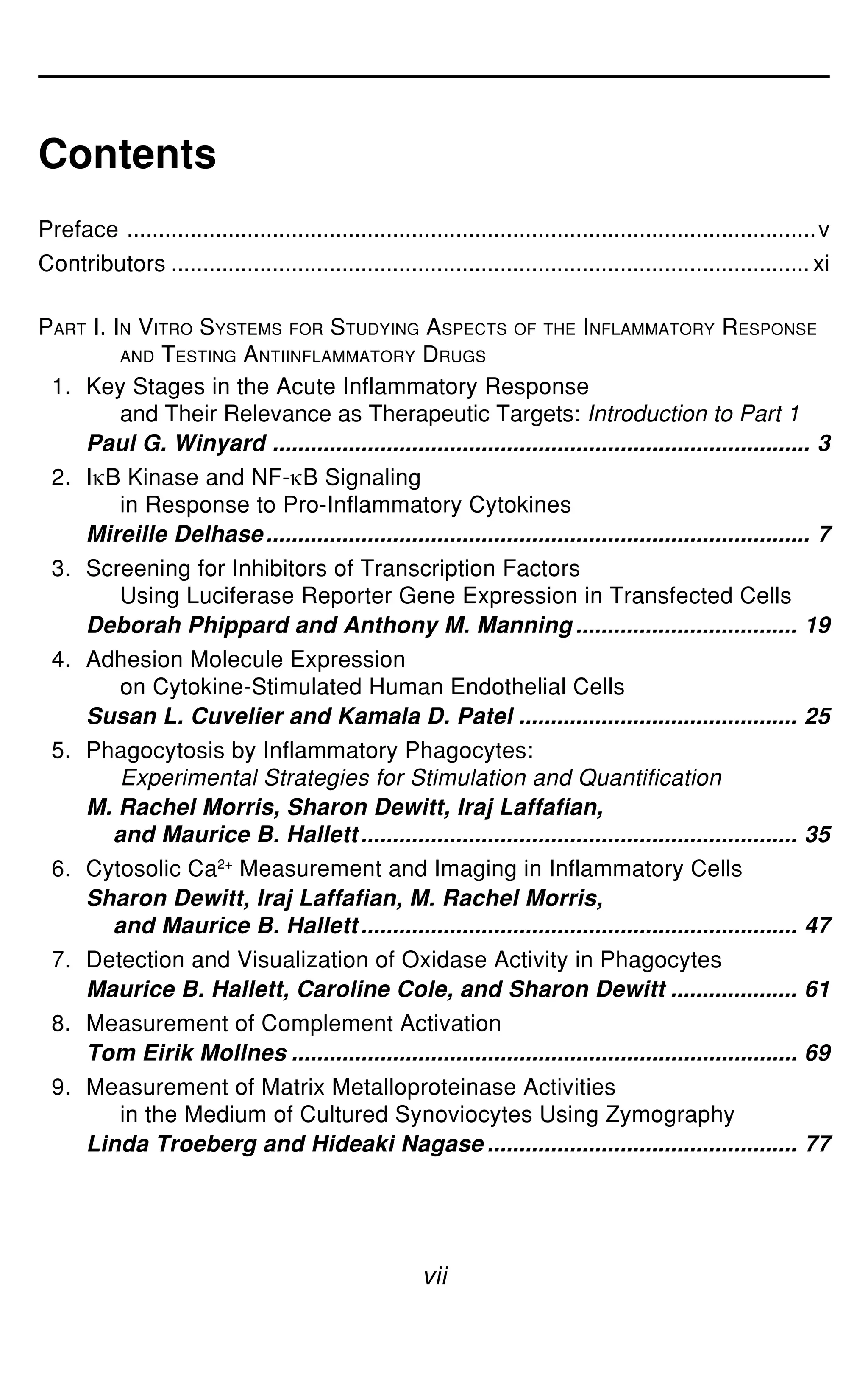

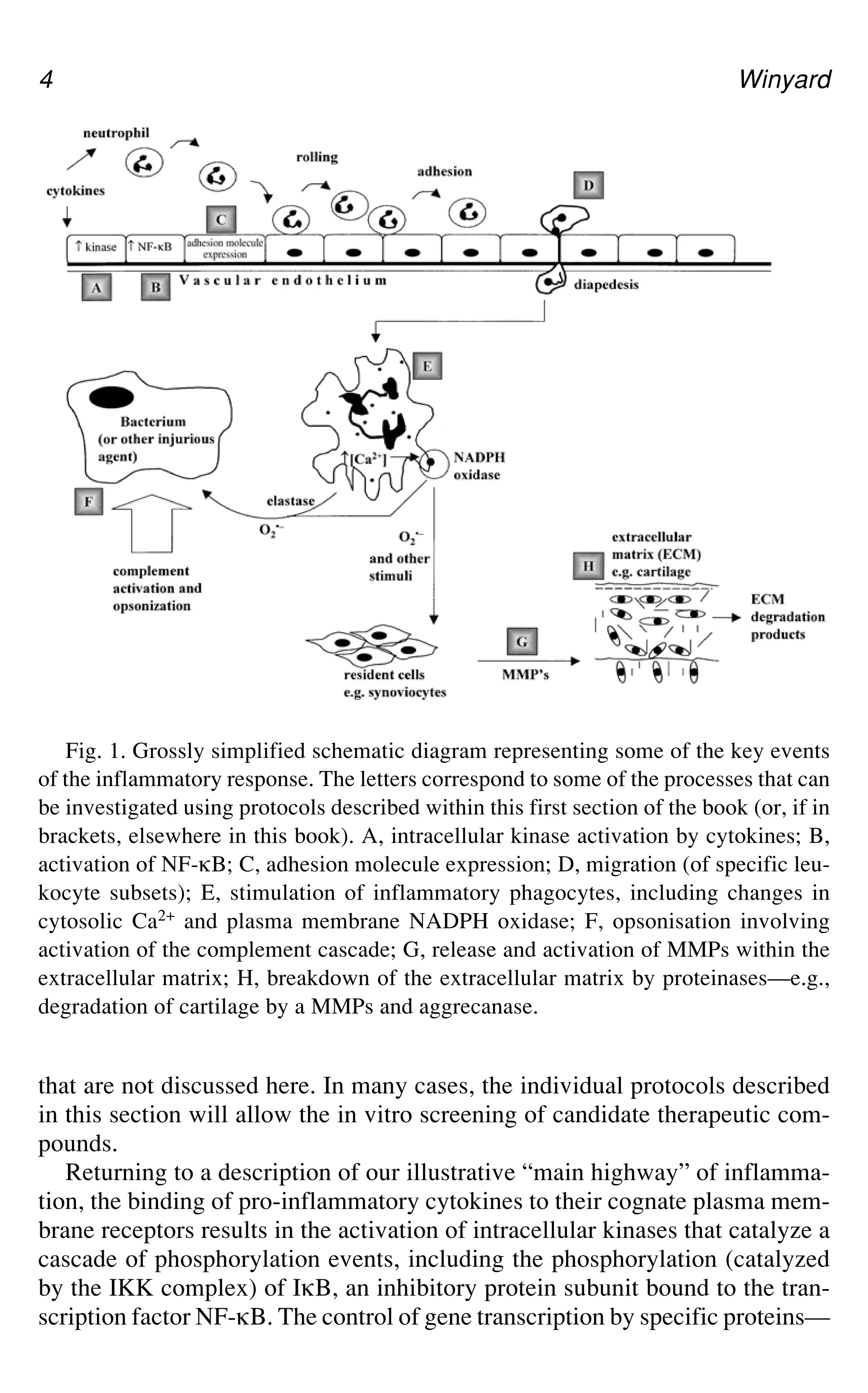
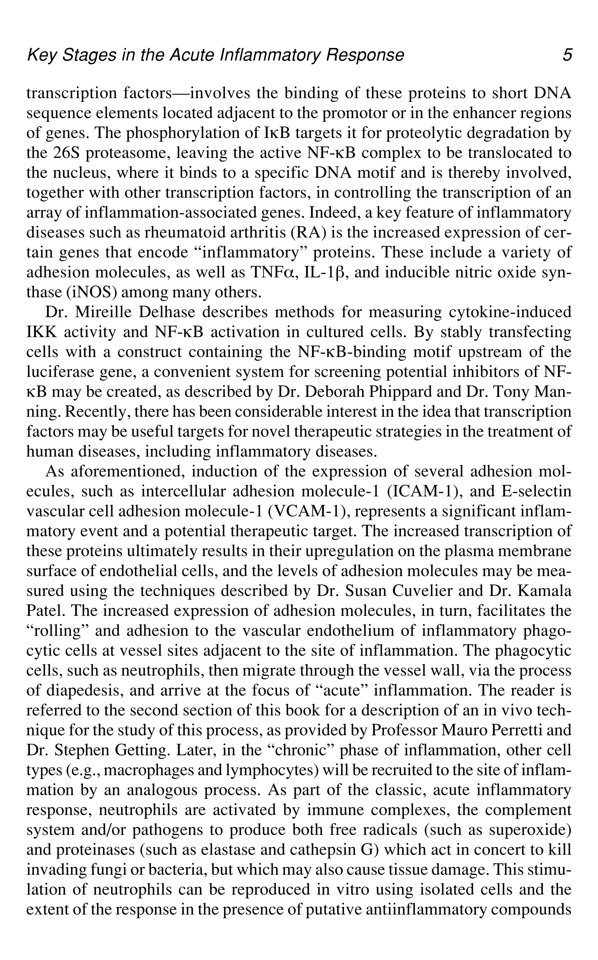
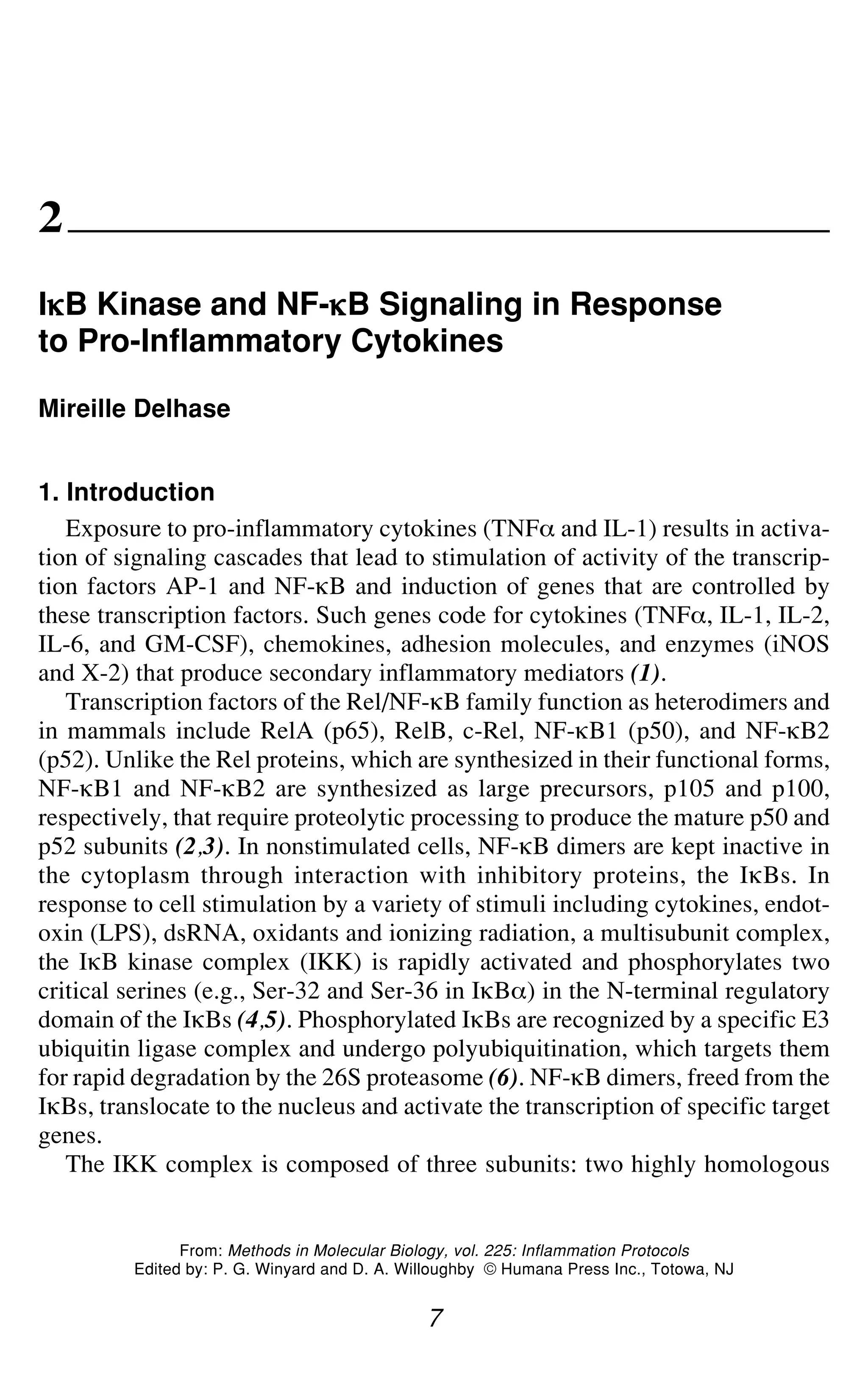
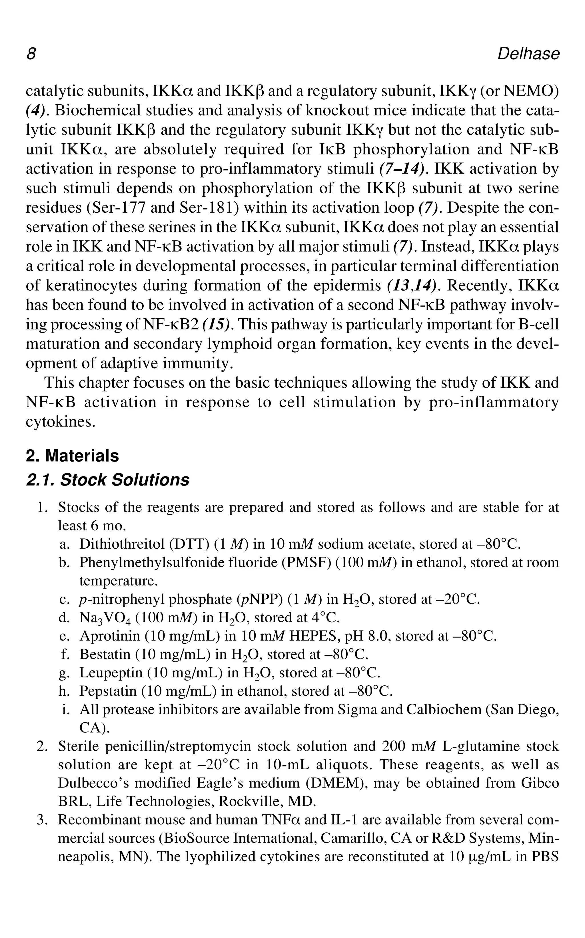
![IgB Kinase and NF-gB Signaling 9
plus 0.1% bovine serum albumin (BSA, tissue culture grade) and stored at –80°C
in 100-μL aliquots.
4. ATP stock solution: 100 mM in H2O, stored at –80°C in 50-μL aliquots.
5. 32P-orthophosphate (cat. no. Nex-53C) is from NEN Life Science Products, Bos-
ton, MA).
6. [a-32P]ATP is from Amersham Pharmacia Biotech, Piscataway, NJ.
2.2. Cell Lysis Buffers
1. Lysis buffer: 50 mM Tris-HCl, pH 7.6, 250 mM sodium chloride (NaCl), 3 mM
ethylenediamine tetraacetic acid (EDTA), 3 mM ethyleneglycol tetraacetic acid
(EGTA), 1% (v/v) Triton X-100, 0.5% (v/v) Nonidet (P-40) (NP40), 10% glyc-
erol, 20 mM sodium fluoride (NaF), 40 mM `-glycero-3-phosphate. This buffer
is prepared as a stock solution, filtered through a 0.45-μm filter unit, and stored
at 4°C. Before use, the following should be added: 2 mM DTT, 1 mM PMSF,
2 mM pNPP, 1 mM sodium orthovanadate (Na3VO4), and 10 μg/mL each of
aprotinin, bestatin, leupeptin, and pepstatin (see Subheading 2.1.).
2. RIPA buffer: 20 mM Tris-HCl, pH 7.6, 150 mM NaCl, 1 mM EDTA, 1 mM
EGTA, 1% NP40, 0.5% (w/v) sodium deoxycholate (DOC), 0.05% sodium
dodecyl sulfate (SDS), 10% glycerol, 20 mM NaF, 40 mM `-glycero-3-phos-
phate, 2.5 mM sodium metabisulfite, 5 mM benzamidine, 2 mM DTT, 1 mM
PMSF, 20 mM pNPP, 1 mM Na3VO4, and 10 μg/mL each of aprotinin, bestatin,
leupeptin, and pepstatin (see Subheading 2.1.).
2.3. Bacterial Culture Medium and Bacteria Lysis Buffers
1. Lbroth: bacto-tryptone 10 g/L, bacto-yeast extract 5 g/L, NaCl 5 g/L, adjusted to
pH 7.5 with NaOH. 100 μg/mL ampicillin and 2% glucose are added.
2. Base buffer: PBS, pH 8.0, 1 mM EDTA, 0.5% (v/v) Triton X-100, 5 mM DTT,
store at 4°C. This buffer is stable for several months if prepared without DTT.
DTT can be added just before use (see Subheading 2.1.).
3. Buffer I : Base buffer containing 0.3 M ammonium sulfate [(NH4)2SO4].
4. Lysis buffer: Buffer I supplemented with 1 mM PMSF and 10 μg/mL of protease
inhibitors (aprotinin, bestatin, leupeptin, and pepstatin) (see Subheading 2.1.).
5. Elution buffer: 100 mM Tris-HCl, pH 8.0, 20 mM glutathione.
6. Dialysis buffer: 20 mM Tris-HCl, pH 8.0, 100 mM NaCl, 0.2 mM EDTA, 10 mM
`-glycero-3-phosphate and 10% glycerol.
2.4. Kinase Assay
1. Kinase buffer (10X): 200 mM HEPES, pH 7.5, 100 mM magnesium chloride
(MgCl2).
2. Reaction buffer: kinase buffer [1X] supplemented with 20 mM `-glycero-3-phos-
phate, 10 mM pNPP, 1 mM DTT, and 20 μM “unlabeled” ATP. This buffer should
be prepared just before use.](https://image.slidesharecdn.com/2163577-250515100949-fe8e5c38/75/Inflammation-Protocols-1st-Edition-Paul-G-Winyard-Auth-Paul-G-Winyard-24-2048.jpg)
![IgB Kinase and NF-gB Signaling 11
Fig. 1. Cytokine-induced IKK phosphorylation. HeLa cells incubated for 5 h with
[32P]orthophosphate were stimulated with TNF_ (20 ng/mL) for the indicated times,
then lysed. The IKK complex was immunoprecipitated (IP) with an antibody to IKK_
(clone # B78-1 from PharMingen), resolved by gel electrophoresis and transferred onto
a PVDF membrane. Phosphoproteins were detected by autoradiography. IKK_ and
IKK` were identified by immunoblotting (IB) using specific antibodies (anti-IKK_,
clone # B78-1 from PharMingen and anti-IKK` H470 from Santa Cruz Biotechnology).
5. The supernatant is transferred to a clean tube and the protein concentration is
determined by Bradford assay (17).
6. The lysates (WCE, whole cell extracts) are stored at –80°C until use. WCEs can
be used for analysis of protein composition by western blotting, immune-com-
plex kinase assay and electrophoretic mobility shift assay (EMSA).
3.2. Detection of IKK Phosphorylation by 32P Metabolic Labeling
(see Note 1)
1. Cells at subconfluence (70–80%) (one 60-mm dish per assay) are washed twice
in phosphate-free DMEM and incubated for 1 h in the same medium.
2. The medium is then replaced by labeling medium (4 mL per dish) consisting of
phosphate-free DMEM supplemented with 10% dialyzed FBS and 1–2 mCi/mL
32P-orthophosphate. Cells are labeled for 4–5 h at 37°C.
3. TNF_ or IL-1 stimulation of the cells is performed as described in Subheading](https://image.slidesharecdn.com/2163577-250515100949-fe8e5c38/75/Inflammation-Protocols-1st-Edition-Paul-G-Winyard-Auth-Paul-G-Winyard-26-2048.jpg)
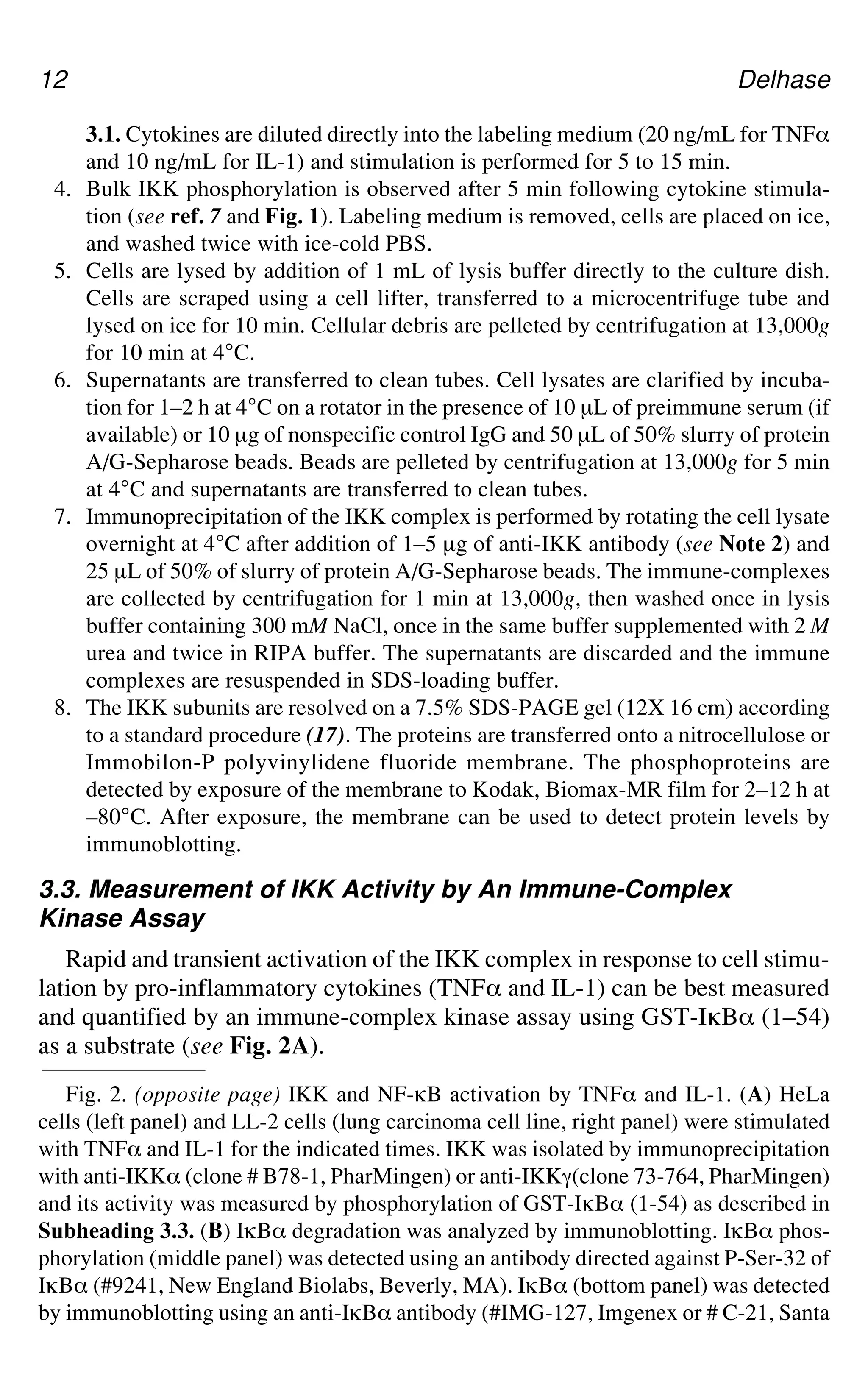
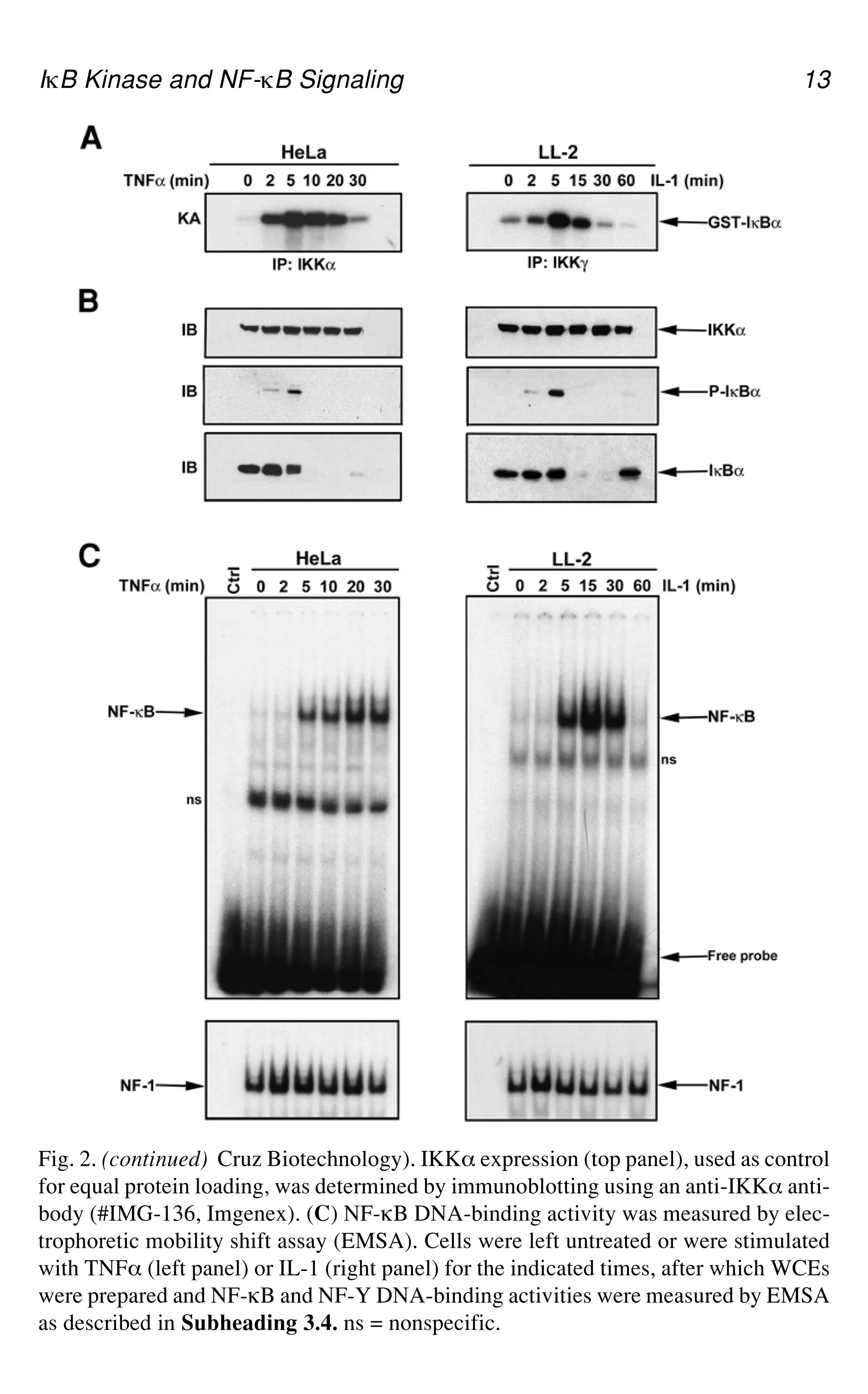
![IgB Kinase and NF-gB Signaling 15
5. The immune-complexes are resuspended in a 30-μL reaction buffer as described
in Subheading 2.4. containing 5μCi of [a-32P]ATP (6000 Ci/mmole) and 1 μg of
GST-IgB_(1–54) substrate (see Subheading 3.3.1). The kinase reaction is
allowed to proceed for 30 min at 30°C.
6. The reactions are stopped by addition of SDS sample loading buffer (16). The
samples are heated at 100ºC for 5 min to denature the proteins and then analyzed
by SDS-PAGE (10% gel). Electrophoresis is stopped before the free isotope
(which migrates on the gel together with the yellow pNPP band) runs off the gel.
7. The bottom of the gel containing the free isotope is cut off and discarded. The
gel is stained with Coomassie blue (17), destained in 10% acetic acid, 10%
methanol in H2O and dried. This step allows visualization of the substrate to
verify equal loading.
8. The dried gel is exposed to Kodak X-OMAT-AR films. Alternatively, the kinase
assay gel can be directly transferred to a PVDF membrane, which is then exposed
to film. This allows further detection of IKK in the immune-complexes by
immunoblotting.
9. The IKK activity is determined by the amount of radioactive 32P incorporated
into the GST-IgB_(1-54) substrate. Quantification can be performed on a
phosphoimager (preferred) or by densitometric scanning of the films.
3.4. Measurement of NF-g
g
g
g
gB Activation
by Electrophoretic Mobility Shift Assay
3.4.1. Oligonucleotide Probe Labeling
1. Complementary oligonucleotides are synthesized according to consensus seq-
uences for binding of transcription factors (see Subheading 2.5.).
2. 10 μL of both oligonucleotide stock solutions (at 1 μg/μL) are diluted into 100 μL
of TE in a microcentrifuge tube. The tube is heated at 80°C for 10 min in a beaker
of water, then slowly cooled down to room temperature (about 1 h) to allow
annealing of the oligonucleotides. The double-stranded (ds) oligonucleotide stock
solution is stored at –20°C.
3. The EMSA probe is prepared by incubation for 20 min at room temperature of
1 μL of ds oligonucleotide stock solution in a 20-μL reaction mixture containing
1X Klenow buffer (see Subheading 2.5.). 25 μM of each dATP, dTTP, and
dGTP, 50 μCi [_-32P]dCTP and 5 units of Klenow enzyme (see Note 4).
4. The labeling reaction is stopped by addition of 25 μM of unlabelled dCTP and
incubation is continued for 5 min at room temperature.
5. The probe is purified on a Nu-Clean D25 spin column (see Note 5) and stored at
–20°C.
3.4.2. Electrophoretic Mobility Shift Assay
1. Aliquots of WCEs containing 10 μg of protein (adjusted to 10 μL with cell lysis
buffer) are incubated for 30 min at room temperature in a 20-μL volume reaction
containing 2 μL of 10X EMSA buffer (see Subheading 2.5.), 1 μL of 20 mM
DTT, 2 μg of poly(dI.dC) and 15,000–30,000 cpm of probe. H2O is added to
bring the volume to 20 μL.](https://image.slidesharecdn.com/2163577-250515100949-fe8e5c38/75/Inflammation-Protocols-1st-Edition-Paul-G-Winyard-Auth-Paul-G-Winyard-30-2048.jpg)
![16 Delhase
2. The reactions are stopped by addition of 2 μL of 10X gel loading buffer (see
Subheading 2.5.) and the samples are run on a 4% nondenaturing polyacryla-
mide gel (20 × 20 cm).
3. Electrophoresis is stopped when the free probe (which migrates together with the
bromophenol blue band) is 2.5 cm from the bottom of the gel. The gel is fixed
(optional) for 15 min in a solution of 10% acetic acid, 10% methanol in H2O,
then dried and exposed to Kodak X-OMAT-AR film for autoradiography (see
Fig. 2C).
4. Notes
1. Precautions must be taken when performing metabolic labeling with 32P-ortho-
phosphate. Work must be carried out behind a 2.5-cm-thick acrylic shield. A
thick layer of lead can be attached to the outside of the shield to provide addi-
tional protection against radiation. A 1-cm-thick acrylic cell house should be
used to provide shielding when the cell dishes are in the incubator or when they
are moved in the laboratory. Researchers should wear two layers of gloves dur-
ing all steps of the labeling procedure.
2. Immunoprecipitation of the entire IKK complex can be performed using an anti-
body against any of the IKK subunits. Several commercial antibodies have been
tested and found to quantitatively immunoprecipitate the IKK complex. We cur-
rently use anti-IKK_ [clone # B78-1, PharMingen (San Diego, CA), this mono-
clonal antibody recognizes the human IKK_ but not the mouse IKK_], anti-IKK_
M280 [Santa Cruz Biotechnology (Santa Cruz, CA)], anti-IKKa (clones # C73-764
and C73-1794, PharMingen) and anti-IKK` H470 (Santa Cruz Biotechnology)
(7,13). Intensive washings of the immunoprecipitates in RIPA buffer may disrupt
interaction between IKKa and the IKK_/IKK` dimers. Therefore, to examine IKKa
phosphorylation, we recommend immunoprecipitating the IKK complex using an
antibody against IKKa.
3. This recombinant plasmid as well as a mutant version in which Ser 32 and Ser 36
have been replaced by Ala (GST-IgB_(1-54)AA) are available from the author’s
laboratory upon request.
4. If the annealed oligonucleotides do not contain 5' protruding ends, but instead are
blunt ended, labeling cannot be performed using Klenow enzyme. Instead T4
polynucleotide kinase in the presence of [a-32P] ATP should be used (17).
5. Unincorporated nucleotides can also be removed by centrifugation through a “self
made” 1-mL Sephadex G25 column.
Acknowledgment
The author would like to thank D. Rothwarf for helpful discussions and critical
reading of the manuscript. This work was supported by the Sontag Foundation Fel-
lowship of the Arthritis National Research Foundation and a research grant from the
San Diego Chapter of the Arthritis Foundation.](https://image.slidesharecdn.com/2163577-250515100949-fe8e5c38/75/Inflammation-Protocols-1st-Edition-Paul-G-Winyard-Auth-Paul-G-Winyard-31-2048.jpg)
![Screening for Inhibitors of Transcription Factors 23
References
1. Tijan, R. and Maniatis, T. (1994) Transcriptional activation: a complex puzzle
with few easy pieces. Cell 77, 5–8.
2. Manning, A. M. (1996) Transcription factors: a new frontier for drug discovery.
Drug Discovery Today 1, 151–160.
3. Palanki, M. S. S., Erdman, P. E., Gayo-Fung, L. J., Shevlin, G. I., Sullivan, R. W.,
Suto, M. J., et al. (2000) Inhibitors of NF-gB and AP-1 gene expression: SAR
studies on the pyrimidine portion of 2-chloro-4-trifluoromethylpyrimidine-5-[N-
(3',5'-bis(trifluoromethyl)phenyl carboxamide. J. Med. Chem. 43(21), 3995–4004.
4. Palanki, M. S. S., Erdman, P. E., Manning, A. M., Ow, A., Ransone, L. J., Spooner,
C. et al. (2000) Novel inhibitors of AP-1 and NF-gB mediated gene expression.
Structure-activity relationship studies of ethyl-[(methyl-2,5-dioxo(3-
pyrrolinyl))amino]-2-(trifluoromethyl)pyrimidine-5-carboxylate. Bioorg. Med.
Chem. Lett. 10, 1645–1648.
5. Gerlag, D. M., Ransone-Fong, L., Palanki, M., Han, Z., Tak, P. P., Manning, A.
M., et al. (2000) The effect of a T cell-specific NF-gB inhibitor on cytokine
production in vitro and on murine collagen induced arthritis. J. Immunol. 165,
1652–1658.
6. Al-Moslih, M. I. and Dubes, G. R. (1973) The kinetics of DEAE-dextran-induced
cell sensitization to transfection. J. Gen. Virol. 18, 189.](https://image.slidesharecdn.com/2163577-250515100949-fe8e5c38/75/Inflammation-Protocols-1st-Edition-Paul-G-Winyard-Auth-Paul-G-Winyard-37-2048.jpg)