Molecular Toxicology Protocols David H Phillips Alan Hewer Volker M Arlt Auth
Molecular Toxicology Protocols David H Phillips Alan Hewer Volker M Arlt Auth
Molecular Toxicology Protocols David H Phillips Alan Hewer Volker M Arlt Auth
Molecular Toxicology Protocols David H Phillips Alan Hewer Volker M Arlt Auth
Molecular Toxicology Protocols David H Phillips Alan Hewer Volker M Arlt Auth
1.
Molecular Toxicology ProtocolsDavid H Phillips
Alan Hewer Volker M Arlt Auth download
https://ebookbell.com/product/molecular-toxicology-protocols-
david-h-phillips-alan-hewer-volker-m-arlt-auth-11749374
Explore and download more ebooks at ebookbell.com
2.
Here are somerecommended products that we believe you will be
interested in. You can click the link to download.
Molecular Toxicology Protocols3rd Edition 3rd Ed 2020 Edition
Phouthone Keohavong
https://ebookbell.com/product/molecular-toxicology-protocols3rd-
edition-3rd-ed-2020-edition-phouthone-keohavong-47706466
Molecular Toxicology Protocols 2nd Edition Phouthone Keohavong
https://ebookbell.com/product/molecular-toxicology-protocols-2nd-
edition-phouthone-keohavong-4661998
Molecular Toxicology In Caenorhabditis Elegans 1st Ed Dayong Wang
https://ebookbell.com/product/molecular-toxicology-in-caenorhabditis-
elegans-1st-ed-dayong-wang-9961284
Advances In Molecular Toxicology 5 1st Edition James C Fishbein Eds
https://ebookbell.com/product/advances-in-molecular-toxicology-5-1st-
edition-james-c-fishbein-eds-2383384
3.
Molecular Clinical AndEnvironmental Toxicology Volume 1 Molecular
Toxicology 1st Edition Antoinette N Hayes
https://ebookbell.com/product/molecular-clinical-and-environmental-
toxicology-volume-1-molecular-toxicology-1st-edition-antoinette-n-
hayes-4405420
Advances In Molecular Toxicology James C Fishbein Eds
https://ebookbell.com/product/advances-in-molecular-toxicology-james-
c-fishbein-eds-4426698
Advances In Molecular Toxicology 7 James C Fishbein And Jacqueline M
Heilman Eds
https://ebookbell.com/product/advances-in-molecular-
toxicology-7-james-c-fishbein-and-jacqueline-m-heilman-eds-4426700
Advances In Molecular Toxicology Volume 8 First Edition Fishbein
https://ebookbell.com/product/advances-in-molecular-toxicology-
volume-8-first-edition-fishbein-5431748
Advances In Molecular Toxicology Volume 9 1st Ed Heilman Jacqueline M
Fishbein
https://ebookbell.com/product/advances-in-molecular-toxicology-
volume-9-1st-ed-heilman-jacqueline-m-fishbein-5431750
M E TH O D S I N M O L E C U L A R B I O L O G Y™
John M. Walker, SERIES EDITOR
307. Phosphodiesterase Mehtods and Protocols,
edited by Claire Lugnier, 2005
306. Receptor Binding Techniques: Second Edition,
edited by Anthony P. Davenport, 2005
305. Protein–Ligand Interactions: Methods and
Protocols, edited by G. Ulrich Nienhaus, 2005
304. Human Retrovirus Protocols: Virology and
Molecular Biology, edited by Tuofu Zhu, 2005
303. NanoBiotechnology Protocols, edited by Sandra
J. Rosenthal and David W. Wright, 2005
302. Handbook of ELISPOT: Methods and Protocols,
edited by Alexander E. Kalyuzhny, 2005
301. Ubiquitin–Proteasome Protocols, edited by
Cam Patterson and Douglas M. Cyr, 2005
300. Protein Nanotechnology: Protocols,
Instrumentation, and Applications, edited by Tuan
Vo-Dinh, 2005
299. Amyloid Proteins: Methods and Protocols,
edited by Einar M. Sigurdsson, 2005
298. Peptide Synthesis and Application, edited by
John Howl, 2005
297. Forensic DNA Typing Protocols, edited by
Angel Carracedo, 2005
296. Cell Cycle Protocols, edited by Tim Humphrey
and Gavin Brooks, 2005
295. Immunochemical Protocols, Third Edition,
edited by Robert Burns, 2005
294. Cell Migration: Developmental Methods and
Protocols, edited by Jun-Lin Guan, 2005
293. Laser Capture Microdissection: Methods and
Protocols, edited by Graeme I. Murray and
Stephanie Curran, 2005
292. DNA Viruses: Methods and Protocols, edited by
Paul M. Lieberman, 2005
291. Molecular Toxicology Protocols, edited by
Phouthone Keohavong and Stephen G. Grant, 2005
290. Basic Cell Culture, Third Edition, edited by
Cheryl D. Helgason and Cindy Miller, 2005
289. Epidermal Cells, Methods and Applications,
edited by Kursad Turksen, 2005
288. Oligonucleotide Synthesis, Methods and Appli-
cations, edited by Piet Herdewijn, 2005
287. Epigenetics Protocols, edited by Trygve O.
Tollefsbol, 2004
286. Transgenic Plants: Methods and Protocols,
edited by Leandro Peña, 2005
285. Cell Cycle Control and Dysregulation
Protocols: Cyclins, Cyclin-Dependent Kinases,
and Other Factors, edited by Antonio Giordano
and Gaetano Romano, 2004
284. Signal Transduction Protocols, Second Edition,
edited by Robert C. Dickson and Michael D.
Mendenhall, 2004
283. Bioconjugation Protocols, edited by Christof
M. Niemeyer, 2004
282. Apoptosis Methods and Protocols, edited by
Hugh J. M. Brady, 2004
281. Checkpoint Controls and Cancer, Volume 2:
Activation and Regulation Protocols, edited by
Axel H. Schönthal, 2004
280. Checkpoint Controls and Cancer, Volume 1:
Reviews and Model Systems, edited by Axel H.
Schönthal, 2004
279. Nitric Oxide Protocols, Second Edition, edited
by Aviv Hassid, 2004
278. Protein NMR Techniques, Second Edition,
edited by A. Kristina Downing, 2004
277. Trinucleotide Repeat Protocols, edited by
Yoshinori Kohwi, 2004
276. Capillary Electrophoresis of Proteins and
Peptides, edited by Mark A. Strege and
Avinash L. Lagu, 2004
275. Chemoinformatics, edited by Jürgen Bajorath, 2004
274. Photosynthesis Research Protocols, edited by
Robert Carpentier, 2004
273. Platelets and Megakaryocytes, Volume 2:
Perspectives and Techniques, edited by
Jonathan M. Gibbins and Martyn P. Mahaut-
Smith, 2004
272. Platelets and Megakaryocytes, Volume 1:
Functional Assays, edited by Jonathan M.
Gibbins and Martyn P. Mahaut-Smith, 2004
271. B Cell Protocols, edited by Hua Gu and Klaus
Rajewsky, 2004
270. Parasite Genomics Protocols, edited by Sara
E. Melville, 2004
269. Vaccina Virus and Poxvirology: Methods and
Protocols,edited by Stuart N. Isaacs, 2004
268. Public Health Microbiology: Methods and
Protocols, edited by John F. T. Spencer and
Alicia L. Ragout de Spencer, 2004
267. Recombinant Gene Expression: Reviews and
Protocols, Second Edition, edited by Paulina
Balbas and Argelia Johnson, 2004
266. Genomics, Proteomics, and Clinical
Bacteriology: Methods and Reviews, edited by
Neil Woodford and Alan Johnson, 2004
265. RNA Interference, Editing, and
Modification: Methods and Protocols, edited
by Jonatha M. Gott, 2004
264. Protein Arrays: Methods and Protocols,
edited by Eric Fung, 2004
263. Flow Cytometry, Second Edition, edited by
Teresa S. Hawley and Robert G. Hawley, 2004
262. Genetic Recombination Protocols, edited by
Alan S. Waldman, 2004
307
306
305
304
303
302
301
300
299
298
297
296
295
294
293
292
291
290
289
288
287
286
285
284
283
282
281
280
279
278
277
276
275
274
273
272
271
270
269
268
267
266
265
264
263
262
7.
M E TH O D S I N M O L E C U L A R B I O L O G Y™
Molecular Toxicology
Protocols
Edited by
Phouthone Keohavong
Stephen G. Grant
Department of Environmental and Occupational Health
University of Pittsburgh, Pittsburgh, PA
v
Preface
It seems fashionabletoday to simply place the word “molecular” in front of a
traditional field and consider it reinvented. This, without a clear consensus on what
the “molecular” actually means. Certainly chemists working in the field of toxicology
have always considered that they worked at the “molecular” level. It has not been so
clear on the biological side, however, where there has been a history of ongoing
discovery and characterization of toxic mechanisms. In other biological fields,
“molecular” really implies using the tools of “molecular” biology, i.e., recombinant
DNA. Just as the adoption of molecular biological techniques first invaded, then
transformed such biological fields as genetics, physiology, and developmental biology,
so too have these new methods begun to transform toxicology.
Molecular Toxicology Protocols is a book about science on the interface, and a
science that is about to explode upon the clinical and popular horizon. Toxicology, a
subdiscipline of pharmacology, is actually the interface of chemistry and biology. As
most practice it, this field also extends into nonchemical “agents” of deleterious
biological effects, especially radiation, the purview of the radiobiologist and health
physicist. With the huge increase in computational power made available over the last
ten years it has become possible to model and predict the potential toxicity of as yet
unmade chemicals. Perhaps the greatest change in the practice of toxicology has been
application of the the tools of the trade directly to the human population, in what are
known clinically as “translational” studies, opening the new frontier of epidemiology
through the more conventional portal of biostatistics. These studies expand the
traditional public health aspect of toxicology, screening of synthetic agents for
toxicological potential prior to their introduction into the environment, attempting
to define “normal” or “background,” perhaps unavoidable, exposures as mechanisms
of human disease, and to design methods of preclinical intervention (“chemo-
prevention”).
Thus, for our purposes, we will define “molecular” toxicology as either any study
of toxicological mechanism, or any translation of toxicological practice into the human
population.
Today, such “molecular” toxicology is mostly genetic toxicology, where the genetic
material itself, the DNA, is the target molecule. Of course DNA is found throughout
the human body, such that all of the traditional modulators of toxicological effect,
uptake, distribution, metabolism, and so on, must be taken into account. Although
genetic damage can have many outcomes, the one outcome most clearly linking
exposure and disease is cancer.
During the past several years, important progress has been made in the
understanding of the molecular biology of the cell, the responses of cells to genotoxic
agents, and the molecular biology of human cancer. This progress has been achieved
thanks to the ongoing development of new state-of-the-art techniques, as well as
10.
vi Preface
improvements madeupon existing methods to study changes not only in cellular
morphology, but also in the cellular genetic material, the DNA, the cellular transcript,
the mRNA, and the translated product, proteins. These molecular methods are now
opening up many areas to potential clinical applications. Several books are currently
available on the applications of molecular methods to various types of technology.
However, to our knowledge, there is no book emphasizing the application of molecular
methods to genetic toxicology.
Therefore, the aim of Molecular Toxicology Protocols is to bring together a series
of articles, each describing commonly used methods to elucidate specific molecular
aspects of toxicology. With such content, this book addresses not only molecular
biologists and molecular toxicologists, but also all individuals interested in applying
molecular methods to clinical populations, including geneticists, pathologists,
biochemists, and epidemiologists. The volume is divided into seven parts, roughly
corresponding to the spectrum of biomarkers intermediate between exposure and
disease outcome as proposed in molecular epidemiological models. Thus, Part I
includes chapters describing methods of detecting premutagenic lesions in the genetic
material, while Part II contains chapters describing the applications of methods to
assess gross or macroscopic genetic damage. Parts III and IV focus on detection and
characterization of viable mutations, in surrogate markers and cancer-related genes,
respectively. The chapters of Part V describe methods for the analysis of the various
pathways of DNA repair, an important modulator of genotoxicity. Part VI addresses
the application of the new array technologies to genetic toxicology, including methods
for the analysis of individual variation in biotransformation and the effects of genotoxic
exposure on gene expression. Finally, Part VII describes methods for analysis of
cytotoxicity caused by the induction of apoptosis, because cell death can either protect
the organism from a transforming cell or cause distinct health effects itself.
We have no doubt that as time goes by “molecular” approaches will play an
expanding role in all types of toxicology, not just genetic toxicology. Moreover,
genetic toxicology will undoubtedly be found to play a role in many more diseases of
aging than cancer alone; it is probably a fundamental mechanism of aging itself.
Therefore, while the current focus of Molecular Toxicology Protocols is genetic
toxicology, and more specifically the genetic toxicology of cancer, we believe this
represents just the tip of the iceberg with respect to how the field of molecular
toxicology will eventually be understood.
Phouthone Keohavong
Stephen G. Grant
11.
Contents
vii
Preface ..............................................................................................................v
Contributors .....................................................................................................xi
PARTI: ANALYSIS OF DNA ADDUCTS
1 32
P-Postlabeling Analysis of DNA Adducts
David H. Phillips, Alan Hewer, and Volker M. Arlt ............................. 3
2 Modification of the 32
P-Postlabeling Method to Detect a Single
Adduct Species as a Single Spot
Masako Ochiai, Takashi Sugimura, and Minako Nagao ..................... 13
3 DNA Isolation and Sample Preparation for Quantification
of Adduct Levels by Accelerator Mass Spectrometry
Karen H. Dingley, Esther A. Ubick, John S. Vogel,
and Kurt W. Haack ......................................................................... 21
4 Fluoroimaging-Based Immunoassay of DNA Photoproducts
in Ultraviolet-B-Irradiated Tadpoles
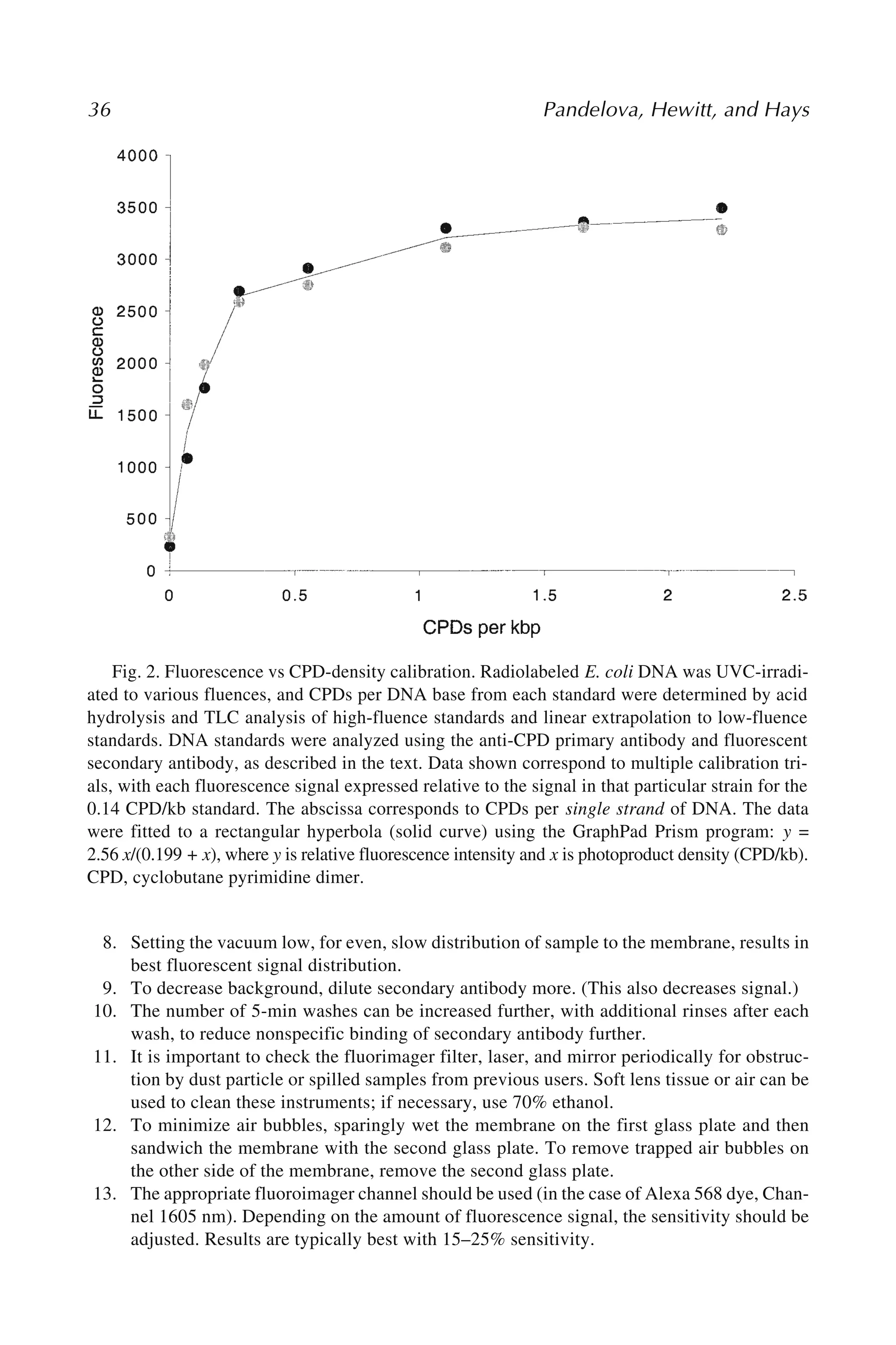
Iovanna Pandelova, Stephen R. Hewitt, and John B. Hays ................ 29
5 Analysis of DNA Strand Cleavage at Abasic Sites
Walter A. Deutsch and Vijay Hegde................................................... 39
PART II: DETECTION OF CHROMOSOMAL AND GENOME-WIDE DAMAGE
6 Premature Chromosome Condensation in Human Resting Peripheral
Blood Lymphocytes for Chromosome Aberration Analysis Using
Specific Whole-Chromosome DNA Hybridization Probes
Pataje G. S. Prasanna and William F. Blakely .................................... 49
7 Mutagen-Induced Chromatid Breakage as a Marker of Cancer Risk
Xifeng Wu, Yun-Ling Zheng, and T. C. Hsu........................................ 59
8 Flow Cytometric Analysis of Micronuclei in Erythrocytes
Jan Grawé ........................................................................................... 69
9 The Comet Assay: A Sensitive Genotoxicity Test
for the Detection of DNA Damage
Günter Speit and Andreas Hartmann ................................................. 85
10 Computerized Image Analysis Software for the Comet Assay
R. C. Chaubey ..................................................................................... 97
12.
viii Contents
11 TheComet–FISH Technique: A Tool for Detection of Specific
DNA Damage and Repair
Alexander Rapp, Michael Hausmann, and Karl Otto Greulich ........ 107
12 DNA Double-Strand Break Damage and Repair
Assessed by Pulsed-Field Gel Electrophoresis
Nina Joshi and Stephen G. Grant ..................................................... 121
PART III: DETECTION AND CHARACTERIZATION OF SURROGATE GENE MUTATION
13 Analysis of In Vivo Mutation in the Hprt and Tk Genes
of Mouse Lymphocytes
Vasily N. Dobrovolsky, Joseph G. Shaddock,
and Robert H. Heflich................................................................. 133
14 Quantifying In Vivo Somatic Mutations
Using Transgenic Mouse Model Systems
Roy R. Swiger .................................................................................... 145
15 Methods for Detecting Somatic Mutations In Vitro:
The Human T-Cell Cloning Assay Selecting for HPRT Mutants
Sai-Mei Hou ..................................................................................... 155
16 Molecular Analysis of Mutations in the Human HPRT Gene
Phouthone Keohavong, Liqiang Xi, and Stephen G. Grant .............. 161
17 Simultaneous Quantification of t(14;18) and
HPRT Exon 2/3 Deletions in Human Lymphocytes
James C. Fuscoe ................................................................................ 171
18 The GPA In Vivo Somatic Mutation Assay
Stephen G. Grant .............................................................................. 179
19 Flow Cytometric Measurement of Mutant T Cells With Altered
Expression of TCR: Detecting Somatic Mutations in Humans
and Mice
Seishi Kyoizumi, Yoichiro Kusunoki, and Tomonori Hayashi .......... 197
PART IV: DETECTION AND CHARACTERIZATION OF CANCER GENE MUTATION
20 Mutation Screening of the TP53 Gene
by Temporal Temperature Gradient Gel Electrophoresis
Therese Sørlie, Hilde Johnsen, Phuong Vu, Guro Elisabeth Lind,
Ragnhild Lothe, and Anne-Lise Børresen-Dale............................. 207
21 Analysis of K-RAS and P53 Mutations in Sputum Samples
Weimin Gao and Phouthone Keohavong ......................................... 217
22 Allele-Specific Competitive Blocker–PCR Detection
of Rare Base Substitution
Barbara L. Parsons, Page B. McKinzie, and Robert H. Heflich ........ 235
13.
23 Gel-Based NonradioactiveSingle-Strand Conformational
Polymorphism and Mutation Detection: Limitations and Solutions
Vibhuti Gupta, Reetakshi Arora, Anand Ranjan,
Narendra K. Bairwa, Dheeraj K. Malhotra,
P. T. Udhayasuriyan, Anjana Saha, and Ramesh Bamezai .............. 247
24 Detection and Characterization of Oncogene Mutations
in Preneoplastic and Early Neoplastic Lesions
Toshinari Minamoto.......................................................................... 263
25 Detection of DNA Double-Strand Breaks and Chromosome
Translocations Using Ligation-Mediated PCR and Inverse PCR
Michael J. Villalobos, Christopher J. Betti,
and Andrew T. M. Vaughan.......................................................... 279
PART V: ANALYSIS OF DNA REPAIR MECHANISMS
26 Microsatellite Instability: An Indirect Assay to Detect Defects
in the Cellular Mismatch Repair Machinery
Anjana Saha, Narendra K. Bairwa, and Ramesh Bamezai ................ 293
27 Unscheduled DNA Synthesis: A Functional Assay
for Global Genomic Nucleotide Excision Repair
Crystal M. Kelly and Jean J. Latimer ................................................. 303
28 Analysis of DNA Repair Using Transfection-Based
Host Cell Reactivation
Jennifer M. Johnson and Jean J. Latimer........................................... 321
29 An Immunoassay for Measuring Repair of Ultraviolet Photoproducts
Shirley McCready.............................................................................. 337
30 Analysis of DNA Double-Strand Break Repair by Nonhomologous
End Joining in Cell-Free Extracts From Mammalian Cells
Petra Pfeiffer, Elke Feldmann, Andrea Odersky,
Steffi Kuhfittig-Kulle, and Wolfgang Goedecke ........................... 351
31 Measuring Recombination Proficiency
in Mouse Embryonic Stem Cells
Andrew J. Pierce and Maria Jasin ..................................................... 373
PART VI: ARRAY TECHNOLOGIES
32 Strategies for Measurement of Biotransformation
Enzyme Gene Expression
Marjorie Romkes and Shama C. Buch .............................................. 387
33 Genotyping Technologies: Application to Biotransformation
Enzyme Genetic Polymorphism Screening
Marjorie Romkes and Shama C. Buch .............................................. 399
Contents ix
14.
34 TaqManTM
Fluorogenic DetectionSystem
to Analyze Gene Transcription in Autopsy Material
Kaori Shintani-Ishida, Bao-Li Zhu, and Hitoshi Maeda .................... 415
35 Development of Quantitative Reverse Transcriptase PCR Assays
for Measuring Gene Expression
Tony E. Godfrey and Lori A. Kelly .................................................... 423
PART VII: APOPTOSIS
36 Quantification of Selective Phosphatidylserine Oxidation
During Apoptosis
James P. Fabisiak, Yulia Y. Tyurina, Vladimir A. Tyurin,
and Valerian E. Kagan................................................................... 449
37 Quantitative Method of Measuring Phosphatidylserine
Externalization During Apoptosis Using Electron Paramagnetic
Resonance Spectroscopy and Annexin-Conjugated Iron
James P. Fabisiak, Grigory G. Borisenko, and Valerian E. Kagan..... 457
38 Detection of Programmed Cell Death in Cells Exposed
to Genotoxic Agents Using a Caspase Activation Assay
Michael E. Gehring and Patrick P. Koty ........................................... 465
Index ............................................................................................................ 473
x Contents
15.
xi
Contributors
VOLKER M. ARLT• Section of Molecular Carcinogenesis, Institute of Cancer
Research, Sutton, Surrey, UK
REETAKSHI ARORA • National Centre of Applied Human Genetics, Jawaharlal Nehru
University, Delhi, India
NARENDRA K. BAIRWA • National Centre of Applied Human Genetics, Jawaharlal
Nehru University, Delhi, India
RAMESH BAMEZAI • National Centre of Applied Human Genetics, Jawaharlal Nehru
University, Delhi, India
CHRISTOPHER J. BETTI • Program in Molecular Biology, Loyola University Chicago
Medical Center, Maywood, IL
WILLIAM F. BLAKELY • Armed Forces Radiobiology Research Institute, Bethesda,
MD
GRIGORY G. BORISENKO • Department of Environmental and Occupational Health,
University of Pittsburgh, Pittsburgh, PA
ANNE-LISE BØRRESEN-DALE • Department of Genetics, The Norwegian Radium
Hospital, Oslo, Norway
SHAMA C. BUCH • Center for Clinical Pharmacology, University of Pittsburgh,
Pittsburgh, PA
R. C. CHAUBEY • Genetic Toxicology and Chromosome Studies Section, Radiation
Biology and Health Sciences Division, Bhabha Atomic Research Centre,
Mumbai, India
WALTER A. DEUTSCH • Pennington Biomedical Research Center, Louisiana State
University, Baton Rouge, LA
KAREN H. DINGLEY • Biology and Biotechnology Research Program, Lawrence
Livermore National Laboratory, Livermore, CA
VASILY N. DOBROVOLSKY • Division of Genetic and Reproductive Toxicology,
National Center for Toxicological Research, US Food and Drug Administration,
Jefferson, AR
JAMES P. FABISIAK • Department of Environmental and Occupational Health,
University of Pittsburgh, Pittsburgh, PA
ELKE FELDMANN • Institute of Genetics, University of Essen, Essen, Germany
JAMES C. FUSCOE • Center for Functional Genomics, National Center for
Toxicological Research, US Food and Drug Administration, Jefferson, AR
WEIMIN GAO • Department of Environmental and Occupational Health, University
of Pittsburgh, Pittsburgh, PA
MICHAEL E. GEHRING • Department of Pediatrics, Wake Forest University School
of Medicine, Winston-Salem, NC
TONY E. GODFREY • Department of Medicine, Mount Sinai School of Medicine,
NewYork, NY
16.
xii Contributors
WOLFGANG GOEDECKE• Institute of Genetics, University of Essen, Essen, Germany
STEPHEN G. GRANT • Department of Environmental and Occupational Health,
University of Pittsburgh, Pittsburgh, PA
JAN GRAWÉ • Cell Analysis Core Facility, Uppsala University, Rudbeck Laboratory,
Uppsala, Sweden
KARL OTTO GREULICH • Department for Single Cell and Single Molecule Techniques,
Institute for Molecular Biotechnology Jena, Jena, Germany
VIBHUTI GUPTA • National Centre of Applied Human Genetics, Jawaharlal Nehru
University, Delhi, India
KURT W. HAACK • Center for Accelerator Mass Spectrometry, Lawrence Livermore
National Laboratory, Livermore, CA
ANDREAS HARTMANN • Novartis Pharma AG, Basel, Switzerland
MICHAEL HAUSMANN • Kirchhoff-Institute for Physics, University of Heidelberg,
Heidelberg, Germany
TOMONORI HAYASHI • Laboratory of Immunology, Department of Radiobiology/
Molecular Epidemiology, Radiation Effects Research Foundation, Hiroshima,
Japan
JOHN B. HAYS • Department of Environmental and Molecular Toxicology, Oregon
State University, Corvallis, OR
ROBERT H. HEFLICH • Division of Genetic and Reproductive Toxicology, National
Center for Toxicological Research, US Food and Drug Administration,
Jefferson, AR
VIJAY HEGDE • Pennington Biomedical Research Center, Louisiana State University,
Baton Rouge, LA
ALAN HEWER • Section of Molecular Carcinogenesis, Institute of Cancer Research,
Sutton, Surrey, UK
STEPHEN R. HEWITT • Department of Environmental and Molecular Toxicology,
Oregon State University, Corvallis, OR
SAI-MEI HOU • Department of Biosciences, Karolinska Institute, Huddinge, Sweden
T. C. HSU • Department of Cancer Biology, M. D. Anderson Cancer Center,
Houston, TX
MARIA JASIN • Cell Biology Program, Memorial Sloan-Kettering Cancer Center and
Cornell University Graduate School of Medical Sciences, New York, NY
HILDE JOHNSEN • Department of Genetics, The Norwegian Radium Hospital, Oslo,
Norway
JENNIFER M. JOHNSON • Department of Molecular Genetics and Biochemistry,
University of Pittsburgh School of Medicine, Pittsburgh, PA
NINA JOSHI • Department of Environmental and Occupational Health, University
of Pittsburgh, Pittsburgh, PA
VALERIAN E. KAGAN • Department of Environmental and Occupational Health,
University of Pittsburgh, Pittsburgh, PA
CRYSTAL M. KELLY • Department of Obstetrics, Gynecology and Reproductive
Sciences, University of Pittsburgh School of Medicine, Pittsburgh, PA
17.
Contributors xiii
LORI A.KELLY • Department of Surgery, University of Pittsburgh School
of Medicine, Pittsburgh, PA
PHOUTHONE KEOHAVONG • Department of Environmental and Occupational Health,
University of Pittsburgh, Pittsburgh, PA
PATRICK P. KOTY • Department of Pediatrics, Wake Forest University School
of Medicine, Winston-Salem, NC
STEFFI KUHFITTIG-KULLE • Institute of Genetics, University of Essen, Essen, Germany
YOICHIRO KUSUNOKI • Laboratory of Immunology, Department of Radiobiology/
Molecular Epidemiology, Radiation Effects Research Foundation, Hiroshima,
Japan
SEISHI KYOIZUMI • Laboratory of Immunology, Department of Radiobiology/
Molecular Epidemiology, Radiation Effects Research Foundation, Hiroshima,
Japan
JEAN J. LATIMER • Department of Obstetrics, Gynecology and Reproductive
Sciences, University of Pittsburgh School of Medicine, Pittsburgh, PA
GURO ELISABETH LIND • Department of Genetics, The Norwegian Radium Hospital,
Oslo, Norway
RAGNHILD LOTHE • Department of Genetics, The Norwegian Radium Hospital, Oslo,
Norway
HITOSHI MAEDA • Department of Legal Medicine, Osaka City University Medical
School, Osaka, Japan
DHEERAJ K. MALHOTRA • National Centre of Applied Human Genetics, Jawaharlal
Nehru University, Delhi, India
TOSHINARI MINAMOTO • Divisions of Diagnostic Molecular Oncology and Surgical
Oncology, Cancer Research Institute, Kanazawa University, Kanazawa, Japan
SHIRLEY MCCREADY • School of Biological and Molecular Sciences, Oxford Brookes
University, Oxford, UK
PAGE B. MCKINZIE • Division of Genetic and Reproductive Toxicology, National
Center for Toxicological Research, US Food and Drug Administration,
Jefferson, AR
MINAKO NAGAO • Biochemistry Division, National Cancer Center Research Institute,
Tokyo, Japan
MASAKO OCHIAI • Biochemistry Division, National Cancer Center Research
Institute, Tokyo, Japan
ANDREA ODERSKY • Institute of Genetics, University of Essen, Essen, Germany
IOVANNA PANDELOVA • Department of Environmental and Molecular Toxicology,
Oregon State University, Corvallis, OR
BARBARA L. PARSONS • Division of Genetic and Reproductive Toxicology, National
Center for Toxicological Research, US Food and Drug Administration,
Jefferson, AR
PETRA PFEIFFER • Institute of Genetics, University of Essen, Essen, Germany
DAVID H. PHILLIPS • Section of Molecular Carcinogenesis, Institute of Cancer
Research, Sutton, Surrey, UK
18.
xiv Contributors
ANDREW J.PIERCE • Markey Cancer Center, University of Kentucky, Lexington, KY
PATAJE G. S. PRASANNA • Armed Forces Radiobiology Research Institute, Bethesda,
MD
ANAND RANJAN • National Centre of Applied Human Genetics, Jawaharlal Nehru
University, Delhi, India
ALEXANDER RAPP • Department for Single Cell and Single Molecule Techniques,
Institute for Molecular Biotechnology Jena, Jena, Germany
MARJORIE ROMKES • Center for Clinical Pharmacology, University of Pittsburgh,
Pittsburgh, PA
ANJANA SAHA • National Centre of Applied Human Genetics, Jawaharlal Nehru
University, Delhi, India
JOSEPH G. SHADDOCK • Division of Genetic and Reproductive Toxicology, National
Center for Toxicological Research, US Food and Drug Administration,
Jefferson, AR
KAORI SHINTANI-ISHIDA • Department of Forensic Medicine, Graduate School
of Medicine, University of Tokyo, Tokyo, Japan
THERESE SØRLIE • Department of Genetics, The Norwegian Radium Hospital, Oslo,
Norway
GÜNTER SPEIT • Abteilung Humangenetik, Universitätsklinkum Ulm, Ulm, Germany
TAKASHI SUGIMURA • National Cancer Center, Tokyo, Japan
ROY R. SWIGER • Midwest Research Institute, Palm Bay, FL
VLADIMIR A. TYURIN • Department of Environmental and Occupational Health,
University of Pittsburgh, Pittsburgh, PA
YULIA Y. TYURINA • Department of Environmental and Occupational Health,
University of Pittsburgh, Pittsburgh, PA
ESTHER A. UBICK • Biology and Biotechnology Research Program, Lawrence
Livermore National Laboratory, Livermore, CA
P. T. UDHAYASURIYAN • National Centre of Applied Human Genetics, Jawaharlal
Nehru University, Delhi, India
ANDREW T. M. VAUGHAN • Department of Radiation Oncology, Loyola University
Chicago Medical Center, Maywood, IL
MICHAEL J. VILLALOBOS • Program in Molecular Biology, Loyola University Chicago
Medical Center, Maywood, IL
JOHN S. VOGEL • Center for Accelerator Mass Spectrometry, Lawrence Livermore
National Laboratory, Livermore, CA
PHUONG VU • Department of Genetics, The Norwegian Radium Hospital, Oslo,
Norway
XIFENG WU • Department of Epidemiology, M. D. Anderson Cancer Center,
Houston, TX
LIQIANG XI • Hillman Cancer Center, University of Pittsburgh, Pittsburgh PA
YUN-LING ZHENG • Laboratory of Human Carcinogenesis, National Cancer Institute,
Bethesda, MD
BAO-LI ZHU • Department of Legal Medicine, Osaka City University Medical
School, Osaka, Japan
4 Phillips, Hewer,and Arlt
genomic integrity, such as a proto-oncogene, tumor suppressor gene, or DNA repair
gene, the result can be a progeny cell that lacks the normal growth restraints of that
cell’s lineage, which can then undergo clonal expansion into a tumor. Thus, the moni-
toring of DNA adduct formation in human tissues and experimental systems is an
important component of studies on the etiology of cancer and on hazard identification
for carcinogens and mutagens.
Because of the low levels of DNA adduct formation that can result in tumor initia-
tion, sensitive methods are required for detecting these events. A number of methods
are currently available that fulfil some or all of the necessary criteria (2,3). These
include earlier approaches in which the carcinogen itself was radiolabeled, so that its
binding to DNA could be detected by means of its radioactive decay. However, this
method was not applicable to monitoring human exposure to carcinogens, nor to any
situation in which exposure occurs over a prolonged period, owing to the economic
and safety issues of long-term exposure to radioactive substances. In another approach,
antibodies have been prepared to a selected range of carcinogen–DNA adducts, and
these have been used to detect adducts of various classes in human and animal tissues
(3). Sensitive fluorescence detection methods have also been developed for those
classes of carcinogen–DNA adducts that are naturally fluorescent (such as etheno
adducts and those formed by polycyclic aromatic hydrocarbons), and mass spectrom-
etry has been applied in a number of instances. The latter method provides the most
definitive characterization of adduct structure of all the methods available.
The basis of the 32P-postlabeling method for DNA adduct detection is that the
radiolabel is introduced into the adduct after it is formed. This overcomes the problem
of radioactive containment during the initial experiment and allows retrospective
analysis for DNA adducts, which is particularly important for human studies. Further-
more, the use of 32P as the isotope allows for a level of sensitivity not achievable with
longer-lived isotopes. For most applications, the principal stages of the 32P-
postlabeling assay are digestion of the DNA to nucleoside 3'-monophosphates, enrich-
ment of the adducts to enhance the sensitivity of the assay, 5'-labeling of the
nucleotides with 32P-orthophosphate (catalyzed by T4 polynucleotide kinase); chro-
matographic and/or electrophoretic separation of the labeled species, and their detec-
tion and quantitation.
The method was originally developed for simple alkyl-modified DNA adducts (4)
but was then adapted for the detection of bulky aromatic and/or hydrophobic adducts
with high sensitivity (5). One of the two principal enhancement procedures that fol-
lowed was digestion of the nucleotides with nuclease P1 prior to labeling, resulting in
normal nucleotides, but not many adducts, being converted to nucleosides, not sub-
strates for T4 polynucleotide kinase (6). The other enhancement method involved ex-
traction of the aromatic/hydrophobic adducts into butanol as a means of separating
them from the unadducted normal nucleotides (7). For these methods, the labeled ad-
ducts are resolved and detected as 3',5'-nucleoside bisphosphates. However, alterna-
tive digestion strategies, carried out both before and after 32P-postlabeling, can lead to
the production of labeled nucleoside 5'-monophosphates (8) (Fig. 1). For some appli-
22.
Postlabeling Analysis ofDNA Adducts 5
cations, the possession of only one charged phosphate group can result in better reso-
lution of carcinogen–DNA adducts and, additionally, improve confidence in the as-
signment of structures to unknown species on the basis of cochromatography with
synthetic standards. Since the earliest days of the assay’s use, resolution of DNA ad-
ducts has been most commonly performed by multidirectional thin-layer chromatog-
raphy (TLC), using polyethyleneimine (PEI)-cellulose plates (5). Alternatively,
high-performance liquid chromatography (HPLC) offers a technique of higher resolu-
tion that is being used more frequently (9,10). For small DNA lesions, such as those
resulting from oxidative damage to DNA, polyacrylamide gel electrophoresis (PAGE)
of DNA digests has also proved useful for resolving the 32P-postlabeled species (11).
The 32P-postlabeling assay currently has multiple applications that include moni-
toring human exposure to environmental carcinogens, mechanistic investigations of
carcinogen activation and tumor initiation, monitoring DNA repair, testing new com-
pounds for genotoxicity, investigating endogenous DNA damage and oxidative pro-
cesses, monitoring marine pollution through measurement of DNA adducts in aquatic
species, and assessing patient response to cytotoxic cancer drugs (12,13).
There are several advantages of 32P-postlabeling over other methods. It does not
require the use of radiolabeled test compounds, making it useful in experiments in
Fig. 1. Methods of 32P-postlabeling. Protocols are given in this article for procedures indi-
cated by bold arrows. X, adducted or modified nucleosides; N, normal nucleosides; p, phos-
phate; *p,[32P]phosphate; MN, micrococcal nuclease; SPD, spleen phosphodiesterase; NP1,
nuclease P1; PAP, prostatic acid phosphatase; PNK, T4 polynucleotide kinase; ATP,
[γ-32P]ATP; VPD, venom phosphodiesterase.
23.
6 Phillips, Hewer,and Arlt
which multiple dosing is required, and it is applicable to a wide range of chemicals
and types of DNA lesion. Prior structural characterization of adducts is not required,
although some assumptions about their likely chromatographic properties may be nec-
essary. It has been used to detect DNA adducts formed by polycyclic aromatic hydro-
carbons, aromatic amines, heterocyclic amines (food mutagens), small aromatic
compounds (such as benzene, styrene, and alkenylbenzenes), alkylating agents, prod-
ucts of lipid peroxidation, reactive oxygen species, and ultraviolet radiation. It requires
only microgram quantities of DNA and is capable of detecting some types of DNA
adducts at levels as low as one adduct in 1010 normal nucleotides in this amount of
material. It can be applied to the assessment of the genotoxic potential of complex
mixtures of chemicals, such as environmental airborne combustion products, particu-
lates, and industrial or environmental pollutants.
The method is not without its limitations, however (14). DNA lesions that are not
chemically stable as mononucleotides will not be detected reliably. The method does
not provide structural information on adducts, and identification of adducts often re-
lies on demonstrating their cochromatography with characterized synthetic standards.
Such standards can provide the means for determining the efficiency of labeling and
detection, whereas in the absence of standards adduct levels may be underestimated.
In the case of complex mixtures, suitable standards often cannot be defined. The
method has also been found to detect endogenously derived DNA adducts that may, in
some instances, mask the formation of adducts formed by the compound under inves-
tigation (14).
In applying 32P-postlabeling to an investigation of the DNA binding activity of a
compound or mixture, the investigator is faced with a number of decisions concerning
the choice of enhancement procedure to be used and the chromatography conditions to
be applied. The protocols given here should be regarded as providing guidance for an
initial investigation. In many cases it may be necessary to try several different
approaches before the best procedure is found.
Following the adoption of 32P-postlabeling by many laboratories and its use for an
increasingly diverse number of applications, it became apparent that protocols varied
widely from laboratory to laboratory, even for analysis of the same types of carcino-
gen–DNA adducts (15). Therefore, an international interlaboratory trial was initiated
to establish a set of standardized protocols that would allow comparisons between
studies from different laboratories, particularly in the interpretation of human
biomonitoring studies (16). Furthermore, validated modified DNA samples, contain-
ing adducts derived from benzo[a]pyrene, from 4-aminobiphenyl, and from 2-amino-
1-methyl-6-phenylimidazo[4,5-b]pyridine (PhIP)were prepared, as was DNA
containing O6-methylguanine (16–18), and these standards have been made available
to investigators to enable them to determine the efficiency of the 32P-postlabeling assay
in their hands.
The protocols described here can be considered an introductory approach to the
method and are based on validated studies of known carcinogen–DNA adducts. When
a new or unknown type of DNA adduct is being investigated, it cannot be asserted
that these conditions will be optimal for its detection and quantitation. Indeed, differ-
24.
Postlabeling Analysis ofDNA Adducts 7
ent carcinogen–DNA adducts have been shown to be postlabeled with different effi-
ciencies (16,19).
2. Materials
2.1. DNA Digestion
1. Use double-distilled water or equivalent throughout.
2. Micrococcal nuclease (MN; cat. no. N3755, Sigma, Poole, Dorset, UK). Dissolve con-
tents in water to give 2 U/μL (see Note 1).
3. Spleen phosphodiesterase (SPD; from calf spleen, Type II; Calbiochem cat. no. 524711,
through CN Biosciences, Nottingham, UK) (see Note 1).
Mix to give final concentrations of 36 mU/μL MN and 6 mU/μL SPD (see Note 2).
4. Digestion buffer: 100 mM sodium succinate, pH 6.0, 50 mM CaCl2.
5. All solutions can be stored at –20°C in small aliquots.
2.2. Nuclease P1 Digestion
1. 0.25 M Sodium acetate buffer, pH 5.0.
2. 2.0 mM ZnCl2.
3. 1.25 mg/mL Nuclease P1 (Sigma, cat. no. N8630; see Note 3).
4. 0.5 M Tris base.
5. All solutions can be stored at –20°C in small aliquots.
2.3. Butanol Extraction
1. Buffer A: 100 mM ammonium formate, pH 3.5.
2. Buffer B: 10 mM tetrabutylammonium chloride.
3. 1-Butanol (redistilled, water-saturated).
4. 1-mL Syringe with blunt-ended needle.
5. 200 mM Tris-HCl, pH 9.5.
6. All solutions should be stored at 4°C.
2.4. DNA Postlabeling
1. T4 polynucleotide kinase (with or without 3'-phosphatase activity; Epicentre, Madison,
WI).
2. Kinase buffer: 200 mM bicine, pH 9.0, 100 mM MgCl2, 100 mM dithiothreitol, 10 mM
spermidine.
3. [γ-32P]ATP: >3000 Ci/mmol.
4. All solutions must be stored at –20°C in small aliquots.
2.5. Thin-Layer Chromatography
1. 20 × 20 cm PEI-impregnated cellulose TLC sheet (cat. no. 801053 Macherey-Nagel,
Middleton Cheney, UK; see Note 4).
2. Whatman no.1 filter sheets.
3. D1: 1 M sodium phosphate, pH 6.0 (see Note 5).
4. D2: 3.5 M lithium formate, 8.5 M urea, pH 3.5 (see Notes 6 and 7).
5. D3: 0.8 M lithium chloride, 0.5 M Tris-HCl, 8.5 M urea, pH 8.0 (see Note 7).
6. Efficiency solvent: 250 mM ammonium sulfate, 40 mM sodium phosphate.
7. Specific activity solvent: 0.5 M sodium phosphate, pH 6.0.
25.
8 Phillips, Hewer,and Arlt
2.6. Detection and Quantification
1. 2 pmol/μL 2'-Deoxyadenosine 3'-monophosphate, in distilled water.
2. Autoradiography film or an electronic imaging device (e.g., Canberra Packard
InstantImager, Downers Grove, IL).
2.7. HPLC Cochromatography
1. 4 M Pyridinium formate, pH 4.5.
2. Ammonium buffer: 2 M ammonium formate, pH 4.5.
3. Acetonitrile.
4. Phenyl-modified reversed-phase column (e.g., 250 × 4.6 mm, particle size 5 mm, Zorbax
Phenyl).
5. HPLC system with in-line radioactivity monitor.
3. Methods
3.1. DNA Digestion
1. Take 4 μg DNA solution in a 1.5-mL tube and evaporate to dryness in a Speedvac evapo-
rator.
2. Add 4 μL MN/SPD mix and 0.8 μL digestion buffer/sample. Vortex and centrifuge to
ensure complete mixing.
3. Incubate at 37°C overnight.
3.2. Nuclease P1 Digestion
1. To the above digest add 2.4 μL sodium acetate buffer, 1.44 μL ZnCl2, and 0.96 μL
nuclease P1/sample. Incubate at 37°C for 1 h.
2. Stop the reaction by addition of 1.92 μL Tris base.
3.3. Butanol Extraction
1. Increase volume of DNA digest from 4.8 to 50 μL with water.
2. Premix 15 μL buffer A, 15 μL buffer B, and 70 μL water/sample.
3. Add 100 μL of premix to side of tube.
4. Immediately add 150 μL of butanol and vortex for 60 s at high speed.
5. Microcentrifuge at 8000g for 90 s. Remove upper butanol layer and keep.
6. Repeat steps 4 and 5.
7. To pooled butanol extracts add 400 μL butanol-saturated water. Vortex for 60 s.
Microcentrifuge as above.
8. Remove water through the butanol layer using a syringe, being careful not to remove any
of the butanol.
9. Repeat step 7 twice, discarding the water each time.
10. Add 3 μL 200 mM Tris-HCI to washed butanol. Vortex briefly.
11. Speedvac to dryness. Redissolve in 50 μL water by vortexing and Speedvac to dryness again.
12. Redissolve in 11.5 μL of water.
3.4. Labeling of Adducts (see Note 8)
1. Premix stock labeling mixture (number of samples + 2) for each sample from: 1.0 μL
kinase buffer, 6 U T4 polynucleotide kinase and 50 μCi [γ-32P]ATP/sample. Add appro-
priate volume to each solution remaining from Subheadings 3.2. or 3.3., step 12.
26.
Postlabeling Analysis ofDNA Adducts 9
2. Incubate at 37°C for 30 min.
3. Staple a 10 × 12-cm Whatman no.1 paper wick to the top edge of a 10 × 20 cm PEI-
cellulose TLC sheet as shown in Fig. 2. Spot the whole of each sample onto the origin of
this sheet. Keep tube for efficiency test (see Subheading 3.5.).
4. Run in D1 overnight with wick hanging outside tank (see Note 9).
5. Cut plates down to 10 × 10 cm as shown in Fig. 2.
6. Wash plates twice in water and dry plates with cool air.
7. Run in D2 and D3 in directions shown in Fig. 2. Before each run, dip lower edge of plate
in water to give an even solvent front. Lids should be taken off tanks for 15 min at end of
run.
8. Wash plates twice in water and cool air-dry between solvents.
3.5. Test for Efficiency of Enrichment Techniques
1. Wash bottom of tube (see Subheading 3.4., step 3) with 50 μL water.
2. Vortex and microcentifuge.
3. Spot 5 μL near lower edge of 10 × 20-cm PEI-cellulose TLC sheet.
4. Run in efficiency solvent from Subheading 2.5., item 6 to top edge (see Note 10).
3.6. Determining the Specific Activity of [γ-32P]ATP
1. Take 3 μL of 2'-deoxyadenosine 3'-monophosphate (6 pmol) + premix from Subheading
3.4., step 1. Incubate at 37°C for 30 min.
2. Dilute to 1.0 mL. Spot 2 × 5 μL 2 cm from the lower edge of a 10 × 20-cm PEI-cellulose
TLC sheet. Run in specific activity solvent from Subheading 2.5., item 7 to top edge.
3. Visualize and count adenosine bisphosphate spot (see Note 11).
4. This will give you dpm/pmol after allowing for dilution and efficiency of counting. Divide
by 2.22 × 10³ to give Ci/mmol.
Fig. 2. Diagram showing multidirectional thin-layer chromatography procedures for the
resolution of 32P-labeled adducts on polyethyleneimine-cellulose.
27.
10 Phillips, Hewer,and Arlt
3.7. Imaging and Quantification
1. Adducts can be visualized by placing plates in cassettes with autoradiography film and
keeping at –80°C for several h, for up to 4 d. Adduct spots can then be cut from the plate
and quantitated in a scintillation counter. Alternatively, an InstantImager can be used,
which will give a result in a few minutes.
2. Counts per minute of the adduct should be corrected for efficiency of the counting proce-
dure and divided by the amount of DNA labeled to give dpm/μg. Dividing this by the
specific activity figure of dpm/fmol will give fmol of adduct/μg DNA. Results can be
expressed in this way or as adducts per 108 normal nucleotides. To arrive at this latter
figure, divide the number of fmols by 0.03, as 33 adducts per 108 nucleotides are equiva-
lent to 1 fmol/μg DNA.
3.8. Extraction of Adducts for HPLC Cochromatography
1. Cut the adduct spot out of the PEI-cellulose TLC sheet and place it in a scintillation vial
(see Note 12).
2. Add 500 μL pyridinium formate and shake gently overnight (see Note 13).
3. Microcentrifuge extracts at 8000g for 90 s to remove small particles.
4. Speedvac to dryness. Redissolve in 100 μL water and methanol (mix 1:1).
3.9. HPLC Cochromatography
1. Analyze aliquots (e.g., 50 μL) of the above extract on a phenyl-modified reversed-phase
column with a linear gradient of acetonitrile (from 0 to 35% in 70 min) in aqueous ammo-
nium buffer. Measure the radioactivity eluting from the column by monitoring Cerenkov
radiation through a radioactivity detector (see Note 14).
4. Notes
1. It is necessary to dialyze the MN solution to remove residual oligonucleotides. Use a 10K
Slide-A-Lyzer from Pierce (cat. no. 66425, through Perbio Science UK, Tattenhall,
Cheshire, UK) suspended in 5 L distilled water at 4°C for 24 h. Change water once. The
SPD must also be dialyzed to remove the ammonium salts, which may inhibit the label-
ing. The enzymes should be stored at –20°C and may be kept for at least 6 mo without
loss of enzyme activity.
2. Originally, Roche SPD was used at 2 mU/μL. This product was discontinued in the year
2000, and alternative sources (e.g., bovine phosphodiesterase from Sigma or Worthington)
have not proved to be as active. It may be necessary to vary the amount used depending
on the type of adducts to be detected.
3. Nuclease P1 solutions (in water) should be stored at –20°C.
4. Plates should be prerun with distilled water and dried to remove a yellow contaminant,
which may lead to an increased background.
5. 1 M Sodium phosphate is usually sufficient for many bulky adducts, but smaller or more
polar adducts may require higher concentrations (1.7–2.3 M sodium phosphate) to avoid
streaking.
6. Use lithium hydroxide to adjust pH to 3.5.
7. D2 and D3 are suitable for many lipophilic bulky adducts, but considerable variation is
possible in both concentration and content.
8. Caution:[γ-32P]ATP is a high-energy β-particle emitter and due regard should be given
to handling the material. Exposure to 32P should be avoided by working in a confined
laboratory area, with protective clothing, shielding, Geiger counters, and body dosimeter.
28.
Postlabeling Analysis ofDNA Adducts 11
We normally use 1-cm-thick Perspex or glass shielding between the operator and the
source material throughout. We routinely wear two pairs of medium-weight rubber gloves
and handle the tubes with 30-cm forceps. An appropriate Geiger counter should be on
during the whole procedure, and working areas should be monitored before and after
work. All apparatus should be checked for contamination and cleaned when appropriate
by immersion in a suitable decontamination fluid (RBS 35 or Decon). Waste must be
discarded according to appropriate local safety procedures.
9. The top of the tank and the lid should be wrapped around with cling film to avoid con-
tamination with radioactivity.
10. Poor efficiency of either enrichment procedure will be demonstrated by the appearance of
the four normal nucleotide spots (see Fig. 3). If there is no indication of excess ATP, then
the sample should be discarded.
11. When quantifying the standard, it is necessary to run a blank using water instead of stan-
dard and to subtract the value obtained as background.
12. The origin after D1 can also be cut out of the PEI-cellulose TLC sheet.
13. The extraction can be monitored by measuring Cerenkov radiation in a scintillation
counter before and after the extraction procedure.
14. Depending on the adduct type, other HPLC conditions may be more suitable.
References
1. Phillips, D. H. (2002) The formation of DNA adducts, in The Cancer Handbook (Allison,
M. R., ed.), Macmillan, London, pp. 293–306.
2. Strickland, P. T., Routledge, M. N., and Dipple, A. (1993) Methodologies for measuring
carcinogen adducts in humans. Cancer Epidemiol. Biomarkers Prev. 2, 607–619.
3. Poirier, M. C., Santella, R. M., and Weston, A. (2000) Carcinogen macromolecular ad-
ducts and their measurement. Carcinogenesis 21, 353–359.
Fig. 3. One-dimensional chromatography of 32P-labeled normal nucleotides on
polyethyleneimine-cellulose.
2
3
29.
12 Phillips, Hewer,and Arlt
4. Randerath, K., Reddy, M. V., and Gupta, R. C. (1981) 32P-labeling test for DNA damage.
Proc. Natl. Acad. Sci. USA 78, 6126–6129.
5. Gupta, R. C., Reddy, M. V., and Randerath, K. (1982) 32P-postlabeling analysis of non-
radioactive aromatic carcinogen-DNA adducts. Carcinogenesis 3, 1081–1092.
6. Reddy, M. V. and Randerath, K. (1986) Nuclease P1-mediated enhancement of sensitivity
of 32P-postlabeling test for structurally diverse DNA adducts. Carcinogenesis 7, 1543–1551.
7. Gupta, R. C. (1985) Enhanced sensitivity of 32P-postlabeling analysis of aromatic
carcinogen:DNA adducts. Cancer Res. 45, 5656–5662.
8. Randerath, K., Randerath, E., Danna, T. F., van Golen, K. L., and Putman, K. L. (1989) A
new sensitive 32P-postlabeling assay based on the specific enzymatic conversion of bulky
DNA lesions to radiolabeled dinucleotides and nucleoside 5'-monophosphates. Carcino-
genesis 10, 1231–1239.
9. Pfau, W., Lecoq, S., Hughes, N. C., et al. (1993) Separation of 32P-labelled 3',5'-
bisphosphate adducts by HPLC, in Postlabelling Methods for Detection of DNA Adducts
(Phillips, D. H., Castegnaro, M., and Bartsch, H., eds.), IARC, Lyon, pp. 233–242.
10. Phillips, D. H., Hewer, A., Horton, M. N., et al. (1999) N-demethylation accompanies
α-hydroxylation in the metabolic activation of tamoxifen in rat liver cells. Carcinogenesis
20, 2003–2009.
11. Jones, G. D., Dickinson, L., Lunec, J., and Routledge, M. N. (1999) SVPD-post-labeling
detection of oxidative damage negates the problem of adventitious oxidative effects dur-
ing 32P-labeling. Carcinogenesis 20, 503–7.
12. Beach, A. C. and Gupta, R. C. (1992) Human biomonitoring and the 32P-postlabeling assay.
Carcinogenesis 13, 1053–1074.
13. Phillips, D. H. (1997) Detection of DNA modifications by the 32P-postlabelling assay.
Mutat. Res. 378, 1–12.
14. Phillips, D. H., Farmer, P. B., Beland, F. A., et al. (2000) Methods of DNA adduct deter-
mination and their application to testing compounds for genotoxicity. Environ. Mol.
Mutagen. 35, 222–233.
15. Phillips, D. H. and Castegnaro, M. (1993) Results of an interlaboratory trial of 32P-
postlabeling, in Postlabelling Methods for Detection of DNA Damage (Phillips, D. H.,
Castegnaro, M., and Bartsch, H., eds.), IARC, Lyon, pp. 35–49.
16. Phillips, D. H. and Castegnaro, M. (1999) Standardization and validation of DNA adduct
postlabelling methods: report of interlaboratory trials and production of recommended
protocols. Mutagenesis 14, 301–315.
17. Beland, F. A., Doerge, D. R., Churchwell, M. I., Poirier, M. C., Schoket, B., and Marques,
M. M. (1999) Synthesis, characterization, and quantitation of a 4-aminobiphenyl-DNA
adduct standard. Chem. Res. Toxicol. 12, 68–77.
18. Osborne, M. R. and Phillips, D. H. (2000) Preparation of a methylated DNA standard, and
its stability on storage. Chem. Res. Toxicol. 13, 257–261.
19. Mourato, L. L. G., Beland, F. A., and Marques, M. M. (1999) 32P-postlabeling of N-
(deoxyguanosin-8-yl)arylamine adducts: a comparative study of labeling efficiencies.
Chem. Res. Toxicol. 12, 661–669.
4
5
6
7
8
10
11
12
13
14
15
17
18
14 Ochiai, Sugimura,and Nagao
In this chapter, approaches are introduced that allow for the detection of single ad-
ducted forms as single spots, with modifications to the original Randerath method. In
these methods, DNA is first digested with microccocal nuclease (MN) and phosphodi-
esterase II (PDE II), and labeled with [γ-32P]ATP under standard (2) or adduct-intensi-
fication conditions (3), the labeled digests obtained are further treated with NP1, T4
polynucleotide kinase (PNK), and phophodiesterase I (PDE I), before analysis of ad-
ducted deoxynucleoside 5'-phosphate formation by thin-layer chromatography (TLC).
In some cases, the labeled DNA digests include mono-, di-, and/or oligo-
deoxynucleotides: [32P]pX(pN)np, where X is an adducted deoxynucleoside, and N is
a normal deoxynucleoside. By treatment with NP1, the 3'-phosphate of [32P]pX(pN)np
can be removed to yield [32P]pX(pN)n (4,5), and further treatment with PDE I may
then produce [32P]pX and n(pN). Some types of adducted deoxynucleoside 3', 5'-
diphosphate are, however, resistant to the phosphatase activity of NP1, although they
are sensitive to that of PNK. Thus, in method I, labeled digests are treated with NP1,
and in method II, with PNK and NP1 and then treated with PDE I in both cases, as
shown in Fig. 1. It is known that the optimum pH for the 3'-phosphatase activity of
PNK is 5.9, whereas that for its kinase activity is 6.5–8.5 (6).
To give a concrete example, when DNA from rats treated with the food-borne mu-
tagenic/carcinogenic heterocyclic amine 2-amino-1-methyl-6-phenylimidazo[4,5-
b]pyridine (PhIP) was analyzed by the 32P-postlabeling method under standard or
intensification conditions, several adduct spots were detected on TLC, and the spot for
authentic N-(deoxyguanosin-8-yl)-2-amino-1-methyl-6-phenylimidazo[4,5-b]pyridine
3', 5'-diphosphate (3', 5'-pdGp-C8-PhIP) coincided with a minor adduct spot, although
it has been demonstrated to be the sole adduct of PhIP by high performance liquid
chromatography (HPLC) analysis using [3H]PhIP (7,8). The additional spots were
demonstrated to be owing to incomplete digestion of DNA (8). A similar result was
also obtained with a second heterocyclic amine, 2-amino-3,4-dimethylimidazo[4,5-
f]quinoline (MeIQ) (9). Single spots thus were generated with DNA from animals
treated with PhIP or MeIQ by method I (8,9).
In the case of the another heterocyclic amine, 2-amino-3-methylimidazo[4,5-
f]quinoline (IQ), five spots were detected on TLC by the standard method (10), and for
two of them their structures were tentatively identified as N-(deoxyguanosin-8-yl)-
2-amino-3-methylimidazo[4,5-f]quinoline 3',5'-diphosphate (pdGp-C8-IQ) (11) and
5-(deoxyguanosin-N2-yl)-2-amino-3-methylimidazo[4,5-f]quinoline (pdGp-N2-IQ)
(12). However, relatively large amounts of radioactivity were also present in the
remaining three spots. When method I was applied for the analysis of IQ-DNA adducts,
four spots were detected, and pdGp-N2-IQ was demonstrated to be resistant to the
phosphatase activity of NP1. However, it was converted to pdG-N2-IQ with PNK,
with or without NP1 (10). Thus, for IQ-DNA adducts, method II is appropriate, and by
this method, spots of pdG-C8-IQ and pdG-N2-IQ, as well as a very small radioactive
spot representing an unknown form of adduct, could be detected (10).
Recoveries with the modified methods are very close to those with the standard and
intensification methods; in other words, very high after treatment with PNK, NP1, and
PDE I. These results indicate that 32P-labeled oligonucleotides have modified bases at
the 5'-most position.
32.
Single Adducts asSingle Spots 15
A major advantage of these methods is that the number of adduct species formed in
vivo and/or under in vitro conditions can be estimated by TLC.
2. Materials
2.1. DNA Digestion
1. 0.01X SSC, 0.1 mM EDTA: make as 1X SSC (0.15 M NaCl, 0.015 M Na-citrate), 10 mM
EDTA. The solution can be stored at 4°C (see Note 1).
2. MN (Worthington, Freehold, NJ): dissolve in water to give 4 U/μL (see Note 2).
3. PDE II from bovine spleen (Worthington): dissolve in water to give 40 mU/µL.
4. Nuclease mixture: mix MN and PDE II solutions at a ratio of 1:1 to give a final concentra-
tion of 2 U/µL for MN and 20 mU/µL for PDE II.
5. Digestion buffer: 0.1 M sodium succinate and 0.05 M CaCl2, pH 6.0. This can be stored
at 4°C.
2.2. Postlabeling
1. [γ-32P]ATP with a specific activity of approx 260 TBq/mmol (~370 MBq/60 µL, e.g.,
ICN Biomedical, Irvine, CA).
2. 10 U/µL PNK (e.g., Takara Shuzo, Kyoto, Japan).
3. 10X Kination buffer: 0.3 M Tris-HCl, pH 9.5, 0.1 M dithiothreitol, 0.1 M MgCl2, 0.01 M
spermidine.
4. ATP solution: 200 µM ATP.
5. Kination solution A (10 µL used for each tube): 1.5 µL of 10X kination buffer, 1 µL of
PNK, 5 µL of [γ-32P] ATP, and 3 µL of ATP solution (see Notes 3 and 4).
6. Kination solution B (5 µL used for each tube): 1.5 µL of 10X kination buffer, 0.5 µL of
PNK, 1.5 µL of [γ-32P] ATP, and 1.5 µL of water.
2.3. Total Nucleotide Analysis
1. 20 mU/µL Potato apyrase (Sigma, St. Louis, MO).
2. Polyethyleneimine (PEI)-cellulose TLC sheets (95-mm height; Polygram CEL 300 PEI,
Machery-Nagel, Duren, Germany; see Note 5).
3. 0.5 M LiCl.
4. Scintillation counter or BioImaging Analyzer (BIA; e.g., BAS2000 Fuji, Tokyo,
Japan).
Fig.1. Principle of the modified method. *, 32P-label; X and Y, modified deoxynucleosides;
N, normal deoxynucleoside. 3' Phosphates of pXp and pX(pN)np seem to show the same sus-
ceptibility to the enzymes. In method I, NP1 and PDE I treatments and in method II, PNK, NP1,
and PDE I treatments are performed. NP1, nuclease P1; PDE I, phosphodiesterase I; PNK, T4
polynucleotide kinase.
33.
16 Ochiai, Sugimura,and Nagao
2.4. Digestion of Adducted Oligonucleotides by Method I
1. NP1: dissolve in water to give 1.6 U/µL (Yamasa Shoyu, Choshi, Japan).
2. PDE I: dissolve in water to give 20 mU/µL (Worthington).
3. Digestion buffer I: 0.3 M sodium acetate (or 0.13 M sodium citrate), pH 5.3. This can be
stored at 4°C.
4. 1 mM ZnCl2. This can be stored at 4°C.
5. 0.3 N HCl. This can be stored at room temperature.
6. 0.5 M Tris-base. This can be stored at 4°C.
2.5. Digestion of Adducted Oligonucleotides by Method II
1. NP1: same as in Subheading 2.4., item 1.
2. PNK: dissolve in water to give 10 U/µL (New England BioLabs, Beverly, MA).
3. PDE I: same as in Subheading 2.4., item 2.
4. Digestion buffer II: 0.2 M sodium citrate buffer, pH 5.7. This can be stored at 4°C.
5. 1 mM ZnCl2:same as in Subheading 2.4., item 4.
6. 0.3 N HCl: same as in Subheading 2.4., item 5.
7. 0.5 M Tris-base: same as in Subheading 2.4., item 6.
2.6. Thin-Layer Chromatography of Labeled Adducts
1. PEI-cellulose TLC sheet (Po CEL 300 PEI, Macherey-Nagel), 20 × 20 cm. Keep at 4°C.
2. Whatman no. 1 filter sheets.
3. D1: 2.3 M sodium phosphate buffer, pH 6.0.
4. D2: 3.4 M lithium formate, 6.4 M urea, pH 3.5 (see Note 6).
5. D3: 0.7 M NaH2PO4, 8.5 M urea, pH 8.0 (see Note 6).
6. D4: 1.7 M sodium phosphate buffer, pH 6.0
3. Methods
3.1. DNA Digestion
1. Dissolve DNA in 0.01X SSC, 0.1 mM EDTA at a concentration of 2 µg/µL.
2. Transfer 5 µL of the DNA solution, 1.5 µL of water, 1.5 µL of the nuclease mixture, and
2 µL of digestion buffer to a 1.5-mL tube.
3. Incubate at 37°C for 3–3.5 h and centrifuge at 12,000g for 5 min at 4°C. Dilute a 2-µL
aliquot of the supernatant with 58 µL of water (see Note 7).
3.2. 32P-Postlabeling by the Standard Method or Adduct-Intensification
Method
1. Standard condition: transfer an aliquot of 5 µL of the diluted DNA digest and 10 µL of
kination solution A to a 1.5-mL tube, and incubate at 37°C for 1 h. Spin down in a
microcentrifuge at 4°C (see Note 8). Proceed to Subheading 3.3.
2. Adduct intensification condition: transfer an aliquot of 5 µL of DNA digest, 5 µL of
water, and 5 µL of kination solution B to a 1.5-mL tube, and incubate at 37°C for 1 h.
3.3. Total Nucleotide Analysis
1. Transfer an aliquot of 2 µL of the 32P-labeled sample to a 0.5-mL tube, add 5.4 mU (3 µL,
1.8 mU/µL) of apyrase, and incubate at 37°C for 45 min (see Note 9).
2. Add water to make a total of 250 µL.
3. On a PEI-cellulose sheet, draw 1-cm2 grids, 3 cm from the base.
4. Spot an aliquot of 5 µL on a PEI-cellulose sheet and dry.
34.
Single Adducts asSingle Spots 17
5. Develop with LiCl solution to the top edge.
6. Check separation of nucleotides (origin) from phosphate (Rf: approx 0.2) by exposure to
an X-ray film for approx 3 min.
7. Carefully cut out the squares containing nucleotides. Place in scintillation vials, add 3 mL
toluene cocktail, and count over the entire energy window (see Note 10).
3.4. Adduct Analysis by Method I
1. Adjust the pH of the remaining sample (13 µL) of the incubate from Subheadings 3.2.,
step 1 or 3.2., step 2 to approx 6.0 by adding 1.8 µL of 0.3 N HCl.
2. To the tube, add 1 µL of NP1 solution (1.6 U), 1 µL of ZnCl 2 solution, and 1.5 µL of
digestion buffer I (pH 5.3) and incubate at 37°C for 10 min (see Note 11).
3. Adjust the pH to 8.0–9.0 by adding 3 µL of 0.5 M Tris-base.
4. Add 1.5 μL of PDE I solution (30 mU) to this tube and incubate at 37°C for 30 min.
Proceed to Subheading 3.6.
3.5. Adduct Analysis by Method II
The 3'-phosphate of some adducts is resistant to NP1 phosphatase activity. In this
case, prior PNK treatment is useful.
1. Adjust the pH of the remaining sample (13 µL) of the incubate from Subheading 3.2.,
step 1 or step 2 to approx 6.0 by adding 1.3 µL of 0.3 N HCl.
2. Add 3 µL of PNK solution (30 U) and 1.5 µL of digestion buffer II pH 5.7 (the final
concentration of citrate is 16 mM) to the tube and incubate at 37°C for 30 min.
3. Add another 1 µL of PNK solution (10 U) and incubate at 37°C for 30 min.
4. Add 0.7 µL of NP1 solution (1.1 U) and 1 µL of ZnCl 2 solution, and incubate at 37°C for
10 min.
5. Adjust the pH to approx 8.0 by adding 3 µL of 0.5 M Tris-base.
6. Add 1.5 µL of PDE I solution (30 mU), and incubate at 37°C for 30 min.
3.6. TLC Analysis
Almost the same TLC conditions as those described in Chapter 1 are applicable,
although migration distances differ from the adducted deoxynucleoside diphosphate
case. Run D3 twice (see Note 12). Run D4 in the same direction as D3 after attaching
a 35-mm Whatman filter paper to the top edge of the TLC sheet.
3.7. Imaging and Quantification
1. Visualize and quantify adduct spots as described in Chapter 1, Subheading 3.7., step 1.
2. Under standard conditions, calculate relative adduct labeling (RAL) according to the fol-
lowing equation:
3. For analysis under intensification conditions, calculate intensification factor (IF) accord-
ing to the following equation:
where RALint is RAL under intensification conditions, and RALstd is RAL under standard
conditions.
RAL =
adduct radioactivity (cpm)
radioactivit
ty of total deoxynucleotide (cpm) fold d
× i
ilution
IF =
RAL
RAL
int
std
35.
18 Ochiai, Sugimura,and Nagao
4. Notes
1. Ultrapure water prepared by passing through Milli-Q spUF is used.
2. All solutions should be stored at –20°C, except where otherwise stated.
3. To ensure thorough mixing, it is recommended that all tubes containing different compo-
nents be vortexed and spun down in a microcentrifuge.
4. Protection from radioactivity during handling of radioactive samples is crucially impor-
tant and should be performed as described in Chapter 1, Note 8.
5. Sheets can be stored at 4°C. Plates should be prerun with water overnight after attach-
ment of a 12-cm filter paper wick and dried at room temperature.
6. The solvent used for IQ-DNA adduct analysis is indicated as an example. For D2, 4.5 M
lithium formate, 8.5 M urea, pH 3.5 is prepared and diluted appropriately. For MeIQ-
adducts 60% and for PhIP-adducts 80% solutions were used. For D3, 1.0 M LiCl, 0.5 M
Tris-HCl, 8.5 M urea, pH 8.0, was used for MeIQ and PhIP.
7. When analysis is performed under intensification conditions (see Subheading 3.2., step
2), it is also necessary to perform the procedure outlined in Subheading 3.2., step 1, to
determine the IF value of each adduct as written in Subheading 3.7.
8. After incubation in a tube, the contents should be spun down at 4°C.
9. Potato apyrase (20 mU/µL) is diluted with water to give a concentration of 1.8 mU/µL.
10. When BIA is used for quantification, a 500X dilution for the standard method (and a 50X
dilution for the adduct intensification method), and exposure to X-ray film for approx 30
min are recommended. For BIA analysis, total and adduct analyses should be made on the
same imaging plates.
11. This condition is appropriate for PhIP and MeIQ adducts, but the optimum pH may differ
depending on specific adducts and it is necessary to check the optimum pH, which should
be between 5.3 and 7.0.
12. The background usually becomes clean by running twice. Running once is enough in
some cases.
References
1. Randerath, K., Reddy, M. V., and Gupta, R. C., (1981) 32P-Labeling test for DNA damage.
Proc. Natl. Acad. Sci. USA 78, 6126–6129.
2. Gupta, R. C., Reddy M. V., and Randerath, K., (1982) 32P-Postlabeling analysis of non-
radioactive aromatic carcinogen-DNA adducts. Carcinogenesis 3, 1081–1092.
3. Randerath E., Atrawal H. P., Weaver, J. A., Bordelon C. B., and Randerath K., (1985) 32P-
postlabeling analysis of DNA adducts persisting for up to 42 weeks in the skin, epidermis
and dermis of mice treated topically with 7,12-dimethylbenz[a]anthracene. Carcinogen-
esis 6, 1117–1126.
4. Wilson, V. L., Basu, A. K., Essigmann, J. M., Smith, R. A., and Harris, C. C., (1988) O6-
alkyldeoxygunosine detection by 32P-postlabeling and nucleotide chromatographic analy-
sis. Cancer Res. 48, 2156–2161.
5. Randerath, K., Randerath, E., Danna, T. F., van Golden, K. L., and Putaman, L. L., (1989)
A new sensitive 32P-postlabeling assay based on the specific enzymatic conversion of
bulky DNA lesions to radiolabeled dinucleotides and nucleoside 5'-monophosphates. Car-
cinogenesis 10, 1231–1239.
6. Cameron, V. and Uhlenbeck, O. C., (1977) 3'-Phosphatase activity in T4 polynucleotide
kinase. Biochemistry 16, 5120–5126.
1
2
3
4
5
6
36.
Single Adducts asSingle Spots 19
7. Frandsen, H., Grivas, S., Andersson, R., Dragsted, L., and Larsen J. C., (1992) Reaction of
the N2-acetoxy derivative of 2-amino-1-methyl-6-phenylimidazo[4,5-b]pyridine with 2'-
deoxyguanosine and DNA. Synthesis and identification of N2-(2'-deoxyguanosin-8-yl)-
PhIP. Carcinogenesis 13, 629–635.
8. Fukutome, K., Ochiai, M., Wakabayashi, K., Watanabe S., Sugimura, T., and Nagao, M.,
(1994) Detection of guanine-C8-2-amino-1-methyl-6-phenylimidazo[4,5-b]pyridine
adduct as a single spot on thin-layer chromatography by modification of the 32P-
postlabeling method. Jpn. J. Cancer Res. 85, 113–117.
9. Tada, A., Ochiai, M., Wakabayashi, K., Nukaya, H., Sugimura T., and Nagao, M., (1994)
Identification of N-(deoxyguanosin-8-yl)-2-amino-3,4-dimethylimidazo[4,5-f]quinoline
(dG-C8-MeIQ) as a major adduct formed by MeIQ with nucleotides in vitro and with
DNA in vivo. Carcinogenesis 15, 1275–1278.
10. Ochiai, M., Nakagama, H., Turesky, R. J., Sugimura, T., and Nagao, M., (1999) A new
modification of the 32P-post-labeling method to recover IQ-DNA adducts as mononucle-
otides. Mutagenesis 14, 239–242.
11. Snyderwine, E. G., Yamashita, K., Adamson, R. H., et al., (1988) Use of the 32P-
postlabeling method to detect DNA adducts of 2-amino-3-methylimidazo[4,5-f]quinoline
(IQ) in monkeys fed IQ: identification of the N-(deoxyguanosin-8-yl)-IQ adduct. Car-
cinogenesis 8, 1739–1743.
12. Turesky, R. J. and Markovic, J., (1994) DNA adduct formation of the food carcinogen 2-
amino-3-methylimidazo[4,5-f]quinoline at the C-8 and N2 atoms of guanine. Chem. Res.
Toxicol. 7, 752–761.
7
8
9
10
22 Dingley etal.
levels (10–10–10–12 nucleotides range) through the use of carbon-14 (14C)- or tritium
(3H)-labeled compounds (reviewed in ref. 3). Thus, DNA adduct levels can be estab-
lished following low chemical doses or using compounds that have a low covalent
binding index (e.g., refs. 4–7). Because the amounts of chemicals and radioactivity
required are low, such studies can be conducted safely in humans (4,8,9).
AMS is a nuclear physics technique that can measure isotopes with a low natural
abundance and a long half-life (e.g., 14C and 3H) with high sensitivity and precision
(reviewed in ref. 10). It was originally developed for use in the earth sciences but has
now found widespread use in biology, with applications in areas such as cancer, nutri-
tion, and pharmaceutical research (for review, see refs. 1, 5, and 11). However, as
AMS is so sensitive, cross-contamination of samples by isotope from equipment and
laboratory supplies can be a major problem (12). Therefore, certain methods have
been chosen to avoid contamination. For example, we have found that phenol/chloro-
form extraction, a process that is used frequently in DNA isolations, can be a major
source of 14C contamination. All gloves, tubes, forceps, and containers for buffers and
other substances used in sample preparation must be disposable. Furthermore, before
analysis by AMS, samples must be converted to graphite (for 14C analysis) or titanium
hydride (for 3H analysis) (13,14). Therefore, the samples must be compatible with this
process. For example, this necessitates the complete removal of sodium salts from the
extracted DNA samples by repeated washing with 70% ethanol.
This chapter describes a protocol for extracting DNA from tissues for analysis of 14C
or 3H content by AMS. The method is based on the use of Qiagen columns. Procedures
for contamination avoidance in samples are included throughout. After the section on
DNA isolation, a description of the process for conversion of the biological material to
graphite for 14C analysis is presented. AMS is then used to quantitate the amount of 14C
in the graphite samples. Owing to the size and cost of an AMS instrument, this tech-
nique is not yet a routine tool in many laboratories. However, there are several facilities
in the United States where samples can be sent for analysis. One such facility, The
Center for Accelerator Mass Spectrometry at Lawrence Livermore National Laboratory,
has a compact AMS system for the analysis of biological samples (15).
2. Materials
2.1. Tissue Homogenization and Protein Digestion
1. Plastic wrap (e.g., Saran wrap).
2. Aluminum foil.
3. Parafilm.
4. 50-mL Polypropylene tubes (e.g., Falcon, BD Biosciences, Franklin Lakes, NJ).
5. Hammer and plastic bag to cover.
6. Disposable scalpels.
7. Disposable forceps (Cole-Parmer, Vernon Hills, IL).
8. Lysis buffer: 4 M urea, 1% Triton X-100, 10 mM EDTA, 100 mM NaCl, 10 mM Tris-HCl,
pH 8.0, 10 mM dithiothreitol.
9. 40 mg/mL Proteinase K in double-distilled water.
10. Shaking water bath or other mixer that will incubate at 37°C.
39.
Adduct Measurement UsingAMS 23
2.2. RNA Digestion
1. RNAse T1: 100 μg/mL in double-distilled water.
2. RNAse A (DNAse-free): 10 mg/mL in double-distilled water.
2.3. Column Purification
1. Qiagen Genomic tip 500 columns (Qiagen, Valencia, CA; see Note 1).
2. 5 M Sodium chloride.
3. 1 M 3-Morpholinopropanesulfonic acid (MOPS), pH 7.0.
4. Buffer B: 750 mM NaCl, 50 mM MOPS, 15% ethanol, 0.15% Triton X-100, pH 7.0.
5. Buffer C: 1 M NaCl, 50 mM MOPS, 15% ethanol, pH 7.0.
6. Buffer λ: 1.25 M NaCl, 50 mM MOPS, 15% ethanol, pH 8.0.
7. 50-mL Polypropylene tubes.
8. Holders for 50-mL polypropylene tubes.
2.4. DNA Precipitation, Washing, Redissolution, Concentration, and Purity
1. Isopropanol.
2. Ice-cold 70% ethanol.
3. Double-distilled water.
4. UV spectrophotometer.
2.5. Conversion to Graphite
1. 4 (Inner dimension) × 50-mm quartz sample tube (special order from Scientific Glass of
Florida, Sanford, FL).
2. Copper oxide (wire form; Aldrich, Milwaukee, WI).
3. 7 (Inner dimension) ×155-mm quartz combustion tube with breakable tip (Scientific Glass
of Florida).
4. 9 (Outer dimension) × 155-mm borosilicate tube with dimple 2 cm from the sealed end
(Scientific Glass of Florida).
5. 6 × 50-mm Borosilicate culture tube (Kimble/Kontes, Vineland, NJ).
6. Tributyrin (ICN Pharmaceutical, Costa Mesa, CA).
7. Zinc (powder; Aldrich).
8. Titanium hydride (powder; Aldrich).
9. Cobalt (powder; Aldrich).
10. Vacuum source (Varian, Lexington, MA).
11. Disposable vacuum manifold made from 5/16-inch plastic Y-connector and 1/2-inch outer
dimension × 5/16-inch inner dimension plastic tubing (Nalgene, Rochester, NY). For
schematic of manifold, see ref. 13.
12. Torch for tube sealing (oxyacetylene-type preferred).
13. Muffle furnace (e.g., NDI Vulcan 3-550, Neytech, Bloomfield, CT).
14. Vacuum concentrator (e.g., RC 10.10, Jouan, Winchester, VA).
15. Liquid nitrogen bath (consisting of liquid nitrogen in a Dewar flask).
16. Dry ice/isopropanol bath (consisting of a slurry made of dry ice and isopropanol).
3. Methods
3.1. Tissue Homogenization and Protein Digestion
1. Place 400 mg of fresh tissue in the middle of a piece of the plastic wrap and fold the top
half of the plastic wrap over the bottom half. Cover the wrapped tissue in a layer of
aluminum foil (see Notes 1 and 2).
40.
24 Dingley etal.
2. Cover hammer with a plastic bag or plastic wrap. Pound tissue with the hammer until
tissue is well homogenized. Scrape homogenized tissue into a 50-mL polypropylene tube
using a clean, disposable scalpel.
3. Add 25 mL fresh lysis buffer.
4. Add 500 μL proteinase K. Mix by vortexing for 5 s.
5. Wrap the top of the tubes with parafilm to prevent leakage and contamination.
6. Place tubes in a 37°C shaking water bath overnight, or until tissue appears to be fully
digested (i.e., there are no visible lumps of tissue).
7. Centrifuge at 20°C, 2000g, 20 min to remove any undigested tissue.
8. Pour supernatant into a clean 50-mL polypropylene tube. Discard pellet.
3.2. RNA Digestion
1. Add 1.25 mL RNAse A and 1.25 mL RNAse T1 to sample.
2. Mix by vortexing for 5 s.
3. Incubate for 30–60 min at room temperature.
3.3. Column Purification
1. Add 1.25 mL 1 M MOPS and 4.5 mL 5 M NaCl to the sample.
2. Mix by vortexing for 5 s.
3. Stand the required number of Qiagen columns in a rack, with a disposable 50-mL polypro-
pylene tube under each one.
4. Add 25 mL buffer B to each of the columns.
5. Let the buffer run through the column completely and then discard the buffer.
6. Pour the samples into the columns. Discard the eluate. If column becomes clogged, see
Note 3.
7. Add 25 mL buffer C to each of the columns. Discard the eluate. Repeat and discard the
eluate.
8. Add 25 mL buffer λ to each of the columns. Collect the eluate in a clean, new 50-mL
polypropylene tube. Keep the eluate, as this will contain the DNA.
3.4. DNA Precipitation
1. Add 25 mL of ice-cold 100% isopropanol to the eluate containing the DNA.
2. Mix by vortexing for 5 s.
3. Wrap the lid in parafilm and place at –20°C overnight to precipitate the DNA.
4. Centrifuge at –4°C, 2000g for 3 h.
5. Carefully pour off supernatant. Save the pelleted DNA, and discard the supernatant.
3.5. Sample Washing and Redissolving
1. Add 5 mL 70% ethanol to the pellet.
2. Mix by vortexing for 5 s.
3. Centrifuge at 4°C, 2000g for 10 min.
4. Save the pellet, and discard the supernatant.
5. Repeat wash step with 70% ethanol and centrifuge.
6. Carefully pour off the supernatant, taking care not to dislodge the pellet. If the pellet
becomes loose, recentrifuge.
7. Carefully invert the tube on laboratory bench paper to drain excess solvent. Leave the
tube inverted for 10–15 min.
8. Add double-distilled water to the pellet (see Note 4).
41.
Adduct Measurement UsingAMS 25
3.6. DNA Concentration and Purity
1. Dilute an aliquot of the DNA with double-distilled water (see Note 4).
2. Measure and record the UV absorbance of the diluted DNA at 260 and 280 nm.
3. A 50 μg/mL solution of DNA has a UV absorbance of 1 at 260 nm. Therefore, DNA
concentration in μg/mL = absorbance at 260 nm × 50 × dilution factor.
4. DNA purity = absorbance at 260 nm/absorbance at 280 nm. Pure DNA should have a
ratio of 1.7–1.9 (see Note 5).
5. DNA should be prepared for AMS analysis as soon as possible to prevent contamination
(see Note 6).
3.7. AMS Sample Preparation (Conversion to Graphite)
1. Place all quartz components into muffle furnace and heat to 900°C for 2 h. Remove com-
ponents after they have cooled; handle 6 × 50-mm tubes with disposable forceps only (to
prevent contamination).
2. Pipet known amount of DNA into clean, uncontaminated quartz sample tube, and add
tributyrin if necessary (see Note 7). The quartz tube should then be placed within a test
tube to protect the sample during vacuum concentration.
3. Remove all volatile components by completely drying with vacuum concentration.
4. Remove quartz sample tube from test tube using disposable forceps. Add 150–200 mg of
copper oxide to dried DNA.
5. Place quartz sample tube in larger quartz combustion tube and evacuate. Seal evacuated
combustion tube with torch.
6. Place combustion tube in muffle furnace at 900°C for 2 h. Remove after it has cooled.
7. Place all borosilicate components in the muffle furnace and heat to 500°C for 2 h. Remove
components after they have cooled; handle 6 × 50-mm tubes with disposable forceps only
(to prevent contamination).
8. Place 100–150 mg zinc powder and 10–20 mg titanium hydride powder into larger boro-
silicate tube.
9. Place 5–8 mg cobalt powder into 6 × 50 mm borosilicate tube.
10. Drop smaller tube into larger tube so that smaller tube rests on dimple in larger tube,
thereby suspending it above the zinc and titanium hydride levels.
11. Place disposable vacuum manifold on vacuum source with the arms of the Y hanging
down.
12. Push the breakable tip end of the quartz tube into one arm of the Y and push the open end
of the borosilicate tube onto the other arm.
13. Evacuate the manifold with the tubes attached.
14. Place the quartz tube into the dry ice/isopropanol bath.
15. Keeping the quartz tube in the dry ice/isopropanol bath, place the borosilicate tube into
the liquid nitrogen bath.
16. Isolate the manifold from the vacuum source (do not remove from the vacuum source).
17. Crack the breakable tip of the quartz tube, allowing the carbon dioxide inside to transfer
to the borosilicate tube.
18. Evacuate the manifold again without removing the tubes from either bath.
19. Using the torch, seal off the borosilicate tube above level of the top of the 6 × 50-mm
tube, trapping the carbon dioxide in the larger borosilicate tube with its other contents.
20. Place the sealed tube in the furnace and heat to 500°C for 4 h. Remove after cooling.
21. Break open the larger borosilicate tube to remove the smaller tube. The black powder in
the small tube is the graphite, which can be analyzed by AMS.
42.
26 Dingley etal.
22. AMS will establish the 14C/12C ratio of the sample. This is then used to calculate the
adduct level (see Note 8).
4. Notes
1. The protocol described is for isolation of up to 400 μg DNA using Qiagen Genomic tip
500 DNA isolation columns. This is approximately equivalent to the amount of DNA ob-
tained from 400 mg wet tissue. Other column sizes are Qiagen Genomic tip 20 for up to 20
μg DNA and tip 100 for up to 90 μg DNA. The protocol can be scaled down for the smaller
columns. (Refer to manufacturer’s instructions for column loading and wash volumes.)
2. When handling samples, it is essential to avoid cross-contamination of isotope by the use
of disposable plastic ware, scalpels, forceps, and gloves. These should be changed in
between samples. Liquid samples should be pipeted using filter tips and clean pipets.
When homogenizing the tissue, there should be disposable barriers (plastic wrap and foil)
in between the sample and hammer.
3. The flow rate of the column will depend on the viscosity of the sample. If the column
exhibits a very slow flow rate or becomes completely clogged, the flow can be assisted by
attaching a small amount of tubing to the bottom of the column and withdrawing eluate
slowly with a syringe. Troubleshooting tips are also described in the Qiagen literature.
4. The volume of water required will depend on the amount of DNA extracted. Typically in
this procedure, 300 μL of water will result in well-dissolved DNA with a concentration of
1–2 mg/mL. A 1:20 dilution of this solution should then be within the range suitable for
DNA concentration determination by UV spectrophotometry. The AMS sample tubes
used in our laboratory have a sample capacity of about 400 μL, so larger samples may
need to be concentrated prior to AMS preparation.
5. If A260/A280-nm absorbance ratios are not within the range of 1.7 to 1.9, this is probably
because of incomplete removal of protein or RNA. The DNA should be repurified prior to
analysis by AMS.
6. We try to submit DNA for analysis by AMS within 24 h of extraction. This reduces the
chance of cross-contamination of samples with radioisotope. However, if this is not pos-
sible, samples should be stored in a refrigerator or freezer that is not used for storage of
high levels of radioisotope.
7. Tributyrin is a nonvolatile hydrocarbon that contains depleted levels of carbon-14. It is
used in AMS to increase the size of small samples for efficient graphitization. Typically,
using this method, samples that contain less than 0.5 mg carbon require the addition of
carrier. For DNA (29% carbon), this would be equal to 1.7 mg. A 40 mg/mL solution of
tributyrin in methanol is made, and then 50 μL is added to each DNA sample. Carrier
controls should be prepared at the same time (see Note 8).
8. The isotope ratios determined by AMS are converted to adduct levels first by subtracting
the natural radiocarbon content of the sample and the radiocarbon contributed from addi-
tion of any carrier. The natural radiocarbon content of the DNA is determined using con-
trol DNA samples from subjects or rodents not given the [14C]-labeled compound. Adduct
levels (ratio of moles of compound/moles nucleotide) are then calculated based on the
percent carbon of DNA (29%) and the compound specific activity (16).
Acknowledgments
The authors thank Kristin Stoker for help with preparation of the manuscript. This
work was performed under the auspices of the US DOE (W-7405-ENG-48) with sup-
port from the NIH/National Center for Research Resources (RR13461).
43.
Adduct Measurement UsingAMS 27
References
1. Garner, R. C. (1998) The role of DNA adducts in chemical carcinogenesis. Mutat. Res.
402, 67–75.
2. Poirier, M. C., Santella, R. M., and Weston, A. (2000) Carcinogen macromolecular ad-
ducts and their measurement. Carcinogenesis 21, 353–359.
3. Turteltaub, K. W. and Dingley, K. H. (1998) Application of accelerated mass spectrom-
etry (AMS) in DNA adduct quantification and identification. Toxicol. Lett. 102–103, 435–439.
4. Lightfoot, T. J., Coxhead, J. M., Cupid, B. C., Nicholson, S., and Garner, R. C. (2000)
Analysis of DNA adducts by accelerator mass spectrometry in human breast tissue after
administration of 2-amino-1-methyl-6-phenylimidazo[4,5-b]pyridine and benzo[a]pyrene.
Mutat. Res. 472, 119–127.
5. Turteltaub, K. W. and Vogel, J. S. (2000) Bioanalytical applications of accelerator mass
spectrometry for pharmaceutical research. Curr. Pharm. Design 6, 991–1007.
6. Dingley, K. H., Roberts, M. L., Velsko, C. A., and Turteltaub, K. W. (1998) Attomole
detection of 3H in biological samples using accelerator mass spectrometry: application in
low-dose, dual-isotope tracer studies in conjunction with 14C accelerator mass spectrom-
etry. Chem. Res. Toxicol. 11, 1217–1222.
7. Creek, M. R., Mani, C., Vogel, J. S., and Turteltaub, K. W. (1997) Tissue distribution and
macromolecular binding of extremely low doses of [14C]-benzene in B6C3F1 mice. Car-
cinogenesis 18, 2421–2427.
8. Dingley, K. H., Curtis, K. D., Nowell, S., Felton, J. S., Lang, N. P., and Turteltaub, K. W.
(1999) DNA and protein adduct formation in the colon and blood of humans after expo-
sure to a dietary-relevant dose of 2-amino-1-methyl-6-phenylimidazo[4,5-b]pyridine. Can-
cer Epidemiol. Biomarkers Prev. 8, 507–512.
9. Mauthe, R. J., Dingley, K. H., Leveson, S. H., et al. (1999) Comparison of DNA-adduct
and tissue-available dose levels of MeIQx in human and rodent colon following adminis-
tration of a very low dose. Int. J. Cancer 80, 539–545.
10. Vogel, J. S., Turteltaub, K. W., Finkel, R., and Nelson, D. E. (1995) Accelerator mass-
spectrometry—isotope quantification at attomole sensitivity. Anal. Chem. 67, A353–A359.
11. Vogel, J. S. and Turteltaub, K. W. (1998) Accelerator mass spectrometry as a bioanalytical
tool for nutritional research. Adv. Exp. Med. Biol. 445, 397–410.
12. Buchholz, B. A., Freeman, S. P. H. T., Haack, K. W., and Vogel, J. S. (2000) Tips and
traps in the C-14 bio-AMS preparation laboratory. Nucl. Instr. Methods Phys. Res. B 172,
404–408.
13. Vogel, J. S. (1992) Rapid production of graphite without contamination for biomedical
AMS. Radiocarbon. 34, 344–350.
14. Roberts, M. L., Velsko, C., and Turteltaub, K. W. (1994) Tritium AMS for biomedical
applications. Nucl. Instr. Methods Phys. Res. B 92, 459–462.
15. Ognibene, T. J., Bench, G., Brown, T. A., Peaslee, G. F., and Vogel, J. S. (2002) A new
accelerator mass spectrometry system for 14C-quantification of biochemical samples. Int.
J. Mass Spectr. 218, 255–264.
16. Vogel J. S., Grant, P. G., Bucholz, B. A., Dingley, K., and Turteltaub, K. W. (2001)
Attomole quantitation of protein separations with accelerator mass spectrometry. Electro-
phoresis 22, 2037–2045.
1
2
4
5
6
7
8
9
10
12
14
15
30 Pandelova, Hewitt,and Hays
whose drastic global population declines have been linked in some cases with solar
UVB radiation (4,5), we have employed cohorts of inbred Xenopus leavis tadpoles.
Their genetic and chronological homogeneity maximizes biological uniformity, and
their small size and semitransparency make even interior organs vulnerable to UV
light. When parallel tadpole subpopulations are exposed to UVB light regimes of
increasing chronic intensity, survival decreases abruptly at fluences that still induce
relatively low levels of photoproducts in DNA (6). Thus, photoproduct assays that are
sensitive, accurate, and reasonably reproducible from one animal to another are needed
to evaluate lethal UVB thresholds.
Assays that depend on cleavage of DNA strands at photoproduct sites and analysis
of the size distributions of the resulting fragments can be highly sensitive. Size distri-
butions are typically measured by alkaline sedimentation of DNA previously radiola-
beled in vivo (2), or alkaline gel electrophoresis and electronic imaging (7).
Radiolabeling of tadpole DNA in vivo is not practical, since the animals are grown in
1-gallon tanks, and the electrophoresis video-imaging technique requires highly spe-
cialized apparatus. Furthermore, extraction of DNA by all but the gentlest techniques
breaks DNA, thus distorting size distributions. In any case, determinations of average
photoproduct levels from fragment analyses typically involve assumptions about the
randomness of photoproduct distribution that may not be warranted for thicker tissues.
Lastly, efficient and highly specific incising enzymes are available only for one major
class of UV photoproducts, cyclobutane pyrimidine dimers (CPDs); for the other major
class, pyrimidine-[6-4']-pyrimidinone photoproducts ([6-4]PPs), precise and efficient
incision is difficult to achieve.
In the past few years, polyclonal (8) and monoclonal (9) antibodies specific for
CPDs and [6-4]PPs have been used for photoproduct-specific immunoassays.
Radioimmune dilution (8) and enzyme-linked immunosorbent assay (10) techniques
have been employed with considerable success, but the relatively large variability in
such measurements has limited sensitivity. More recently, secondary antibodies, able
to bind tightly and specifically to photoproduct-specific primary antibodies and them-
selves linked to signal-generating moieties, have been used for sensitive and repro-
ducible assays (11,12). Antibody-linked horseradish peroxidase can directly generate
a color signal on a membrane, which may then be quantitated by densitometry, or can
generate a chemiluminescent signal from a proprietary peracid/luminol/enhancer cock-
tail that registers on film. Disadvantages of the chemiluminescence-film technique are
limited linear response and the requirement that membranes must be exposed to film
immediately after reactions; the signals slowly decline with time, limiting reproduc-
ibility at low signal levels. All these techniques yield signals that can be used for more
or less sensitive and reproducible direct comparisons among samples, but absolute
determinations of photoproduct frequencies (per DNA bp) require calibration against
irradiated DNA standards in which absolute numbers of photoproducts can be determined.
Here we describe a modified photoproduct-immunoassay approach that addresses
both sensitivity/reproducibility and calibration issues (see Note 1). DNA is vigorously
extracted from irradiated tadpoles so as to minimize contaminants that might engender
spurious fluorescence, and aliquots quantitatively spotted onto membranes, to which
46.
Fluoroimmunoassay of UVPhotoproducts in DNA 31
anti-CPD or anti-[6-4]PP primary antibodies and fluorescent secondary antibodies are
bound. Fluorescence intensities are determined by fluoroimaging of washed mem-
branes, and sample and background signals are analyzed with computer software. CPD
signals are calibrated against DNA standards whose CPD contents can be chromato-
graphically determined (13).
2. Materials
2.1. Isolation of Total DNA From Whole Tadpoles
1. Frog tranquilizer (0.1% tricane).
2. Liquid N2.
3. GenomicPrep Cells and Tissue DNA isolation kit (Amersham, Piscataway, NJ).
4. 20 mg/mL Proteinase K (Worthington, Lakewood, NJ).
5. Phaselock Gel (5 Prime-3 Prime, Inc., Boulder, CO; optional).
6. Phenol/chloroform/isoamyl-alcohol, 25:24:1.
7. Chloroform/isoamyl-alcohol, 24:1.
8. 3 M Sodium acetate.
9. Absolute ethanol.
10. TE buffer: 10 mM Tris-HCl, pH 7.5, 1 mM Na2EDTA.
11. Filter paper.
12. Mortar and pestle.
13. 1.5-mL Microcentrifuge tubes.
14. Tabletop microcentrifuge.
2.2. Photoproduct Immunoassay
1. Irradiated [3H]DNA standards:
a. DNA is extracted from thymine-requiring E. coli (thy–, deo–) cells grown in broth
plus [3H]thymidine and then irradiated with UVC light fluences from 15 to 120 J/m2
(see Note 1).
b. Frequencies of T-T cyclobutane dimers (T[CPD]Ts) are determined by acid hydrolysis
and thin-layer chromatography, as previously described (13), and total numbers of CPDs
are estimated from known or estimated relative frequencies of T[CPD]T, T[CPD]C,
C[CPD]T, and C[CPD]C photoproducts (14). The same methods are used to measure
sample and standard DNA concentrations, as described in Subheading 3.1., step 16.
c. Standards for [6-4]PPs require a more complicated approach. Treatment of UV-irra-
diated plasmids with purified CPD-photolyase will leave only [6-4]PPs, whose aver-
age density (per plasmid or per DNA bp) can be estimated by transfection of
completely repair-incompetent (uvr–, phr–, recA–) E. coli, or by quantitative poly-
merase chain reaction (PCR) amplification.
d. In each case, comparison with unirradiated controls yields the fraction of irradiated
targets with no [6-4]PPs, and the Poisson distribution yields the average number per
plasmid or per bp.
2. 20X SSPE buffer: 3.6 M NaCl, 2 M NaH2PO4·H2O, 0.02 M Na2EDTA·2H2O, pH 7.4 (174
g NaCl, 27.6 g NaH2PO4, and 7.4 g Na2EDTA, dissolved in 800 mL H2O); pH adjusted to
7.4 with 10 N NaOH; volume made up to 1000 mL.
3. 0.4 M NaOH.
47.
32 Pandelova, Hewitt,and Hays
4. 10X PBS buffer: 1.4 M NaCl, 26 mM KCl, 10 mM Na2HPO4, 17.6 mM KH2PO4, pH 7.4
(80 g NaCl, 2 g KCl, 14.48 g Na2HPO4, 2.4 g KH2PO4); pH adjusted to 7.4; volume made
up to 1000 mL.
5. Tween-20 detergent.
6. PBS-T buffer: 0.2% Tween-20 in PBS buffer (1000 mL volume).
7. Nonfat dried milk.
8. Primary monoclonal anti-CPD or anti-[6-4]PP antibody (kind gift from T. Mori [9]),
diluted respectively to 1:2000 or 1:1000 in sterile PBS (1X).
9. Fluorescently labeled secondary antibody, Alexa 568 goat anti-mouse (Molecular Probes,
Eugene, OR). The excitation and emission capabilities of the fluoroimager must be taken
into account.
10. Microcentrifuge tubes or PCR tubes.
11. Vortex mixer.
12. Slot-blot apparatus (BioRad, Hercules, CA).
13. Hybond N+ membrane (Amersham/Pharmacia).
14. Cellulose filter paper for gel blots, Island Scientific grade 238 or equivalent (Bainbridge
Island, WA).
15. Shallow rectangular plastic containers, with lids: two 10-oz containers for soaking of
nitrocellulose membrane and for reactions with primary and secondary antibodies; one
48-oz container for incubation with blocking solution.
16. Rocking platform(s) (used at both room temperature and at 4°C, i.e., in the cold room).
2.3. Detection and Quantification of Fluorescent Signal
From Bound Secondary Antibody
1. Fluoroimager (Hitachi FMBIOII multiview scanner), with software (FMBIO ReadImage
1.1, FMBIO V.6.0.11, Promega, Madison, WI).
2. Software for data analysis (ImageQuant V.1.2, Molecular Dynamics, Sunnyvale, CA).
3. Methods
3.1. Isolation of Total DNA From Whole Tadpoles
1. Anesthetize tadpole in 0.1% Tricane.
2. Place tadpole on a filter paper to drain excess water.
3. Add liquid N2 to a mortar to chill it, then add more liquid N2 and submerge tadpole.
4. Grind tadpole to a fine powder and transfer into microcentrifuge tube. At this point the
sample can be stored at –80°C until further processing.
5. Isolate DNA using GenomicPrep Cells and Tissue DNA isolation kit, supplemented with
proteinase K, according to the manufacturer’s instructions, beginning with 20–50 mg of
frozen sample. Resuspend DNA in 200 μL of TE buffer.
6. Centrifuge samples for 1 min. If a pellet (particulate debris) is formed, transfer the super-
natant into a fresh tube and discard the pellet.
7. Add an equal volume of phenol/chloroform/isoamyl-alcohol, and vigorously invert tube
10 times (if using Phase-Lock Gel, add roughly 200 µL to each tube).
8. Centrifuge for 5 min in microcentrifuge at 12,000–14,000 rpm (~12,000g).
9. To the supernatant add an equal volume of chloroform/isoamyl-alcohol, and vigorously
invert tube 10 times to remove residual phenol from the sample.
10. Centrifuge for 5 min in microcentrifuge at 12,000–14,000 rpm (~12,000g).
11. Transfer the supernatant to a fresh tube.
48.
Fluoroimmunoassay of UVPhotoproducts in DNA 33
12. Add 0.05 vol of 3 M sodium acetate and 2.5 vol of absolute ethanol. Mix well.
13. Centrifuge 1 min in microcentrifuge at 12,000–14,000 rpm (~12,000g).
14. Discard the supernatant. Resuspend pellet in 50–100 μL TE buffer.
15. Vortex vigorously and pipet several times up and down with an automatic pipet to
shear the DNA. Alternatively, digest DNA with restriction enzyme (see Note 2). Store
samples at 4°C.
16. Determine DNA concentration of each sample (see Note 3).
3.2. Photoproduct Immunoassay
Sample preparation and blotting
1. Make dilutions so as to place 100 ng of sample or standard DNA (see Subheading 2.2.,
item 2) in each tube in a volume of 200 μL or less (if using PCR tubes, 100 μL or less);
for [6-4]PP assays, use 1 μg.
2. Boil tubes with samples or standards for 5 min. Use PCR thermocycler, if possible, to
avoid condensation and sample contamination.
3. Chill on ice for 5 min.
4. Add equal volumes of 20X SSPE buffer and vortex. Microcentrifuge (1 min at 12,000–
14,000 rpm [~12,000g]) to remove any particulate matter (see Note 4).
5. Saturate membrane with deionized water and then with 20X SSPE buffer (see Note 5).
6. Place membrane on a plastic gasket and cut one corner for orientation. Set up slot-blot
apparatus according to the manufacturer’s instructions (see Note 6).
7. Add 200 μL of 20X SSPE buffer to each well, and pull through with a vacuum.
8. Add samples and standards (maximum volume 400 μL to each well; see Note 7) and let
stand for 15 min. Pull through with vacuum, adjusted to slow flow (see Note 8).
9. Add 200 μL 20X SSPE buffer to rinse wells, and pull through with vacuum.
Fixing and probing of membrane with antibody:
10. Soak filter paper in 0.4 M NaOH and pour off excess.
11. Place membrane on NaOH-soaked filter paper for 15 min.
12. Rinse membrane briefly with 5X SSPE buffer.
13. Block membrane by covering with 5% nonfat milk solution in PBS-T buffer (100 mL), in
shallow rectangular plastic container with lid.
14. Rock gently at room temperature for at least 2 h, or overnight, at 4°C.
15. Rinse off the milk with PBS-T buffer quickly twice and then for 15 min with rocking.
16. React with primary CPD antibody: place washed membrane in 10 mL of primary anti-
body solution diluted with PBS buffer, typically 1:2000 for CPDs and 1:1000 for [6-
4]PPs (test solutions beforehand with irradiated DNA standards); incubate for 2 h at room
temperature in 10-oz plastic container, rocking gently. Remove and store antibody solu-
tion at 4°C; solution can be used once more within 2 wk.
17. Wash membrane in PBS-T buffer twice quickly and then once with 20 min of rocking,
and twice with 5 min of rocking.
18. React membrane with secondary antibody, diluted 1:2500 in PBS buffer: place washed
membrane in 10 mL of secondary antibody solution (see Note 9). Incubate for 1 h at room
temperature, rocking gently. Remove secondary antibody solution and discard.
19. Wash membrane as in step 17, but increase the number of 5-min washes (with rocking) to
six (see Note 10).
20. Cover membrane with plastic wrap to keep from drying and with foil to prevent
photobleaching.
49.
34 Pandelova, Hewitt,and Hays
3.3. Detection and Quantification of Photoproducts (see Note 11)
1. For detection of the fluorescent signal from bound secondary antibody use excitation and
emission settings recommended by the antibody supplier and compatible with the
fluorimager. Commonly used lasers excite at 405 nm, 488 nm, 543 nm, and 633 nm, with
tunability offering some flexibility around each line. Separation between excitation and
emission maxima should be maximal, consistent with filtering characteristics of the instru-
ment. Extinction coefficients and quantum yields should be high enough to generate ade-
quate signals. Molecular Probes Inc. describes properties of its wide range of dye-labeled
secondary antibodies at http://www.probes.com. Obviously, the immunospecificity of the
secondary must match the primary antibody.
2. Membrane must be wet and flattened out on a glass plate. All bubbles must be removed.
3. Place second glass plate on top of the membrane (see Note 12). Scan according to instruc-
tions for fluoroimager (see Notes 13 and 14).
4. For analysis, scan must first be opened using attached FMBIO Analysis V.6.0.11 soft-
ware. Invert image under “Mode” dropdown menu and select “Reverse Analysis” option
to return to light spots on dark background (opposite of membrane shown in Fig. 1).
Next, “Save Copy As” choosing a unique file name, and change file format to TIFF
(Tagged Image File Format). This TIFF file can then be analyzed using ImageQuant soft-
ware, which gives better control of background and is easier to use. Analyze spots using
ImageQuant software. It is usually best to generate a peak and valley trace using a line
scan (Fig. 1), because backgrounds are determined unambiguously. Comparison of box
scans of spots and background (no spot) regions is less reliable.
3.4. Calculations
1. Construct a standard curve of fluorescence (arbitrary units) vs known amounts of CPDs,
using irradiated DNA standards (see Subheading 2.2., item 2; see Note 15).
2. Use a standard (nonlinear) calibration to determine the CPD amounts from relative fluo-
rescence values of samples (see Note 16). To determine relative sample fluorescence
values, multiple the ratio (observed absolute sample fluorescence)/(observed absolute
standard fluorescence) by the appropriate relative standard fluorescence from the stan-
dard calibration curve. Using Fig. 2, for example, values would be 1.0 for 0.14 CPD/kb
and 0.57 for 0.07 CPD/kb. Then read the corresponding CPD values from the calibration
curve. Alternatively, use an equation fitted to the curve, e.g. (Fig. 2), y = 2.56 x/(0.199 +
x), where y is relative fluorescence intensity and x is a photoproduct density (CPD/kb), to
obtain x for a given y.
4. Notes
1. Linearity between photoproduct induction and UVC or UVB light fluence is well known.
Typically only the highest fluences induce enough photoproducts to be measured quanti-
tatively by the acid hydrolysis/TLC technique. Photoproduct levels in lower fluence DNA
standards are estimated by linear extrapolation.
2. DNA fragmented to average lengths of 10 kbp or less works best with the slot-blot appa-
ratus.
3. For dsDNA, we use PicoGreen dye (Molecular Probes), with a microplate fluorescence
reader. Samples and DNA standards are measured in duplicate or triplicate. Dilute at least
5 μL of sample into a final volume of 200 μL. If DNA concentration is too high, make
successive dilutions, always using at least 5 μL.
50.
Fluoroimmunoassay of UVPhotoproducts in DNA 35
4. If particulate matter is observed, pipet off the top 80% (as much as can be cleanly removed
from the pellet) of the supernatant and transfer to a clean tube for subsequent processing.
Particulate matter to some extent is typical, so this may be done routinely.
5. Never touch membrane with fingers; always use forceps.
6. The slot-blot apparatus should be cleaned with soap and then soaked in 0.4 M NaOH to
remove any residual DNA from previous experiments. If this is still a problem, a prelimi-
nary membrane can be applied to the apparatus (without sample), washed with 20X SSPE
buffer, and then discarded, before applying a new membrane for sample spotting.
7. DNA from each sample (tadpole) is spotted in duplicate or triplicate, along with DNA
from unirradiated control tadpoles and a series of irradiated DNA standards (see Sub-
heading 2.2., item 2). The same amounts of sample and standard DNA are applied, typi-
cally 100 ng (CPDs) or 1 μg ([6-4]PPs). When signals are expected to be weak, 150 ng
(CPDs) can be used for all spots without loss of linearity.
Fig. 1. Analysis of fluorescent-antibody signals. Lower panel: membrane image was
obtained by successively binding anti-CPD primary antibody and Alexa 568-labeled secondary
antibody to spotted DNA samples, followed by fluoroimaging and analysis with an FMBio
scanner and software, as described in the text. Upper panel: light/dark inverted TIFF file from
FMBio analysis of indicated row (brackets) of membrane was line-scan-analyzed using
ImageQuant software, as described in the text.
51.
36 Pandelova, Hewitt,and Hays
8. Setting the vacuum low, for even, slow distribution of sample to the membrane, results in
best fluorescent signal distribution.
9. To decrease background, dilute secondary antibody more. (This also decreases signal.)
10. The number of 5-min washes can be increased further, with additional rinses after each
wash, to reduce nonspecific binding of secondary antibody further.
11. It is important to check the fluorimager filter, laser, and mirror periodically for obstruc-
tion by dust particle or spilled samples from previous users. Soft lens tissue or air can be
used to clean these instruments; if necessary, use 70% ethanol.
12. To minimize air bubbles, sparingly wet the membrane on the first glass plate and then
sandwich the membrane with the second glass plate. To remove trapped air bubbles on
the other side of the membrane, remove the second glass plate.
13. The appropriate fluoroimager channel should be used (in the case of Alexa 568 dye, Chan-
nel 1605 nm). Depending on the amount of fluorescence signal, the sensitivity should be
adjusted. Results are typically best with 15–25% sensitivity.
Fig. 2. Fluorescence vs CPD-density calibration. Radiolabeled E. coli DNA was UVC-irradi-
ated to various fluences, and CPDs per DNA base from each standard were determined by acid
hydrolysis and TLC analysis of high-fluence standards and linear extrapolation to low-fluence
standards. DNA standards were analyzed using the anti-CPD primary antibody and fluorescent
secondary antibody, as described in the text. Data shown correspond to multiple calibration tri-
als, with each fluorescence signal expressed relative to the signal in that particular strain for the
0.14 CPD/kb standard. The abscissa corresponds to CPDs per single strand of DNA. The data
were fitted to a rectangular hyperbola (solid curve) using the GraphPad Prism program: y =
2.56 x/(0.199 + x), where y is relative fluorescence intensity and x is photoproduct density (CPD/kb).
CPD, cyclobutane pyrimidine dimer.
52.
Fluoroimmunoassay of UVPhotoproducts in DNA 37
14. It is very important to turn the auto focus off and to adjust the focus manually. Manual
focus is done by scanning a small area (which includes the fluorescence signal), then
changing the focus setting up or down in 0.2-mm increments (until the fluorescence sig-
nal reaches a maximum and then starts to decline) and then using the setting for maxi-
mum signal.
15. Antibody signals show a linear response only at low photoproduct levels (Fig. 2), at which
presumably one (divalent) primary antibody reacts with one photoproduct. The curvature
at higher levels is thought to reflect binding of two neighboring photoproducts by one
antibody. For this reason, apparently linear calibration curves obtained by diluting a single
heavily irradiated sample (even with unirradiated DNA added to keep total concentra-
tions the same) are spurious.
16. We have routinely determined CPD levels as low as 0.01–0.02 per kbp, at a signal/noise
level of roughly 3:1.
Acknowledgments
This work was supported by USEPA grant R-821275 to John B. Hays.
References
1. Freeman, S. E., Hacham, H., Gange, R. W., Maytum, D. J., Sutherland, J. C., and Sutherland,
B. M. (1989) Wavelength dependence of pyrimidine dimer formation in DNA of human
skin irradiated in situ with ultraviolet light. Proc. Natl. Acad. Sci. USA 86, 5605–5609.
2. Pang, Q. and Hays, J. B. (1991) UV-B-inducible and temperature-sensitive photoreactiva-
tion of cyclobutane pyrimidine dimers in Arabidopsis thaliana. Plant Physiol. 95, 536–543.
3. Quaite, F. E., Sutherland, B. M., and Sutherland, J. C. (1992) Action spectrum for DNA
damage in alfalfa lowers predicted impact of ozone depletion. Nature 358, 576–578.
4. Blaustein, A. R., Hoffman, P. D., Hokit, D. G., Kiesecker, J. M., Walls, S. C., and Hays, J.
B. (1994) UV repair and resistance to solar UV-B in amphibian eggs: a link to population
declines? Proc. Natl. Acad. Sci. USA 91, 1791–1795.
5. Blaustein, A. R., Kiesecker, J. M., Hoffman, P. D., and Hays, J. B. (1997) The significance
of ultraviolet-B radiation to amphibian population declines. Rev. Toxicol. 1, 147–165.
6. Pandelova, I., Hewitt, S. R., Rollins-Smith, L., and Hays, J. B. UVB-dose toxicity thresh-
olds and steady-state DNA-photoproduct levels during chronic irradiation of inbred Xeno-
pus laevis tadpoles. Photochem. Photobiol., submitted.
7. Sutherland, J. C., Monteleone, D. C., Trunk, J. G., Bennett, P. V., and Sutherland, B. M.
(2001) Quantifying DNA damage by gel electrophoresis, electronic imaging and number
average length analysis. Electrophoresis 22, 843–854.
8. Mitchell, D. L. (1996) Radioimmune assay of DNA damaged by ultraviolet light, in Tech-
nologies for Detection of DNA Damage and Mutation (Pfeifer, G., ed.), Plenum, New
York, pp. 73–85.
9. Mori, T., Nakane, M., Hattori, T., Matsunaga, T., Ihara, M., and Nikaido, O. (1991)
Simultaneous establishment of monoclonal antibodies specific for either cyclobutane
pyrimidine dimer or (6-4)photoproduct from the same mouse immunized with ultraviolet-
irradiated DNA. Photochem. Photobiol. 54, 225–232.
10. Matsunaga, T., Mori, T., and Nikaido, O. (1990) Base sequence specificity of a mono-
clonal antibody binding to (6-4)photoproducts. Mutat. Res. 235, 187–194.
11. Stapleton, A. E., Mori, T., and Walbot, V. (1993) A simple and sensitive antibody-based
method to measure UV-induced DNA damage in Zea mays. Plant Mol. Biol. Rep. 11,
230–236.
1
4
5
7
9
10
53.
38 Pandelova, Hewitt,and Hays
12. Crowley, D. J. and Hanawalt, P. C. (1998) Induction of the SOS response increases the
efficiency of global nucleotide excision repair of cyclobutane pyrimidine dimers, but not
6-4 photoproducts, in UV-irradiated Escherichia coli. J. Bacteriol. 180, 3345–3352.
13. Hays, J. B. and Hoffman, P. (1999) Measurement of activities of cyclobutane-pyrimidine-
dimer and (6-4)-photoproduct photolyases, in DNA Repair Protocols: Eukaryotic Systems
(Henderson, D. S., ed.), Humana, Totowa, NJ., pp. 133–146.
14. Friedberg, E. C., Walker, A., and Siede, W. (1995) DNA Repair and Mutagenesis. Ameri-
can Society of Microbiology, Washington, DC.
12
13
Raimondo
(accosta una sediaa quella di Luciano, sicchè le due sedie sono a
contatto, e, sedutosi proprio accanto a lui ma un po' più indietro,
continua a parlargli, quasi sulla spalla, in tono di mistero e d'intimità,
suggestivamente.) Nel provarmi ad aiutarti un poco, ho, anzitutto,...
non so perchè... l'ispirazione di richiamare alla tua memoria un
episodio dell'anno passato. Era... la festa di mia moglie. Abitavamo
in città; ed io m'ero recato apposta, la mattina, qui, in campagna,
per cogliere con le mie mani le rose più belle del mio giardino. Non
ne avevo trovate che cinque degne di lei, e glie le avevo offerte. La
sera, tu e gli altri miei discepoli veniste a vedermi per fare gli auguri
a lei ed a me. Mia moglie si era adornata di quelle cinque rose. Se
l'era messe alla cintola, e per tutta la serata le tenne. Verso il tardi,
però, qualche minuto avanti che voialtri vi congedaste, io notai
ch'ella aveva una rosa di meno. Non sospettai neppure per un
istante che l'avesse donata. Sapevo bene di non dover concepire un
sospetto così ingiurioso. Dissi fra me: «sarà caduta». E quando tutti
eravate usciti, io mi detti a farne ricerca. Fu una ricerca paziente,
minuziosa, come se si fosse trattato d'una perla rarissima.
Impossibile trovarla! Quella rosa era sparita. E allora?... Donata, no,
indubbiamente no. Ne ero sicuro come della vista dei miei occhi.
Risultava quindi chiaro che uno di voi aveva raccolta la rosa caduta.
(Breve pausa) Ed ora fammi tu il favore di aiutare me nella
ricostruzione di questo episodio. La vedesti tu, nella sera stessa,
quella rosa, sul petto di qualcuno dei tuoi compagni?
Luciano
... No... non la vidi.
Raimondo
(guardandolo fisso, con una intensità magnetica) Ciò significa... che
chi ebbe cura di prenderla... ebbe anche cura di nasconderla.
Luciano
57.
(con l'emozione d'unadifesa imprudentemente affrettata) Il
prendere e il nascondere una rosa caduta a una signora è un fatto
innocuo, è un fatto puerile, che non ha nessuna conseguenza, che
non ha nessuna importanza.
Raimondo
(scoppiando e levandosi in un fulmineo scatto bieco e trionfale) Ah!
Tu difendi la causa tua! Ecco, finalmente, l'indizio preciso!
Luciano
Ma l'indizio di che?!
Raimondo
L'indizio che mi basta! (Poi, terribile, ma senza voce, quasi temesse
che anche le mura potessero ascoltare) Non è forse giusto che io
abbia terrore della mia chiaroveggenza?
Luciano
(affranto e umiliato) Vi ho offeso, è vero, ma è stato uno
sconvolgimento del mio povero cervello: una povera follia solitaria,
che un uomo come voi può guardare con compassione.
Raimondo
(avidamente) E racconta adesso; racconta la storia del tuo amore. Io
avrò compassione di te, ma tu capirai che ora mi spetta di sapere
tutto!
Luciano
La storia di una follia non si racconta, non si ricorda. La storia d'una
follia non c'è. Che cosa avrei da raccontarvi? Io non vedevo, io non
discernevo....
Raimondo
E, inavvedutamente, svelavi a lei il tuo segreto....
Luciano
58.
Questo, mai! Benchèaccecato e impazzito, io tenevo per delitto il
mio peccato di pensiero. Facevo appunto quello che fa il delinquente,
che è tutto dedito a disperdere le tracce del delitto commesso.
Raimondo
E che ne sai tu d'esserci riuscito?
Luciano
Ne ho la certezza qui, qui, nella mia coscienza; ne ho avuto sempre
la certezza anche dall'inalterato contegno di lei....
Raimondo
In altri termini, tu credi che se ella ti avesse compreso....
Luciano
(interrompendo) Mi avrebbe mostrato il suo sdegno, mi avrebbe
mostrata la sua collera....
Raimondo
E non ti pare verosimile che ella abbia dissimulata a te la sua
compiacenza, non la sua collera, come tu hai cercato di dissimulare a
lei il tuo amore?
Luciano
Ma che dite?! La virtù di vostra moglie è così congiunta a voi che
tutto quello che è estraneo alla vostra persona non può nemmeno
sfiorare l'animo di lei.
Raimondo
Converrai che l'elogio tributato da te alla virtù di mia moglie non
debba avere, nella logica mia, un grande valore. (Risolutamente)
Saprò da lei stessa ciò che non ho potuto sapere da te! (Andando
veloce verso la porta a destra e moderando la concitazione, chiama:)
Giulia! Giulia!
Luciano
59.
(levandosi spaventato) Chevolete fare adesso?
Raimondo
(biecamente) Non ti allarmare. Non sarò certo così ingenuo da dirle
di che cosa voglio essere informato.
SCENA IV.
RAIMONDO, LUCIANO e GIULIA.
Giulia
(serenamente premurosa, entrando) Hai bisogno di me?
Raimondo
(celando per quanto gli è possibile la straordinaria tensione dei nervi,
concentra su lei l'udito e la vista affinchè non il più piccolo
mutamento del volto e della voce gli sfugga.) Ti ho chiamata...
perchè c'è qui Luciano che desidera di salutarti. Ne ha il diritto. Fra i
miei discepoli, è stato quello che per i suoi impegni professionali ha
più frequentata la nostra casa ed era diventato quindi... un nostro
amico intimo. Ora egli parte, e va a stabilirsi molto lontano. Da me,
egli si separa certamente per sempre. Da te, non si sa mai! Il mondo
non è così grande come sembra. Potrete ancora incontrarvi.
Giulia
(seria, cortese, inalterata) A rivederci, signor Luciano. (Gli porge la
mano.)
Luciano
(stringendogliela appena con le dita quasi inerti) Addio, signora. (Poi,
dopo una pausa, a Raimondo, come chiedendogli l'ultimo abbraccio)
Non volete dirmi null'altro?
Raimondo
60.
(tenta di vincereun senso di repulsione, ma non può, e gli risponde
fiocamente:) No, Luciano.
Luciano
(esita ad andarsene.)
Giulia
(li osserva tutti e due e comprende che qualche cosa essi le celano.)
Luciano
Allora... me ne vado?...
Raimondo
(intensamente vigile, non distoglie un istante la sua attenzione da
Giulia, e per giustificarsi dinanzi a lei del freddo commiato finge il
proposito di evitare nuove effusioni.) Noi due ci siamo già salutati,
Luciano.... Ci siamo già abbracciati.... Non bisogna prolungare una
commozione che c'infiacchirebbe. Va.
Luciano
(dopo un estremo breve indugio, rapidamente esce.)
Giulia
(lo segue un po' con lo sguardo.)
SCENA V.
RAIMONDO e GIULIA.
(Un silenzio.)
Raimondo
(ostentando una certa disinvoltura e continuando a vigilare) Non ti
addolora che Luciano ci lasci?
61.
Giulia
Non lo avevamopiù visto da molto tempo. Tu ti eri già distaccato da
tutti.
Raimondo
È nondimeno triste ch'egli parta così, all'impensata, senza neppure
ripromettersi di tornare.
Giulia
(si stringe un po' nelle spalle.)
(Pausa.)
Raimondo
Tu, naturalmente, non conosci la ragione della sua partenza.
Giulia
No.
Raimondo
E non sei curiosa?
Giulia
Tutto ciò che non riguarda noi due m'interessa così poco!
Raimondo
E, difatti, la sua partenza non ci riguarda punto. Egli... ha vinto un
concorso... all'estero, e si reca ad assumere l'ufficio che gli è stato
destinato. Questo è il motivo che mi ha addotto. Credi che abbia
potuto ingannarmi?
Giulia
Non lo credo.
(Pausa.)
62.
Raimondo
Che opinione tisei formata di lui?
Giulia
Mi pare una brava persona. Ma non mi sono mai data la pena di
formarmene una opinione precisa.
Raimondo
Egli, invece,... mi ha molto parlato di te.
Giulia
Di me?!
Raimondo
Te ne meravigli?
Giulia
Non capisco a che proposito si sia permesso di parlare di me.
Raimondo
Io gli dicevo che la tua assistenza è inappuntabile, ed egli... ha
lodata la nobiltà del tuo animo, la tua intemeratezza.... Non c'è da
aversela a male, e soprattutto non c'è da meravigliarsene. Eravate
buoni amici.
Giulia
Eravamo buoni amici?!
Raimondo
Lo suppongo.
Giulia
Quando egli era il tuo coadiutore, non ci scambiavamo più di dieci
parole al giorno.
63.
Raimondo
Abbi pazienza,... nonè così. Io ricordo che volontieri conversavate
insieme.
Giulia
Tutt'al più, conversavamo tutti e due con te.
Raimondo
C'ero anch'io, sì; ed è perciò che me ne ricordo.
Giulia
Con quel giovane, come con ogni altro tuo discepolo o conoscente,
io non mi sono mai trovata sola. Tu non volevi, ed io obbedivo
volentieri.
Raimondo
Precisamente. Voi... non aveste mai l'occasione di....
Giulia
(con un accento di malinconico rimprovero) Di che?! Di che?!
Raimondo
Non aveste mai l'occasione di creare fra voi una vera amicizia.
Questo volevo dire. Ti dispiace che io ti dia ragione?
Giulia
Ciò che mi dispiace, tu lo sai. Quel giovane avrà avuto delle parole
gentili per me, e tu stai per infliggerti una tortura più dilaniatrice dì
quante te ne sei inflitte sinora. Per questo mi hai chiamata, e per
questo continui ad occuparti di lui. Sei veramente di una ferocia
senza limite con te stesso e con me.
Raimondo
E, a tuo avviso, la mia ferocia, anche questa volta, non ha altra
causa che la mia fantasia, non ha altra causa che la mia mente
64.
esaltata?
Giulia
Sì, Raimondo mio.Fin da stamattina, ti è parso di vedere non so
quale minaccia nella schiera di quei tuoi discepoli che venivano a
farti visita.
Raimondo
(facendo gli ultimi sforzi per contenersi) Ed era più che una
minaccia, Giulia!
Giulia
Allucinazione, Raimondo! Allucinazione!
Raimondo
(ruggendo con impeto selvaggio) Realtà viva ed indistruttibile! Quel
giovane ha confessato....
Giulia
(violentemente alterata) Che cosa?
Raimondo
No! no!... Non è vero, non è vero.... Sono io che invento..., sono io
che oso ricorrere ai più bassi sotterfugi per indagare come al
solito.... Tu comprendi facilmente che una confessione di tal genere
non si fa ad un marito.... E poi Luciano è così preso dalla sua
scienza, è così assorbito dai suoi ideali e aveva tanta soggezione di
me... che non si concepirebbe come egli avrebbe potuto cominciare
ad amarti....
Giulia
(con gli sguardi limpidi e con la voce ferma e vibrante) E se anche
mi amasse, che temeresti tu? Nessuna donna è spinta ad amare
solamente dal sapersi amata!
Raimondo
65.
(quasi timido) Ma...quando l'amore d'una persona non indegna
fosse costante a traverso il tempo, a traverso lo spazio, quando
quest'amore portasse i segni dei sacrificii compiuti, sublimi come i
tuoi propositi di resistenza, non potresti finire con l'esserne
soggiogata?
Giulia
Disgraziatamente, mi ripeti le interrogazioni che mi facevi stamane!
Raimondo
(abbandonandosi tutto alla sincerità dolorosa) No, Giulia, non sono
le interrogazioni di stamane, perchè in questo momento noi non
parliamo più d'un'ombra senza contorni, non parliamo più d'un caso
vago ed ipotetico: parliamo bensì d'un uomo esistente che tutti e
due conosciamo e d'un fatto flagrante di cui tutti e due siamo
convinti.
Giulia
Chi ti dice che ne sia convinta anch'io?!
Raimondo
Quando mi sono affrettato a negartelo per riparare all'imprudenza
d'averti apprestata io stesso l'esca tentatrice, tu hai ritenuta falsa la
mia smentita; e ciò significa che t'eri convinta immediatamente
d'essere amata. Alle mie interrogazioni non più fantastiche, dunque,
tu devi dare risposte concrete. E affinchè tu veda chiaro
nell'avvenire, affinchè tu sia in condizione di misurare le tue forze
prima di rispondermi, io ti faccio sapere che l'uomo che ti ama è
dotato d'una indole eletta; io ti faccio sapere che per la purezza del
suo animo egli si è dibattuto fra pene indicibili e mi ha quasi pregato
di strappargli dalla bocca la sua confessione; io ti faccio sapere che
la vera ragione per cui egli parte, rovinando la sua carriera, è che ha
sentito verso di me e verso di te il dovere di fuggire. Ed ora che sai
chi è lui e di che cosa è capace, riunisco in una le mie interrogazioni.
66.
Ti senti tucosì forte da non vacillare, in nessun evento, dinanzi a
quell'uomo?
Giulia
Ma sì, Raimondo. Per me quell'uomo non è e non sarà diverso da un
altro. E poi, tutti gli eventi immaginabili non sarebbero forse eliminati
dalla muraglia che innalzerei intorno a me se davvero la crudeltà del
destino m'imponesse di sopravviverti?... (Indi con un gesto quasi di
nausea) Guarda a quali orribili discorsi mi trascini!
Raimondo
Non avere alcun ritegno e dimmi con precisione quello che faresti,
quello che farai, sopravvivendomi.
Giulia
Ebbene, potrei ridurmi in un ritiro, potrei chiudermi in un
eremitaggio; oppure, che so io?..., per non lasciare inaridire il mio
cuore, istituirei, a poco a poco, un ospizio. Sì, mi dedicherei, per
esempio, a sollevare dal dolore e dall'indigenza le donne rimaste sole
al mondo, senza appoggio e senza speranza di averne: quelle
specialmente che, come me, non avessero vanità, non avessero
ambizioni, quelle che portassero un eterno lutto nell'animo.
M'intendi, Raimondo? Facendo questo, io eleverei a religione il
sentimento della fedeltà, e, come confortata da una religione vera,
vivrei serena, assorta, devota.
Raimondo
(ha ascoltato con tenera emozione, quasi che in quell'onda di bontà
fossero stati per assopirsi i suoi tormenti; ma come ella termina di
parlare egli è ripreso dalla desolazione angosciosa.) Non ti è venuta
ancora alle labbra la sola parola che rende indissolubile un vincolo!
Giulia
Quale?!
Raimondo
67.
(con un fremitodi volontà risoluta) Tu devi giurare, Giulia! Devi
giurare che mai, mai, mai ti lascerai commuovere dall'amore di
quell'uomo: neppure il giorno in cui egli, dopo una battaglia lunga,
venisse a morire presso la tua porta!
Giulia
Mi chiedi un giuramento!?
Raimondo
(in un delirio d'implorazione) Te lo chiedo perchè soltanto così la tua
fedeltà mi sarebbe incondizionatamente vincolata.
Giulia
E non preferisci mille volte che io ti offra intera la mia vita senza
esserci costretta da un giuramento?
Raimondo
(con gli occhi di fuoco, a voce bassissima) Tu hai paura di giurare!
Giulia
No, Raimondo.
Raimondo
Sì, tu hai paura di giurare e la nascondi nel sottile pretesto di volermi
essere fedele per tua elezione.
Giulia
Io non ho paura, ti dico! Non continuare a macerarti, sventurato che
sei, anche nella fatica inaudita che fai cercando di abbattere la
fiducia che io ripongo in me stessa! Con una pertinacia senza riposo,
hai già tentato di persuadermi che quello che sento per te non sia
l'amore genuino e perfetto; hai già tentato d'infondermi il dubbio che
io non abbia tanta forza da poter trionfare dei pericoli inevitabili; ed
hai fatto di più, hai fatto di più: hai tentato di cacciarmi davanti un
uomo del quale non m'ero mai sognata d'occuparmi, pure avendone
68.
sempre intraveduti iturbamenti puerili. Tu hai voluto circondarlo
dell'aureola del martire, hai voluto avvertirmi ch'egli è capace di
sacrifizi sublimi, ed, essendoti accorto che tutto ciò, com'era
naturale, non mi ha menomamente turbata, ora, per questa tua sete
di spasimi, ti sforzi d'insinuare nella mia stessa coscienza il sospetto
che io abbia paura di pronunziare il giuramento che mi chiedi! Ma,
Dio misericordioso, Raimondo, non lo sai, non lo comprendi che il
farmi giurare un patto sarebbe una miserabile cosa per me e per te?
E non comprendi, non comprendi che un giuramento non è il mezzo
migliore per impossessarsi di un'anima?
Raimondo
(invaso dallo spavento, coprendosi le orecchie) Taci! Taci! Questa è
la voce della ribellione!
Giulia
È la voce della verità sacrosanta, Raimondo; e se nel momento in cui
siamo io ti mentissi, mi parrebbe di spezzare l'anello d'acciaio che ci
congiunge!
Raimondo
(con uno scroscio orribile di dolore e di furore) Tu ti lasci aperto il
cammino del tradimento, maledetta!
Giulia
(trascinata dalla frenesia ch'egli le comunica) Senti, Raimondo, senti,
senti.... Vuoi tu un mezzo sicuro per tenermi con te anche dopo la
tua morte?
Raimondo
Sì!
Giulia
Per prendermi tutta quanta sin da ora, sin da ora, senza darmi il
tempo di offrirti la mia vita?
69.
Raimondo
Sì, sì, senzadarti il tempo di offrirmela!
Giulia
(con un grido raccapricciante) Devi uccidermi! Questo è l'unico
mezzo possibile. Questo è l'unico mezzo sicuro. Uccidimi! Raimondo!
Uccidimi!
Raimondo
(irrompendo disperatamente) Non so ucciderti! Se avessi saputo
farlo, non avrei aspettata la tua esortazione!
Giulia
E allora che altro puoi volere da me?!
Raimondo
Voglio la menzogna! Ecco quello che voglio, perchè solamente nella
menzogna potrò trovare un'ultima illusione. Cancella sùbito la verità
che hai detta. Sappila cancellare, te ne supplico. Mentiscimi bene!
Mentiscimi bene!...
Giulia
(prorompendo in un pianto di pietà infinita e andando a lui per
afferrarselo fra le braccia) Povero Raimondo! Povero Raimondo mio!
Raimondo
(cadendo in ginocchio e avvinghiandosi a lei pazzamente) Mentiscimi
bene!...
(Sipario.)
70.
ATTO TERZO.
Lo stessosalotto. — Sulla tavola non ci sono più nè i libri nè il
piccolo vaso con i pochi fiori. Anche la grande poltrona di pelle scura
è sparita.
SCENA I.
GIUSEPPE e GIULIA.
(Nella camera non c'è nessuno, ma una sonora risata femminile, che
entra dalla finestra aperta, vi mette un po' d'animazione.)
Giuseppe
(compare dalla comune e va verso la finestra affrettando il passo alla
meglio e sbuffando.) (Ha le spalle un po' più curve, la testa più
bianca.)
(Un'altra risata più rumorosa risuona.)
Giuseppe
(affacciandosi alla finestra) Carolina.... Sei tu che ridi in questo modo
indecente?...
(Si ode ancora ridere.)
Giuseppe
Ma un po' di rispetto non lo hai, contadinaccia che sei? Non vedi che
c'è in giardino la signora Giulia?
71.
Giulia
(da basso) No,Giuseppe. Lasciate che rida, lei. È ragazza. Lasciate
che stia allegra.
Giuseppe
Io poi dico: perchè non vi aiuta a cogliere fiori invece di fare la
sciocca?
Giulia
(nel cui accento, anche da lontano, si nota un suono diverso, più
limpido, più fermo) Sono io che non ho voluto. Non l'ho permesso
neanche a voi. Del resto, ecco: bell'e finito.
Giuseppe
(scostandosi dalla finestra, mormora con affettuoso compiacimento:)
Che donna! Che donna!... Se lui dall'altro mondo la vedesse! (Si
ferma e riflette con mestizia) Mah!...
Giulia
(entra dalla comune. Ha una leggera vestaglia bianca con qualche
nastro e qualche nodo nero, ed ha il collo scoperto, le maniche
rimboccate, i capelli in iscompiglio, appena annodati. Porta sulle
braccia, pressochè tutte nude, un canestrello pieno di fiori. Entrando,
va difilata a mostrare i fiori a Giuseppe.) Voi dicevate che non ce
n'erano abbastanza? (Per fargliene vedere la quantità li riversa tutti
sulla tavola capovolgendo il canestrello.) Che ne dite?
Giuseppe
Per la corona che ci abbisogna, sono pochi, difatti. E poi voi avete
presi anche i garofani, anche le rose. A me pareva che i fiori di
questo genere non fossero adatti....
Giulia
Ma io voglio comporne un bel mazzetto: non una corona. Oggi si
onora la sua memoria. Si scoprirà il suo busto all'Università.
72.
S'inneggerà al suonome per un trionfo della scienza! Una corona
mortuaria sarebbe una brutta cosa! Non sarà egli come un uomo
vivo, oggi, in mezzo ai suoi colleghi, in mezzo ai suoi allievi? E non è
sempre vivo presso di me?
Giuseppe
L'idea è giusta, e non vi si può dare torto.
Giulia
E piccolina deve essere la mia offerta d'omaggio. Mi dispiacerebbe
molto che fosse notata. Piccolina e tutta sorrisi, perchè i sorrisi de'
fiori egli li amava.
Giuseppe
Ah, signora Giulia, il giorno della vostra festa, tre anni fa!...
Giulia
Parlate di quando venne qui, quasi alla chetichella, per cogliere delle
rose da offrirmi?
Giuseppe
Poveretto! Tornò a casa, in città, con gli occhi che gli lucevano di
contentezza. E diceva di aver saputo trovare per voi le rose più belle
sbocciate sotto il sole.
Giulia
Nello stesso giardino e dalle stesse piante ne ho colte oggi per lui.
Giuseppe
Non erano che cinque, signora Giulia, ma grandi così! Mi pare di
vedergliele ancora nella mano. Le portava col braccio allungato, in
alto, come si porta... una bandiera,... come si porta una torcia
accesa!
Giulia
73.
E che pene,che preoccupazioni gli dettero anche quelle rose!
Giuseppe
Mi ricordo che volle farvele tenere addosso tutta la giornata.
Giulia
Ma il male fu che la sera io ne perdetti una, ed egli non se ne dette
pace. La cercò fino a notte tarda, e, non avendola ritrovata, rimase
triste, nervoso.... Chi sa quali sospetti faceva!
Giuseppe
(meravigliandosi) Quali sospetti poteva fare?!
Giulia
(scacciando la nube nera che ad un tratto le è scesa dinanzi) Non ne
parliamo, Giuseppe! (Poi, mutando e rianimandosi e prendendo dei
fiori) Dunque, mettiamoci al lavoro.
Giuseppe
Domando scusa, ma a lavorare mi ci debbo mettere io solo. Se il
cogliere i fiori era diritto vostro, il combinarli insieme spetta a me. Un
po' per uno. Il mio padrone l'ho conosciuto una trentina d'anni prima
che lo conoscesse vossignoria e gli sono stato e gli sarò fedele non
meno di voi.
Giulia
Va bene: un po' per uno.
Giuseppe
E vi dichiaro che alla cerimonia d'oggi non mancherei neppure se
non avessi più l'uso delle gambe.
Giulia
Siete proprio voi che mi ci dovete accompagnare. Quei signori del
Comitato mi manderanno la carrozza. Ma non mi farò accompagnare
74.
da nessuno diloro.
Giuseppe
E io approvo pienamente. I giovanotti... sempre a debita distanza!
Giulia
(ha una scossa: lo guarda, e, invasa dalla triste reminiscenza,
tentenna un po' il capo. Un lieve sorriso profondamente malinconico
le sfiora le labbra.)
Giuseppe
Eh! (Con una fisonomia e un gesto tra di devozione e d'intransigenza
pare che le dica: «perdonatemi, ma faccio il mio dovere».)
SCENA II.
GIULIA, GIUSEPPE e FAUSTINA.
Faustina
(di dentro) Chi è di casa? Chi è di casa?
Giulia
Andate a vedere, Giuseppe. Portando il canestrello dei fiori, non ho
chiuso l'uscio di scala.
Faustina
A porta che trovi aperta non bussare! Ed io non ho bussato. (Entra
dalla comune.) (È una donna che può avere un po' più di
sessant'anni. È vestita con un certo sfarzo, molto goffamente. Ha un
aspetto bonario e ridanciano. Il cappello un po' di traverso accentua i
connotati del buonumore.)
Giuseppe
75.
(andando verso dilei bruscamente come per impedirle di avanzarsi)
Ma chi siete?!
Faustina
Domandatelo a lei chi sono.
Giulia
(riconoscendola subito) Oh! Faustina!... Faustina!
Faustina
Più vecchiotta, questo non si può negare; ma sempre Faustina mi
chiamo, e sempre la serva che vi ha vista nascere sono.
Giulia
(aprendole le braccia con vivissima cordialità) Vieni qua, Faustina
mia!
Faustina
(si slancia per baciarla: poi si arresta) Mi permettete, eh?
Giulia
Ma sì!
(Si abbracciano e si baciano.)
Faustina
Non ci vediamo da circa tredici anni, sapete!
Giulia
Sono di più, sono di più, Faustina.
Faustina
Dal giorno delle vostre nozze dovete contare. O non venni a vedervi
vestita da sposa?
Giulia
76.
Welcome to ourwebsite – the perfect destination for book lovers and
knowledge seekers. We believe that every book holds a new world,
offering opportunities for learning, discovery, and personal growth.
That’s why we are dedicated to bringing you a diverse collection of
books, ranging from classic literature and specialized publications to
self-development guides and children's books.
More than just a book-buying platform, we strive to be a bridge
connecting you with timeless cultural and intellectual values. With an
elegant, user-friendly interface and a smart search system, you can
quickly find the books that best suit your interests. Additionally,
our special promotions and home delivery services help you save time
and fully enjoy the joy of reading.
Join us on a journey of knowledge exploration, passion nurturing, and
personal growth every day!
ebookbell.com


![© 2005 Humana Press Inc.
999 Riverview Drive, Suite 208
Totowa, New Jersey 07512
www.humanapress.com
All rights reserved. No part of this book may be reproduced, stored in a retrieval system, or transmitted in
any form or by any means, electronic, mechanical, photocopying, microfilming, recording, or otherwise
without written permission from the Publisher. Methods in Molecular BiologyTM
is a trademark of The
Humana Press Inc.
All papers, comments, opinions, conclusions, or recommendations are those of the author(s), and do not
necessarily reflect the views of the publisher.
This publication is printed on acid-free paper. ∞
ANSI Z39.48-1984 (American Standards Institute)
Permanence of Paper for Printed Library Materials.
Cover design by Patricia F. Cleary
Cover illustrations: Detection of mutant K-RAS in human colon aberrant crypt foci (Fig. 4, Chap. 24; see full
caption on p. 269 and discussion on p. 268). Photomicrograph of human lymphocyte in the comet assay
(Fig. 1, Chap. 9; see full caption and discussion on p. 86).
For additional copies, pricing for bulk purchases, and/or information about other Humana titles, contact
Humana at the above address or at any of the following numbers: Tel.: 973-256-1699; Fax: 973-256-8341;
E-mail: humana@humanapr.com; or visit our Website: www.humanapress.com
Photocopy Authorization Policy:
Authorization to photocopy items for internal or personal use, or the internal or personal use of specific
clients, is granted by Humana Press Inc., provided that the base fee of US $25.00 per copy is paid directly
to the Copyright Clearance Center at 222 Rosewood Drive, Danvers, MA 01923. For those organizations
that have been granted a photocopy license from the CCC, a separate system of payment has been arranged
and is acceptable to Humana Press Inc. The fee code for users of the Transactional Reporting Service is:
[1-58829-084-0/05 $25.00].
Printed in the United States of America. 10 9 8 7 6 5 4 3 2 1
ISSN: 1064-3745
eISBN:1-59259-840-4
Library of Congress Cataloging-in-Publication Data
Molecular toxicology protocols / edited by Phouthone Keohavong, Stephen G. Grant.
p. ; cm. -- (Methods in molecular biology ; 291)
Includes bibliographical references and index.
ISBN 1-58829-084-0 (alk. paper)
1. Molecular toxicology--Laboratory manuals. [DNLM: 1. DNA Adducts--analysis. 2. DNA Mutational
Analysis. 3. Apoptosis. 4. DNA Repair. QU 58.5 M7185 2005] I. Keohavong, Phouthone. II. Grant, Stephen
G. III. Series: Methods in molecular biology (Clifton, N.J.) ; v. 291.
RA1220.3.M66 2005
615.9--dc22
2004005911](https://image.slidesharecdn.com/5874687-250603083210-8978989d/75/Molecular-Toxicology-Protocols-David-H-Phillips-Alan-Hewer-Volker-M-Arlt-Auth-8-2048.jpg)

![Postlabeling Analysis of DNA Adducts 3
3
From: Methods in Molecular Biology, vol. 291: Molecular Toxicology Protocols
Edited by: P. Keohavong and S. G. Grant © Humana Press Inc., Totowa, NJ
1
32P-Postlabeling Analysis of DNA Adducts
David H. Phillips, Alan Hewer, and Volker M. Arlt
Summary
32P-Postlabeling analysis is an ultrasensitive method for the detection of DNA
adducts, such as those formed directly by the covalent binding of carcinogens and mu-
tagens to bases in DNA, as well as other DNA lesions resulting from modification of
bases by endogenous or exogenous agents (e.g., oxidative damage). The procedure
involves four main steps: enzymatic digestion of a DNA sample; enrichment of the ad-
ducts; radiolabeling of the adducts by T4 kinase-catalyzed transference of 32P-ortho-
phosphate from [γ-32P]ATP; and chromatographic separation of labeled adducts and
detection and quantification by means of their radioactive decay. Using 10 μg or less of
DNA, this technique is capable of detecting adduct levels as low as one adduct in 109–
1010 normal nucleotides. It is applicable to a wide range of investigations, including
monitoring human exposure to environmental or occupational carcinogens, determining
whether a chemical has genotoxic properties, analysis of the genotoxicity of complex
mixtures, elucidation of the activation pathways of carcinogens, and monitoring of DNA
repair.
Key Words: DNA adducts; 32P-postlabeling; T4 polynucleotide kinase; nuclease P1;
complex mixtures; DNA repair; oxidative DNA damage; carcinogens; mutagens;
genotoxicity; environmental carcinogens; thin-layer chromatography; HPLC; DNA
digestion.
1. Introduction
A common mechanism by which carcinogens initiate the process of malignant trans-
formation is through damaging the DNA in the target organ in such a way that errors in
replication can be induced. In many cases, this damage is in the form of a chemical
modification of one of the nucleotides in DNA, in which the carcinogen becomes
covalently bound to form a stably modified nucleotide (a DNA adduct) (1). When a
replication error occurs at a critical site in a gene essential for cell cycle control or](https://image.slidesharecdn.com/5874687-250603083210-8978989d/75/Molecular-Toxicology-Protocols-David-H-Phillips-Alan-Hewer-Volker-M-Arlt-Auth-20-2048.jpg)
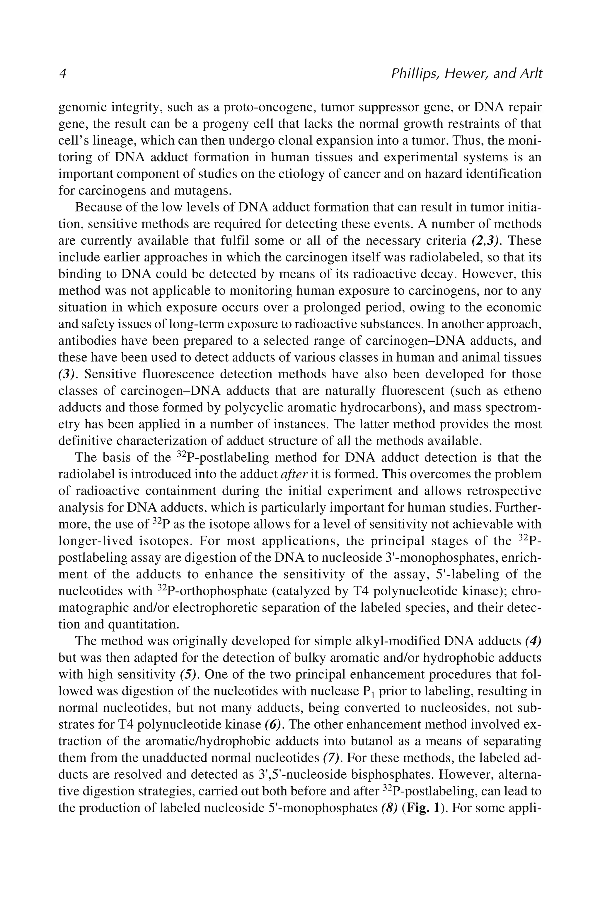
![Postlabeling Analysis of DNA Adducts 5
cations, the possession of only one charged phosphate group can result in better reso-
lution of carcinogen–DNA adducts and, additionally, improve confidence in the as-
signment of structures to unknown species on the basis of cochromatography with
synthetic standards. Since the earliest days of the assay’s use, resolution of DNA ad-
ducts has been most commonly performed by multidirectional thin-layer chromatog-
raphy (TLC), using polyethyleneimine (PEI)-cellulose plates (5). Alternatively,
high-performance liquid chromatography (HPLC) offers a technique of higher resolu-
tion that is being used more frequently (9,10). For small DNA lesions, such as those
resulting from oxidative damage to DNA, polyacrylamide gel electrophoresis (PAGE)
of DNA digests has also proved useful for resolving the 32P-postlabeled species (11).
The 32P-postlabeling assay currently has multiple applications that include moni-
toring human exposure to environmental carcinogens, mechanistic investigations of
carcinogen activation and tumor initiation, monitoring DNA repair, testing new com-
pounds for genotoxicity, investigating endogenous DNA damage and oxidative pro-
cesses, monitoring marine pollution through measurement of DNA adducts in aquatic
species, and assessing patient response to cytotoxic cancer drugs (12,13).
There are several advantages of 32P-postlabeling over other methods. It does not
require the use of radiolabeled test compounds, making it useful in experiments in
Fig. 1. Methods of 32P-postlabeling. Protocols are given in this article for procedures indi-
cated by bold arrows. X, adducted or modified nucleosides; N, normal nucleosides; p, phos-
phate; *p,[32P]phosphate; MN, micrococcal nuclease; SPD, spleen phosphodiesterase; NP1,
nuclease P1; PAP, prostatic acid phosphatase; PNK, T4 polynucleotide kinase; ATP,
[γ-32P]ATP; VPD, venom phosphodiesterase.](https://image.slidesharecdn.com/5874687-250603083210-8978989d/75/Molecular-Toxicology-Protocols-David-H-Phillips-Alan-Hewer-Volker-M-Arlt-Auth-22-2048.jpg)
![6 Phillips, Hewer, and Arlt
which multiple dosing is required, and it is applicable to a wide range of chemicals
and types of DNA lesion. Prior structural characterization of adducts is not required,
although some assumptions about their likely chromatographic properties may be nec-
essary. It has been used to detect DNA adducts formed by polycyclic aromatic hydro-
carbons, aromatic amines, heterocyclic amines (food mutagens), small aromatic
compounds (such as benzene, styrene, and alkenylbenzenes), alkylating agents, prod-
ucts of lipid peroxidation, reactive oxygen species, and ultraviolet radiation. It requires
only microgram quantities of DNA and is capable of detecting some types of DNA
adducts at levels as low as one adduct in 1010 normal nucleotides in this amount of
material. It can be applied to the assessment of the genotoxic potential of complex
mixtures of chemicals, such as environmental airborne combustion products, particu-
lates, and industrial or environmental pollutants.
The method is not without its limitations, however (14). DNA lesions that are not
chemically stable as mononucleotides will not be detected reliably. The method does
not provide structural information on adducts, and identification of adducts often re-
lies on demonstrating their cochromatography with characterized synthetic standards.
Such standards can provide the means for determining the efficiency of labeling and
detection, whereas in the absence of standards adduct levels may be underestimated.
In the case of complex mixtures, suitable standards often cannot be defined. The
method has also been found to detect endogenously derived DNA adducts that may, in
some instances, mask the formation of adducts formed by the compound under inves-
tigation (14).
In applying 32P-postlabeling to an investigation of the DNA binding activity of a
compound or mixture, the investigator is faced with a number of decisions concerning
the choice of enhancement procedure to be used and the chromatography conditions to
be applied. The protocols given here should be regarded as providing guidance for an
initial investigation. In many cases it may be necessary to try several different
approaches before the best procedure is found.
Following the adoption of 32P-postlabeling by many laboratories and its use for an
increasingly diverse number of applications, it became apparent that protocols varied
widely from laboratory to laboratory, even for analysis of the same types of carcino-
gen–DNA adducts (15). Therefore, an international interlaboratory trial was initiated
to establish a set of standardized protocols that would allow comparisons between
studies from different laboratories, particularly in the interpretation of human
biomonitoring studies (16). Furthermore, validated modified DNA samples, contain-
ing adducts derived from benzo[a]pyrene, from 4-aminobiphenyl, and from 2-amino-
1-methyl-6-phenylimidazo[4,5-b]pyridine (PhIP)were prepared, as was DNA
containing O6-methylguanine (16–18), and these standards have been made available
to investigators to enable them to determine the efficiency of the 32P-postlabeling assay
in their hands.
The protocols described here can be considered an introductory approach to the
method and are based on validated studies of known carcinogen–DNA adducts. When
a new or unknown type of DNA adduct is being investigated, it cannot be asserted
that these conditions will be optimal for its detection and quantitation. Indeed, differ-](https://image.slidesharecdn.com/5874687-250603083210-8978989d/75/Molecular-Toxicology-Protocols-David-H-Phillips-Alan-Hewer-Volker-M-Arlt-Auth-23-2048.jpg)
![Postlabeling Analysis of DNA Adducts 7
ent carcinogen–DNA adducts have been shown to be postlabeled with different effi-
ciencies (16,19).
2. Materials
2.1. DNA Digestion
1. Use double-distilled water or equivalent throughout.
2. Micrococcal nuclease (MN; cat. no. N3755, Sigma, Poole, Dorset, UK). Dissolve con-
tents in water to give 2 U/μL (see Note 1).
3. Spleen phosphodiesterase (SPD; from calf spleen, Type II; Calbiochem cat. no. 524711,
through CN Biosciences, Nottingham, UK) (see Note 1).
Mix to give final concentrations of 36 mU/μL MN and 6 mU/μL SPD (see Note 2).
4. Digestion buffer: 100 mM sodium succinate, pH 6.0, 50 mM CaCl2.
5. All solutions can be stored at –20°C in small aliquots.
2.2. Nuclease P1 Digestion
1. 0.25 M Sodium acetate buffer, pH 5.0.
2. 2.0 mM ZnCl2.
3. 1.25 mg/mL Nuclease P1 (Sigma, cat. no. N8630; see Note 3).
4. 0.5 M Tris base.
5. All solutions can be stored at –20°C in small aliquots.
2.3. Butanol Extraction
1. Buffer A: 100 mM ammonium formate, pH 3.5.
2. Buffer B: 10 mM tetrabutylammonium chloride.
3. 1-Butanol (redistilled, water-saturated).
4. 1-mL Syringe with blunt-ended needle.
5. 200 mM Tris-HCl, pH 9.5.
6. All solutions should be stored at 4°C.
2.4. DNA Postlabeling
1. T4 polynucleotide kinase (with or without 3'-phosphatase activity; Epicentre, Madison,
WI).
2. Kinase buffer: 200 mM bicine, pH 9.0, 100 mM MgCl2, 100 mM dithiothreitol, 10 mM
spermidine.
3. [γ-32P]ATP: >3000 Ci/mmol.
4. All solutions must be stored at –20°C in small aliquots.
2.5. Thin-Layer Chromatography
1. 20 × 20 cm PEI-impregnated cellulose TLC sheet (cat. no. 801053 Macherey-Nagel,
Middleton Cheney, UK; see Note 4).
2. Whatman no.1 filter sheets.
3. D1: 1 M sodium phosphate, pH 6.0 (see Note 5).
4. D2: 3.5 M lithium formate, 8.5 M urea, pH 3.5 (see Notes 6 and 7).
5. D3: 0.8 M lithium chloride, 0.5 M Tris-HCl, 8.5 M urea, pH 8.0 (see Note 7).
6. Efficiency solvent: 250 mM ammonium sulfate, 40 mM sodium phosphate.
7. Specific activity solvent: 0.5 M sodium phosphate, pH 6.0.](https://image.slidesharecdn.com/5874687-250603083210-8978989d/75/Molecular-Toxicology-Protocols-David-H-Phillips-Alan-Hewer-Volker-M-Arlt-Auth-24-2048.jpg)
![8 Phillips, Hewer, and Arlt
2.6. Detection and Quantification
1. 2 pmol/μL 2'-Deoxyadenosine 3'-monophosphate, in distilled water.
2. Autoradiography film or an electronic imaging device (e.g., Canberra Packard
InstantImager, Downers Grove, IL).
2.7. HPLC Cochromatography
1. 4 M Pyridinium formate, pH 4.5.
2. Ammonium buffer: 2 M ammonium formate, pH 4.5.
3. Acetonitrile.
4. Phenyl-modified reversed-phase column (e.g., 250 × 4.6 mm, particle size 5 mm, Zorbax
Phenyl).
5. HPLC system with in-line radioactivity monitor.
3. Methods
3.1. DNA Digestion
1. Take 4 μg DNA solution in a 1.5-mL tube and evaporate to dryness in a Speedvac evapo-
rator.
2. Add 4 μL MN/SPD mix and 0.8 μL digestion buffer/sample. Vortex and centrifuge to
ensure complete mixing.
3. Incubate at 37°C overnight.
3.2. Nuclease P1 Digestion
1. To the above digest add 2.4 μL sodium acetate buffer, 1.44 μL ZnCl2, and 0.96 μL
nuclease P1/sample. Incubate at 37°C for 1 h.
2. Stop the reaction by addition of 1.92 μL Tris base.
3.3. Butanol Extraction
1. Increase volume of DNA digest from 4.8 to 50 μL with water.
2. Premix 15 μL buffer A, 15 μL buffer B, and 70 μL water/sample.
3. Add 100 μL of premix to side of tube.
4. Immediately add 150 μL of butanol and vortex for 60 s at high speed.
5. Microcentrifuge at 8000g for 90 s. Remove upper butanol layer and keep.
6. Repeat steps 4 and 5.
7. To pooled butanol extracts add 400 μL butanol-saturated water. Vortex for 60 s.
Microcentrifuge as above.
8. Remove water through the butanol layer using a syringe, being careful not to remove any
of the butanol.
9. Repeat step 7 twice, discarding the water each time.
10. Add 3 μL 200 mM Tris-HCI to washed butanol. Vortex briefly.
11. Speedvac to dryness. Redissolve in 50 μL water by vortexing and Speedvac to dryness again.
12. Redissolve in 11.5 μL of water.
3.4. Labeling of Adducts (see Note 8)
1. Premix stock labeling mixture (number of samples + 2) for each sample from: 1.0 μL
kinase buffer, 6 U T4 polynucleotide kinase and 50 μCi [γ-32P]ATP/sample. Add appro-
priate volume to each solution remaining from Subheadings 3.2. or 3.3., step 12.](https://image.slidesharecdn.com/5874687-250603083210-8978989d/75/Molecular-Toxicology-Protocols-David-H-Phillips-Alan-Hewer-Volker-M-Arlt-Auth-25-2048.jpg)
![Postlabeling Analysis of DNA Adducts 9
2. Incubate at 37°C for 30 min.
3. Staple a 10 × 12-cm Whatman no.1 paper wick to the top edge of a 10 × 20 cm PEI-
cellulose TLC sheet as shown in Fig. 2. Spot the whole of each sample onto the origin of
this sheet. Keep tube for efficiency test (see Subheading 3.5.).
4. Run in D1 overnight with wick hanging outside tank (see Note 9).
5. Cut plates down to 10 × 10 cm as shown in Fig. 2.
6. Wash plates twice in water and dry plates with cool air.
7. Run in D2 and D3 in directions shown in Fig. 2. Before each run, dip lower edge of plate
in water to give an even solvent front. Lids should be taken off tanks for 15 min at end of
run.
8. Wash plates twice in water and cool air-dry between solvents.
3.5. Test for Efficiency of Enrichment Techniques
1. Wash bottom of tube (see Subheading 3.4., step 3) with 50 μL water.
2. Vortex and microcentifuge.
3. Spot 5 μL near lower edge of 10 × 20-cm PEI-cellulose TLC sheet.
4. Run in efficiency solvent from Subheading 2.5., item 6 to top edge (see Note 10).
3.6. Determining the Specific Activity of [γ-32P]ATP
1. Take 3 μL of 2'-deoxyadenosine 3'-monophosphate (6 pmol) + premix from Subheading
3.4., step 1. Incubate at 37°C for 30 min.
2. Dilute to 1.0 mL. Spot 2 × 5 μL 2 cm from the lower edge of a 10 × 20-cm PEI-cellulose
TLC sheet. Run in specific activity solvent from Subheading 2.5., item 7 to top edge.
3. Visualize and count adenosine bisphosphate spot (see Note 11).
4. This will give you dpm/pmol after allowing for dilution and efficiency of counting. Divide
by 2.22 × 10³ to give Ci/mmol.
Fig. 2. Diagram showing multidirectional thin-layer chromatography procedures for the
resolution of 32P-labeled adducts on polyethyleneimine-cellulose.](https://image.slidesharecdn.com/5874687-250603083210-8978989d/75/Molecular-Toxicology-Protocols-David-H-Phillips-Alan-Hewer-Volker-M-Arlt-Auth-26-2048.jpg)
![10 Phillips, Hewer, and Arlt
3.7. Imaging and Quantification
1. Adducts can be visualized by placing plates in cassettes with autoradiography film and
keeping at –80°C for several h, for up to 4 d. Adduct spots can then be cut from the plate
and quantitated in a scintillation counter. Alternatively, an InstantImager can be used,
which will give a result in a few minutes.
2. Counts per minute of the adduct should be corrected for efficiency of the counting proce-
dure and divided by the amount of DNA labeled to give dpm/μg. Dividing this by the
specific activity figure of dpm/fmol will give fmol of adduct/μg DNA. Results can be
expressed in this way or as adducts per 108 normal nucleotides. To arrive at this latter
figure, divide the number of fmols by 0.03, as 33 adducts per 108 nucleotides are equiva-
lent to 1 fmol/μg DNA.
3.8. Extraction of Adducts for HPLC Cochromatography
1. Cut the adduct spot out of the PEI-cellulose TLC sheet and place it in a scintillation vial
(see Note 12).
2. Add 500 μL pyridinium formate and shake gently overnight (see Note 13).
3. Microcentrifuge extracts at 8000g for 90 s to remove small particles.
4. Speedvac to dryness. Redissolve in 100 μL water and methanol (mix 1:1).
3.9. HPLC Cochromatography
1. Analyze aliquots (e.g., 50 μL) of the above extract on a phenyl-modified reversed-phase
column with a linear gradient of acetonitrile (from 0 to 35% in 70 min) in aqueous ammo-
nium buffer. Measure the radioactivity eluting from the column by monitoring Cerenkov
radiation through a radioactivity detector (see Note 14).
4. Notes
1. It is necessary to dialyze the MN solution to remove residual oligonucleotides. Use a 10K
Slide-A-Lyzer from Pierce (cat. no. 66425, through Perbio Science UK, Tattenhall,
Cheshire, UK) suspended in 5 L distilled water at 4°C for 24 h. Change water once. The
SPD must also be dialyzed to remove the ammonium salts, which may inhibit the label-
ing. The enzymes should be stored at –20°C and may be kept for at least 6 mo without
loss of enzyme activity.
2. Originally, Roche SPD was used at 2 mU/μL. This product was discontinued in the year
2000, and alternative sources (e.g., bovine phosphodiesterase from Sigma or Worthington)
have not proved to be as active. It may be necessary to vary the amount used depending
on the type of adducts to be detected.
3. Nuclease P1 solutions (in water) should be stored at –20°C.
4. Plates should be prerun with distilled water and dried to remove a yellow contaminant,
which may lead to an increased background.
5. 1 M Sodium phosphate is usually sufficient for many bulky adducts, but smaller or more
polar adducts may require higher concentrations (1.7–2.3 M sodium phosphate) to avoid
streaking.
6. Use lithium hydroxide to adjust pH to 3.5.
7. D2 and D3 are suitable for many lipophilic bulky adducts, but considerable variation is
possible in both concentration and content.
8. Caution:[γ-32P]ATP is a high-energy β-particle emitter and due regard should be given
to handling the material. Exposure to 32P should be avoided by working in a confined
laboratory area, with protective clothing, shielding, Geiger counters, and body dosimeter.](https://image.slidesharecdn.com/5874687-250603083210-8978989d/75/Molecular-Toxicology-Protocols-David-H-Phillips-Alan-Hewer-Volker-M-Arlt-Auth-27-2048.jpg)
![Single Adducts as Single Spots 13
13
From: Methods in Molecular Biology, vol. 291: Molecular Toxicology Protocols
Edited by: P. Keohavong and S. G. Grant © Humana Press Inc., Totowa, NJ
2
Modification of the 32P-Postlabeling Method to Detect
a Single Adduct Species as a Single Spot
Masako Ochiai, Takashi Sugimura, and Minako Nagao
Summary
The original 32P-postlabeling method developed by Randerath and his colleagues has
been modified to detect a single type of adduct as a single spot in thin-layer chromatog-
raphy (TLC), because some types of adducts gave multiple adduct spots by the original
method. In the remodified methods, DNA is first digested with micrococcal nuclease
and phophodiesterase II and then labeled with [γ-32P]ATP under standard or adduct-
intensification conditions. Since the labeled digest includes adducted mono-, di-, and/or
oligo-deoxynucleotides, it is further treated with phosphatase and phosphodiesterase
prior to TLC. The labeled digest is treated with nuclease P1 (NP1) in method I, and with
T4 polynucleotide kinase and NP1 in method II, and then with phosphodiesterase I in
both cases, and subjected to TLC. The advantage of these methods is that the number of
adduct species formed can be estimated by TLC.
Key Words: Adducted deoxynucleoside 5'-phosphate; nuclease P1; T4 polynucle-
otide kinase; phosphatases; phosphodiesterase I; phosphodiesterase II; micrococcal
nuclease; intensification method; standard method; method I; method II.
1. Introduction
The 32P-postlabeling method devised by Randerath and his group (1,2) is widely
used to detect DNA adducts formed in vitro and in vivo with various mutagens and
carcinogens. The advantages of this method (and simple modifications like butanol
extraction and use of nuclease P1 [NP1] to detect adducts with high efficiency) are
introduced in Chapter 1 of this volume. The principle is to detect DNA lesions as
adducted deoxynucleoside 3', 5'-diphosphate elements. However, with the established
methodology, a single type of adduct is not necessarily detected as a single spot owing
to incomplete digestion of adducted DNA.](https://image.slidesharecdn.com/5874687-250603083210-8978989d/75/Molecular-Toxicology-Protocols-David-H-Phillips-Alan-Hewer-Volker-M-Arlt-Auth-30-2048.jpg)
![14 Ochiai, Sugimura, and Nagao
In this chapter, approaches are introduced that allow for the detection of single ad-
ducted forms as single spots, with modifications to the original Randerath method. In
these methods, DNA is first digested with microccocal nuclease (MN) and phosphodi-
esterase II (PDE II), and labeled with [γ-32P]ATP under standard (2) or adduct-intensi-
fication conditions (3), the labeled digests obtained are further treated with NP1, T4
polynucleotide kinase (PNK), and phophodiesterase I (PDE I), before analysis of ad-
ducted deoxynucleoside 5'-phosphate formation by thin-layer chromatography (TLC).
In some cases, the labeled DNA digests include mono-, di-, and/or oligo-
deoxynucleotides: [32P]pX(pN)np, where X is an adducted deoxynucleoside, and N is
a normal deoxynucleoside. By treatment with NP1, the 3'-phosphate of [32P]pX(pN)np
can be removed to yield [32P]pX(pN)n (4,5), and further treatment with PDE I may
then produce [32P]pX and n(pN). Some types of adducted deoxynucleoside 3', 5'-
diphosphate are, however, resistant to the phosphatase activity of NP1, although they
are sensitive to that of PNK. Thus, in method I, labeled digests are treated with NP1,
and in method II, with PNK and NP1 and then treated with PDE I in both cases, as
shown in Fig. 1. It is known that the optimum pH for the 3'-phosphatase activity of
PNK is 5.9, whereas that for its kinase activity is 6.5–8.5 (6).
To give a concrete example, when DNA from rats treated with the food-borne mu-
tagenic/carcinogenic heterocyclic amine 2-amino-1-methyl-6-phenylimidazo[4,5-
b]pyridine (PhIP) was analyzed by the 32P-postlabeling method under standard or
intensification conditions, several adduct spots were detected on TLC, and the spot for
authentic N-(deoxyguanosin-8-yl)-2-amino-1-methyl-6-phenylimidazo[4,5-b]pyridine
3', 5'-diphosphate (3', 5'-pdGp-C8-PhIP) coincided with a minor adduct spot, although
it has been demonstrated to be the sole adduct of PhIP by high performance liquid
chromatography (HPLC) analysis using [3H]PhIP (7,8). The additional spots were
demonstrated to be owing to incomplete digestion of DNA (8). A similar result was
also obtained with a second heterocyclic amine, 2-amino-3,4-dimethylimidazo[4,5-
f]quinoline (MeIQ) (9). Single spots thus were generated with DNA from animals
treated with PhIP or MeIQ by method I (8,9).
In the case of the another heterocyclic amine, 2-amino-3-methylimidazo[4,5-
f]quinoline (IQ), five spots were detected on TLC by the standard method (10), and for
two of them their structures were tentatively identified as N-(deoxyguanosin-8-yl)-
2-amino-3-methylimidazo[4,5-f]quinoline 3',5'-diphosphate (pdGp-C8-IQ) (11) and
5-(deoxyguanosin-N2-yl)-2-amino-3-methylimidazo[4,5-f]quinoline (pdGp-N2-IQ)
(12). However, relatively large amounts of radioactivity were also present in the
remaining three spots. When method I was applied for the analysis of IQ-DNA adducts,
four spots were detected, and pdGp-N2-IQ was demonstrated to be resistant to the
phosphatase activity of NP1. However, it was converted to pdG-N2-IQ with PNK,
with or without NP1 (10). Thus, for IQ-DNA adducts, method II is appropriate, and by
this method, spots of pdG-C8-IQ and pdG-N2-IQ, as well as a very small radioactive
spot representing an unknown form of adduct, could be detected (10).
Recoveries with the modified methods are very close to those with the standard and
intensification methods; in other words, very high after treatment with PNK, NP1, and
PDE I. These results indicate that 32P-labeled oligonucleotides have modified bases at
the 5'-most position.](https://image.slidesharecdn.com/5874687-250603083210-8978989d/75/Molecular-Toxicology-Protocols-David-H-Phillips-Alan-Hewer-Volker-M-Arlt-Auth-31-2048.jpg)
![Single Adducts as Single Spots 15
A major advantage of these methods is that the number of adduct species formed in
vivo and/or under in vitro conditions can be estimated by TLC.
2. Materials
2.1. DNA Digestion
1. 0.01X SSC, 0.1 mM EDTA: make as 1X SSC (0.15 M NaCl, 0.015 M Na-citrate), 10 mM
EDTA. The solution can be stored at 4°C (see Note 1).
2. MN (Worthington, Freehold, NJ): dissolve in water to give 4 U/μL (see Note 2).
3. PDE II from bovine spleen (Worthington): dissolve in water to give 40 mU/µL.
4. Nuclease mixture: mix MN and PDE II solutions at a ratio of 1:1 to give a final concentra-
tion of 2 U/µL for MN and 20 mU/µL for PDE II.
5. Digestion buffer: 0.1 M sodium succinate and 0.05 M CaCl2, pH 6.0. This can be stored
at 4°C.
2.2. Postlabeling
1. [γ-32P]ATP with a specific activity of approx 260 TBq/mmol (~370 MBq/60 µL, e.g.,
ICN Biomedical, Irvine, CA).
2. 10 U/µL PNK (e.g., Takara Shuzo, Kyoto, Japan).
3. 10X Kination buffer: 0.3 M Tris-HCl, pH 9.5, 0.1 M dithiothreitol, 0.1 M MgCl2, 0.01 M
spermidine.
4. ATP solution: 200 µM ATP.
5. Kination solution A (10 µL used for each tube): 1.5 µL of 10X kination buffer, 1 µL of
PNK, 5 µL of [γ-32P] ATP, and 3 µL of ATP solution (see Notes 3 and 4).
6. Kination solution B (5 µL used for each tube): 1.5 µL of 10X kination buffer, 0.5 µL of
PNK, 1.5 µL of [γ-32P] ATP, and 1.5 µL of water.
2.3. Total Nucleotide Analysis
1. 20 mU/µL Potato apyrase (Sigma, St. Louis, MO).
2. Polyethyleneimine (PEI)-cellulose TLC sheets (95-mm height; Polygram CEL 300 PEI,
Machery-Nagel, Duren, Germany; see Note 5).
3. 0.5 M LiCl.
4. Scintillation counter or BioImaging Analyzer (BIA; e.g., BAS2000 Fuji, Tokyo,
Japan).
Fig.1. Principle of the modified method. *, 32P-label; X and Y, modified deoxynucleosides;
N, normal deoxynucleoside. 3' Phosphates of pXp and pX(pN)np seem to show the same sus-
ceptibility to the enzymes. In method I, NP1 and PDE I treatments and in method II, PNK, NP1,
and PDE I treatments are performed. NP1, nuclease P1; PDE I, phosphodiesterase I; PNK, T4
polynucleotide kinase.](https://image.slidesharecdn.com/5874687-250603083210-8978989d/75/Molecular-Toxicology-Protocols-David-H-Phillips-Alan-Hewer-Volker-M-Arlt-Auth-32-2048.jpg)
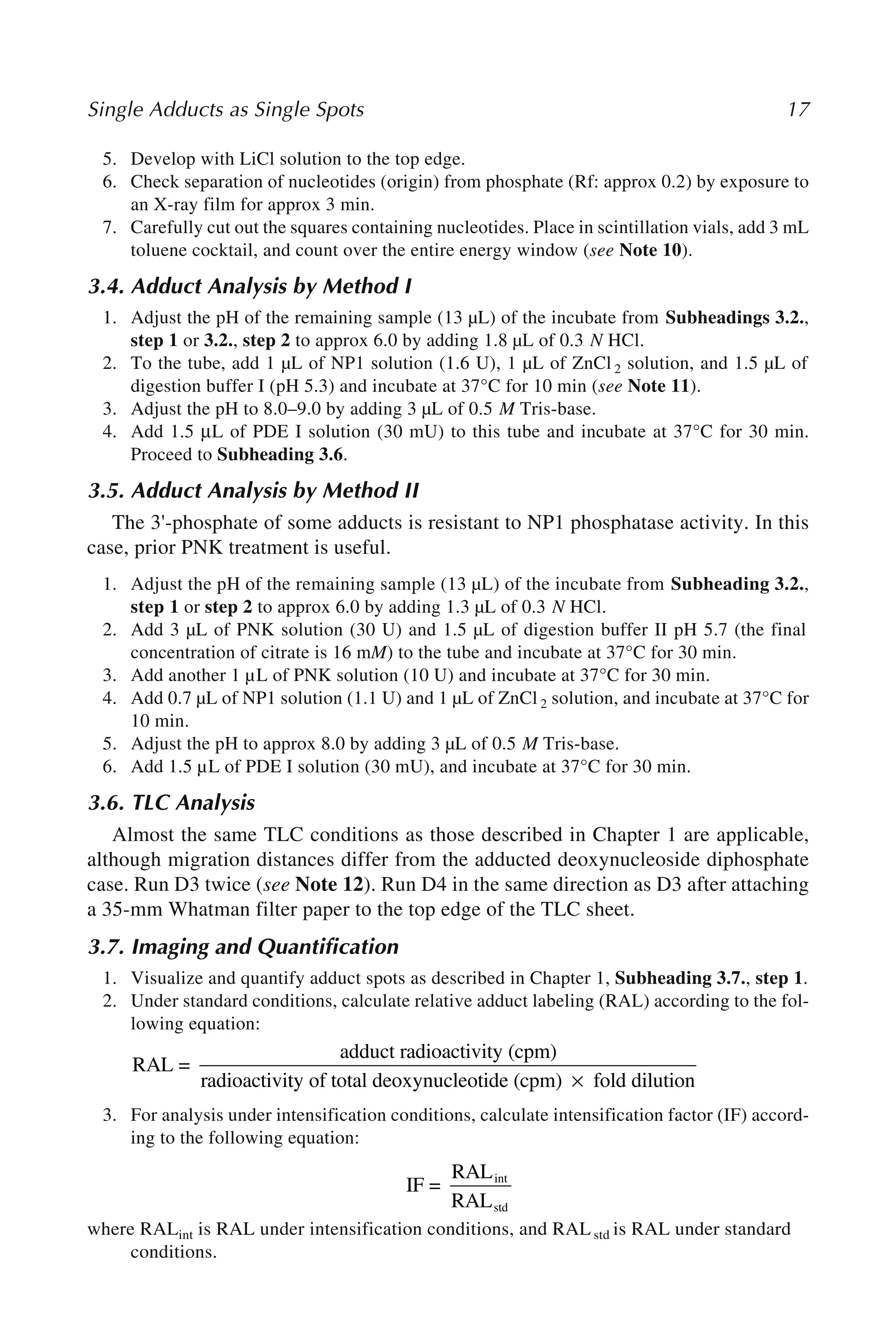
![18 Ochiai, Sugimura, and Nagao
4. Notes
1. Ultrapure water prepared by passing through Milli-Q spUF is used.
2. All solutions should be stored at –20°C, except where otherwise stated.
3. To ensure thorough mixing, it is recommended that all tubes containing different compo-
nents be vortexed and spun down in a microcentrifuge.
4. Protection from radioactivity during handling of radioactive samples is crucially impor-
tant and should be performed as described in Chapter 1, Note 8.
5. Sheets can be stored at 4°C. Plates should be prerun with water overnight after attach-
ment of a 12-cm filter paper wick and dried at room temperature.
6. The solvent used for IQ-DNA adduct analysis is indicated as an example. For D2, 4.5 M
lithium formate, 8.5 M urea, pH 3.5 is prepared and diluted appropriately. For MeIQ-
adducts 60% and for PhIP-adducts 80% solutions were used. For D3, 1.0 M LiCl, 0.5 M
Tris-HCl, 8.5 M urea, pH 8.0, was used for MeIQ and PhIP.
7. When analysis is performed under intensification conditions (see Subheading 3.2., step
2), it is also necessary to perform the procedure outlined in Subheading 3.2., step 1, to
determine the IF value of each adduct as written in Subheading 3.7.
8. After incubation in a tube, the contents should be spun down at 4°C.
9. Potato apyrase (20 mU/µL) is diluted with water to give a concentration of 1.8 mU/µL.
10. When BIA is used for quantification, a 500X dilution for the standard method (and a 50X
dilution for the adduct intensification method), and exposure to X-ray film for approx 30
min are recommended. For BIA analysis, total and adduct analyses should be made on the
same imaging plates.
11. This condition is appropriate for PhIP and MeIQ adducts, but the optimum pH may differ
depending on specific adducts and it is necessary to check the optimum pH, which should
be between 5.3 and 7.0.
12. The background usually becomes clean by running twice. Running once is enough in
some cases.
References
1. Randerath, K., Reddy, M. V., and Gupta, R. C., (1981) 32P-Labeling test for DNA damage.
Proc. Natl. Acad. Sci. USA 78, 6126–6129.
2. Gupta, R. C., Reddy M. V., and Randerath, K., (1982) 32P-Postlabeling analysis of non-
radioactive aromatic carcinogen-DNA adducts. Carcinogenesis 3, 1081–1092.
3. Randerath E., Atrawal H. P., Weaver, J. A., Bordelon C. B., and Randerath K., (1985) 32P-
postlabeling analysis of DNA adducts persisting for up to 42 weeks in the skin, epidermis
and dermis of mice treated topically with 7,12-dimethylbenz[a]anthracene. Carcinogen-
esis 6, 1117–1126.
4. Wilson, V. L., Basu, A. K., Essigmann, J. M., Smith, R. A., and Harris, C. C., (1988) O6-
alkyldeoxygunosine detection by 32P-postlabeling and nucleotide chromatographic analy-
sis. Cancer Res. 48, 2156–2161.
5. Randerath, K., Randerath, E., Danna, T. F., van Golden, K. L., and Putaman, L. L., (1989)
A new sensitive 32P-postlabeling assay based on the specific enzymatic conversion of
bulky DNA lesions to radiolabeled dinucleotides and nucleoside 5'-monophosphates. Car-
cinogenesis 10, 1231–1239.
6. Cameron, V. and Uhlenbeck, O. C., (1977) 3'-Phosphatase activity in T4 polynucleotide
kinase. Biochemistry 16, 5120–5126.
1
2
3
4
5
6](https://image.slidesharecdn.com/5874687-250603083210-8978989d/75/Molecular-Toxicology-Protocols-David-H-Phillips-Alan-Hewer-Volker-M-Arlt-Auth-35-2048.jpg)
![Single Adducts as Single Spots 19
7. Frandsen, H., Grivas, S., Andersson, R., Dragsted, L., and Larsen J. C., (1992) Reaction of
the N2-acetoxy derivative of 2-amino-1-methyl-6-phenylimidazo[4,5-b]pyridine with 2'-
deoxyguanosine and DNA. Synthesis and identification of N2-(2'-deoxyguanosin-8-yl)-
PhIP. Carcinogenesis 13, 629–635.
8. Fukutome, K., Ochiai, M., Wakabayashi, K., Watanabe S., Sugimura, T., and Nagao, M.,
(1994) Detection of guanine-C8-2-amino-1-methyl-6-phenylimidazo[4,5-b]pyridine
adduct as a single spot on thin-layer chromatography by modification of the 32P-
postlabeling method. Jpn. J. Cancer Res. 85, 113–117.
9. Tada, A., Ochiai, M., Wakabayashi, K., Nukaya, H., Sugimura T., and Nagao, M., (1994)
Identification of N-(deoxyguanosin-8-yl)-2-amino-3,4-dimethylimidazo[4,5-f]quinoline
(dG-C8-MeIQ) as a major adduct formed by MeIQ with nucleotides in vitro and with
DNA in vivo. Carcinogenesis 15, 1275–1278.
10. Ochiai, M., Nakagama, H., Turesky, R. J., Sugimura, T., and Nagao, M., (1999) A new
modification of the 32P-post-labeling method to recover IQ-DNA adducts as mononucle-
otides. Mutagenesis 14, 239–242.
11. Snyderwine, E. G., Yamashita, K., Adamson, R. H., et al., (1988) Use of the 32P-
postlabeling method to detect DNA adducts of 2-amino-3-methylimidazo[4,5-f]quinoline
(IQ) in monkeys fed IQ: identification of the N-(deoxyguanosin-8-yl)-IQ adduct. Car-
cinogenesis 8, 1739–1743.
12. Turesky, R. J. and Markovic, J., (1994) DNA adduct formation of the food carcinogen 2-
amino-3-methylimidazo[4,5-f]quinoline at the C-8 and N2 atoms of guanine. Chem. Res.
Toxicol. 7, 752–761.
7
8
9
10](https://image.slidesharecdn.com/5874687-250603083210-8978989d/75/Molecular-Toxicology-Protocols-David-H-Phillips-Alan-Hewer-Volker-M-Arlt-Auth-36-2048.jpg)
![26 Dingley et al.
22. AMS will establish the 14C/12C ratio of the sample. This is then used to calculate the
adduct level (see Note 8).
4. Notes
1. The protocol described is for isolation of up to 400 μg DNA using Qiagen Genomic tip
500 DNA isolation columns. This is approximately equivalent to the amount of DNA ob-
tained from 400 mg wet tissue. Other column sizes are Qiagen Genomic tip 20 for up to 20
μg DNA and tip 100 for up to 90 μg DNA. The protocol can be scaled down for the smaller
columns. (Refer to manufacturer’s instructions for column loading and wash volumes.)
2. When handling samples, it is essential to avoid cross-contamination of isotope by the use
of disposable plastic ware, scalpels, forceps, and gloves. These should be changed in
between samples. Liquid samples should be pipeted using filter tips and clean pipets.
When homogenizing the tissue, there should be disposable barriers (plastic wrap and foil)
in between the sample and hammer.
3. The flow rate of the column will depend on the viscosity of the sample. If the column
exhibits a very slow flow rate or becomes completely clogged, the flow can be assisted by
attaching a small amount of tubing to the bottom of the column and withdrawing eluate
slowly with a syringe. Troubleshooting tips are also described in the Qiagen literature.
4. The volume of water required will depend on the amount of DNA extracted. Typically in
this procedure, 300 μL of water will result in well-dissolved DNA with a concentration of
1–2 mg/mL. A 1:20 dilution of this solution should then be within the range suitable for
DNA concentration determination by UV spectrophotometry. The AMS sample tubes
used in our laboratory have a sample capacity of about 400 μL, so larger samples may
need to be concentrated prior to AMS preparation.
5. If A260/A280-nm absorbance ratios are not within the range of 1.7 to 1.9, this is probably
because of incomplete removal of protein or RNA. The DNA should be repurified prior to
analysis by AMS.
6. We try to submit DNA for analysis by AMS within 24 h of extraction. This reduces the
chance of cross-contamination of samples with radioisotope. However, if this is not pos-
sible, samples should be stored in a refrigerator or freezer that is not used for storage of
high levels of radioisotope.
7. Tributyrin is a nonvolatile hydrocarbon that contains depleted levels of carbon-14. It is
used in AMS to increase the size of small samples for efficient graphitization. Typically,
using this method, samples that contain less than 0.5 mg carbon require the addition of
carrier. For DNA (29% carbon), this would be equal to 1.7 mg. A 40 mg/mL solution of
tributyrin in methanol is made, and then 50 μL is added to each DNA sample. Carrier
controls should be prepared at the same time (see Note 8).
8. The isotope ratios determined by AMS are converted to adduct levels first by subtracting
the natural radiocarbon content of the sample and the radiocarbon contributed from addi-
tion of any carrier. The natural radiocarbon content of the DNA is determined using con-
trol DNA samples from subjects or rodents not given the [14C]-labeled compound. Adduct
levels (ratio of moles of compound/moles nucleotide) are then calculated based on the
percent carbon of DNA (29%) and the compound specific activity (16).
Acknowledgments
The authors thank Kristin Stoker for help with preparation of the manuscript. This
work was performed under the auspices of the US DOE (W-7405-ENG-48) with sup-
port from the NIH/National Center for Research Resources (RR13461).](https://image.slidesharecdn.com/5874687-250603083210-8978989d/75/Molecular-Toxicology-Protocols-David-H-Phillips-Alan-Hewer-Volker-M-Arlt-Auth-42-2048.jpg)
![Adduct Measurement Using AMS 27
References
1. Garner, R. C. (1998) The role of DNA adducts in chemical carcinogenesis. Mutat. Res.
402, 67–75.
2. Poirier, M. C., Santella, R. M., and Weston, A. (2000) Carcinogen macromolecular ad-
ducts and their measurement. Carcinogenesis 21, 353–359.
3. Turteltaub, K. W. and Dingley, K. H. (1998) Application of accelerated mass spectrom-
etry (AMS) in DNA adduct quantification and identification. Toxicol. Lett. 102–103, 435–439.
4. Lightfoot, T. J., Coxhead, J. M., Cupid, B. C., Nicholson, S., and Garner, R. C. (2000)
Analysis of DNA adducts by accelerator mass spectrometry in human breast tissue after
administration of 2-amino-1-methyl-6-phenylimidazo[4,5-b]pyridine and benzo[a]pyrene.
Mutat. Res. 472, 119–127.
5. Turteltaub, K. W. and Vogel, J. S. (2000) Bioanalytical applications of accelerator mass
spectrometry for pharmaceutical research. Curr. Pharm. Design 6, 991–1007.
6. Dingley, K. H., Roberts, M. L., Velsko, C. A., and Turteltaub, K. W. (1998) Attomole
detection of 3H in biological samples using accelerator mass spectrometry: application in
low-dose, dual-isotope tracer studies in conjunction with 14C accelerator mass spectrom-
etry. Chem. Res. Toxicol. 11, 1217–1222.
7. Creek, M. R., Mani, C., Vogel, J. S., and Turteltaub, K. W. (1997) Tissue distribution and
macromolecular binding of extremely low doses of [14C]-benzene in B6C3F1 mice. Car-
cinogenesis 18, 2421–2427.
8. Dingley, K. H., Curtis, K. D., Nowell, S., Felton, J. S., Lang, N. P., and Turteltaub, K. W.
(1999) DNA and protein adduct formation in the colon and blood of humans after expo-
sure to a dietary-relevant dose of 2-amino-1-methyl-6-phenylimidazo[4,5-b]pyridine. Can-
cer Epidemiol. Biomarkers Prev. 8, 507–512.
9. Mauthe, R. J., Dingley, K. H., Leveson, S. H., et al. (1999) Comparison of DNA-adduct
and tissue-available dose levels of MeIQx in human and rodent colon following adminis-
tration of a very low dose. Int. J. Cancer 80, 539–545.
10. Vogel, J. S., Turteltaub, K. W., Finkel, R., and Nelson, D. E. (1995) Accelerator mass-
spectrometry—isotope quantification at attomole sensitivity. Anal. Chem. 67, A353–A359.
11. Vogel, J. S. and Turteltaub, K. W. (1998) Accelerator mass spectrometry as a bioanalytical
tool for nutritional research. Adv. Exp. Med. Biol. 445, 397–410.
12. Buchholz, B. A., Freeman, S. P. H. T., Haack, K. W., and Vogel, J. S. (2000) Tips and
traps in the C-14 bio-AMS preparation laboratory. Nucl. Instr. Methods Phys. Res. B 172,
404–408.
13. Vogel, J. S. (1992) Rapid production of graphite without contamination for biomedical
AMS. Radiocarbon. 34, 344–350.
14. Roberts, M. L., Velsko, C., and Turteltaub, K. W. (1994) Tritium AMS for biomedical
applications. Nucl. Instr. Methods Phys. Res. B 92, 459–462.
15. Ognibene, T. J., Bench, G., Brown, T. A., Peaslee, G. F., and Vogel, J. S. (2002) A new
accelerator mass spectrometry system for 14C-quantification of biochemical samples. Int.
J. Mass Spectr. 218, 255–264.
16. Vogel J. S., Grant, P. G., Bucholz, B. A., Dingley, K., and Turteltaub, K. W. (2001)
Attomole quantitation of protein separations with accelerator mass spectrometry. Electro-
phoresis 22, 2037–2045.
1
2
4
5
6
7
8
9
10
12
14
15](https://image.slidesharecdn.com/5874687-250603083210-8978989d/75/Molecular-Toxicology-Protocols-David-H-Phillips-Alan-Hewer-Volker-M-Arlt-Auth-43-2048.jpg)
![Fluoroimmunoassay of UV Photoproducts in DNA 29
29
From: Methods in Molecular Biology, vol. 291, Molecular Toxicology Protocols
Edited by: P. Keohavong and S. G. Grant © Humana Press Inc., Totowa, NJ
4
Fluoroimaging-Based Immunoassay of DNA Photoproducts
in Ultraviolet-B-Irradiated Tadpoles
Iovanna Pandelova, Stephen R. Hewitt, and John B. Hays
Summary
Sensitive and accurate measurement of photoproducts induced in DNA by natural or
artificial ultraviolet-B (UVB; and UVC) light is essential to evaluate the toxic and
mutagenic effects of this radiation. Monoclonal antibodies specific for the two major
classes of photoproducts—cyclobutane pyrimidine dimers (CPDs) and pyrimidine-[6-4]-
pyrimidinone photoproducts ([6-4]PPs)—have made possible highly specific and sensi-
tive assays. Described here is the use of these primary antibodies with fluorescent
secondary antibodies to generate 96-spot arrays. Stable fluorescence signals are rapidly
and sensitively scored by fluoroimaging and computer analysis of peak-and-valley traces.
CPD levels in a series of calibration standards are determined by acid hydrolysis/thin-
layer chromatography analyses of radiolabeled bacterial DNA, UV-irradiated to known
high fluences, and linear extrapolation to known lower fluences. The nonlinear fluores-
cence vs CPD curve reflects the effect of photoproduct concentration on single vs double
binding by divalent antibody proteins. This technique is applied to photoproducts in
whole inbred Xenopus laevis tadpoles, chronically irradiated at a series of UVB fluences
that reach a lethality threshold when in vivo steady-state photoproduct levels are still
quite low. As few as 0.01–0.02 CPDs per DNA kbp can be reliably detected, at signal/
noise ratios of roughly 3:1.
Key Words: DNA adducts; UV; cyclobutane pyrimidine dimers (CPD); immunoas-
say; monoclonal antibody; fluorescent antibody; tadpole; DNA repair; Xenopus leavis;
[6-4]photoproduct; fluoroimaging.
1. Introduction
Sensitive and accurate detection of photoproducts induced in cellular DNA by
ultraviolet (UV) light is essential to interpretation of viability and survival data. Most
studies have used cultures of prokaryotic or eukaryotic cells, because of their unifor-
mity and amenability to statistical analyses. Notable exceptions are analyses of photo-
product accumulation and “sunburn” response in outer layers of mammalian skin (1),
as well as measurements of photoproducts in thin leaves of alfalfa and Arabidopsis
seedlings (2,3). To analyze quantitatively ultraviolet-B (UVB) effects on amphibians,](https://image.slidesharecdn.com/5874687-250603083210-8978989d/75/Molecular-Toxicology-Protocols-David-H-Phillips-Alan-Hewer-Volker-M-Arlt-Auth-44-2048.jpg)
![30 Pandelova, Hewitt, and Hays
whose drastic global population declines have been linked in some cases with solar
UVB radiation (4,5), we have employed cohorts of inbred Xenopus leavis tadpoles.
Their genetic and chronological homogeneity maximizes biological uniformity, and
their small size and semitransparency make even interior organs vulnerable to UV
light. When parallel tadpole subpopulations are exposed to UVB light regimes of
increasing chronic intensity, survival decreases abruptly at fluences that still induce
relatively low levels of photoproducts in DNA (6). Thus, photoproduct assays that are
sensitive, accurate, and reasonably reproducible from one animal to another are needed
to evaluate lethal UVB thresholds.
Assays that depend on cleavage of DNA strands at photoproduct sites and analysis
of the size distributions of the resulting fragments can be highly sensitive. Size distri-
butions are typically measured by alkaline sedimentation of DNA previously radiola-
beled in vivo (2), or alkaline gel electrophoresis and electronic imaging (7).
Radiolabeling of tadpole DNA in vivo is not practical, since the animals are grown in
1-gallon tanks, and the electrophoresis video-imaging technique requires highly spe-
cialized apparatus. Furthermore, extraction of DNA by all but the gentlest techniques
breaks DNA, thus distorting size distributions. In any case, determinations of average
photoproduct levels from fragment analyses typically involve assumptions about the
randomness of photoproduct distribution that may not be warranted for thicker tissues.
Lastly, efficient and highly specific incising enzymes are available only for one major
class of UV photoproducts, cyclobutane pyrimidine dimers (CPDs); for the other major
class, pyrimidine-[6-4']-pyrimidinone photoproducts ([6-4]PPs), precise and efficient
incision is difficult to achieve.
In the past few years, polyclonal (8) and monoclonal (9) antibodies specific for
CPDs and [6-4]PPs have been used for photoproduct-specific immunoassays.
Radioimmune dilution (8) and enzyme-linked immunosorbent assay (10) techniques
have been employed with considerable success, but the relatively large variability in
such measurements has limited sensitivity. More recently, secondary antibodies, able
to bind tightly and specifically to photoproduct-specific primary antibodies and them-
selves linked to signal-generating moieties, have been used for sensitive and repro-
ducible assays (11,12). Antibody-linked horseradish peroxidase can directly generate
a color signal on a membrane, which may then be quantitated by densitometry, or can
generate a chemiluminescent signal from a proprietary peracid/luminol/enhancer cock-
tail that registers on film. Disadvantages of the chemiluminescence-film technique are
limited linear response and the requirement that membranes must be exposed to film
immediately after reactions; the signals slowly decline with time, limiting reproduc-
ibility at low signal levels. All these techniques yield signals that can be used for more
or less sensitive and reproducible direct comparisons among samples, but absolute
determinations of photoproduct frequencies (per DNA bp) require calibration against
irradiated DNA standards in which absolute numbers of photoproducts can be determined.
Here we describe a modified photoproduct-immunoassay approach that addresses
both sensitivity/reproducibility and calibration issues (see Note 1). DNA is vigorously
extracted from irradiated tadpoles so as to minimize contaminants that might engender
spurious fluorescence, and aliquots quantitatively spotted onto membranes, to which](https://image.slidesharecdn.com/5874687-250603083210-8978989d/75/Molecular-Toxicology-Protocols-David-H-Phillips-Alan-Hewer-Volker-M-Arlt-Auth-45-2048.jpg)
![Fluoroimmunoassay of UV Photoproducts in DNA 31
anti-CPD or anti-[6-4]PP primary antibodies and fluorescent secondary antibodies are
bound. Fluorescence intensities are determined by fluoroimaging of washed mem-
branes, and sample and background signals are analyzed with computer software. CPD
signals are calibrated against DNA standards whose CPD contents can be chromato-
graphically determined (13).
2. Materials
2.1. Isolation of Total DNA From Whole Tadpoles
1. Frog tranquilizer (0.1% tricane).
2. Liquid N2.
3. GenomicPrep Cells and Tissue DNA isolation kit (Amersham, Piscataway, NJ).
4. 20 mg/mL Proteinase K (Worthington, Lakewood, NJ).
5. Phaselock Gel (5 Prime-3 Prime, Inc., Boulder, CO; optional).
6. Phenol/chloroform/isoamyl-alcohol, 25:24:1.
7. Chloroform/isoamyl-alcohol, 24:1.
8. 3 M Sodium acetate.
9. Absolute ethanol.
10. TE buffer: 10 mM Tris-HCl, pH 7.5, 1 mM Na2EDTA.
11. Filter paper.
12. Mortar and pestle.
13. 1.5-mL Microcentrifuge tubes.
14. Tabletop microcentrifuge.
2.2. Photoproduct Immunoassay
1. Irradiated [3H]DNA standards:
a. DNA is extracted from thymine-requiring E. coli (thy–, deo–) cells grown in broth
plus [3H]thymidine and then irradiated with UVC light fluences from 15 to 120 J/m2
(see Note 1).
b. Frequencies of T-T cyclobutane dimers (T[CPD]Ts) are determined by acid hydrolysis
and thin-layer chromatography, as previously described (13), and total numbers of CPDs
are estimated from known or estimated relative frequencies of T[CPD]T, T[CPD]C,
C[CPD]T, and C[CPD]C photoproducts (14). The same methods are used to measure
sample and standard DNA concentrations, as described in Subheading 3.1., step 16.
c. Standards for [6-4]PPs require a more complicated approach. Treatment of UV-irra-
diated plasmids with purified CPD-photolyase will leave only [6-4]PPs, whose aver-
age density (per plasmid or per DNA bp) can be estimated by transfection of
completely repair-incompetent (uvr–, phr–, recA–) E. coli, or by quantitative poly-
merase chain reaction (PCR) amplification.
d. In each case, comparison with unirradiated controls yields the fraction of irradiated
targets with no [6-4]PPs, and the Poisson distribution yields the average number per
plasmid or per bp.
2. 20X SSPE buffer: 3.6 M NaCl, 2 M NaH2PO4·H2O, 0.02 M Na2EDTA·2H2O, pH 7.4 (174
g NaCl, 27.6 g NaH2PO4, and 7.4 g Na2EDTA, dissolved in 800 mL H2O); pH adjusted to
7.4 with 10 N NaOH; volume made up to 1000 mL.
3. 0.4 M NaOH.](https://image.slidesharecdn.com/5874687-250603083210-8978989d/75/Molecular-Toxicology-Protocols-David-H-Phillips-Alan-Hewer-Volker-M-Arlt-Auth-46-2048.jpg)
![32 Pandelova, Hewitt, and Hays
4. 10X PBS buffer: 1.4 M NaCl, 26 mM KCl, 10 mM Na2HPO4, 17.6 mM KH2PO4, pH 7.4
(80 g NaCl, 2 g KCl, 14.48 g Na2HPO4, 2.4 g KH2PO4); pH adjusted to 7.4; volume made
up to 1000 mL.
5. Tween-20 detergent.
6. PBS-T buffer: 0.2% Tween-20 in PBS buffer (1000 mL volume).
7. Nonfat dried milk.
8. Primary monoclonal anti-CPD or anti-[6-4]PP antibody (kind gift from T. Mori [9]),
diluted respectively to 1:2000 or 1:1000 in sterile PBS (1X).
9. Fluorescently labeled secondary antibody, Alexa 568 goat anti-mouse (Molecular Probes,
Eugene, OR). The excitation and emission capabilities of the fluoroimager must be taken
into account.
10. Microcentrifuge tubes or PCR tubes.
11. Vortex mixer.
12. Slot-blot apparatus (BioRad, Hercules, CA).
13. Hybond N+ membrane (Amersham/Pharmacia).
14. Cellulose filter paper for gel blots, Island Scientific grade 238 or equivalent (Bainbridge
Island, WA).
15. Shallow rectangular plastic containers, with lids: two 10-oz containers for soaking of
nitrocellulose membrane and for reactions with primary and secondary antibodies; one
48-oz container for incubation with blocking solution.
16. Rocking platform(s) (used at both room temperature and at 4°C, i.e., in the cold room).
2.3. Detection and Quantification of Fluorescent Signal
From Bound Secondary Antibody
1. Fluoroimager (Hitachi FMBIOII multiview scanner), with software (FMBIO ReadImage
1.1, FMBIO V.6.0.11, Promega, Madison, WI).
2. Software for data analysis (ImageQuant V.1.2, Molecular Dynamics, Sunnyvale, CA).
3. Methods
3.1. Isolation of Total DNA From Whole Tadpoles
1. Anesthetize tadpole in 0.1% Tricane.
2. Place tadpole on a filter paper to drain excess water.
3. Add liquid N2 to a mortar to chill it, then add more liquid N2 and submerge tadpole.
4. Grind tadpole to a fine powder and transfer into microcentrifuge tube. At this point the
sample can be stored at –80°C until further processing.
5. Isolate DNA using GenomicPrep Cells and Tissue DNA isolation kit, supplemented with
proteinase K, according to the manufacturer’s instructions, beginning with 20–50 mg of
frozen sample. Resuspend DNA in 200 μL of TE buffer.
6. Centrifuge samples for 1 min. If a pellet (particulate debris) is formed, transfer the super-
natant into a fresh tube and discard the pellet.
7. Add an equal volume of phenol/chloroform/isoamyl-alcohol, and vigorously invert tube
10 times (if using Phase-Lock Gel, add roughly 200 µL to each tube).
8. Centrifuge for 5 min in microcentrifuge at 12,000–14,000 rpm (~12,000g).
9. To the supernatant add an equal volume of chloroform/isoamyl-alcohol, and vigorously
invert tube 10 times to remove residual phenol from the sample.
10. Centrifuge for 5 min in microcentrifuge at 12,000–14,000 rpm (~12,000g).
11. Transfer the supernatant to a fresh tube.](https://image.slidesharecdn.com/5874687-250603083210-8978989d/75/Molecular-Toxicology-Protocols-David-H-Phillips-Alan-Hewer-Volker-M-Arlt-Auth-47-2048.jpg)
![Fluoroimmunoassay of UV Photoproducts in DNA 33
12. Add 0.05 vol of 3 M sodium acetate and 2.5 vol of absolute ethanol. Mix well.
13. Centrifuge 1 min in microcentrifuge at 12,000–14,000 rpm (~12,000g).
14. Discard the supernatant. Resuspend pellet in 50–100 μL TE buffer.
15. Vortex vigorously and pipet several times up and down with an automatic pipet to
shear the DNA. Alternatively, digest DNA with restriction enzyme (see Note 2). Store
samples at 4°C.
16. Determine DNA concentration of each sample (see Note 3).
3.2. Photoproduct Immunoassay
Sample preparation and blotting
1. Make dilutions so as to place 100 ng of sample or standard DNA (see Subheading 2.2.,
item 2) in each tube in a volume of 200 μL or less (if using PCR tubes, 100 μL or less);
for [6-4]PP assays, use 1 μg.
2. Boil tubes with samples or standards for 5 min. Use PCR thermocycler, if possible, to
avoid condensation and sample contamination.
3. Chill on ice for 5 min.
4. Add equal volumes of 20X SSPE buffer and vortex. Microcentrifuge (1 min at 12,000–
14,000 rpm [~12,000g]) to remove any particulate matter (see Note 4).
5. Saturate membrane with deionized water and then with 20X SSPE buffer (see Note 5).
6. Place membrane on a plastic gasket and cut one corner for orientation. Set up slot-blot
apparatus according to the manufacturer’s instructions (see Note 6).
7. Add 200 μL of 20X SSPE buffer to each well, and pull through with a vacuum.
8. Add samples and standards (maximum volume 400 μL to each well; see Note 7) and let
stand for 15 min. Pull through with vacuum, adjusted to slow flow (see Note 8).
9. Add 200 μL 20X SSPE buffer to rinse wells, and pull through with vacuum.
Fixing and probing of membrane with antibody:
10. Soak filter paper in 0.4 M NaOH and pour off excess.
11. Place membrane on NaOH-soaked filter paper for 15 min.
12. Rinse membrane briefly with 5X SSPE buffer.
13. Block membrane by covering with 5% nonfat milk solution in PBS-T buffer (100 mL), in
shallow rectangular plastic container with lid.
14. Rock gently at room temperature for at least 2 h, or overnight, at 4°C.
15. Rinse off the milk with PBS-T buffer quickly twice and then for 15 min with rocking.
16. React with primary CPD antibody: place washed membrane in 10 mL of primary anti-
body solution diluted with PBS buffer, typically 1:2000 for CPDs and 1:1000 for [6-
4]PPs (test solutions beforehand with irradiated DNA standards); incubate for 2 h at room
temperature in 10-oz plastic container, rocking gently. Remove and store antibody solu-
tion at 4°C; solution can be used once more within 2 wk.
17. Wash membrane in PBS-T buffer twice quickly and then once with 20 min of rocking,
and twice with 5 min of rocking.
18. React membrane with secondary antibody, diluted 1:2500 in PBS buffer: place washed
membrane in 10 mL of secondary antibody solution (see Note 9). Incubate for 1 h at room
temperature, rocking gently. Remove secondary antibody solution and discard.
19. Wash membrane as in step 17, but increase the number of 5-min washes (with rocking) to
six (see Note 10).
20. Cover membrane with plastic wrap to keep from drying and with foil to prevent
photobleaching.](https://image.slidesharecdn.com/5874687-250603083210-8978989d/75/Molecular-Toxicology-Protocols-David-H-Phillips-Alan-Hewer-Volker-M-Arlt-Auth-48-2048.jpg)
![Fluoroimmunoassay of UV Photoproducts in DNA 35
4. If particulate matter is observed, pipet off the top 80% (as much as can be cleanly removed
from the pellet) of the supernatant and transfer to a clean tube for subsequent processing.
Particulate matter to some extent is typical, so this may be done routinely.
5. Never touch membrane with fingers; always use forceps.
6. The slot-blot apparatus should be cleaned with soap and then soaked in 0.4 M NaOH to
remove any residual DNA from previous experiments. If this is still a problem, a prelimi-
nary membrane can be applied to the apparatus (without sample), washed with 20X SSPE
buffer, and then discarded, before applying a new membrane for sample spotting.
7. DNA from each sample (tadpole) is spotted in duplicate or triplicate, along with DNA
from unirradiated control tadpoles and a series of irradiated DNA standards (see Sub-
heading 2.2., item 2). The same amounts of sample and standard DNA are applied, typi-
cally 100 ng (CPDs) or 1 μg ([6-4]PPs). When signals are expected to be weak, 150 ng
(CPDs) can be used for all spots without loss of linearity.
Fig. 1. Analysis of fluorescent-antibody signals. Lower panel: membrane image was
obtained by successively binding anti-CPD primary antibody and Alexa 568-labeled secondary
antibody to spotted DNA samples, followed by fluoroimaging and analysis with an FMBio
scanner and software, as described in the text. Upper panel: light/dark inverted TIFF file from
FMBio analysis of indicated row (brackets) of membrane was line-scan-analyzed using
ImageQuant software, as described in the text.](https://image.slidesharecdn.com/5874687-250603083210-8978989d/75/Molecular-Toxicology-Protocols-David-H-Phillips-Alan-Hewer-Volker-M-Arlt-Auth-50-2048.jpg)