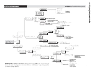
Hyperphosphatemia is a condition characterized by abnormally high levels of phosphorus in the blood. The document provides an algorithmic approach for evaluating and treating hyperphosphatemia. It outlines potential causes of hyperphosphatemia including increased phosphorus intake, decreased renal excretion or filtration, and redistribution of phosphorus from bones and cells. Laboratory tests that should be checked include levels of calcium, parathyroid hormone, bicarbonate, and creatinine clearance. Treatment depends on the underlying cause but may involve restricting phosphorus intake, using phosphate-binding medications, or treating any acidosis or kidney problems present.
This document discusses hyperphosphatemia, which occurs when phosphate levels in the blood are abnormally high. It provides information on the causes of hyperphosphatemia, which include kidney disease, and discusses guideline target levels and treatment options. Treatment involves phosphate restriction, phosphate binders such as aluminum hydroxide, calcium salts, and newer non-calcium binders. Novel therapies being researched include nicotinamide and iron-based compounds. The goal is to manage phosphate levels through diet, medication and dialysis to prevent complications in patients with chronic kidney disease.
The document discusses phosphate homeostasis and hypophosphatemia. Phosphate is essential for cell function and structure. Hypophosphatemia can be caused by intracellular shifts, increased urinary excretion, decreased absorption, or low intake. Severe hypophosphatemia can cause muscle weakness, respiratory issues, and neurological problems. Treatment involves correcting underlying causes and replacing phosphate through diet, oral supplements, or intravenous administration depending on severity.
This document discusses hyperphosphatemia in chronic kidney disease. It causes include reduced kidney function and phosphate retention. Target phosphate levels are 3.5-5.5 mg/dL. Treatment includes phosphate restriction, phosphate binders like calcium salts, and novel therapies like nicotinamide. Management involves controlling diet, medications, and dialysis in end-stage patients to control phosphate levels and prevent complications.
Hypophosphatemia is a condition defined by a serum phosphorus level lower than 2.7 mg/dL, which can be caused by insufficient phosphorus intake, increased phosphorus excretion, or intracellular shifts. Symptoms include decreased cardiac and respiratory function, weakness, impaired reflexes, bone abnormalities, irritability, and bleeding issues. Treatment involves monitoring for symptoms, discontinuing contributing medications, supplementing with phosphorus and vitamin D orally or intravenously, careful movement to prevent fractures, and dietary adjustments to intake phosphorus-rich and limit calcium-rich foods.
This document discusses hyperphosphatemia and its causes and effects. It begins by defining hyperphosphatemia as a serum phosphate level greater than 5.2 mg/dL. It then discusses pseudohyperphosphatemia which can result from conditions like multiple myeloma or hyperlipidemia. The document outlines the clinical features of hyperphosphatemia which can include hypocalcemic symptoms. It also discusses acute phosphate nephropathy and treatments which involve restricting phosphate intake, using phosphate binders, and dialysis for more severe cases. The regulation of phosphate excretion and potential causes of hyperphosphatemia both renal and non-renal are explored in detail.
Phospate restiction in renal failure (by low phosphate whey protein powder)Reijo Laatikainen
The study evaluated replacing part of the dietary protein intake of 27 hemodialysis patients with hyperphosphataemia with a low-phosphorus whey protein concentrate. Patients were randomly assigned to either continue their usual diet or partially replace morning milk and lunch meats/proteins with the concentrate. At 3 months, patients using the concentrate showed significant decreases in serum phosphate and PTH levels, while control patients did not. Further research is needed to verify the results and ensure long-term protein sufficiency, but low-phosphate protein supplements may help control phosphorus levels in renal failure patients.
This document discusses the serum phosphorus test, which measures the level of phosphorus in the blood. Phosphorus is essential for bone growth, energy storage, and muscle and nerve function. Too much or too little phosphorus can cause health issues. The serum phosphorus test can detect hyperphosphatemia (high phosphorus) or hypophosphatemia (low phosphorus), but further tests are needed to determine the underlying cause. A doctor may order this test if symptoms indicate abnormal phosphorus levels, if other blood tests show abnormalities, or if the patient has conditions like kidney disease that could affect phosphorus. High or low levels can be caused by dietary, medication, or health issues.
A new perspective on hyperphosphatemiastevechendoc
This document discusses hyperphosphatemia, providing 3 key points:
1. Hyperphosphatemia is defined as a serum phosphate level greater than 5.2 mg/dL and can be caused by decreased renal excretion, shifts in phosphate from intracellular to extracellular fluid, or increased phosphate intake. It most commonly occurs with renal dysfunction.
2. Symptoms of hyperphosphatemia are generally nonspecific but can include bone and joint pain, pruritus, and rash. Severe hyperphosphatemia can cause hypocalcemic symptoms due to precipitation of calcium phosphate in soft tissues.
3. Treatment of hyperphosphatemia depends on the cause but may include restricting phosphate intake, using phosphate binders like aluminum hydrox
Magnesium is absorbed in the small intestine and plasma levels are normally between 1.7-2.3 mg/dL. Hypermagnesemia occurs when levels exceed the normal range, with the most common cause being renal failure. Early symptoms include nausea and drowsiness, while levels over 10 mg/dL can cause muscle paralysis, respiratory failure, and cardiac arrest. Treatment involves withdrawing magnesium supplementation if mild, or using diuretics, calcium gluconate, or dialysis for severe cases.
This document discusses the case of a 65-year-old man who presented with bilateral stress fractures and generalized bone and muscle pain. Laboratory tests found that he had hypophosphatemia. The document then provides an extensive overview on the evaluation, causes, and management of hypophosphatemia. Specifically for this patient, workup found elevated FGF23 levels and a bone tumor was discovered and resected, resolving the hypophosphatemia. The tumor was determined to be the cause of tumor-induced osteomalacia.
This document discusses hypokalemia and hypomagnesemia. It defines hypokalemia as a low serum potassium level below 3.5-5.0 mEq/L, which can be caused by vomiting, diarrhea, diuretics or gastric suctioning. Signs of hypokalemia include muscle weakness, polyuria, fatigue and arrhythmias. Causes include decreased intake, redistribution into cells, and increased losses from the gastrointestinal tract or kidneys. Treatment focuses on correcting the potassium deficit and minimizing ongoing losses, often through oral or IV supplementation. Hypomagnesemia is defined as a low blood magnesium level, which can be present with or without an actual magnesium deficiency. C
This document provides dietary guidelines and recommendations for hemodialysis patients. It discusses controlling intake of phosphorus, potassium, sodium, protein, and fluids. It lists foods to limit and include for each nutrient. It also provides grocery shopping lists, fast food nutrition facts, tips for dining out, and holiday food suggestions while on a renal diet. The goal is to help patients feel better and avoid complications by following a diet tailored to their renal needs and individual factors.
Calcium and phosphate metabolism is tightly regulated in the body. Calcium is mainly stored in bones while phosphate is found intracellularly and extracellularly. Vitamin D, parathyroid hormone, and calcitonin control calcium and phosphate levels by impacting absorption in the gut and kidneys. An imbalance can result in hypercalcemia with symptoms like nausea and fatigue, or hypocalcemia which can cause tetany.
This document summarizes electrolyte disturbances, specifically disorders of sodium and potassium balance. It discusses the causes, types, clinical features, diagnosis, and treatment of hyponatremia, hypernatremia, hypokalemia, and hyperkalemia. The key points covered include normal sodium and potassium levels, how they are regulated, complications that can arise from imbalances, and goals and principles of correcting electrolyte abnormalities.
Chronic Kidney Disease, CKD, Nephrology, Dee Evardone
This document provides an overview of chronic kidney disease (CKD). It defines CKD as the presence of kidney damage or decreased kidney function for three or more months. Key points include:
- CKD is defined based on evidence of kidney damage through structural abnormalities found on biopsy, imaging, or urine tests, or decreased glomerular filtration rate (GFR) below 60 mL/min/1.73m2.
- Common causes of CKD include diabetes, hypertension, glomerulonephritis, cystic kidney diseases, and vascular diseases.
- The document outlines clinical and laboratory manifestations of CKD and approaches to evaluating and managing patients with CKD.
Rheumatoid arthritis (RA) is an autoimmune disease that causes inflammation of the joints. While the cause is unknown, genetic factors may increase susceptibility. RA commonly affects adults between 30-50 years old, with women being affected more than men. Symptoms include morning joint stiffness, swelling of joints on both sides of the body, and lumps under the skin. Regular exercise and medications like NSAIDs and DMARDs can help reduce pain and inflammation. Lifestyle changes and stress management may also help control symptoms.
Rheumatoid arthritis (RA) is a chronic autoimmune disease that causes symmetrical joint inflammation. It affects around 1% of the population, predominantly women aged 30-50. Genetic and environmental factors contribute to its development. RA is characterized by synovitis and infiltration of inflammatory cells into the synovium, resulting in cartilage and bone destruction. Common clinical features include pain, stiffness, and swelling of small joints. Left untreated, RA can cause joint deformity and damage to other body systems. Investigations reveal elevated inflammatory markers and autoantibodies. Management involves a multidisciplinary approach including medications to relieve symptoms and slow disease progression.
Rheumatoid arthritis current diagnosis and treatmentAnkur Varshney
This document provides information on the diagnosis and management of rheumatoid arthritis (RA). It begins with an introduction to RA, noting that it is a chronic inflammatory joint disease affecting approximately 1% of the population. It then discusses the clinical presentation and manifestations of RA, including onset, patterns of joint involvement, and articular and extra-articular symptoms. The document reviews the diagnostic criteria for RA and covers laboratory investigations and radiographic features. It concludes with an overview of the goals and various treatment modalities for RA, including NSAIDs, corticosteroids, disease-modifying antirheumatic drugs (DMARDs), immunosuppressive therapies, and biological therapies.
Rheumatoid arthritis is a chronic disease that damages joints, causing inflammation and stiffness. It affects approximately 1% of the world's population, mostly women. While the exact cause is unknown, it is an autoimmune disorder where the immune system attacks the body's own tissues. Symptoms include fatigue, lack of appetite, fever, and joint pain. It is diagnosed through blood tests and x-rays. Treatment involves medications like NSAIDs, DMARDs, and corticosteroids to reduce inflammation and manage symptoms.
Rasamanikya is a excellent preparation in the field of Rasashastra, it is used in various Kushtha Roga, Shwasa, Vicharchika, Bhagandara, Vatarakta, and Phiranga Roga. In this article Preparation& Comparative analytical profile for both Formulationon i.e Rasamanikya prepared by Kushmanda swarasa & Churnodhaka Shodita Haratala. The study aims to provide insights into the comparative efficacy and analytical aspects of these formulations for enhanced therapeutic outcomes.
Clinic ^%[+27633867063*Abortion Pills For Sale In Tembisa Central19various
Clinic ^%[+27633867063*Abortion Pills For Sale In Tembisa Central Clinic ^%[+27633867063*Abortion Pills For Sale In Tembisa CentralClinic ^%[+27633867063*Abortion Pills For Sale In Tembisa CentralClinic ^%[+27633867063*Abortion Pills For Sale In Tembisa CentralClinic ^%[+27633867063*Abortion Pills For Sale In Tembisa Central
Muktapishti is a traditional Ayurvedic preparation made from Shoditha Mukta (Purified Pearl), is believed to help regulate thyroid function and reduce symptoms of hyperthyroidism due to its cooling and balancing properties. Clinical evidence on its efficacy remains limited, necessitating further research to validate its therapeutic benefits.
Promoting Wellbeing - Applied Social Psychology - Psychology SuperNotesPsychoTech Services
A proprietary approach developed by bringing together the best of learning theories from Psychology, design principles from the world of visualization, and pedagogical methods from over a decade of training experience, that enables you to: Learn better, faster!
Our backs are like superheroes, holding us up and helping us move around. But sometimes, even superheroes can get hurt. That’s where slip discs come in.