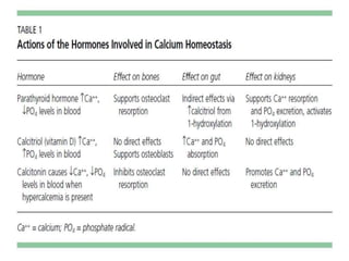
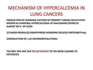
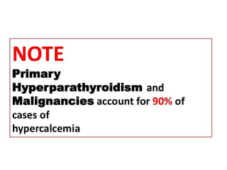
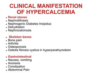
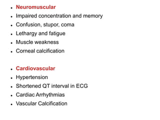
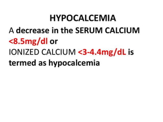
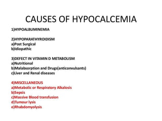
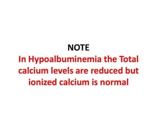
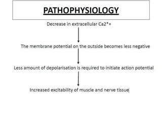
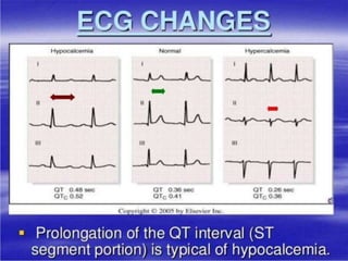
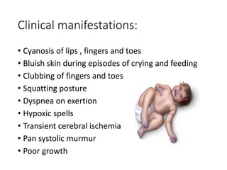
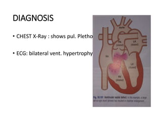
HYPERCALCEMIA
AND
HYPOCALCEMIA
ROLL NO. 113-117
CLINICAL MANIFESTATION
OF HYPERCALCEMIA
 Renal stones
 Nephrolithiasis
 Nephrogenic Diabetes Insipidus
 Dehydration
 Nephrocalcinosis
 Skeleton bones
 Bone pain
 Arthritis
 Osteoporosis
 Osteitis fibrosis cystica in hyperparathyroidism
 Gastrointestinal
 Nausea, vomiting
 Anorexia
 Constipation
 Abdominal Pain
 Neuromuscular
 Impaired concentration and memory
 Confusion, stupor, coma
 Lethargy and fatigue
 Muscle weakness
 Corneal calcification
 Cardiovascular
 Hypertension
 Shortened QT interval in ECG
 Cardiac Arrhythmias
 Vascular Calcification
Cyanotic congenital
Heart disease
Classification
• Pulmonary blood flow – tetralogy of fallot
• —Tricuspid atresia
• Mixed blood flow — transposition of great
arteries
• —total anomalous
pulmonary venous return
• — hypoplastic left heart
syndrome
Tetralogy of fallot
• It is the most common type of cyanotic
congenital heart disease .
• Classical form includes 4 defects i.e.
• Ventricular septal defect
• Pulmonary stenosis
• Overriding aorta
• Right ventricular hypertrophy
Pathophysiology
• Blood normally returns from systemic circulation
to right atrium and right ventricles .
• Outflow of the blood from right ventricle is
resisted by pulmonary stenosis so that blood
flows through ventricular septal defect into the
aorta .
• There is right to left shunt , hypertrophy of right
ventricles occur .
• Due to this unoxygenated blood from right
ventricles cyanosis occurs .
Clinical manifestations:
• Cyanosis of lips , fingers and toes
• Bluish skin during episodes of crying and feeding
• Clubbing of fingers and toes
• Squatting posture
• Dyspnea on exertion
• Hypoxic spells
• Transient cerebral ischemia
• Pan systolic murmur
• Poor growth
Diagnostic evaluation :
• On Auscultation – loud harsh systolic murmur may be
heard at left middle to lower sternal border .
• Radiography – The heart shows enlarged ventricle on
right side , large aorta and decrease in size of
pulmonary artery . Unusual shape of heart – wooden
shoe or boot shaped heart .
• ECG : shows evidence of right ventricular
hypertrophy.
• Echocardiography: shows evidence of aortic override
, thick anterior right ventricular wall and large aorta .
Management :
• Oxygen administration for cyanosis .
• Surgical management:
• Palliative shunt – if pulmonary arteries
are too hypoplastic then preferred
procedure is modified Blalock Taussig shunt ,
with an anastomoses created between
pulmonary artery and subclavian artery .
• Complete repair : Elective repair is usually
performed in the first year of life . Indications
for repair includes increasing cyanosis and
valve closure of the VSD and resection of
infundibular stenosis with a pericardial patch
to enlarge the right ventricular outflow tract .
• Normally ductus atreriosus closes soon after birth
but in this anomaly it fails to do so due to which
there is continuous AV shunt from aorta to
pulmonary artery .
• Clinical features — dyspnoea , machinery murmur.
( maximum in 2nd left inter coastal space below
clavicle) , accompanied her thrill , pulse is increased
in volume .
• Investigations – echocardiogram is IOC . Enlargement
of pulmonary artery may be detected radiologically .
• ECG – evidence of right ventricular hypertrophy.
Persistent ductus atreriosus
Management:
• Persistent ductus can be closed at cardiac
catheterisation with an implantable occlusive
device
• Closure should be undertaken in infancy if
shunt is significant otherwise it can be
delayed until childhood.
• When ductus is structurally intact ,
prostaglandin synthetase inhibitor may be
used in first week of life to induce closure .
ATRIAL SEPTAL DEFECT
.
ETIOPATHOGENESIS
• One of most common CHD.
• FREQUENCY: twice in females
• Due to defect in ‘ostium secundum ’ involving fossa ovalis
• Large volume of blood shunts through defect from LA to RA
• From RA to pulmonary arteries
CLINICAL FEATURES
• Mostly asymptomatic for years & detected at routine
examination.
• Dyspnea
• Chest infection
• Arrythmias esp. atrial fibrillation
• characteristic physical signs are result of volume
overload of RV
• In children with large shunt : have diastolic murmur
over tricuspid valve.
.
INVESTIGATIONS
• CHEST X- Ray : enlargement of heart , pul. Artery & pul.
Plethora
• ECG: incomplete right bundle branch block
• Echocardiography: demonstrate defect and RV dilation,
RV hypertrophy and pul. Artery dilation
• Transesophageal Echocardiography: show precise size
and location of defect
MANAGEMENT
• ASD with pul. Flow is increased by
50% above systemic flow : closed
surgically
• Closure can accomplished at cardiac
catheterization using implant closure
devices
• Contraindication to surgery: Shunt
reversal and severe pul. Hypertension
VENTRICULAR SEPTAL
DEFECT
•
.
ETIOPATHOGENESIS
• It is most common congenital cardiac defect
• Defect may be isolated or part of compex congenital
heart dis.
• Occurs as result of incomplete septation of ventricles.
• Most defects are perimembranous (junction of
membrane & muscular portion)
• Acquired defect may occur due to rupture as in acute
MI or rarely trauma
CLINICAL FEATURES
• Pansystolic murmur
• Prominent parasternal pulsation
• tachypnea
• Indrawing of ribs on inspiration
• May present as: cardiac failure in infants
As murmur with only minor hemodynamic
disturbance in older children & adults
DIAGNOSIS
• CHEST X-Ray : shows pul. Plethora
• ECG: bilateral vent. hypertrophy
MANAGEMENT
• Small defect : no specific treatment
• Cardiac failure in infancy: treated with digoxin and
diuretics
• Persistent failure indication for surgical repair of defect.
• Except in Eisenmenger’s syndrome ,long term
prognosis is very good in VSD
HYPERCALCEMIA-V-HYPOCALCEMIA.pptx

HYPERCALCEMIA-V-HYPOCALCEMIA.pptx

  • 1.
  • 17.
    CLINICAL MANIFESTATION OF HYPERCALCEMIA Renal stones  Nephrolithiasis  Nephrogenic Diabetes Insipidus  Dehydration  Nephrocalcinosis  Skeleton bones  Bone pain  Arthritis  Osteoporosis  Osteitis fibrosis cystica in hyperparathyroidism  Gastrointestinal  Nausea, vomiting  Anorexia  Constipation  Abdominal Pain
  • 18.
     Neuromuscular  Impairedconcentration and memory  Confusion, stupor, coma  Lethargy and fatigue  Muscle weakness  Corneal calcification  Cardiovascular  Hypertension  Shortened QT interval in ECG  Cardiac Arrhythmias  Vascular Calcification
  • 48.
  • 49.
    Classification • Pulmonary bloodflow – tetralogy of fallot • —Tricuspid atresia • Mixed blood flow — transposition of great arteries • —total anomalous pulmonary venous return • — hypoplastic left heart syndrome
  • 50.
    Tetralogy of fallot •It is the most common type of cyanotic congenital heart disease . • Classical form includes 4 defects i.e. • Ventricular septal defect • Pulmonary stenosis • Overriding aorta • Right ventricular hypertrophy
  • 51.
    Pathophysiology • Blood normallyreturns from systemic circulation to right atrium and right ventricles . • Outflow of the blood from right ventricle is resisted by pulmonary stenosis so that blood flows through ventricular septal defect into the aorta . • There is right to left shunt , hypertrophy of right ventricles occur . • Due to this unoxygenated blood from right ventricles cyanosis occurs .
  • 52.
    Clinical manifestations: • Cyanosisof lips , fingers and toes • Bluish skin during episodes of crying and feeding • Clubbing of fingers and toes • Squatting posture • Dyspnea on exertion • Hypoxic spells • Transient cerebral ischemia • Pan systolic murmur • Poor growth
  • 53.
    Diagnostic evaluation : •On Auscultation – loud harsh systolic murmur may be heard at left middle to lower sternal border . • Radiography – The heart shows enlarged ventricle on right side , large aorta and decrease in size of pulmonary artery . Unusual shape of heart – wooden shoe or boot shaped heart . • ECG : shows evidence of right ventricular hypertrophy. • Echocardiography: shows evidence of aortic override , thick anterior right ventricular wall and large aorta .
  • 54.
    Management : • Oxygenadministration for cyanosis . • Surgical management: • Palliative shunt – if pulmonary arteries are too hypoplastic then preferred procedure is modified Blalock Taussig shunt , with an anastomoses created between pulmonary artery and subclavian artery .
  • 55.
    • Complete repair: Elective repair is usually performed in the first year of life . Indications for repair includes increasing cyanosis and valve closure of the VSD and resection of infundibular stenosis with a pericardial patch to enlarge the right ventricular outflow tract .
  • 56.
    • Normally ductusatreriosus closes soon after birth but in this anomaly it fails to do so due to which there is continuous AV shunt from aorta to pulmonary artery . • Clinical features — dyspnoea , machinery murmur. ( maximum in 2nd left inter coastal space below clavicle) , accompanied her thrill , pulse is increased in volume . • Investigations – echocardiogram is IOC . Enlargement of pulmonary artery may be detected radiologically . • ECG – evidence of right ventricular hypertrophy. Persistent ductus atreriosus
  • 57.
    Management: • Persistent ductuscan be closed at cardiac catheterisation with an implantable occlusive device • Closure should be undertaken in infancy if shunt is significant otherwise it can be delayed until childhood. • When ductus is structurally intact , prostaglandin synthetase inhibitor may be used in first week of life to induce closure .
  • 58.
  • 59.
    ETIOPATHOGENESIS • One ofmost common CHD. • FREQUENCY: twice in females • Due to defect in ‘ostium secundum ’ involving fossa ovalis • Large volume of blood shunts through defect from LA to RA • From RA to pulmonary arteries
  • 60.
    CLINICAL FEATURES • Mostlyasymptomatic for years & detected at routine examination. • Dyspnea • Chest infection • Arrythmias esp. atrial fibrillation • characteristic physical signs are result of volume overload of RV • In children with large shunt : have diastolic murmur over tricuspid valve.
  • 61.
  • 62.
    INVESTIGATIONS • CHEST X-Ray : enlargement of heart , pul. Artery & pul. Plethora • ECG: incomplete right bundle branch block • Echocardiography: demonstrate defect and RV dilation, RV hypertrophy and pul. Artery dilation • Transesophageal Echocardiography: show precise size and location of defect
  • 63.
    MANAGEMENT • ASD withpul. Flow is increased by 50% above systemic flow : closed surgically • Closure can accomplished at cardiac catheterization using implant closure devices • Contraindication to surgery: Shunt reversal and severe pul. Hypertension
  • 64.
  • 65.
    ETIOPATHOGENESIS • It ismost common congenital cardiac defect • Defect may be isolated or part of compex congenital heart dis. • Occurs as result of incomplete septation of ventricles. • Most defects are perimembranous (junction of membrane & muscular portion) • Acquired defect may occur due to rupture as in acute MI or rarely trauma
  • 66.
    CLINICAL FEATURES • Pansystolicmurmur • Prominent parasternal pulsation • tachypnea • Indrawing of ribs on inspiration • May present as: cardiac failure in infants As murmur with only minor hemodynamic disturbance in older children & adults
  • 67.
    DIAGNOSIS • CHEST X-Ray: shows pul. Plethora • ECG: bilateral vent. hypertrophy
  • 68.
    MANAGEMENT • Small defect: no specific treatment • Cardiac failure in infancy: treated with digoxin and diuretics • Persistent failure indication for surgical repair of defect. • Except in Eisenmenger’s syndrome ,long term prognosis is very good in VSD