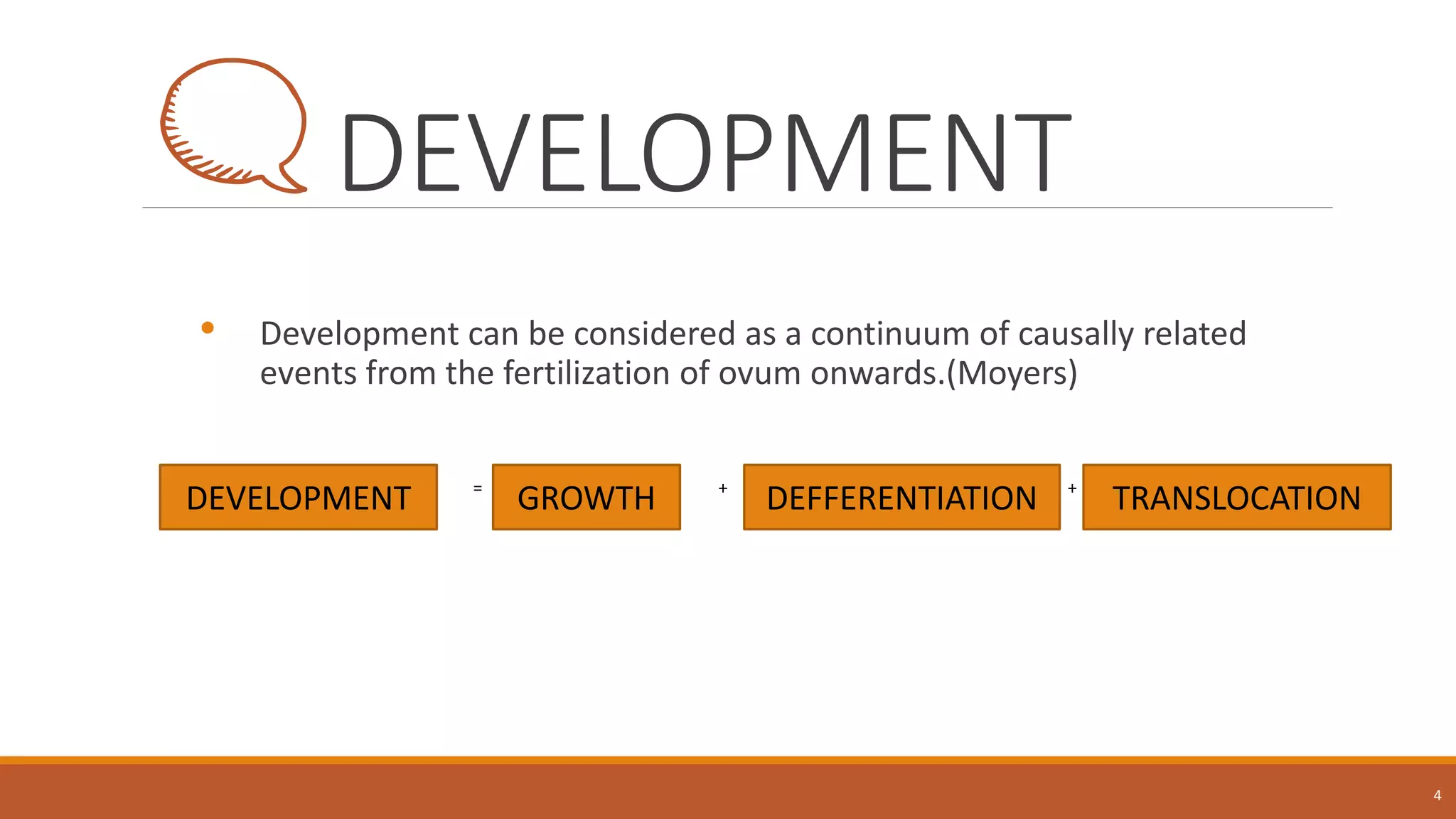
Growth basics
By- Dr Kanika Singh
CONTENTS
• DEFINITION OF GROWTH AND DEVELOPMENT
• BONE GROWTH :
A) INTRAMEMBRANOUS OSSIFICATION
B) ENDOCHONDRAL OSSIFICATION
• MECHANISM OF BONE GROWTH :
A) DEPOSITION AND RESORPTION
B) ENDOSTEAL AND PERIOSTEAL BONE GROWTH
C) REMODELING
• GROWTH PATTERN :
A) CEPHALOCAUDAL GROWTH
B) SCAMMON’S GROWTH GRADIENT
C) VARIABILITY
D) TIMING, DISTANCE AND VELOCITY CURVES
• METHODS OF STUDYING GROWTH
A) MEASUREMENT APPROACH
B) EXPERIMENTAL APPROACH
• GROWTH MOVEMENTS :
A) DRIFT AND DISPLACEMENT
B) V - PRINCIPLE
C) SURFACE PRINCIPLE
• ENLOW COUNTERPART PRINCIPLE
2
GROWTH
• Refers to increase in size and number , but tends to be
linked more to change than anything else . (Proffit)
• Process of remodeling to provide enlargement (Enlow)
• Any change in morphology which is within measurable
parameter. ( Moss)
• Increase in size or number-(TODD)
3
DEVELOPMENT
• Development can be considered as a continuum of causally related
events from the fertilization of ovum onwards.(Moyers)
• DDE == + +
+
4
DEVELOPMENT GROWTH TRANSLOCATIONDEFFERENTIATION
BONE GROWTH
• Bone formation occurs by three co-ordinated processes:
the production ,
the maturation of osteoid matrix or skeletogenesis ,
subsequent mineralization of the matrix.
• bone tissue arises through two processes:
a)intramembranous ossification - flat bones of the skull and face, the mandible
b)endochondral ossification - weight-bearing bones of the axial skeleton
and bones of extremities.
5
INTRAMEMBRANOUS BONE
FORMATION
• Increased vascularity of tissue.
• Active proliferation of mesenchymal cells. The
mesenchymal cells give rise to osteogenic cells, which
develop into osteoblasts.
• Osteoblasts begin to lay down osteoid. Osteoid is the
organic part of bone without the inorganic constituent.
• Osteoblasts either retreat or become entrapped as
osteocytes in the osteoid.
• The osteoid calcifies to form spicules of spongy bone.
• The spicules unite to form trabeculae. The inorganic
salts carried in by the blood vessels supposedly bring
about calcification
6
Intramembranous Ossification
(Source: Gartner and Hiatt, Color Textbook of Histology)
Textbook of Craniofacial Growth by Sridhar Premkumar
ENDOCHONDRAL OSSIFICATION
7Textbook of Craniofacial Growth by Sridhar Premkumar
MECHANISM OF BONE GROWTH
Takes place in the following three ways:
• Chondral growth
• Sutural growth
• Periosteal growth
8
CHONDRAL GROWTH –
• The organic matrix in cartilage is not normally
calcified and because the tissue is avascular,
metabolites must enter and leave by simple
diffusion.
• Also cartilage like soft tissue has interstitial as well as
appositional growth. Interstitial growth is possible
because the matrix is not calcified and can therefore
expand to accommodate the chondrocytes resulting
from cell division.
• The growth of cartilage is also bidirectional
MECHANISM OF BONE GROWTH
• Sutural Growth -
Sutural growth occurs due to osteoblasts and
is similar to periosteal growth.
Bone apposition takes place at the edges of
bone.
The middle zone contains numerous blood
vessels and connects both the fibrous layer to
one another.
The active growth of suture is found only at
the bone edges.
9
Premature closure of various
sutures and their effects
Sutures Skull deformity
Sagittal suture Scaphocephaly
Symmetric fusion
of coronal suture Oxycephaly
Asymmetric fusion of
coronal suture Plagiocephaly
Metopic suture Trigonocephaly
MECHANISM OF BONE GROWTH
• Periosteal Growth
Periosteum controls the resorption and deposition of the bone during maturity.
The growth direction of periosteal growth is on one side only and bone growth or
deposition takes place only on the surface.
10
GROWTH PATTERN
• In Studies of growth and development, the concept of pattern is an important
one.
• In a general sense, pattern reflects proportionality, usually of a complex set of
proportions rather than just a single proportional relationship.
• Because it refers not just to a set of proportional relationships at a point in time,
but to the change in these proportional relationships over time.
11
A) Cephalocaudal Growth
B) Scammon’s Growth Gradient
C) Variability
D) Timing, Distance And Velocity Curves
Proffit W.R. , Fields H.W. , Sarver D.M. (2007). Contemporary Orthodontics. St. Louis, mo: Mosby Elsevier .
1. Cephalocaudal growth :
12
Redrawn from Robbins WJ, et al. Growth. New Haven: Yale University Press;
1928
Illustrates the change in overall body
proportions that occurs during normal growth
and development.
At 3rd month of intrauterine life -
• Head constitutes 50% of
total body length.
• At this stage, the cranium
is large relative to the
face and represents more
than half the total head
• Limbs are Rudimentary
13
At the time of birth -
• Portion of the head is
decreased to 30%.
• the trunk and limbs have
grown faster than the
head and face.
• The overall pattern of
growth thereafter will
follows this course.
14
In the adult-
• Progressive reduction of in
relative size of head which is 12 %
of the total body length.
• There is more growth of the
lower limbs than the upper limbs
during postnatal life.
15
Cephalocaudal growth :
• Axis of increased growth extending from
the head toward the feet.
• This increased axis of growth in the
caudal direction is called cephalocaudal
growth gradient.
16
Evidence on face -
17
At birth jaws and face are
less developed than skull.
Maxilla being closure to skull
develop faster than
mandible.
LoweryGH. Growth and Development of Children. 6th ed. Chicago: Year Book
Medical Publishers; 1973
2. Scammon’s growth curve :
18
Human body comprise of four major
tissues:
1. neural
2.somatic
3.lymphoid
4. genital and sexual tissues
19
Scammon RD. The measurement of the body in childhood. In: Harris JA, ed.
The Measurement of Man. Minneapolis: University of Minnesota Press; 1930
Scammon's curves for growth of the four major tissue
systems of the body. As the graph indicates, growth of the
neural tissues is nearly complete by 6 or 7 years of age.
General body tissues, including muscle, bone, and viscera,
show an S-shaped curve, with a definite slowing of the rate
of growth during childhood and an acceleration at puberty.
Lymphoid tissues proliferate far beyond the adult amount in
late childhood and then undergo involution at the same
time that growth of the genital tissues accelerates rapidly.
3. Variability :
As deviation from the usual pattern and
express it in a quantitative manner.
20
VARIABILITY -
21
More appropriate to put
forth variability as deviation
from the usual pattern, and
express it in a quantitative
manner
Can be done with the help of
a growth chart.
Charts can be used to
determine whether the
growth is normal or abnormal.
1. Location of an individual
relative to the group .
2. Can be used to follow a
child over time to evaluate
whether there is an
unexpected change of
growth
• 50% - individual who are exactly at
the midpoint.
• One larger than 90% would plot
above 90% line.
• One who was smaller than 90% of
the population would plot below
the 10% line.
• A general guideline is that a child
who falls outside the range of 97%
of the population should receive a
special study before being
accepted as extreme of a
population
22
23
Growth of a boy who developed a medical
problem that affected growth, plotted on
the male chart.
Note the change in pattern (crossover of
lines on the chart) between ages 10 and
11.
This reflects the impact of serious illness
beginning at that time, with partial recovery
after age 13 but a continuing effect on
growth.
4. Timing
Variations in timing arises because
the same events happens for different
individuals at different time.
Or
The biological clocks of different
individuals are set differently.
24
Periods during
which the growth
process are turned
on are called
growth spurts
a) Just before birth
b) One year after birth
c) Mixed dentition growth
spurt:
BOYS- 8-11 years
GIRLS – 7-9 years
d) Pre-pubertal growth spurt:
BOYS – 14-16 years
GIRLS – 11-13 years
Velocity and distance curves
• These are obtained by measuring individual children
repeatedly during their growth.
• Height and weight are the most commonly used
measurements.
25
DISTANCE CURVE :
The curve obtained by
joining successive points
of a child’s height from
birth to adulthood is called
distance curve.
VELOCITY CURVE :
Amount of increment in
height per year is plotted
against age in years.
Are more revealing than
distance curve.
Velocity and distance curves
26
 In velocity curve, it is easier to see
when accelerations and decelerations
in the rate of growth occurred.
 Timing variability can be reduced by
using developmental age rather than
chronological age as an expression of
an individual’s growth status .
(Data from Scammon, 1927,Amer F Phys Anthrop.)
METHODS FOR STUDYING
PHYSICAL GROWTH
• Two basic approaches to studying physical growth.
• 1st based on techniques for measuring living animals, measurement itself does no
harm.
• 2nd approach uses experiments in which growth is manipulated in some way. This
implies that the subject of the experiment will be available in some details and
the detailed study may be distructive.
27
MEASUREMENT
APPROACH
ACQUIRING ANALYSIS
Proffit W.R. , Fields H.W. , Sarver D.M. (2007). Contemporary Orthodontics. St. Louis, mo: Mosby Elsevier
.
EXPERIMENTAL
APPROACH
A) B)
AQUIRING MEASUREMENT
DATA : CRANIOMETRY
28
• The 1st of the measurement approaches for studying growth.
• Craniometry was originally used to study the Neanderthal and Cromagnon
peoples whose skulls were found in European caves in the 18th and 19th
centuries.
• Craniometry has the advantage that rather precise measurement can be
made on dry skulls.
• Disadvantages is these growth data must be cross-sectional.
Proffit W.R. , Fields H.W. , Sarver D.M. (2007). Contemporary Orthodontics. St. Louis, mo: Mosby Elsevier .
CRANIOMETRY
29
• eg: Kranioti E. F. et al in 2008 did craniometric analysis of the modern
Cretan population.
• The aim of this study is to develop a sex determination technique using
osteometric data from skeletal remains of a contemporary Cretan
cemetery population.
• A total of 178 well-preserved , adult skulls of Cretan origin were
measured. And were compared with several populations geographically
and time wise distant from Cretans.
• Using stepwise method involving 5 dimensions, the result accuracy was
88.2%.
Craniometric analysis of the modern Cretan population by Elena F. Kranioti et al in Forensic Science
International 180 (2008) 110.e1-110.e5
AQUIRING MEASUREMENT
DATA : ANTHROPOMETRY
30
• Made it possible to measure skeletal dimensions on living individuals.
• It involves various landmarks establishment in living individuals simply
by using soft tissue points overlying these bony landmarks.
• Although the landmarks measured on the soft tissue introduces some
variation because of the soft-tissue thickness overlying both
landmarks. But it is possible to follow the growth of an individuals
directly, by making the same measurements repeatedly at different
times.
Proffit W.R. , Fields H.W. , Sarver D.M. (2007). Contemporary Orthodontics. St. Louis, mo: Mosby Elsevier .
ANTHROPOMETRY
31
• In recent years , Leslie G. Farkas in his study introduced anthropometric methods
into clinical practice to quantify changes in the craniofacial framework, features
distinguishing various races/ethnic groups were discovered.
• Normal range in each resultant database was established by the set of
anthropometric measurements of the face in the population gathered by various
international scientist.
• Study group consisted of 1470 healthy subjects (18 to 30 years) , 750 males and
720 females.
• The largest group come from Europe. 3 were drawn from Middle-East , 5 from
Asia and 4 from African origin
• Morphological characteristics were determined by 14 anthropometric
measurements.
Farkas, LG:Anthropometry of the Head and Face. 1994, Raven Press, New York.
ANTHROPOMETRY
32
Measurements on the lateral aspect of the face:
Head: tr-n (forehead height) Face: tr-gn
(physiognomical
face height) n-gn (morphological face height) sn-gn
(lower
face height) Nose: n-sn (nose height), in: (inclination
of the
nasal bridge) Ear: sa-sba (length of the auricle).
Measurements on the frontal aspect of the face:
Orbits: en-en (intercanthal width) ex-ex (biocular
width)
en-ex (eye fissure length) Face: zy-zy (face width) go-
go
(mandible width) Nose: al-al (morphological nose
width)
Labio-oral region: ch-ch (mouth width
Farkas, LG:Anthropometry of the Head and Face. 1994, Raven Press, New York.
ANTHROPOMETRY
33
• The results concluded, regions with single measurements showed
identical values to NAW in forehead height, mouth width and ear
height were found in 99.7% in both sexes.
• The orbital regions exhibited the greatest variations in identical and
contrasting measurements in comparison to NAW.
Farkas, LG:Anthropometry of the Head and Face. 1994, Raven Press, New York.
AQUIRING MEASUREMENT DATA
: CEPHALOMETRIC RADIOLOGY
• Technique depends on precisely orienting the head before making a radiograph,
with equally precise control of magnification.
• This approach can combine the advantages of craniometry and anthropometry.
• Allows direct measurement of bony skeletal dimensions, since the bone can be
seen through the soft tissue covering in a radiograph, but it also allows the same
individual to be followed over time.
• Disadvantage of a standard cephalometric radiograph is that it produces a two-
dimensional representation of a three-dimensional structure.
• This can be overcome by making more than one radiograph at different
orientations and using triangulation to calculate oblique distances.
• Current picture of craniofacial growth is based on cephalometric studies.
34
Proffit W.R. , Fields H.W. , Sarver D.M. (2007). Contemporary Orthodontics. St. Louis, mo: Mosby Elsevier .
AQUIRING MEASUREMENT DATA
: THREE-DIMENSIONAL IMAGING
• Information now is being obtained with the application 3-D imaging techniques.
• Computer axial tomography(CT) allows 3-D reconstructions of the cranium and
face, and this method has been applied for several years to plan surgical
treatment for patient with facial deformities.
• Recently, Cone beam rather than CT has been applied to facial scans. This
significantly reduces the radiation dose and cost. CBCT allows scans of patients
with radiation exposure that is much closer to dose from cephalograms.
• MAGNETIC RESONANCE IMAGING also provides 3-D images , with the advantage
that there is no radiation exposure with this techniques.
35
Proffit W.R. , Fields H.W. , Sarver D.M. (2007). Contemporary Orthodontics. St. Louis, mo: Mosby Elsevier .
THREE-DIMENSIONAL
IMAGING
36
• THREE –DIMENSIONAL PHOTGRAPHY also makes it possible much more accurate
measurements of facial soft tissue dimensions
• Images from a single photograph with a 3dMD camera. Both profile and oblique and
frontal views can be captured at the same head position, and measurements of soft
tissue dimensions and proportions can be made with great accuracy at any orientation
of the face, which makes such a 3- D camera a valuable research tool
Proffit W.R. , Fields H.W. , Sarver D.M. (2007). Contemporary Orthodontics. St. Louis, mo: Mosby Elsevier .
ANALYSIS OF
MEASUREMENT DATA
37
• Both anthropometric and cephalometric data can be expressed cross-
sectionally rather than longitudinally
• It is easier and quicker to do a cross-sectional study, gathering data once
for any individual and including subjects of different ages, rather than
spending many years on a study in which the same individuals are
measured repeatedly. For this reason, most studies are cross-sectional.
• Longitudinal studies are efficient in the sense that a great deal of
information can be gained from a relatively small number of subjects,
fewer than would be needed in a cross-sectional study. In addition, the
longitudinal data highlight individual variations, particularly variations
caused by timing effects.
Proffit W.R. , Fields H.W. , Sarver D.M. (2007). Contemporary Orthodontics. St. Louis, mo: Mosby Elsevier
MATHEMATICAL TRANSFORMATIONS
• Various other mathematical transformations can be used with growth data to make them easier
to understand.
• more complex mathematical transformation was used by D’Arcy Thompson
38
In the early 1900s, D'Arcy Thompson showed that
mathematical transformation of a grid could
account for the changes in the shape of the face
from man (A) to chimpanzee (B), monkey (C),
dog (D), or other animals.
Application of this method revealed previously
unsuspected similarities among
various species
Redrawn from Thompson DT. On Growth and Form. Cambridge: Cambridge University Press; 1961
MATHEMATICAL TRANSFORMATIONS
39
 Data for the increase in weight of early
embryos, with the raw data plotted in green
and the same data plotted after logarithmic
transformation in blue.
 At this stage, the weight of the embryo
increases dramatically, but, as shown by the
straight line after transformation, the rate of
multiplication of individual cells remains fairly
constant.
 When more cells are present, more divisions
can occur, and the weight increases faster.
Experimental
Approaches
40
Vital Staining:
• dyes that stain mineralizing tissues (or occasionally, soft tissues) are
injected into an animal. These dyes remain in the bones and teeth and
can be detected later after sacrifice of the animal.
• This method was originated by the great English anatomist John Hunter
in the eighteenth century.
textile waste pigs occasionally fed on textile waste.
observed that the bones of
pigs
were often stained in an
interesting way
Proffit W.R. , Fields H.W. , Sarver D.M. (2007). Contemporary Orthodontics. St.
Louis, mo: Mosby Elsevier .
Experimental
Approaches
41
ALIZARIN
adsorption of ALIZARIN
hydroxyapatite, Ca10(PO4)6(OH)2 (HAP)
ionized 1-, 2-OH groups of Alizarin can be
electrostatically bound to Ca2+ on HAP
Moriguchi, T., Yano, K., Nakagawa, S., & Kaji, F. (2003). Elucidation of adsorption mechanism of bone-staining agent alizarin red S on
hydroxyapatite by FT-IR microspectroscopy. Journal of colloid and interface science, 260(1), 19-25.
• Alizarin reacts strongly with
calcium at sites where bone
calcification is occurring.
• Bone remodels rapidly, and
areas from which bone is
being removed also can be
identified by the fact that vital
stained material has been
removed from these location.
Experimental
Approaches
42
• Many children born in the late 1950s and early 1960s were treated for recurrent infections
with the antibiotic tetracycline. It was discovered too late that tetracycline is an excellent
vital stain that binds to calcium at growth sites in the same way as alizarin.
RADIOACTIVE TRACERS :
• it has become possible to use almost any radioactively labeled metabolite that becomes
incorporated into the tissues as a sort of vital stain.
• The location is detected by the weak radioactivity given off at the site where the material
was incorporated.
• The gamma-emitting isotope 99mTc can be used to detect areas of rapid bone growth in
humans.
• But these images are more useful in diagnosis of localized growth problems than for studies
of growth patterns.
• For most studies of growth, radioactively labeled materials in the tissues of experimental
animals are detected by the technique of AUTORADIOGRAPHY.
Proffit W.R. , Fields H.W. , Sarver D.M. (2007). Contemporary Orthodontics. St.
Louis, mo: Mosby Elsevier .
Experimental
Approaches
43
• Emulsion differ from the standard
photographic film in terms of having higher
ratio of silver halide to gelatin and small size
of grain.
• Photography emulsion is placed over a thin
section of tissue containing the isotope
• then is exposed in the dark by the radiation
• the location of the radiation that indicates
where growth is occurring can be observed by
looking at the tissue section through the film
Autoradiograph of fetal rat bones growing in organ culture, with
14C-proline and 3H-thymidine incorporated in the culture medium.
Thymidine is incorporated into DNA, which is replicated when a cell
divides, so labeled nuclei are those of cells that underwent mitosis in
culture. Because proline is a major constituent of collagen,
cytoplasmic labeling indicates areas where proline was incorporated,
primarily into extracellularly secreted collagen.
Experimental
Approaches
44
Implant Radiography :
• In this technique, inert metal pins are
placed in bones anywhere in the skeleton,
including the face and jaws.
• metal pins are well tolerated by the
skeleton, become permanently
incorporated into the bone without
causing any problems, and are easily
visualized on a cephalogram.
• This method of study was developed by
Professor Arne Björk and coworkers at the
Royal Dental College in Copenhagen.
 In his study , small pins of hard
tantalum were hammered into
the bone under local analgesia.
 With a pencil-shaped
instruments, in the tip of which
the implant is placed.
Björk, A. (1968). The use of metallic implants in the study of facial growth in children: method and
application. American Journal of Physical Anthropology, 29(2), 243-254.
Diagram illustrating method of inserting the metallic implants.
Experimental
Approaches
45
implants are inserted in four regions
of the mandible:
(1) One in the midline of the symphysis;
(2) two under the first or second premolar or first
molar, the right side;
(3) One on the external aspect of the ramus, right
side,
(4) one under the second premolar, left side.
Implants are inserted in four zones in the maxilla.
(1) Before eruption of the permanent incisors, one on each side of the hard
palate, behind the deciduous canines;
(2) after eruption of the permanent incisors, one on each side of the median
suture, under the anterior nasal spine;
(3-4) two on each side in the zygomatic process of the maxilla.
Björk, A. (1968). The use of metallic implants in the study of facial growth in children: method and
application. American Journal of Physical Anthropology, 29(2), 243-254.
GROWTH MOVEMENTS:
REMODELING
46
Remodeling involves deposition of bone on any surface pointed toward the
direction of enlargement of a given area; resorption usually occurs on the
opposite side of that particular bony Cortex.
Addition of new bone on one
side is known as DEPOSITION
Elimination of bone from the
other side is known as
RESORPTION.
Essentials of facial growth, 2nd ed By Donald H. Enlow and Mark G. Hans
Needham Press; 2008
REMODELING
• The surface that faces toward the direction
of movement is depository (+). The opposite
surface, facing away from the growth
direction, is resorptive (- ).
• If the rates of deposition and resorption are
equal, the thickness of the cortex remains
constant. If deposition exceeds resorption,
overall size and cortical thickness gradually
increase.
47
Essentials of facial growth, 2nd ed By Donald H. Enlow and Mark G. Hans
Needham Press; 2008
REMODELING
48
Functions of remodeling are :
(1) progressively create the changing size of each whole bone
(2) sequentially relocate each of the component regions of the
whole bone to allow for overall enlargement
(3) Progressively shape the bone to accommodate its various
functions
(4) Provide progressive fine-tune fitting of all the separate
bones to each other and to their contiguous, growing,
functioning soft tissues
(5) carry out continuous structural adjustments to adapt to the
intrinsic and extrinsic changes in conditions
Essentials of facial growth, 2nd ed By Donald H. Enlow and Mark G. Hans
Needham Press; 2008
RELOCATION
49
• To illustrate, in the stack of chips is at the
right end in a at the level of the condyle
in the smallest mandibular stage.
• This location then becomes translocated
"across" the ramus to lie at the level of
the anterior margin in the third stage.
• The black chip became progressively
"relocated" not by its own movement,
but because new chips have been added
on one side and removed from the other.
Essentials of facial growth, 2nd ed By Donald H. Enlow and Mark G. Hans Needham Press; 2008
RELOCATION
50
• maxillary arch and palate move
downward.
• Inferior direction of remodeling by
the hard palate and the bony
maxillary arch.
• Bone deposition occurs on the
downward-facing oral surface,
together with resorption from the
superior-facing nasal surface of the
palate.
Essentials of facial growth, 2nd ed By Donald H. Enlow and Mark G. Hans Needham Press; 2008
DISPLACEMENT
51
• Process of displacement, the whole bone is carried by mechanical
force as it simultaneously enlarges.
• Displacement is a separate movement of the whole bone by some
physical force that carries it, in toto, away from its contacts with
other bones, which are also growing and increasing in overall size at
the same time. This two-phase remodeling-displacement process
takes place virtually simultaneously.
Essentials of facial growth, 2nd ed By Donald H. Enlow and Mark G. Hans
Needham Press; 2008
DISPLACEMENT
52
PRIMARY DISPLACEMENT
• As a bone enlarges, it is simultaneous carried away
from the other bones in direct contact with it.
• This creates space within which bony enlargement
takes place.
• Two principal remodeling vectors in the maxilla, for
example, are posterior and superior.
• Because primary displacement takes place at an
interface with other, contiguous skeletal elements,
joint contacts are thus important growth sites
involved in this kind of remodeling change.
Essentials of facial growth, 2nd ed By Donald H. Enlow and Mark G. Hans
Needham Press; 2008
DISPLACEMENT
53
SECONDARY DISPLACEMENT
• The movement of a bone and its
soft tissues is not directly related
to its own enlargement.
• the anterior direction of growth
by the middle cranial fossae and
the temporal lobes of the
cerebrum secondarily displaces
the entire nasomaxillary complex
anteriorly and inferiorly.
Essentials of facial growth, 2nd ed By Donald H. Enlow and Mark G. Hans
Needham Press; 2008
DISPLACEMENT
54
"domino effect." That is, growth changes
can be passed on from region to region to
produce a secondary (spinoff) effect in
areas quite distant. Such effects are
cumulative.
Thus, as any bone develops, remodels, and
becomes displaced in conjunction with its
own growth process, it is also secondarily
displaced, in addition, resulting from the
growth of other bones and their soft
tissues.
Eg. anterior part of the midfacial region is
resorptive in nature.
Yet the face grows forward.
The forward movement is a composite result of
growth changes
(1) by resorption and deposition that cause
the maxilla to enlarge backward and
(2) by primary and secondary displacement
movements that cause it to be carried forward.
Essentials of facial growth, 2nd ed By Donald H. Enlow and Mark G. Hans
Needham Press; 2008
GROWTH ROTATIONS
55
Two basic categories of rotations:
(1) remodeling rotations (2) displacement rotations
The pattern of growth fields results in a rotation of
the skeletal part shown.
Such rotations are a significant part of the
developmental process of the face and cranium.
Essentials of facial growth, 2nd ed By Donald H. Enlow and Mark G. Hans
Needham Press; 2008
GROWTH ROTATIONS
56
• REMODELING ROTATIONS –
Ramus of the mandible addition to
providing insertion for masticatory
muscles, is to properly position the lower
dental arch in occlusion with the upper.
To do this, it usually becomes
more upright in alignment as
development proceeds, closing the
ramus-to-corpus ("gonial") angle.
Essentials of facial growth, 2nd ed By Donald H. Enlow and Mark G. Hans
Needham Press; 2008
GROWTH ROTATIONS
57
DISPLACEMENT ROTATION-
1. As remodelling rotations growth
change proceeds, the entire
mandible can also become rotated
more downward and backward or
upward and forward.
2. nasomaxillarycomplex is rotated by
displacement in either a clockwise
or counterclockwise direction,
depending on growth activities of
the overlying basicraniumand also
the extent of growth by the sutural
system attachingthe midface to the
cranial floor.
Essentials of facial growth, 2nd ed By Donald H. Enlow and Mark G. Hans Needham Press; 2008
BJORK: Based on centre of rotation
Forward Rotation
Rotation of either jaw is considered “forward” when there is more growth posteriorly than
anteriorly and given a negative sign.
Backward Rotation
Rotation is considered “backward” when there is more growth anteriorly than posteriorly
and given a positive sign.
GROWTH ROTATIONS
WILLAMR.PROFITT
Rotation of mandible occurs as a result of combined interaction between
growth at the condyles and surface remodelling of various areas of the
mandible.
•Internal rotation : Rotation which occurs in the core of each jaw.
This tends to be masked by surface changes and alterations in the rate of
tooth eruption.
External rotation : Surface changes produce external rotation.
GROWTH ROTATIONS
GROWTH ROTATIONS
BJORK AND SKEILLER’S METHOD
Bjork and Skeiller subsequently together carried out extensive implant studies and introduced various
terminologies to understand the rotational pattern of mandible. They divided the rotation into three
components:
• Total rotation. The rotation of the mandibular corpus measured as a change in inclination of an
implant line in the mandibular corpus relative to the anterior cranial base.
• Matrix rotation. This is the rotation of the soft-tissue matrix of the mandible relative to the cranial
base. The soft-tissue matrix is defined by the tangential mandibular line. The matrix rotation has its
center at the condyles.
• Intramatrix rotation. Change in reference/implant line in mandibular corpus relative to the tangential
mandibular line. Difference between total and matrix rotation.
.
60
GROWTH ROTATIONS
SCHUDY’S CONCEPT :
According to Schudy, the rotation of the mandible is the result of disharmony
between vertical growth and anteroposterior or horizontal growth of jaws.
Accordingly, he describes two type of growth rotation of mandible, namely,
clockwise and counter clockwise rotations. If the condylar growth is greater than
vertical growth in the molar area, the mandible rotates counterclockwise and
results in more horizontal change of the chin and less increase in anterior facial height.
Extremes of this condition causes closed bites.
Conversely, if vertical growth in the molar region is greater than that at the condyles,
the mandible rotates clockwise resulting in more anterior facial height and less horizontal
change of the chin. Extremes of this condition cause open bites. Schudy states that the rotation
of mandible has more implications in vertical growth of the craniofacial complex.
61
GROWTH ROTATIONS
DIBBETS’ CONCEPT :
Dibbets hypothetically constructed two possible divergent patterns of mandibular growth:
(1) a circular growth pattern which postulates condylar growth as a segment of a circle with its
center at the chin. The whole mandible would then rotate around itself within its periosteal
contours, resulting in “intramatrix rotation” without enlargement of the mandible.
(2) The other pattern is conceived as a linear growth pattern of the condyle, without an
“intramatrix rotation,” and maximum enlargement of the mandible. Most children will be
observed to fall in between these two postulated extreme patterns.
62
DRIFT
63
(A) Cortical plate of bone
(B) increase in thickness due to apposition on one of the
surfaces
(C) When the resorption process on one side of the bone
exceeds the apposition process on the opposing side, the
thickness of the bone will be reduced
(D) When resorption on one side of the bone corresponds
in magnitude to apposition on the opposing side, the bone
will drift without changing its size
(E) The cortical plate has drifted completely
to the right when compared to its original position in ‘A’ by
the process of remodeling
V Principle
• The V principle is an important facial
skeleton growth mechanism.
• The areas grow by bone deposition on
the inner side due to the concept of
surface growth depending on growth
direction.
• also called expanding ‘V’ principle.
64
Bone is deposited on the inner surface of ‘V’
shaped bone and resorbed on the outer
surface. Thus, the ‘V’ moves away from
its narrow end (direction of the arrow) and
enlarges in overall
Essentials of facial growth, 2nd ed By Donald H. Enlow and Mark G. Hans
Needham Press; 2008
V Principle
• Longitudinal section through the right
and left coronoid processes of a
mandible reveals that the processes are
enlarged during growth.
• The structures increase in height, the tips
of the coronoid processes diverge
further, and their bony bases converge.
65
The ‘V’ principle—horizontal expansion. Mandibular
configuration of a five year old and an adult viewed from
above. The mandible is viewed from above, including a
horizontal section through the base of the coronoid process.
Bone is deposited on the lingual side of the mandibular
structures up to the ramal surface. Thus, the coronoid process
move—despite bone deposition on the inner surfaces in
backward direction and the posterior parts of the mandible widen (Enlow
1982)
Surface principle
• The surface principle states that bone
sides which face the direction of the
growth are subject to deposition and
those opposed to it undergo resorption.
• Reversals direction of growth can result
in bone deposition and resorption
processes taking place directly adjacent
to one another on the same cortex.
66
Surface principle. The areas marked ‘X’ on the outer
surface of the bone and those marked ‘B’ on the inner
surfaces are in the direction of growth and are
depositor.
Accordingly, areas ‘A’ and ‘Y’ resorb in the opposite
direction
Enlow counterpart
principle
67
• According to Enlow, the growth activity in one region is invariably accompanied by
complementary growth in other regions.
• Both the dimensions and alignment of the craniofacial components are important in
determining the overall facial balance.
• Thus if the anterior facial height is long, facial balance is preserved if the posterior
facial height and mandibular ramus height are also relatively large.
Essentials of facial growth, 2nd ed By Donald H. Enlow and Mark G. Hans
Needham Press; 2008
Enlow counterpart
principle
68
• Enlow stresses the importance of complementary growth of facial skeleton to
preserve the facial harmony. Based on this concept of growth equivalents.
• Nasomaxillary complex elongation is the counter part for elongation of anterior
cranial fossa.
• spheno-occipital region is the growth equivalent of the underlying pharyngeal
region and the increasing length of ramus.
• Maxilla and mandible corpus are mutual counterparts
Essentials of facial growth, 2nd ed By Donald H. Enlow and Mark G. Hans
Needham Press; 2008
Enlow counterpart
principle
69
Each regional growth change is presented as two separate processes.
First, the changes produced by deposition and resorption (remodeling)
are described and are shown by fine arrows in the illustrations.
Second, the changes produced by displacement are described and are represented
by heavy arrows.
These two processes, it is understood, take place at-the same time, but they must be
described separately because their effects are quite different.
Essentials of facial growth, 2nd ed By Donald H. Enlow and Mark G. Hans
Needham Press; 2008
Enlow counterpart principle
An expandable photographic tripod is used here as an
analogy:
Eg: Segment x, is short relative to y, thus causing a
retrusion of z .
In addition, the relative (not actual) length of a whole
leg can also be altered by changing its alignment.
70
Developmental
sequence
71
Regional Change (Stage) 1
• Two reference lines are used, a horizontal
and a vertical,“ so that directions and
amounts of growth changes can be
visualized.
• The bony maxillary arch lengthens
horizontally in a posterior direction.
• This is schematized by showing a
posterior movement of the
pterygomaxillary fissure (PTM).
Developmental
sequence
72
Regional Change (Stage) 2 :
• First of the two-part growth process
described for each region, that is,
remodeling by deposition and resorption.
The second part involves displacement,
described in the present stage.
• As the maxillary tuberosity grows and
lengthens posteriorly, the whole maxilla is
simultaneously carried anteriorly. The
amount of this forward displacement exactly
equals the amount of posterior lengthening
Developmental
sequence
73
Regional Change (Stage) 3:
• Changes of mandible is described in this stage
• has two major parts, the corpus (body) and the
ramus.
• the body of the mandible is the structural
counterpart to the body of the maxilla.
• anterior part of the ramus remodels posteriorly, a
relocation process that produces a corresponding
elongation of the corpus to match the elongation of
the maxilla.
Developmental
sequence
74
Regional Change (Stage) 4
• The displacement of the mandible is seen.
• The whole mandible is displaced anteriorly, just as the
maxilla also becomes carried anteriorly while it
simultaneously grows posteriorly.
• To do this, the condyle and the posterior part of the
ramus remodel posteriorly
• This returns the horizontal dimension of the ramus to
the same breadth present in Stages 1 and 2 above; the
amount of anterior ramus resorption is equaled by the
amount of posterior ramus addition.
• This purpose is not to increase the width of the ramus
itself, but to relocate it posteriorly for lengthening the
corpus.
Developmental
sequence
75
Regional Change (Stage) 5:
obliquely upward and backward direction of
ramus remodeling must also lengthen its
vertical dimension in order to
provide for horizontal enlargement. This
separates the occlusion (contacts
between the upper and lower teeth) because
the mandibular arch is displaced
inferiorly as well as anteriorly.
Developmental
sequence
76
Regional Change (Stage) 6:
• the dimensions of the temporal lobes of the
cerebrum and the middle cranial fossae will also
increase.
• resorption on the endocranial side and deposition
of bone on the ectocranial side of the cranial floor.
• The total growth expansion of the middle fossa
would now project it anteriorly beyond the vertical
reference line, except that this line itself is moved
in the next stage
Developmental
sequence
77
Regional Change (Stage) 7
• All cranial and facial parts lying anterior to the
middle cranial fossa (in front of the vertical
reference line) become displaced in a forward
direction as a result .
• The whole vertical reference line moves
anteriorly to the same extent that the middle
cranial fossa expands in a forward direction.
• The maxillary tuberosity remains in a constant
position on the vertical reference line as this
interface line moves forward. The forehead,
anterior cranial fossa, cheekbone, palate, and
maxillary arch all undergo protrusive
displacement in an anterior direction.
Developmental
sequence
78
Regional Change (Stage) 8:
• Greater part of middle cranial fossa growth
occurs in front of the condyle and between
the condyle and the maxillary tuberosity.
• The spheno-occipital synchondrosis also lies
between the condyle and the anterior
boundary of the middle cranial fossa.
Developmental
sequence
79
Regional Change (Stage) 9:
• The horizontal extent of middle cranial fossa
elongation is matched by the corresponding
extent of horizontal increase by the ramus.
• The horizontal dimension of the ramus now
equals the horizontal dimension of the
middle cranial fossa.
• The effective span of the latter, as it relates
to the ramus, is the straight line distance
from the cranial floor-condyle articulation to
the vertical reference line.
Developmental
sequence
80
Regional Change (Stage) 10:
The oblique manner of condylar growth
necessarily produces an upward
and backward projection of the condyle with a
corresponding downward as well as forward
direction of mandibular displacement.
The ramus thus becomes vertically as well as
horizontally enlarged.
This results in a further descent of the
mandibular arch and separation of the
occlusion.
Developmental
sequence
81
Regional Change (Stage) 11:
• The floor of the anterior cranial fossa and
the forehead grow by deposition on the
ectocranial side with resorption from the
endocranial side. The nasal bones are
displaced anteriorly.
• The posterior-anterior length of the anterior
cranial fossa is now in balance with the
extent of horizontal lengthening by its
structural counterpart, the maxillary arch.
Developmental
sequence
82
Regional Change (Stage) 12:
• The labial (external) side of the premaxillary region
faces mostly upward and away from the downward
direction of growth, and it is thus largely resorptive.
• The lingual side faces toward the downward growth
directions and is depository.
• The growth pattern also provides for the remodeling
of the alveolar bone as it adapts to the variable
positions of the incisors
Developmental
sequence
83
Regional Change (Stage) 13 :
• Vertical growth by displacement is associated
with bone deposition at the many and
various sutures of the maxilla where it
contacts the multiple, separate bones above
and behind it. Bone is added at these sutures
by amounts equalling whole maxillary
displacement inferiorly
Developmental
sequence
84
Regional Change (Stage) 13 :
vertical drift of the tooth, a process that is
accompanied by the same deposition and
resorption of alveolar bone that works with the
familiar “mesial drift" of the dentition.
Vertical drift takes place in addition to eruption,
which is a separate growth movement. The
vertical drift process is important to the clinician
because it provides a great deal of growth
movement to "work with" during treatment.
Developmental
sequence
85
Regional Change (Stage) 14:
The mandibular teeth and alveolar bone drift upward to
attain full occlusion.
This is produced by a superior drift of each mandibular
tooth, together with a corresponding remodeling increase
in the height of the alveolar bone.
The extent of this upward growth movement plus that
of the downward growth movement by the maxillary arch
equals the combined extent of vertical remodeling by the
ramus and middle cranial fossa if the pattern of the face is
not changed.
Developmental
sequence
86
Regional Change (Stage) 15:
• The lower incisors undergo a lingual tipping
(a "retroclination"), so that the uppers
overlap the lowers for proper overbite.
• Posterior rotational movement of the
mandibular incisors as they simultaneously
drift superiorly.
• The movement of the teeth is accompanied
by resorption on the outside (labial) surface
of the alveolar region just above the chin,
and deposition on the lingual side
Developmental
sequence
87
• Regional Change (Stage) 16:
• The malar area also remodels posteriorly
by continued deposition of new bone on
its posterior side and resorption from its
anterior side.
• The front surface of the whole cheekbone
area is thus actually resorptive.
• This remodeling process keeps this area's
position in proper relationship to the
lengthening maxillary arch as a whole.
Developmental
sequence
88
Regional Change (Stage) 17:
the malar area is moved anteriorly and
inferiorly by primary displacement as it
enlarges.
The cheekbone thereby proportionately
matches the maxilla in
(1) the directions and amount of horizontal and
vertical remodeling relocation and
(2) the directions and amount of primary
displacement.
REFERENCES
• Colored atlas of dental medicine: Thomas rakosi, Irmtrud Jonas, Thomas M.
Graber
• Essentials of facial growth, 2nd ed By Donald H. Enlow and Mark G. Hans
Needham Press; 2008.
• Proffit W.R. , Fields H.W. , Sarver D.M. (2007). Contemporary Orthodontics. St.
Louis, mo: Mosby Elsevier .
• Moriguchi, T., Yano, K., Nakagawa, S., & Kaji, F. (2003). Elucidation of
adsorption mechanism of bone-staining agent alizarin red S on hydroxyapatite
by FT-IR microspectroscopy. Journal of colloid and interface science, 260(1), 19-
25.
• Farkas, LG:Anthropometry of the Head and Face. 1994, Raven Press, New York.
89
REFERENCES
•Gartner and Hiatt, Color Textbook of Histology
•Textbook of Craniofacial Growth : Sridhar Premkumar
•Björk, A. (1968). The use of metallic implants in the study of facial growth in children: method and
application. American Journal of Physical Anthropology, 29(2), 243-254
• Scammon RD. The measurement of the body in childhood. In: Harris JA, ed. The Measurement of
Man. Minneapolis: University of Minnesota Press; 1930
•Craniometric analysis of the modern Cretan population by Elena F. Kranioti et al in Forensic Science
International 180 (2008) 110.e1-110.e5
90
91

Growth basics

  • 1.
  • 2.
    CONTENTS • DEFINITION OFGROWTH AND DEVELOPMENT • BONE GROWTH : A) INTRAMEMBRANOUS OSSIFICATION B) ENDOCHONDRAL OSSIFICATION • MECHANISM OF BONE GROWTH : A) DEPOSITION AND RESORPTION B) ENDOSTEAL AND PERIOSTEAL BONE GROWTH C) REMODELING • GROWTH PATTERN : A) CEPHALOCAUDAL GROWTH B) SCAMMON’S GROWTH GRADIENT C) VARIABILITY D) TIMING, DISTANCE AND VELOCITY CURVES • METHODS OF STUDYING GROWTH A) MEASUREMENT APPROACH B) EXPERIMENTAL APPROACH • GROWTH MOVEMENTS : A) DRIFT AND DISPLACEMENT B) V - PRINCIPLE C) SURFACE PRINCIPLE • ENLOW COUNTERPART PRINCIPLE 2
  • 3.
    GROWTH • Refers toincrease in size and number , but tends to be linked more to change than anything else . (Proffit) • Process of remodeling to provide enlargement (Enlow) • Any change in morphology which is within measurable parameter. ( Moss) • Increase in size or number-(TODD) 3
  • 4.
    DEVELOPMENT • Development canbe considered as a continuum of causally related events from the fertilization of ovum onwards.(Moyers) • DDE == + + + 4 DEVELOPMENT GROWTH TRANSLOCATIONDEFFERENTIATION
  • 5.
    BONE GROWTH • Boneformation occurs by three co-ordinated processes: the production , the maturation of osteoid matrix or skeletogenesis , subsequent mineralization of the matrix. • bone tissue arises through two processes: a)intramembranous ossification - flat bones of the skull and face, the mandible b)endochondral ossification - weight-bearing bones of the axial skeleton and bones of extremities. 5
  • 6.
    INTRAMEMBRANOUS BONE FORMATION • Increasedvascularity of tissue. • Active proliferation of mesenchymal cells. The mesenchymal cells give rise to osteogenic cells, which develop into osteoblasts. • Osteoblasts begin to lay down osteoid. Osteoid is the organic part of bone without the inorganic constituent. • Osteoblasts either retreat or become entrapped as osteocytes in the osteoid. • The osteoid calcifies to form spicules of spongy bone. • The spicules unite to form trabeculae. The inorganic salts carried in by the blood vessels supposedly bring about calcification 6 Intramembranous Ossification (Source: Gartner and Hiatt, Color Textbook of Histology) Textbook of Craniofacial Growth by Sridhar Premkumar
  • 7.
    ENDOCHONDRAL OSSIFICATION 7Textbook ofCraniofacial Growth by Sridhar Premkumar
  • 8.
    MECHANISM OF BONEGROWTH Takes place in the following three ways: • Chondral growth • Sutural growth • Periosteal growth 8 CHONDRAL GROWTH – • The organic matrix in cartilage is not normally calcified and because the tissue is avascular, metabolites must enter and leave by simple diffusion. • Also cartilage like soft tissue has interstitial as well as appositional growth. Interstitial growth is possible because the matrix is not calcified and can therefore expand to accommodate the chondrocytes resulting from cell division. • The growth of cartilage is also bidirectional
  • 9.
    MECHANISM OF BONEGROWTH • Sutural Growth - Sutural growth occurs due to osteoblasts and is similar to periosteal growth. Bone apposition takes place at the edges of bone. The middle zone contains numerous blood vessels and connects both the fibrous layer to one another. The active growth of suture is found only at the bone edges. 9 Premature closure of various sutures and their effects Sutures Skull deformity Sagittal suture Scaphocephaly Symmetric fusion of coronal suture Oxycephaly Asymmetric fusion of coronal suture Plagiocephaly Metopic suture Trigonocephaly
  • 10.
    MECHANISM OF BONEGROWTH • Periosteal Growth Periosteum controls the resorption and deposition of the bone during maturity. The growth direction of periosteal growth is on one side only and bone growth or deposition takes place only on the surface. 10
  • 11.
    GROWTH PATTERN • InStudies of growth and development, the concept of pattern is an important one. • In a general sense, pattern reflects proportionality, usually of a complex set of proportions rather than just a single proportional relationship. • Because it refers not just to a set of proportional relationships at a point in time, but to the change in these proportional relationships over time. 11 A) Cephalocaudal Growth B) Scammon’s Growth Gradient C) Variability D) Timing, Distance And Velocity Curves Proffit W.R. , Fields H.W. , Sarver D.M. (2007). Contemporary Orthodontics. St. Louis, mo: Mosby Elsevier .
  • 12.
    1. Cephalocaudal growth: 12 Redrawn from Robbins WJ, et al. Growth. New Haven: Yale University Press; 1928 Illustrates the change in overall body proportions that occurs during normal growth and development.
  • 13.
    At 3rd monthof intrauterine life - • Head constitutes 50% of total body length. • At this stage, the cranium is large relative to the face and represents more than half the total head • Limbs are Rudimentary 13
  • 14.
    At the timeof birth - • Portion of the head is decreased to 30%. • the trunk and limbs have grown faster than the head and face. • The overall pattern of growth thereafter will follows this course. 14
  • 15.
    In the adult- •Progressive reduction of in relative size of head which is 12 % of the total body length. • There is more growth of the lower limbs than the upper limbs during postnatal life. 15
  • 16.
    Cephalocaudal growth : •Axis of increased growth extending from the head toward the feet. • This increased axis of growth in the caudal direction is called cephalocaudal growth gradient. 16
  • 17.
    Evidence on face- 17 At birth jaws and face are less developed than skull. Maxilla being closure to skull develop faster than mandible. LoweryGH. Growth and Development of Children. 6th ed. Chicago: Year Book Medical Publishers; 1973
  • 18.
    2. Scammon’s growthcurve : 18 Human body comprise of four major tissues: 1. neural 2.somatic 3.lymphoid 4. genital and sexual tissues
  • 19.
    19 Scammon RD. Themeasurement of the body in childhood. In: Harris JA, ed. The Measurement of Man. Minneapolis: University of Minnesota Press; 1930 Scammon's curves for growth of the four major tissue systems of the body. As the graph indicates, growth of the neural tissues is nearly complete by 6 or 7 years of age. General body tissues, including muscle, bone, and viscera, show an S-shaped curve, with a definite slowing of the rate of growth during childhood and an acceleration at puberty. Lymphoid tissues proliferate far beyond the adult amount in late childhood and then undergo involution at the same time that growth of the genital tissues accelerates rapidly.
  • 20.
    3. Variability : Asdeviation from the usual pattern and express it in a quantitative manner. 20
  • 21.
    VARIABILITY - 21 More appropriateto put forth variability as deviation from the usual pattern, and express it in a quantitative manner Can be done with the help of a growth chart. Charts can be used to determine whether the growth is normal or abnormal. 1. Location of an individual relative to the group . 2. Can be used to follow a child over time to evaluate whether there is an unexpected change of growth
  • 22.
    • 50% -individual who are exactly at the midpoint. • One larger than 90% would plot above 90% line. • One who was smaller than 90% of the population would plot below the 10% line. • A general guideline is that a child who falls outside the range of 97% of the population should receive a special study before being accepted as extreme of a population 22
  • 23.
    23 Growth of aboy who developed a medical problem that affected growth, plotted on the male chart. Note the change in pattern (crossover of lines on the chart) between ages 10 and 11. This reflects the impact of serious illness beginning at that time, with partial recovery after age 13 but a continuing effect on growth.
  • 24.
    4. Timing Variations intiming arises because the same events happens for different individuals at different time. Or The biological clocks of different individuals are set differently. 24 Periods during which the growth process are turned on are called growth spurts a) Just before birth b) One year after birth c) Mixed dentition growth spurt: BOYS- 8-11 years GIRLS – 7-9 years d) Pre-pubertal growth spurt: BOYS – 14-16 years GIRLS – 11-13 years
  • 25.
    Velocity and distancecurves • These are obtained by measuring individual children repeatedly during their growth. • Height and weight are the most commonly used measurements. 25 DISTANCE CURVE : The curve obtained by joining successive points of a child’s height from birth to adulthood is called distance curve. VELOCITY CURVE : Amount of increment in height per year is plotted against age in years. Are more revealing than distance curve.
  • 26.
    Velocity and distancecurves 26  In velocity curve, it is easier to see when accelerations and decelerations in the rate of growth occurred.  Timing variability can be reduced by using developmental age rather than chronological age as an expression of an individual’s growth status . (Data from Scammon, 1927,Amer F Phys Anthrop.)
  • 27.
    METHODS FOR STUDYING PHYSICALGROWTH • Two basic approaches to studying physical growth. • 1st based on techniques for measuring living animals, measurement itself does no harm. • 2nd approach uses experiments in which growth is manipulated in some way. This implies that the subject of the experiment will be available in some details and the detailed study may be distructive. 27 MEASUREMENT APPROACH ACQUIRING ANALYSIS Proffit W.R. , Fields H.W. , Sarver D.M. (2007). Contemporary Orthodontics. St. Louis, mo: Mosby Elsevier . EXPERIMENTAL APPROACH A) B)
  • 28.
    AQUIRING MEASUREMENT DATA :CRANIOMETRY 28 • The 1st of the measurement approaches for studying growth. • Craniometry was originally used to study the Neanderthal and Cromagnon peoples whose skulls were found in European caves in the 18th and 19th centuries. • Craniometry has the advantage that rather precise measurement can be made on dry skulls. • Disadvantages is these growth data must be cross-sectional. Proffit W.R. , Fields H.W. , Sarver D.M. (2007). Contemporary Orthodontics. St. Louis, mo: Mosby Elsevier .
  • 29.
    CRANIOMETRY 29 • eg: KraniotiE. F. et al in 2008 did craniometric analysis of the modern Cretan population. • The aim of this study is to develop a sex determination technique using osteometric data from skeletal remains of a contemporary Cretan cemetery population. • A total of 178 well-preserved , adult skulls of Cretan origin were measured. And were compared with several populations geographically and time wise distant from Cretans. • Using stepwise method involving 5 dimensions, the result accuracy was 88.2%. Craniometric analysis of the modern Cretan population by Elena F. Kranioti et al in Forensic Science International 180 (2008) 110.e1-110.e5
  • 30.
    AQUIRING MEASUREMENT DATA :ANTHROPOMETRY 30 • Made it possible to measure skeletal dimensions on living individuals. • It involves various landmarks establishment in living individuals simply by using soft tissue points overlying these bony landmarks. • Although the landmarks measured on the soft tissue introduces some variation because of the soft-tissue thickness overlying both landmarks. But it is possible to follow the growth of an individuals directly, by making the same measurements repeatedly at different times. Proffit W.R. , Fields H.W. , Sarver D.M. (2007). Contemporary Orthodontics. St. Louis, mo: Mosby Elsevier .
  • 31.
    ANTHROPOMETRY 31 • In recentyears , Leslie G. Farkas in his study introduced anthropometric methods into clinical practice to quantify changes in the craniofacial framework, features distinguishing various races/ethnic groups were discovered. • Normal range in each resultant database was established by the set of anthropometric measurements of the face in the population gathered by various international scientist. • Study group consisted of 1470 healthy subjects (18 to 30 years) , 750 males and 720 females. • The largest group come from Europe. 3 were drawn from Middle-East , 5 from Asia and 4 from African origin • Morphological characteristics were determined by 14 anthropometric measurements. Farkas, LG:Anthropometry of the Head and Face. 1994, Raven Press, New York.
  • 32.
    ANTHROPOMETRY 32 Measurements on thelateral aspect of the face: Head: tr-n (forehead height) Face: tr-gn (physiognomical face height) n-gn (morphological face height) sn-gn (lower face height) Nose: n-sn (nose height), in: (inclination of the nasal bridge) Ear: sa-sba (length of the auricle). Measurements on the frontal aspect of the face: Orbits: en-en (intercanthal width) ex-ex (biocular width) en-ex (eye fissure length) Face: zy-zy (face width) go- go (mandible width) Nose: al-al (morphological nose width) Labio-oral region: ch-ch (mouth width Farkas, LG:Anthropometry of the Head and Face. 1994, Raven Press, New York.
  • 33.
    ANTHROPOMETRY 33 • The resultsconcluded, regions with single measurements showed identical values to NAW in forehead height, mouth width and ear height were found in 99.7% in both sexes. • The orbital regions exhibited the greatest variations in identical and contrasting measurements in comparison to NAW. Farkas, LG:Anthropometry of the Head and Face. 1994, Raven Press, New York.
  • 34.
    AQUIRING MEASUREMENT DATA :CEPHALOMETRIC RADIOLOGY • Technique depends on precisely orienting the head before making a radiograph, with equally precise control of magnification. • This approach can combine the advantages of craniometry and anthropometry. • Allows direct measurement of bony skeletal dimensions, since the bone can be seen through the soft tissue covering in a radiograph, but it also allows the same individual to be followed over time. • Disadvantage of a standard cephalometric radiograph is that it produces a two- dimensional representation of a three-dimensional structure. • This can be overcome by making more than one radiograph at different orientations and using triangulation to calculate oblique distances. • Current picture of craniofacial growth is based on cephalometric studies. 34 Proffit W.R. , Fields H.W. , Sarver D.M. (2007). Contemporary Orthodontics. St. Louis, mo: Mosby Elsevier .
  • 35.
    AQUIRING MEASUREMENT DATA :THREE-DIMENSIONAL IMAGING • Information now is being obtained with the application 3-D imaging techniques. • Computer axial tomography(CT) allows 3-D reconstructions of the cranium and face, and this method has been applied for several years to plan surgical treatment for patient with facial deformities. • Recently, Cone beam rather than CT has been applied to facial scans. This significantly reduces the radiation dose and cost. CBCT allows scans of patients with radiation exposure that is much closer to dose from cephalograms. • MAGNETIC RESONANCE IMAGING also provides 3-D images , with the advantage that there is no radiation exposure with this techniques. 35 Proffit W.R. , Fields H.W. , Sarver D.M. (2007). Contemporary Orthodontics. St. Louis, mo: Mosby Elsevier .
  • 36.
    THREE-DIMENSIONAL IMAGING 36 • THREE –DIMENSIONALPHOTGRAPHY also makes it possible much more accurate measurements of facial soft tissue dimensions • Images from a single photograph with a 3dMD camera. Both profile and oblique and frontal views can be captured at the same head position, and measurements of soft tissue dimensions and proportions can be made with great accuracy at any orientation of the face, which makes such a 3- D camera a valuable research tool Proffit W.R. , Fields H.W. , Sarver D.M. (2007). Contemporary Orthodontics. St. Louis, mo: Mosby Elsevier .
  • 37.
    ANALYSIS OF MEASUREMENT DATA 37 •Both anthropometric and cephalometric data can be expressed cross- sectionally rather than longitudinally • It is easier and quicker to do a cross-sectional study, gathering data once for any individual and including subjects of different ages, rather than spending many years on a study in which the same individuals are measured repeatedly. For this reason, most studies are cross-sectional. • Longitudinal studies are efficient in the sense that a great deal of information can be gained from a relatively small number of subjects, fewer than would be needed in a cross-sectional study. In addition, the longitudinal data highlight individual variations, particularly variations caused by timing effects. Proffit W.R. , Fields H.W. , Sarver D.M. (2007). Contemporary Orthodontics. St. Louis, mo: Mosby Elsevier
  • 38.
    MATHEMATICAL TRANSFORMATIONS • Variousother mathematical transformations can be used with growth data to make them easier to understand. • more complex mathematical transformation was used by D’Arcy Thompson 38 In the early 1900s, D'Arcy Thompson showed that mathematical transformation of a grid could account for the changes in the shape of the face from man (A) to chimpanzee (B), monkey (C), dog (D), or other animals. Application of this method revealed previously unsuspected similarities among various species Redrawn from Thompson DT. On Growth and Form. Cambridge: Cambridge University Press; 1961
  • 39.
    MATHEMATICAL TRANSFORMATIONS 39  Datafor the increase in weight of early embryos, with the raw data plotted in green and the same data plotted after logarithmic transformation in blue.  At this stage, the weight of the embryo increases dramatically, but, as shown by the straight line after transformation, the rate of multiplication of individual cells remains fairly constant.  When more cells are present, more divisions can occur, and the weight increases faster.
  • 40.
    Experimental Approaches 40 Vital Staining: • dyesthat stain mineralizing tissues (or occasionally, soft tissues) are injected into an animal. These dyes remain in the bones and teeth and can be detected later after sacrifice of the animal. • This method was originated by the great English anatomist John Hunter in the eighteenth century. textile waste pigs occasionally fed on textile waste. observed that the bones of pigs were often stained in an interesting way Proffit W.R. , Fields H.W. , Sarver D.M. (2007). Contemporary Orthodontics. St. Louis, mo: Mosby Elsevier .
  • 41.
    Experimental Approaches 41 ALIZARIN adsorption of ALIZARIN hydroxyapatite,Ca10(PO4)6(OH)2 (HAP) ionized 1-, 2-OH groups of Alizarin can be electrostatically bound to Ca2+ on HAP Moriguchi, T., Yano, K., Nakagawa, S., & Kaji, F. (2003). Elucidation of adsorption mechanism of bone-staining agent alizarin red S on hydroxyapatite by FT-IR microspectroscopy. Journal of colloid and interface science, 260(1), 19-25. • Alizarin reacts strongly with calcium at sites where bone calcification is occurring. • Bone remodels rapidly, and areas from which bone is being removed also can be identified by the fact that vital stained material has been removed from these location.
  • 42.
    Experimental Approaches 42 • Many childrenborn in the late 1950s and early 1960s were treated for recurrent infections with the antibiotic tetracycline. It was discovered too late that tetracycline is an excellent vital stain that binds to calcium at growth sites in the same way as alizarin. RADIOACTIVE TRACERS : • it has become possible to use almost any radioactively labeled metabolite that becomes incorporated into the tissues as a sort of vital stain. • The location is detected by the weak radioactivity given off at the site where the material was incorporated. • The gamma-emitting isotope 99mTc can be used to detect areas of rapid bone growth in humans. • But these images are more useful in diagnosis of localized growth problems than for studies of growth patterns. • For most studies of growth, radioactively labeled materials in the tissues of experimental animals are detected by the technique of AUTORADIOGRAPHY. Proffit W.R. , Fields H.W. , Sarver D.M. (2007). Contemporary Orthodontics. St. Louis, mo: Mosby Elsevier .
  • 43.
    Experimental Approaches 43 • Emulsion differfrom the standard photographic film in terms of having higher ratio of silver halide to gelatin and small size of grain. • Photography emulsion is placed over a thin section of tissue containing the isotope • then is exposed in the dark by the radiation • the location of the radiation that indicates where growth is occurring can be observed by looking at the tissue section through the film Autoradiograph of fetal rat bones growing in organ culture, with 14C-proline and 3H-thymidine incorporated in the culture medium. Thymidine is incorporated into DNA, which is replicated when a cell divides, so labeled nuclei are those of cells that underwent mitosis in culture. Because proline is a major constituent of collagen, cytoplasmic labeling indicates areas where proline was incorporated, primarily into extracellularly secreted collagen.
  • 44.
    Experimental Approaches 44 Implant Radiography : •In this technique, inert metal pins are placed in bones anywhere in the skeleton, including the face and jaws. • metal pins are well tolerated by the skeleton, become permanently incorporated into the bone without causing any problems, and are easily visualized on a cephalogram. • This method of study was developed by Professor Arne Björk and coworkers at the Royal Dental College in Copenhagen.  In his study , small pins of hard tantalum were hammered into the bone under local analgesia.  With a pencil-shaped instruments, in the tip of which the implant is placed. Björk, A. (1968). The use of metallic implants in the study of facial growth in children: method and application. American Journal of Physical Anthropology, 29(2), 243-254. Diagram illustrating method of inserting the metallic implants.
  • 45.
    Experimental Approaches 45 implants are insertedin four regions of the mandible: (1) One in the midline of the symphysis; (2) two under the first or second premolar or first molar, the right side; (3) One on the external aspect of the ramus, right side, (4) one under the second premolar, left side. Implants are inserted in four zones in the maxilla. (1) Before eruption of the permanent incisors, one on each side of the hard palate, behind the deciduous canines; (2) after eruption of the permanent incisors, one on each side of the median suture, under the anterior nasal spine; (3-4) two on each side in the zygomatic process of the maxilla. Björk, A. (1968). The use of metallic implants in the study of facial growth in children: method and application. American Journal of Physical Anthropology, 29(2), 243-254.
  • 46.
    GROWTH MOVEMENTS: REMODELING 46 Remodeling involvesdeposition of bone on any surface pointed toward the direction of enlargement of a given area; resorption usually occurs on the opposite side of that particular bony Cortex. Addition of new bone on one side is known as DEPOSITION Elimination of bone from the other side is known as RESORPTION. Essentials of facial growth, 2nd ed By Donald H. Enlow and Mark G. Hans Needham Press; 2008
  • 47.
    REMODELING • The surfacethat faces toward the direction of movement is depository (+). The opposite surface, facing away from the growth direction, is resorptive (- ). • If the rates of deposition and resorption are equal, the thickness of the cortex remains constant. If deposition exceeds resorption, overall size and cortical thickness gradually increase. 47 Essentials of facial growth, 2nd ed By Donald H. Enlow and Mark G. Hans Needham Press; 2008
  • 48.
    REMODELING 48 Functions of remodelingare : (1) progressively create the changing size of each whole bone (2) sequentially relocate each of the component regions of the whole bone to allow for overall enlargement (3) Progressively shape the bone to accommodate its various functions (4) Provide progressive fine-tune fitting of all the separate bones to each other and to their contiguous, growing, functioning soft tissues (5) carry out continuous structural adjustments to adapt to the intrinsic and extrinsic changes in conditions Essentials of facial growth, 2nd ed By Donald H. Enlow and Mark G. Hans Needham Press; 2008
  • 49.
    RELOCATION 49 • To illustrate,in the stack of chips is at the right end in a at the level of the condyle in the smallest mandibular stage. • This location then becomes translocated "across" the ramus to lie at the level of the anterior margin in the third stage. • The black chip became progressively "relocated" not by its own movement, but because new chips have been added on one side and removed from the other. Essentials of facial growth, 2nd ed By Donald H. Enlow and Mark G. Hans Needham Press; 2008
  • 50.
    RELOCATION 50 • maxillary archand palate move downward. • Inferior direction of remodeling by the hard palate and the bony maxillary arch. • Bone deposition occurs on the downward-facing oral surface, together with resorption from the superior-facing nasal surface of the palate. Essentials of facial growth, 2nd ed By Donald H. Enlow and Mark G. Hans Needham Press; 2008
  • 51.
    DISPLACEMENT 51 • Process ofdisplacement, the whole bone is carried by mechanical force as it simultaneously enlarges. • Displacement is a separate movement of the whole bone by some physical force that carries it, in toto, away from its contacts with other bones, which are also growing and increasing in overall size at the same time. This two-phase remodeling-displacement process takes place virtually simultaneously. Essentials of facial growth, 2nd ed By Donald H. Enlow and Mark G. Hans Needham Press; 2008
  • 52.
    DISPLACEMENT 52 PRIMARY DISPLACEMENT • Asa bone enlarges, it is simultaneous carried away from the other bones in direct contact with it. • This creates space within which bony enlargement takes place. • Two principal remodeling vectors in the maxilla, for example, are posterior and superior. • Because primary displacement takes place at an interface with other, contiguous skeletal elements, joint contacts are thus important growth sites involved in this kind of remodeling change. Essentials of facial growth, 2nd ed By Donald H. Enlow and Mark G. Hans Needham Press; 2008
  • 53.
    DISPLACEMENT 53 SECONDARY DISPLACEMENT • Themovement of a bone and its soft tissues is not directly related to its own enlargement. • the anterior direction of growth by the middle cranial fossae and the temporal lobes of the cerebrum secondarily displaces the entire nasomaxillary complex anteriorly and inferiorly. Essentials of facial growth, 2nd ed By Donald H. Enlow and Mark G. Hans Needham Press; 2008
  • 54.
    DISPLACEMENT 54 "domino effect." Thatis, growth changes can be passed on from region to region to produce a secondary (spinoff) effect in areas quite distant. Such effects are cumulative. Thus, as any bone develops, remodels, and becomes displaced in conjunction with its own growth process, it is also secondarily displaced, in addition, resulting from the growth of other bones and their soft tissues. Eg. anterior part of the midfacial region is resorptive in nature. Yet the face grows forward. The forward movement is a composite result of growth changes (1) by resorption and deposition that cause the maxilla to enlarge backward and (2) by primary and secondary displacement movements that cause it to be carried forward. Essentials of facial growth, 2nd ed By Donald H. Enlow and Mark G. Hans Needham Press; 2008
  • 55.
    GROWTH ROTATIONS 55 Two basiccategories of rotations: (1) remodeling rotations (2) displacement rotations The pattern of growth fields results in a rotation of the skeletal part shown. Such rotations are a significant part of the developmental process of the face and cranium. Essentials of facial growth, 2nd ed By Donald H. Enlow and Mark G. Hans Needham Press; 2008
  • 56.
    GROWTH ROTATIONS 56 • REMODELINGROTATIONS – Ramus of the mandible addition to providing insertion for masticatory muscles, is to properly position the lower dental arch in occlusion with the upper. To do this, it usually becomes more upright in alignment as development proceeds, closing the ramus-to-corpus ("gonial") angle. Essentials of facial growth, 2nd ed By Donald H. Enlow and Mark G. Hans Needham Press; 2008
  • 57.
    GROWTH ROTATIONS 57 DISPLACEMENT ROTATION- 1.As remodelling rotations growth change proceeds, the entire mandible can also become rotated more downward and backward or upward and forward. 2. nasomaxillarycomplex is rotated by displacement in either a clockwise or counterclockwise direction, depending on growth activities of the overlying basicraniumand also the extent of growth by the sutural system attachingthe midface to the cranial floor. Essentials of facial growth, 2nd ed By Donald H. Enlow and Mark G. Hans Needham Press; 2008
  • 58.
    BJORK: Based oncentre of rotation Forward Rotation Rotation of either jaw is considered “forward” when there is more growth posteriorly than anteriorly and given a negative sign. Backward Rotation Rotation is considered “backward” when there is more growth anteriorly than posteriorly and given a positive sign. GROWTH ROTATIONS
  • 59.
    WILLAMR.PROFITT Rotation of mandibleoccurs as a result of combined interaction between growth at the condyles and surface remodelling of various areas of the mandible. •Internal rotation : Rotation which occurs in the core of each jaw. This tends to be masked by surface changes and alterations in the rate of tooth eruption. External rotation : Surface changes produce external rotation. GROWTH ROTATIONS
  • 60.
    GROWTH ROTATIONS BJORK ANDSKEILLER’S METHOD Bjork and Skeiller subsequently together carried out extensive implant studies and introduced various terminologies to understand the rotational pattern of mandible. They divided the rotation into three components: • Total rotation. The rotation of the mandibular corpus measured as a change in inclination of an implant line in the mandibular corpus relative to the anterior cranial base. • Matrix rotation. This is the rotation of the soft-tissue matrix of the mandible relative to the cranial base. The soft-tissue matrix is defined by the tangential mandibular line. The matrix rotation has its center at the condyles. • Intramatrix rotation. Change in reference/implant line in mandibular corpus relative to the tangential mandibular line. Difference between total and matrix rotation. . 60
  • 61.
    GROWTH ROTATIONS SCHUDY’S CONCEPT: According to Schudy, the rotation of the mandible is the result of disharmony between vertical growth and anteroposterior or horizontal growth of jaws. Accordingly, he describes two type of growth rotation of mandible, namely, clockwise and counter clockwise rotations. If the condylar growth is greater than vertical growth in the molar area, the mandible rotates counterclockwise and results in more horizontal change of the chin and less increase in anterior facial height. Extremes of this condition causes closed bites. Conversely, if vertical growth in the molar region is greater than that at the condyles, the mandible rotates clockwise resulting in more anterior facial height and less horizontal change of the chin. Extremes of this condition cause open bites. Schudy states that the rotation of mandible has more implications in vertical growth of the craniofacial complex. 61
  • 62.
    GROWTH ROTATIONS DIBBETS’ CONCEPT: Dibbets hypothetically constructed two possible divergent patterns of mandibular growth: (1) a circular growth pattern which postulates condylar growth as a segment of a circle with its center at the chin. The whole mandible would then rotate around itself within its periosteal contours, resulting in “intramatrix rotation” without enlargement of the mandible. (2) The other pattern is conceived as a linear growth pattern of the condyle, without an “intramatrix rotation,” and maximum enlargement of the mandible. Most children will be observed to fall in between these two postulated extreme patterns. 62
  • 63.
    DRIFT 63 (A) Cortical plateof bone (B) increase in thickness due to apposition on one of the surfaces (C) When the resorption process on one side of the bone exceeds the apposition process on the opposing side, the thickness of the bone will be reduced (D) When resorption on one side of the bone corresponds in magnitude to apposition on the opposing side, the bone will drift without changing its size (E) The cortical plate has drifted completely to the right when compared to its original position in ‘A’ by the process of remodeling
  • 64.
    V Principle • TheV principle is an important facial skeleton growth mechanism. • The areas grow by bone deposition on the inner side due to the concept of surface growth depending on growth direction. • also called expanding ‘V’ principle. 64 Bone is deposited on the inner surface of ‘V’ shaped bone and resorbed on the outer surface. Thus, the ‘V’ moves away from its narrow end (direction of the arrow) and enlarges in overall Essentials of facial growth, 2nd ed By Donald H. Enlow and Mark G. Hans Needham Press; 2008
  • 65.
    V Principle • Longitudinalsection through the right and left coronoid processes of a mandible reveals that the processes are enlarged during growth. • The structures increase in height, the tips of the coronoid processes diverge further, and their bony bases converge. 65 The ‘V’ principle—horizontal expansion. Mandibular configuration of a five year old and an adult viewed from above. The mandible is viewed from above, including a horizontal section through the base of the coronoid process. Bone is deposited on the lingual side of the mandibular structures up to the ramal surface. Thus, the coronoid process move—despite bone deposition on the inner surfaces in backward direction and the posterior parts of the mandible widen (Enlow 1982)
  • 66.
    Surface principle • Thesurface principle states that bone sides which face the direction of the growth are subject to deposition and those opposed to it undergo resorption. • Reversals direction of growth can result in bone deposition and resorption processes taking place directly adjacent to one another on the same cortex. 66 Surface principle. The areas marked ‘X’ on the outer surface of the bone and those marked ‘B’ on the inner surfaces are in the direction of growth and are depositor. Accordingly, areas ‘A’ and ‘Y’ resorb in the opposite direction
  • 67.
    Enlow counterpart principle 67 • Accordingto Enlow, the growth activity in one region is invariably accompanied by complementary growth in other regions. • Both the dimensions and alignment of the craniofacial components are important in determining the overall facial balance. • Thus if the anterior facial height is long, facial balance is preserved if the posterior facial height and mandibular ramus height are also relatively large. Essentials of facial growth, 2nd ed By Donald H. Enlow and Mark G. Hans Needham Press; 2008
  • 68.
    Enlow counterpart principle 68 • Enlowstresses the importance of complementary growth of facial skeleton to preserve the facial harmony. Based on this concept of growth equivalents. • Nasomaxillary complex elongation is the counter part for elongation of anterior cranial fossa. • spheno-occipital region is the growth equivalent of the underlying pharyngeal region and the increasing length of ramus. • Maxilla and mandible corpus are mutual counterparts Essentials of facial growth, 2nd ed By Donald H. Enlow and Mark G. Hans Needham Press; 2008
  • 69.
    Enlow counterpart principle 69 Each regionalgrowth change is presented as two separate processes. First, the changes produced by deposition and resorption (remodeling) are described and are shown by fine arrows in the illustrations. Second, the changes produced by displacement are described and are represented by heavy arrows. These two processes, it is understood, take place at-the same time, but they must be described separately because their effects are quite different. Essentials of facial growth, 2nd ed By Donald H. Enlow and Mark G. Hans Needham Press; 2008
  • 70.
    Enlow counterpart principle Anexpandable photographic tripod is used here as an analogy: Eg: Segment x, is short relative to y, thus causing a retrusion of z . In addition, the relative (not actual) length of a whole leg can also be altered by changing its alignment. 70
  • 71.
    Developmental sequence 71 Regional Change (Stage)1 • Two reference lines are used, a horizontal and a vertical,“ so that directions and amounts of growth changes can be visualized. • The bony maxillary arch lengthens horizontally in a posterior direction. • This is schematized by showing a posterior movement of the pterygomaxillary fissure (PTM).
  • 72.
    Developmental sequence 72 Regional Change (Stage)2 : • First of the two-part growth process described for each region, that is, remodeling by deposition and resorption. The second part involves displacement, described in the present stage. • As the maxillary tuberosity grows and lengthens posteriorly, the whole maxilla is simultaneously carried anteriorly. The amount of this forward displacement exactly equals the amount of posterior lengthening
  • 73.
    Developmental sequence 73 Regional Change (Stage)3: • Changes of mandible is described in this stage • has two major parts, the corpus (body) and the ramus. • the body of the mandible is the structural counterpart to the body of the maxilla. • anterior part of the ramus remodels posteriorly, a relocation process that produces a corresponding elongation of the corpus to match the elongation of the maxilla.
  • 74.
    Developmental sequence 74 Regional Change (Stage)4 • The displacement of the mandible is seen. • The whole mandible is displaced anteriorly, just as the maxilla also becomes carried anteriorly while it simultaneously grows posteriorly. • To do this, the condyle and the posterior part of the ramus remodel posteriorly • This returns the horizontal dimension of the ramus to the same breadth present in Stages 1 and 2 above; the amount of anterior ramus resorption is equaled by the amount of posterior ramus addition. • This purpose is not to increase the width of the ramus itself, but to relocate it posteriorly for lengthening the corpus.
  • 75.
    Developmental sequence 75 Regional Change (Stage)5: obliquely upward and backward direction of ramus remodeling must also lengthen its vertical dimension in order to provide for horizontal enlargement. This separates the occlusion (contacts between the upper and lower teeth) because the mandibular arch is displaced inferiorly as well as anteriorly.
  • 76.
    Developmental sequence 76 Regional Change (Stage)6: • the dimensions of the temporal lobes of the cerebrum and the middle cranial fossae will also increase. • resorption on the endocranial side and deposition of bone on the ectocranial side of the cranial floor. • The total growth expansion of the middle fossa would now project it anteriorly beyond the vertical reference line, except that this line itself is moved in the next stage
  • 77.
    Developmental sequence 77 Regional Change (Stage)7 • All cranial and facial parts lying anterior to the middle cranial fossa (in front of the vertical reference line) become displaced in a forward direction as a result . • The whole vertical reference line moves anteriorly to the same extent that the middle cranial fossa expands in a forward direction. • The maxillary tuberosity remains in a constant position on the vertical reference line as this interface line moves forward. The forehead, anterior cranial fossa, cheekbone, palate, and maxillary arch all undergo protrusive displacement in an anterior direction.
  • 78.
    Developmental sequence 78 Regional Change (Stage)8: • Greater part of middle cranial fossa growth occurs in front of the condyle and between the condyle and the maxillary tuberosity. • The spheno-occipital synchondrosis also lies between the condyle and the anterior boundary of the middle cranial fossa.
  • 79.
    Developmental sequence 79 Regional Change (Stage)9: • The horizontal extent of middle cranial fossa elongation is matched by the corresponding extent of horizontal increase by the ramus. • The horizontal dimension of the ramus now equals the horizontal dimension of the middle cranial fossa. • The effective span of the latter, as it relates to the ramus, is the straight line distance from the cranial floor-condyle articulation to the vertical reference line.
  • 80.
    Developmental sequence 80 Regional Change (Stage)10: The oblique manner of condylar growth necessarily produces an upward and backward projection of the condyle with a corresponding downward as well as forward direction of mandibular displacement. The ramus thus becomes vertically as well as horizontally enlarged. This results in a further descent of the mandibular arch and separation of the occlusion.
  • 81.
    Developmental sequence 81 Regional Change (Stage)11: • The floor of the anterior cranial fossa and the forehead grow by deposition on the ectocranial side with resorption from the endocranial side. The nasal bones are displaced anteriorly. • The posterior-anterior length of the anterior cranial fossa is now in balance with the extent of horizontal lengthening by its structural counterpart, the maxillary arch.
  • 82.
    Developmental sequence 82 Regional Change (Stage)12: • The labial (external) side of the premaxillary region faces mostly upward and away from the downward direction of growth, and it is thus largely resorptive. • The lingual side faces toward the downward growth directions and is depository. • The growth pattern also provides for the remodeling of the alveolar bone as it adapts to the variable positions of the incisors
  • 83.
    Developmental sequence 83 Regional Change (Stage)13 : • Vertical growth by displacement is associated with bone deposition at the many and various sutures of the maxilla where it contacts the multiple, separate bones above and behind it. Bone is added at these sutures by amounts equalling whole maxillary displacement inferiorly
  • 84.
    Developmental sequence 84 Regional Change (Stage)13 : vertical drift of the tooth, a process that is accompanied by the same deposition and resorption of alveolar bone that works with the familiar “mesial drift" of the dentition. Vertical drift takes place in addition to eruption, which is a separate growth movement. The vertical drift process is important to the clinician because it provides a great deal of growth movement to "work with" during treatment.
  • 85.
    Developmental sequence 85 Regional Change (Stage)14: The mandibular teeth and alveolar bone drift upward to attain full occlusion. This is produced by a superior drift of each mandibular tooth, together with a corresponding remodeling increase in the height of the alveolar bone. The extent of this upward growth movement plus that of the downward growth movement by the maxillary arch equals the combined extent of vertical remodeling by the ramus and middle cranial fossa if the pattern of the face is not changed.
  • 86.
    Developmental sequence 86 Regional Change (Stage)15: • The lower incisors undergo a lingual tipping (a "retroclination"), so that the uppers overlap the lowers for proper overbite. • Posterior rotational movement of the mandibular incisors as they simultaneously drift superiorly. • The movement of the teeth is accompanied by resorption on the outside (labial) surface of the alveolar region just above the chin, and deposition on the lingual side
  • 87.
    Developmental sequence 87 • Regional Change(Stage) 16: • The malar area also remodels posteriorly by continued deposition of new bone on its posterior side and resorption from its anterior side. • The front surface of the whole cheekbone area is thus actually resorptive. • This remodeling process keeps this area's position in proper relationship to the lengthening maxillary arch as a whole.
  • 88.
    Developmental sequence 88 Regional Change (Stage)17: the malar area is moved anteriorly and inferiorly by primary displacement as it enlarges. The cheekbone thereby proportionately matches the maxilla in (1) the directions and amount of horizontal and vertical remodeling relocation and (2) the directions and amount of primary displacement.
  • 89.
    REFERENCES • Colored atlasof dental medicine: Thomas rakosi, Irmtrud Jonas, Thomas M. Graber • Essentials of facial growth, 2nd ed By Donald H. Enlow and Mark G. Hans Needham Press; 2008. • Proffit W.R. , Fields H.W. , Sarver D.M. (2007). Contemporary Orthodontics. St. Louis, mo: Mosby Elsevier . • Moriguchi, T., Yano, K., Nakagawa, S., & Kaji, F. (2003). Elucidation of adsorption mechanism of bone-staining agent alizarin red S on hydroxyapatite by FT-IR microspectroscopy. Journal of colloid and interface science, 260(1), 19- 25. • Farkas, LG:Anthropometry of the Head and Face. 1994, Raven Press, New York. 89
  • 90.
    REFERENCES •Gartner and Hiatt,Color Textbook of Histology •Textbook of Craniofacial Growth : Sridhar Premkumar •Björk, A. (1968). The use of metallic implants in the study of facial growth in children: method and application. American Journal of Physical Anthropology, 29(2), 243-254 • Scammon RD. The measurement of the body in childhood. In: Harris JA, ed. The Measurement of Man. Minneapolis: University of Minnesota Press; 1930 •Craniometric analysis of the modern Cretan population by Elena F. Kranioti et al in Forensic Science International 180 (2008) 110.e1-110.e5 90
  • 91.

Editor's Notes

  • #5 Development includes all the changes in life of a subject from his origin as a single cell till death. Development = Growth + Differentiation + Translocation, where differentiation means change in quality, and translocation means change in position
  • #6 In intramembranous ossification, bone is formed directly from mesenchymal tissue. The flat bones of the skull and face, the mandible and the clavicle develop in this manner. In endochondral ossification, a cartilage model of the bone is formed first, and is later replaced by bone. The weight-bearing bones of the axial skeleton and the bones of the extremities (most of the skeleton) develop in this manner.
  • #8 Endochondral ossification also begins with the aggregation of mesenchyme cells, but these differentiate into chondroblasts which secrete hyaline cartilage matrix. The cartilage is secreted in the general shape of the bone that it will become, and grows by both interstitial (mostly in length) and appositional (mostly in width) growth. Sometimes, during the growth of this cartilage model (starting at about week 12 in the human fetus), some of the inner perichondrial cells begin to give rise to osteoblasts instead of chondroblasts (As a result, the former perichondrium is now called the periosteum). In long bones, this process begins at the mid-region of the bone. The newly formed osteoblasts secrete osteoid, forming a bone collar around the cartilage model. Therefore the very first bone that is formed during endochondral ossification is considered to arise by intramembranous ossification. Development of long bones begins with condensation of the mesenchyme to form a cartilaginous model of the bone to be formed (Fig. 1.9). Mesenchymal cells undergo division and differentiate into prechondroblasts and then into chondroblasts. These cells secrete the cartilaginous matrix. Like osteoblasts, the chondroblasts become progressively embedded within their own matrix, where they lie within lacunae, and they are then called chondrocytes. Unlike osteocytes however, chondrocytes continue to proliferate for some time, this being allowed in part by the gel-like consistency of cartilage. At the periphery of this cartilage (the perichondrium), the mesenchymal cells continue to proliferate and differentiate. This is called appositional growth. Beginning in the center of the cartilage model, at what is to become the primary ossification center. The embryonic cartilage is avascular. During its early development, a ring of woven bone is formed at the periphery by intramembranous ossification in the future midshaft area under the perichondrium (which becomes periosteum). Following calcification of this woven bone, blood vessels, preceded by the osteoclasts entering the primary ossification center, will penetrate this bone and the calcified cartilage, forming the blood supply which will allow seeding of the hematopoietic bone marrow and invasion of osteoclasts to resorb the calcified cartilage. Secondary ossification centers begin to form at the epiphyseal ends
  • #9 Chondral growth, achieved by interstitial growth of cartilage originating in cartilage. Example includes synchondroses.
  • #10 Sutural growth occurs due to osteoblasts and is similar to periosteal growth, the difference being that bone apposition takes place at the edges of bone. The histology of suture shows, a cellular osteoblastic layer bordering the bone, a fibrous layer and a middle zone (Fig. 1.11). The middle zone contains numerous blood vessels and connects both the fibrous layer to one another. The active growth of suture is found only at the bone edges. Premature fusion (synostosis) of suture leads to skull deformities. Table 1.2 shows the different deformities produced due to premature fusion of craniofacial sutures.
  • #18 When the skull of a newborn infant is compared proportionally with that of an adult, it is easy to see that the infant has a relatively much larger cranium and a much smaller face. This change is an important aspect of the pattern of facial growth. Not only is there a cephalocaudal gradient of growth within the body, there also is one within the face. From that perspective, the mandible, being farther away from the brain, tends to grow more and later than the maxilla, which is closer.
  • #19 Human body is comprised of four major tissues. They are neural, somatic—includes muscles and bone, lymphoid and genital/sexual tissue. Not all the tissue systems of the body grow at the same rate.
  • #20 Growth of the neural tissues is complete by 6 or 7 years of age. General body tissues, including muscle bone and viscera show an ‘S’ shaped curve, with a definite slowing down of the rate of growth during childhood and acceleration at puberty. Lymphoid tissues proliferate far beyond the adult amount in late childhood and then undergo involution at the same time when growth of the genital tissues accelerates rapidly.
  • #21 A second important concept in the study of growth and development is variability. Obviously, everyone is not alike in the way that they grow, as in everything else. It can be difficult but clinically very important to decide whether an individual is merely at the extreme of the normal variation or falls outside the normal rang
  • #23 Variability in the bone is due to following reason: Variation within normal range Due to other influences,eg. Illness, malnutrition. Due to timing defects. Growth of a normal girl plotted on the chart for females. Note that this girl remained at about the 75th percentile for height and weight over this entire period of observation
  • #24 Growth of a boy who developed a medical problem that affected growth, plotted on the male chart. Note the change in pattern (crossover of lines on the chart) between ages 10 and 11. This reflects the impact of serious illness beginning at that time, with partial recovery after age 13 but a continuing effect on growth
  • #27 Growth can be plotted in either height or weight at any age (the black line here) or the amount of change in any given interval (the maroon line here, showing the same data as the black line). A curve like the black line is called a “distance curve,” whereas the maroon line is a “velocity curve.” Plotting velocity rather than distance makes it easier to see when accelerations and decelerations in the rate of growth occurred.
  • #29 Cross – sectional means that although different ages are represented in the population, the same individual can be measured at only one point in time.
  • #33 Trichion – sagittal midpoint of the forehead that borders the hairline . Nasion – the point in the middle line located at the nasal root. Subnasal- the point where the upper lip joins the columella. Gnathion – here was the lowest soft tissue point of mandible. Endocanthion , exocanthion , alar base width , chelion point.
  • #41 observed that the bones of pigs that occasionally were fed textile waste were often stained in an interesting way. He discovered that the active agent was a dye called alizarin, which still is used for vital staining studies. Alizarin reacts strongly with calcium at sites where bone calcification is occurring.
  • #50 Let the stack of chips represent a whole growing area having complex topographic shape, such as the ramus, rather than a perfectly cylindrical form. It is apparent that the changing relative positions of the black chip would require continuous remodeling of the shape and sectional dimensions to conform with each successive position the chip comes to occupy. A sequence of continuous remodeling changes is required level by level. Remodeling is a process of reshaping and resizing each level (chip) within a growing bone as it is relocated sequentially into a succession of new levels
  • #51 The combination results in a downward relocation of the whole palate and maxillary arch composite into the progressively lower levels, so that the arch finally comes to lie considerably below the inferior orbital rim. The vertical dimension of the nasal chamber is greatly increased as a result.
  • #53 As this occurs, the whole bone is displaced in opposite anterior and inferior directions. Primary displacement produces the "space" within which the bone continues to enlarge. The amount of this primary displacement exactly equals the amount of new bone deposition that takes place within articular contacts. The respective directions are always opposite in the primary type of displacement.
  • #56 The phrase growth rotation was introduced in 1955 by Bjork, who used it to describe a particular phenomenon occurring during the growth of the head. Ever since the introduction of the term rotation, it has been attended by a considerable amount of confusion. .
  • #58 (as determined by the vertical height of the developing nasomaxillary complex.). This is a displacement rotation of the mandible as a whole as its ramus simultaneously The alignment position of the whole mandible can be up or down at the condylar pivot . The primary reason that this kind of developmental rotation takes place is to adjust to whatever vertical size exists for the midface and the alignment of the middle cranial fossa. The mandible rotates forward and upward to meet a short midface and/or a closed basicranial flexure ), and it rotates down and back rotates to a (usually) more closed position by an adjustive remodeling rotation. to accommodate a vertically long midface and/or a more open basicranial flexure. These are a displacement type of rotation •
  • #60 Overall change in the orientation (Internal and external) of each jaw is judged by palatal plane and mandibular plane
  • #61 The difference between the total rotation and the matrix rotation is an expression of the remodeling at the lower border of the mandible. It is identified by the change in inclination of an implant or reference line in the mandibular corpus relative to the tangential mandibular line. The intramatrix rotation has its center somewhere in the corpus
  • #65 The V principle is an important facial skeleton growth mechanism, since many facial and cranial bones have ‘V’ configuration or ‘V’ shaped regions. The areas grow by bone deposition on the inner side due to the concept of surface growth depending on growth direction. Resorption takes place on the external surface of the ‘V’. The ‘V’ moves away from its tip and enlarges simultaneously. Thus an increase in size and growth movement takes place in a unified process. Hence it is also called expanding ‘V’ principle. The movement of the bone is towards the broad end of the ‘V’
  • #67 As individual parts of the bone grow in different directions, only half of the deposition process is localized in on the outer cortical plate (periosteal bone formation). The other half of the growth process consists of bone deposition on the inner cortical surface (endosteal bone formation).
  • #68 Thus if the anterior facial height is long, facial balance is preserved if the posterior facial height and mandibular ramus height are also relatively large.
  • #71 The tripod has a series of telescopic segments in each leg; the length of each segment matches the length of its "counterpart" segments in the other two legs. If all the segments are extended to exactly the same length, the tripod retains geometric balance and overall symmetry. If, however, anyone segment is not extended equal to the others, the leg as a whole is either shorter or longer, although the remainder of all the segments in that leg match their respective counterparts. One can thus identify which particular segment is different and determine the extent of imbalance
  • #72 Note its new location behind the vertical reference Line PTM is the routine radiographic landmark used to identify the maxillary tuberosity, and it appears on headfilms as an "inverted teardrop" produced by the gap between the pterygoid plates and the maxilla. The overall length of the maxillary arch has increased by the same amount that PTM moves posteriorly. Bone has been deposited on the posterior- facing cortical surface of the maxillary tuberosity. Resorption occurs on the opposite side of the same cortical plate, which is the inside surface of the maxilla within the maxillary sinus.
  • #73 Note that PTM is "returned" to the vertical reference line. Of course, it never actually departed from this line because backward growth (Stage 1) and forward displacement (Stage 2) occur at the same time. This is a primary type of displacement because it occurs in conjunction with the bone's own enlargement; that" is, as the bone is displaced, it undergoes remodeling growth that keeps pace with the amount of displacement. A protrusion of the forward part of the arch now occurs, not because of direct growth in the forward part itself, but rather because of growth in the posterior region of the maxilla as the whole bone is simultaneously displaced anteriorly. Note the Class II position of the molars. What is the source of the biomechanical force producing this maxillary movement? The answer, in brief, involves the developmental expansion of all the enclosing soft tissues which, attached to the maxilla by Sharpey's fibers, carry the maxillary complex anteriorly.
  • #74  The mandibular arch lengthens by an amount that equals the remodeling of the maxillary arch (Stage 1), and both elongate in a posterior direction. However, note that the two arches are still offset; the maxilla is in a protrusive position even though upper and lower arch lengths are the same, as seen in Figure 3-6. A Class II type of relationship still exists between the maxillary and mandibular molars. The proper Class I position is seen in Stage 1; the mandibular posterior tooth shown in the diagram should normally be about one-half cusp ahead of its maxillary antagonist,
  • #76 1. The corpus of the mandible elongates primarily in a posterior direction, just as the maxilla also lengthens posteriorly (Stage 1). It does this by remodeling from what was ramus into what then becomes a posterior addition to the mandibular arch. In this respect, mandibular arch elongation differs from maxillary arch elongation because the maxillary tuberosity is a free surface, unlike the posterior end of the mandibular corpus. 2. The whole ramus has moved posteriorly. However, the only actual change in horizontal dimension involves the mandibular corpus, which becomes longer. The horizontal dimension of the ramus remains constant during this particular remodeling stage (the widening of the ramus itsel1 is part of another stage). 3. The anterior displacement of the whole mandible equals the amount of anterior maxillary displacement assuming everything is perfectly balanced (which is unlikely; see Chapter 10.) This places,the mandibular arch in proper position relative to the maxillary arch just above it. The arch lengths and the positions of the maxilla and mandible are now in balance, and a Class I position of the teeth has been "returned." 4. Note, however, that the obliquely upward and backward direction of ramus remodeling must also lengthen its vertical dimension in order to provide for horizontal enlargement. This separates the occlusion (contacts between the upper and lower teeth) because the mandibular arch is displaced inferiorly as well as anteriorly. 5. In both the maxilla and mandible, the type of displacement is primary because it takes place in conjunction with each bone's own enlargement. The source of this displacement movement is comparable to that just described for the maxilla.
  • #78 This is because the line represents the anterior boundary between the enlarging middle cranial fossa and all of the cranial and facial parts in front of it. The maxillary tuberosity remains in a constant position on the vertical reference line as this interface line moves forward. The forehead, anterior cranial fossa, cheekbone, palate, and maxillary arch all undergo protrusive displacement in an anterior direction. This is a secondary type of displacement because the actual enlargement of these various parts is not directly involved. They are simply moved anteriorly because the middle cranial fossa behind them expands in this direction. The floor of the fossa, however, does not push the anterior cranial fossa and the nasomaxillary complex forward. Rather, they are carried forward as the frontal and temporal lobes of the cerebrum enlarge by respective
  • #79 The expansion of the middle cranial fossa, just described, also has a displacement effect on the mandible (Fig. 3-10). This too is a secondary type of displacement. The extent of the displacement effect, however, is much less than that for the maxilla. This, importantly, is because the greater part of middle cranial fossa growth occurs in front of the condyle and between the condyle and the maxillary tuberosity. The spheno-occipital synchondrosis also lies between the condyle and the anterior boundary of the middle cranial fossa. Thus, the extent of maxillary protrusive displacement far exceeds the amount of mandible protrusive displacement caused by middle fossa enlargement. The result is an offset horizontal placement between the upper and lower arches.. The upper incisors show an "overjet," and the molars are in a Class II position, even though the mandibular and maxillary arch lengths themselves are matched in respective dimensions...
  • #81 Note also that the anterior border of the ramus lies ahead of the vertical reference line. The "real" junction between the ramus and corpus, however, is the lingual tuberosity housing the last Il}olar, not the "anterior border." The lingual tuberosity lies on the vertical reference line behind the anterior border, which overlaps this tuberosity (not shown in the figure because it cannot be seen in a lateral headfilm; observe on a dry mandible). This protruding overlap is an evolutionary result of the distinctive upright remodeling rotation of the ramus among higher primates relating to the more vertically elongate rotation of the midface (see Chapter 9) and the extension of a flange of the anterior border to accommodate the temporalis muscl
  • #83 The vertical lengthening of the nasomaxillary complex, as with its horizontal elongation, is brought about by a composite of (1) growth by deposition and resorption, and (2) a primary displacement movement associated directly with its own enlargement. The latter is considered in a later stage. The combination of resorption on the superior (nasal) side of the palate and deposition on the inferior (oral) side produces a downward remodeling movement of the whole palate from 1 to 2
  • #87 The alveolar bone thus moves backward as the incisors undergo lingual drift. This does not occur to the same extent in individuals having an "end-to-end" incisor relationship or an anterior crossbite.
  • #88  They both relocate backward, thereby maintaining the proper anatomic positions between them. The amount of deposition on the posterior side, however, exceeds resorption on the anterior surface, so that the whole malar protuberance becomes larger. Another way of understanding the rationale for the growth of the zygomatic process of the maxilla is to compare it with the coronoid process of the mandible. Just as the coronoid process relocates backward by anterior resorption and posterior deposition to keep pace with the overall posterior elongation of the whole bone, the zygomatic process similarly remodels posteriorly by anterior resorption and posterior deposition. Note that the vertical length of the lateral orbital rim increases by sutural deposits at the frontozygomatic suture. The zygomatic arch also enlarges considerably by bone deposition along its inferior edge. The arch remodels laterally (not seen, of course, in lateral headfilms) by bone deposition on the lateral surface, together with resorption from the medial side within the temporal fossa.