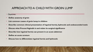
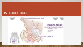
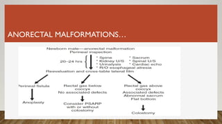
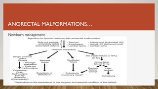
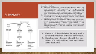
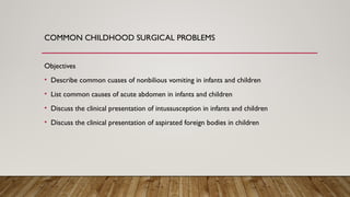
Dr. Addisu Andargie
Assnt .Prof. of pediatric surgery ,FCS (COSECSA )
YEKATIT 12 HOSPITAL MEDICAL COLLEGE
DEPARTMENT OF SURGERY
….
Yekatit 12 Hospital Medical College
Department of Surgery
Dr. Addisu Andargie
Assn’t .Prof. of Pediatric Surgery ,FCS (COSECSA )
APPROACH TO A CHILDWITH GROIN LUMP
• Objectives
• Define anatomy of groin
• List common cuases of groin lump in children
• Describe common clinical presentation of inguinal hernia, hydrocele and undesecended testis
• Discuss what ProcessVaginalis is and relate its surgical significance
• Describe how inguinal hernia can present in an acute abdomen
• Define an acute scrotum
• Discuss how to differentiate inguinal hernia and hydrocele
INTRODUCTION
ANATOMY OF THE GROIN
ANATOMY…
ANATOMY…
SURGICAL IMPORTANCE OF THE GROIN
• Passage zone between the abdominal area, the external
genital region, and the root of lower extremity.
• Region where most hernias of the abdominal wall occur(75%)
• During descent of the testicles, a peritoneal outpouching called the
processus vaginalis follows the testicles to the scrotum
• Failure of closure of the pocessus vaginalis leads to the propensity to
develop a number of abnormalities
COMMON CAUSES OF INGUINAL SWELLING
HISTORY
The following history suggest inguinal hernia
• Sudden apperance of groin swelling whole crying in an infant or while coughing
in older children
• The variability in size of the swelling
• The periodic disappearance of the swelling
• Apparent lack of pain exceipt when it stucks in the sack
• Mathors can point exact area of swelling
PHYSICAL EXAMINATION
Lump present
• See the the position of the lump in relation to the external inguinal ring.
• Palpate the neck of the scrotum b/n index finger and the thump
#‘cannot get above it’- inguinal hernia
# ability to reduce manually-inguinal hernia
Lump absent
• Silk sign
• Ask the parents to mark with pen or take a photo when it reappears
PHYSICAL EXAMINATION. . .
• Cough impulse on standing position for older child
EMBRYOLOGY
• An outpouching of peritonium through the internal ring called the
process vaginalis
• It elongates when the intra-abdominal testis passes through it during
gestation
• Portion of it above the testis obliterates closing the internal ring
• Failure of this to happen results in inguinal hernia (* bowel and other
organs) or hydrocele( peritoneal fluid only)
RISK FACTORS FOR INGUINAL HERNIA
EMBRYOLOGY
CLINICAL PRESENTATION
• Hernias are usually asymptomatic
• Twice more common on the right side and 10%bilateral
• Intermittent bulge in the groin, labia or scrotum often apparent during
crying or straining
• May be present with a tender /painfull firm nonreducable mass in the
groin
• Abdominal distention, bowel obstruction, peritonitis and bloody stool
are late signs
MANAGMENT
 An inguinal hernia will not resolve spontaneously, so surgical closure is always
indicated
 Because of the high risk of incarceration in young infants repair should be done
bimmediately.
 Incarcerated hernia can be reduced under sedation
 An urgent operation is indicated in suspected strangulation or failed reduction
 The principle of pediatric inguinal hernia repair is high ligation of the hernia sac
HYDROCELE
Communicating hydrocele
• Flactuating size (increase during day/uppright postion, decrease during the
night/supine positio
Non communicating hydrocele
• stable size / may be present at birth or develop in older children
Abdomino-scrotal hydrocele
• The hydrocele continues to elarge and extends to abdomen. Can confuse with
inguinal hernia
Encysted hydrocele of the cord-a roundish tense /painless mass in the upper
scrotum or inguinal canal
HYDROCELE..
• The natural history is gradual and complete resolution by 1 year of age
• Indication for surgery include;
- Congenital hydroceles that fails to resolve by age 2 years.
– Non-communicating hydroceles that manifest after 1 year of age.
– Continued discomfort and enlargement.
– Secondary infection (very rare)
THE UNDESCENDEDTESTIS
 Normal testicular descent relies on a complex interplay of numerous factors
 Coordinated interaction among endocrine, paracrine,growth,and mechanical factors
 Happens in two phases
- Transabdominal phase 8-15 wks
- Inguinoscrotal phase 28-35 wks
THE UNDESCENDEDTESTIS…
THE UNDESCENDEDTESTIS
INCIDENCE
 3% of term male infants and in up to 33–45% of premature and/or LBW male infants
 The majority will descend in the first 6-12 monthes, incidence after is 1%
 Testicular descent after 1 year is unlikely
 About 75–80 % of UDTs are unilateral , 10% bilateral
 In 80–90 % of cases, an undescended testis can be felt in the inguinal canal
THE UDT…
Risk factors
• Family history
• Prematurity /Low birth weight
• Abdominal wall defects (Gastroschisis, Omphalocele, extrophy
syndromes
• Neural tube defects
• Chromosomal abnormalities ( down syndrome, prune belly syndrome,
prader-willi syndrome)
UDT CLASSIFICATION AND DIAGNOSIS
THE UDT…
• The diagnosis of undescended testes is clinical.
THE UDT…
Where the testis is truly undescended, the first step is to locate its
position.
The flat of the hand is placed gently over the inguinal region in the
area of the superficial inguinal pouch
By gently rolling the fingertips round and round, the testis usually
can be felt as a mobile, spherical structure between the
subcutaneous fat and the abdominal wall muscles.
Using the fingers of one hand to press firmly against the abdominal
wall and attempt to ‘milk’ the testis towards the scrotum like a
plough
TREATMENT OF UDT
• The treatment of undescended testes is surgical orchidopexy to be done by 9
to 18 months of age.
• This recommendation is based on several factors:
• Spontaneous testicular descent is unlikely after this age
• Histological abnormalities are subsequently more likely
• Orchidopexy also allows early detection of any testicular cancer, reduced risk of
trauma and torsion, and improvement in germ cell function and ultimate
prognosis of fertility
• Treatment of associated hernia when present
THE ACUTE PAINFUL SCROTUM
• Acute scrotum-sudden onset of scrotal pain with or without swelling
and erythema.
• Early recognition and management is imperative as torsion can result it
testicular loss
THE ACUTE SCROTUM….
Testicular Torsion
• An acute vascular event in which spermatic cord twists on its axis
• Is a surgical emergency-outcome depends on duration of ischemia
• The underlying cause is “the bell-clapper deformity” in which the testis hangs freely
with in parietal tunica vaginalis due to extention of tunica high in spermatic cord
• Most frequent among adolscents aged 12-18yrs and neonates
THE ACUTE SCROTUM
Classification
• Intravaginal or extravaginal , acute vs intermittent
• The degree of torsion
varies from 180°
to 720°.
THE ACUTE SCROTUM..
Clinical presentation
• Can occur spontanously, during physical activity , during sleep and can also follow
trauma
• Classic is sudden severe scrotal pain associated with swelling and erythema
• The pain is often associated with nausea and vomiting
THE ACUTE SCROTUM…
Physical Examination
• – A swollen, tender, high-riding testis.
• – Abnormal transverse lie of testis.
• – Loss of the cremasteric refex.
• – Edema involving the entire scrotum.
• – Enlargement and edema of the testis
• --Scrotal erythema
THE ACUTE SCROTUM…
THE ACUTE SCROTUM…
Management
• The goal of emergent surgical exploration are
• Confirmation of the diagnosis of torsion.
• Detorsion of the involved testis
• Assessment of vaibility of involved testis
• Removal of frankly ischmic testis Or fixation of viable testis
• Fixation of the contralateral testis
THE ACUTE SCROTUM..
Epididimo-Orchitis
• Most commonly follows from reflux of infected urine
• – There is marked redness and swelling of the affected testis
• – The patient is often febrile
• – The cremasteric refux is usually present
• --The scrotal pain and swelling typically have a slow onset, worsening over days
THE ACUTE SCROTUM…
Torsion of testicular appendages
• The most common cause of acute scrotum
• Ocurrs most commonly b/n ages of 7 to 10 yrs
• Sudden onset of pain /excusite focal tenderness
• The “blue dot sign”—ischemic appendage may be seen through scrotal skin
• Is self limited and often managed with NSAIDs
REFERRENCES
Yekatit 12 Hospital Medical
College
Department of Surgery
Dr.Addisu Andargie
(Pediatric Surgeon )
COMMON CAUSES OF NEONATAL INTESTINAL
OBSTRUCTION
Objectives
• List most common cause of intestinal obstruction in newborns
• Discuss the clinical manifestetion of Hirschsprung’s diseases
• Discuss the types of Anorectal malformations in males and females
• Discuss the types of intestinal atresias , clinical presentation and classification
INTRODUCTION…
INTRODUCTION
 Neonatal bowel obstruction occurs when the normal passage of meconium or milk from
mouth to anus is interrupted by physical forces or by bowel dysfunction
 Its presentation may be acute or chronic, affect proximal or distal gut
 Obstruction may also be partial, intermittent or complete
 Neonatal bilious vomiting should be considered to be a surgical emergency until proved
otherwise
INTRODUCTION….
Common causes of neonatal intestinal obstruction
• Doudnum --- doudnal atresia,doudnal stenosis,annular pancreas, malrotation
• Jejenum ----Atresia/stenosis/malrotation
• Ileum ---Atresia/stenosis/Muconium ileus/duplication cyst/Hirschsprung’s
diseases/malrotation
• Colon ---Atresia/stenosis, Hirschsprung‘s diseases, mucomium ileus
• Rectum and anus----Anorectal malformations, HSCD , muconium plug
INTRODUCTION…
Clinical presentation
• Bilious vomiting
• Abdominal distention
• Feeding intolerance
• Failure to pass or delayed passage muconium
• Absent or abnormal position of anal opening
• Sepsis
NEONATAL INTESTINAL OBSTRUCTION
History
• Length of pregnancy
• Maternal comorbities
• Result of antenatal ultraslund (polyhydroaminos, double bubble sign)
• Passage of muconium and its timing
• If the baby passed anything rectaly? Color and consistent
NEONATAL INTESTINAL OBSTRUCTION..
Physical examination
• Presence of normal anus
• Extent of abdominal distention
--no distention , upper abdominal or generalized distention
• Visible and palpable bowel loops
• Erythema and abdominal wall tenderness
• Extent of dehydration and associated anomalies
• Associted anomalies (* down syndrome)
Duodenal Atresia and Stenosis
 Frequent cause of intestinal obstruction and occurs in 1 per 5000 to
10,000 live births
 Associated anomalies have been reported in 45–65% of cases
 Trisomy 21 is found in half the cases, cardiac malformations in 25–65%
of cases, and malrotation in30% of cases
 The most common cause of duodenal obstruction is atresia.
EMBRYOLOGY
 The epithelial lining of deodnum undergoes a rapid proliferative phase
that occludes the lumen of the intestinal tube on day 42 of development
 Over the course of the ensuing 2 days, the plug of epithelial tissue
develops cracks that give way, reestablishing the continuity
of the intestinal lumen
Classification
Diagnosis
 Antenatal ultrasound; polyhydroaminos and double bubble sign
 Bilious emesis within the first hours of life in a neonate
 In neonates with duodenal atresia, the abdomen is scaphoid.
 Abdominal radiograph is usually sufficient to confirm the diagnosis
DIADNOSIS AND MANAGEMENT
 In almost all cases of
duodenal atresia, the distal
bowel is gasless
 With duodenal stenosis,a
double bubble sign is often not
present and the diagnosis is
usually made with a contrast
study
MANAGEMENT
 NGT decompression
 Resuscitation with correction of fluid balance and electrolyte
Abnormalities
 Laparoscopic or open duodenoduodenostomy
NEONATAL INTESTINAL OBSTRUCTION…
Jejunoileal Atresias/Stenosis
 Jejunoileal atresia occurs in approximately 1 in 5000 live Births
 It occurs equally in males and females, and about one in three infants is premature
 Jejunoileal atresia occurs as a result of an intrauterine ischemic insult to the midgut
 Intrauterine vascular disruption can lead to ischemic necrosis of the bowel with subsequent
resorption of affected bowel
NEONATAL BOWEL OBSTRUCTION…
Possible inuetero vascular accidents include ;
 intussusception,
 midgut volvulus,
 Thromboembolic occlusions,
 transmesenteric internal hernias,
 incarceration or snaring of bowel in an omphalocele or gastroschisis
NEONATAL INTESTINAL OBSTRUCTION…
Diagnosis
 Bilious emesis
 Abdominal distention
 Failure to pass muconium
 triple bubble sign
 multiple dilated bowel loops
• suggesting a distal obstruction
NEONATAL INTESTINAL OBSTRUCTIO…
Management
• Fluid and electrolyte correction
• NGT decompression
Disorders of Intestinal Rotation and Fixation
 Transformation from a simple, straight alimentary tube into the
mature fixed and folded configuration normally present at birth
 The most accepted model of midgut maturation involves four distinct
stages:
(1) herniation,
(2) rotation,
(3) retraction, and (4) fixation
Embryology
(a).The pre-arterial (duodeno-jejunal) segment initially
growsmore quickly than the post-arterial (ileo-colic)
segment
(b). The pre-arterial segment first rotates 90°
counter-clockwise which places it to the right of the SMA
(c) andthen a second 90° (d) and in doing so the
duodeno-jejunal junction comes to lie posterior to the
SMA.
Rotation ofthe post-arterial segment is in the same direction
as that of the pre-arterial segment (e), initiallythrough 90°
(a), followed by a second 90° b)
Abnormal Embryology

Abnormal Embryology
NEONATAL INTESTINAL OBSTRUCTION
Presentation
 Acute MidgutVolvulus
 CHRONIC MIDGUTVOLVULUS
 Acute duodenal obstruction secondary to congenital bands
 Chronic duodenal obstruction secondary to congenital bands
 Reverse rotation with colonic obstruction
NEONATAL INTESTINAL OBSTRUCTION…
Acute MidgutVolvulus
 Up to 75% of patients present during the first month of life.
 An infant who appeared well at birth who then has sudden onset of bile stained vomiting
 Soon after the onset of vomiting, the lower abdomen may appear scaphoid
 Infants with complete obstruction rapidly develop intestinal ischemia with a firm, distended
abdomen; hypovolemia and shock follows
NEONATAL INTESTINAL OBSTRUCTION…
Diagnosis
 As vascular compromise progresses, intra luminal bleeding may occur and blood is
often passed per rectum
 Laparotomy
 Upper Gastrointestinal Contrast Study
 Ultrasonography
 Incidental finding
NEONATAL INTESTINAL OBSTRUCTION…
Management
 This diagnosis represents one of the few critical emergencies in paediatric surgery.
 Oral feeds are stopped and a nasogastric tube inserted.
 Resuscitation
 broad spectrum antibiotics
 an exploratory laparotomy or laparoscopy
HIRSCHSPRUNG’S DISEASES
Definition
 Absence of ganglion cells in the myenteric and submucosal plexuses of the distal intestine
 Ganglion cells are responsible for normal peristalsis, absence means functional intestinal
obstruction at the level of aganglionosis
 HD occurs in approximately 1 in 5000 live births
 With a male-to-female ratio of 4:1, HD is far more common in boys
HIRSCHSPRUNG’S DISEASES
Clinical presentation
Neonatal Obstruction
 50% to 90% of children with HSD present during the neonatal period
 Delayed passage of meconium beyond the first 24 hours
 Abdominal distension,
 Bilious vomiting, and feeding intolerance
 Peritonitis with cecal or appendiceal perforation
HIRSCHSPRUNG’S DISEASES
Approximately 10% of children with
Hirschsprung disease
present with enterocolitis
HIRSCHSPRUNG’S DISEASES
Diagnosis
RADIOLOGIC EVALUATION---Barium enema
 ANORECTAL MANOMETRY
 RECTAL BIOPSY
HIRSCHSPRUNG’S DISEASES
 The first priority is resuscitation
 Broad-spectrum antibiotics
 Nasogastric tube should be inserted
 Decompression of the colon using digital rectal stimulation,irrigations, or occasionally an
emergency stoma
 Pull through procedure
ANORECTAL MALFORMATIONS
 Anorectal malformations (ARM) present across a range of defects, from isolated malformations
with to complex malformations with associated defects
 A newborn with ARM has no anal opening or has a visible fistula
 Most babies (50% to 60%) with ARMs have one or more abnormalities that affect other systems
 ARMs occurs in one out of every 4000 to 5000 newborns and is more common in males
ANORECTAL MALFORMATIONS…
Embryilogy
• Cloaca is first formed at around 21 days of gestation
• The septum in the middle grows downward until it joins the
cloacal membrane, 6th
wk
• Cloacal membrane ruptures at 7th
wk creating urogenital and
anal openings
• By 9th
wk, all relevant structures are in place
ANORECTAL MALFORMATIONS…
ANORECTAL MALFORMATIONS. ..
Associated Anomalies
 Cardiovascular anomalies (30%)
 Gastrointestinal anomaliesTEF IN I0%
 Spinal, sacral, and vertebral anomalies
 Genitourinary anomalies
 Gynecologic anomalies

Physical Examination
ANORECTAL MALFORMATIONS…
Newborn management
SUMMARY
REFERRENCES
አመሠግናለሁ !
Yekatit 12 Hospital Medical College
Department of Surgery
Unit of Pediatric Surgery
Dr.Addisu Andargie (Pediatric Surgeon)
COMMON CHILDHOOD SURGICAL PROBLEMS
Objectives
• Describe common cuases of nonbilious vomiting in infants and children
• List common causes of acute abdomen in infants and children
• Discuss the clinical presentation of intussusception in infants and children
• Discuss the clinical presentation of aspirated foreign bodies in children
NON-BILE-STAINEDVOMITING
IN INFANCY
• Commonly cuased by feeding problem
• Differentiating trival cuases , excess feeding, from infectious and mechanical causes is a
mejor challege.
• Knowledge of the patient’s age and a careful history usually will provide clues as to the
correct diagnos
• The history should be aimed at answering the following questions
1. Is the infant well and active, and keen to take feeds?
2. What is the duration, nature and timing of the vomiting?
3. Is the vomiting forceful?
CUASES OF NON-BILE-STAINEDVOMITING IN INFANCY
Hidden infections
-Meningitis
-Gastroenteritis
-UTI
Systemic illness-vague systemic
symptoms and signs in unwell baby.
Gastro- esophageal reflux Mechanical problem- well baby with or without
failure to thrive
Pyloric stenosis Mechanical problem- healthy baby with or
without failure to thrive
Feeding problem
excess feeding, inexprienced mother
Gready healthy baby
Associted with colicy pain Intermittent pain with nonbilious vomiting
develops to bilious vomiting
HYPERTHROPHIC PYLORIC STENOSIS
It occurs at a rate of 1–4 per 1000 live births
 4:1 male-to-female ratio
 Risk factors include family history, gender, younger maternal age, being a first-born
infant, medications
 The cause of HPS is unknown
 Genetic and environmental factors appear to play a role
HYPERTHROPHIC PYLORIC STENOSIS…
• DIAGNOSIS
 Nonbilious, projectile vomiting in a term neonate b/n 2 and 8 wks.
 The emesis occurs with every feeding and becomes forceful
 The pylorus is palpable in 70–90% of patients
 Ultrasound has become the standard technique for diagnosing HPS
 A muscle thickness of ≥4 mm and a pyloric length of ≥16 mm
HPS…
HPS…
Management
 IS not a surgical emergency ,resuscitation is of the utmost Priority
 Initially, a 10- to 20-mL/kg bolus of NS should be given
 Then D5/½NS with 20–30 mEq/L of KCl is started
 Electrolytes should be checked every 6 hrs until they normalize
 Ramsted Pyloromyotomy is the standard procedure
ABDOMINAL PAIN IN INFANTS AND CHILDREN
• Abdominal pain is extremely common in infants and children and may reflect a variety of
condition .
• Often , the cuase cannot be determined with certainity., may be unreleated pathology to
the abdomen.
• When a child presents with persistent, lasting longer than 4 to 6 hrs, or worsening
abdominal pain with associted vomiting ,every effort must be made to obtain a clinical
diagnosis
COMMON CUASES OF COLIC IN INFANTS
• Infantile colic
• Gastroenteritis
• Constipation
• Intussusception
• Appendicitis
• Mesentric Lymphadenitis
• Bowel obstruction ; congenital or adhesions
• Henock-schonlein Purpura
COMMON CUASES OF ABDOMINAL PAIN IN
INFANTS AND CHILDREN
ABDOMINAL PAIN IN INFANTS AND CHILDREN
ABDOMINAL PAIN IN INFANTS AND CHILDREN…
History
• Obstruction of a hollow viscus causes sharp spasms of ‘colic’ between which the patient
suffers a dull ache.
• The child with colic is restless and unable to obtain a position of comfort.
• Children with peritonitis will remain still and dislike being disturbed or handled. It will
hurt to cough, sneeze, vomit or to take a deep breath.
• Vomiting commonly accompanies abdominal pain in children .
Physical Examination
• Adequate exposure , chest to upper thigh , is important
• Is peritonitis present? Simple manuevers include
- ---- Ask to puff out or draw in there abdomen
- -----shack the chest or pelvis side to side
- ----Percussion tenderness
- ----excistance of involuntary gaurding
• Digital rectal examination
INTUSSUSCEPTION
 is the most frequent cause of bowel obstruction in infants and toddlers.
 It is an acquired invagination of the proximal bowel (intussusceptum) into the distal
bowel (intussuscipiens)
 There may or may not be a lead point
 The highest incidence occurs in infants b/n ages 4 and 9 months
 Intussusception is uncommon below 3 months and after 3 years of age
ITUSSU…….
The mesentery of the proximal bowel is drawn into the
distal bowel, it is compressed, resulting in venous obstruction
and bowel wall edema.
 If reduction does not occur, arterial insufficiency will
ultimately lead to ischemia and bowel wall necrosis
Primary intussusception
 A majority of cases, especially in infants, do not have a lead point
 The cause is attributed to hypertrophied Peyer patches within the bowel wall
 Intussusception occurs in the wake of an upper respiratory tract
• infection or an episode of gastroenteritis
 Adenovirus and rotavirus has been implicated
Secondary Intussusception
have an identifiable lesion that serves as a lead point
 1.5–12% of cases, and the presence of a lead point increases in proportion with age
 Meckel diverticulum, polyps and duplications.
 Other benign lead points include hemangiomas, carcinoid tumors, foreign bodies,
lymphomas and small bowel tumors
INTUSSUSCEPTION…
Anatomic Classification
 ileocolic - 85% 0f cases
 ileoileocolic – 10% 0f cases
 Appendicocolic, cecocolic, and colocolic intussusceptions are much less common (2.5%)
and are usually associated with PLPs.
 Jejunojejunal and ileoileal intussusceptions occur infrequentl (2.5%) and usually have a PLP
Clinical Presentation
 Abdomianl pain or vomiting
 The sudden onset of severe, colicky, intermittent abdominal pain
 This pain episode typically lasts only a few minutes
 Between attacks, the child may appear comfortable
 Abdominal mass or rectal bleeding
 The often curved abdominal mass can be palpated in the RUQ of the abdomen about 65% cases.
 Rectal bleeding is usually the last sign to occur.
INTUSSUSCEPTION…
Diagnosis
Laboratory – complete blood count and electrolytes
 imaging
 abdominal us –sensitivity is near 100% with experienced radiologist
INTUSSUSCEPTION…
• Nonoperative Management
 HYDROSTATIC AND PNEUMATIC REDUCTION
ileocolic, primary, viable bowel
 Operative Management
 failed nonoperative reduction
 presence of lead point
 evidence of peritonitis
Reading assignments
• Pediatric appendicitis
• Mesentric adenitis
• Infantile colic
FOREIGN BODY ASPIRATION
Airway Foreign Bodies
• Age 1-3 years
o Toddlers are inquisitive & place objects in their mouths
o Don’t distinguish edible from inedible objects
o Relatively immature swallowing mechanism
o Lack dentition for grinding food
o Supervision is challenging
FB ASPIRATION…
FB ASPIRATION…
Clinical Presentation
• Most important factor in evaluation is accurate history
• Choking crisis (95%)…most sensitive clinical parameter
• Clinical findings (50-90% of pts)
o Persistent cough, respiratory compromise, hemoptysis, fever
• A history of choking without physical findings of reduced air exchange requires a high
degree of suspicion
FB ASPIRATION
• FBs in the larynx and trachea can be completely obstructive causing sudden
death
• 20-50% of FBs are diagnosed more than a week after aspiration
• Features that distinguish FBA from other causes
o Witnessed aspiration
o Sudden onset
o Absence of antecedent fever
FB ASPIRATION. ..
• History & physical exam
• Work up
o CBC
o Plain radiography
o CT scan
o MRI
o Bronchoscopy
FB ASPIRATION …
FB ASPIRATION…
Complete obstruction
• Age < 1 yr
I. back blows to a child placed in the prone position
II. Turn child into a supine position and do chest thrusts
• CPR
• Age > 1 yr
I. Heimlich maneuver
II. CPR
FB ASPIRATION…
• Rigid endoscopy
 Preoxygenation with
100% facemask oxygen
 General anesthesia
HYPOSPADIAS
 Urethral meatus that opens on the ventral surface of the penis proximal to the end of the glans.
 The meatus may be located anywhere along the length of the penis from the glans to a proximal
location as low as the perineum
 Ventral curvature of the penis, chordee, has an inconsistent association with hypospadias
HYPOSPADIAS.
 The incidence of hypospadias has been estimated 1 in 250 to 300 boys
 Genetic factors, inadequate hormonal stimulation,maternal/placental factors, and
environmental factors have all been implicated.
 There is a definite familial occurrence of hypospadias.This is estimated to be about 7 %.
HYPOSPADIAS.. .
HYPOSPADIAS…
HYPOSPADIAS….
Management
 Objectives of hypospadia surgery
 Complete straightening of the penis
 Locating the meatus at the tip of the glans
 Forming a symmetric, conically shaped glans
 Constructing a neourethra uniform in caliber
 Completing a satisfactory cosmetic skin coverage
 timing of repair is usually 9-18 months
REFERRENCES
I stand with Russia!!
Stop war in Ukraine!!!

Groin Lump In Children which needs Surgery

  • 1.
    Dr. Addisu Andargie Assnt.Prof. of pediatric surgery ,FCS (COSECSA ) YEKATIT 12 HOSPITAL MEDICAL COLLEGE DEPARTMENT OF SURGERY
  • 2.
    …. Yekatit 12 HospitalMedical College Department of Surgery Dr. Addisu Andargie Assn’t .Prof. of Pediatric Surgery ,FCS (COSECSA )
  • 3.
    APPROACH TO ACHILDWITH GROIN LUMP • Objectives • Define anatomy of groin • List common cuases of groin lump in children • Describe common clinical presentation of inguinal hernia, hydrocele and undesecended testis • Discuss what ProcessVaginalis is and relate its surgical significance • Describe how inguinal hernia can present in an acute abdomen • Define an acute scrotum • Discuss how to differentiate inguinal hernia and hydrocele
  • 4.
  • 5.
  • 6.
  • 7.
  • 8.
    SURGICAL IMPORTANCE OFTHE GROIN • Passage zone between the abdominal area, the external genital region, and the root of lower extremity. • Region where most hernias of the abdominal wall occur(75%) • During descent of the testicles, a peritoneal outpouching called the processus vaginalis follows the testicles to the scrotum • Failure of closure of the pocessus vaginalis leads to the propensity to develop a number of abnormalities
  • 9.
    COMMON CAUSES OFINGUINAL SWELLING
  • 10.
    HISTORY The following historysuggest inguinal hernia • Sudden apperance of groin swelling whole crying in an infant or while coughing in older children • The variability in size of the swelling • The periodic disappearance of the swelling • Apparent lack of pain exceipt when it stucks in the sack • Mathors can point exact area of swelling
  • 11.
    PHYSICAL EXAMINATION Lump present •See the the position of the lump in relation to the external inguinal ring. • Palpate the neck of the scrotum b/n index finger and the thump #‘cannot get above it’- inguinal hernia # ability to reduce manually-inguinal hernia Lump absent • Silk sign • Ask the parents to mark with pen or take a photo when it reappears
  • 12.
    PHYSICAL EXAMINATION. .. • Cough impulse on standing position for older child
  • 14.
    EMBRYOLOGY • An outpouchingof peritonium through the internal ring called the process vaginalis • It elongates when the intra-abdominal testis passes through it during gestation • Portion of it above the testis obliterates closing the internal ring • Failure of this to happen results in inguinal hernia (* bowel and other organs) or hydrocele( peritoneal fluid only)
  • 15.
    RISK FACTORS FORINGUINAL HERNIA
  • 16.
  • 17.
    CLINICAL PRESENTATION • Herniasare usually asymptomatic • Twice more common on the right side and 10%bilateral • Intermittent bulge in the groin, labia or scrotum often apparent during crying or straining • May be present with a tender /painfull firm nonreducable mass in the groin • Abdominal distention, bowel obstruction, peritonitis and bloody stool are late signs
  • 18.
    MANAGMENT  An inguinalhernia will not resolve spontaneously, so surgical closure is always indicated  Because of the high risk of incarceration in young infants repair should be done bimmediately.  Incarcerated hernia can be reduced under sedation  An urgent operation is indicated in suspected strangulation or failed reduction  The principle of pediatric inguinal hernia repair is high ligation of the hernia sac
  • 19.
    HYDROCELE Communicating hydrocele • Flactuatingsize (increase during day/uppright postion, decrease during the night/supine positio Non communicating hydrocele • stable size / may be present at birth or develop in older children Abdomino-scrotal hydrocele • The hydrocele continues to elarge and extends to abdomen. Can confuse with inguinal hernia Encysted hydrocele of the cord-a roundish tense /painless mass in the upper scrotum or inguinal canal
  • 20.
    HYDROCELE.. • The naturalhistory is gradual and complete resolution by 1 year of age • Indication for surgery include; - Congenital hydroceles that fails to resolve by age 2 years. – Non-communicating hydroceles that manifest after 1 year of age. – Continued discomfort and enlargement. – Secondary infection (very rare)
  • 21.
    THE UNDESCENDEDTESTIS  Normaltesticular descent relies on a complex interplay of numerous factors  Coordinated interaction among endocrine, paracrine,growth,and mechanical factors  Happens in two phases - Transabdominal phase 8-15 wks - Inguinoscrotal phase 28-35 wks
  • 22.
  • 23.
    THE UNDESCENDEDTESTIS INCIDENCE  3%of term male infants and in up to 33–45% of premature and/or LBW male infants  The majority will descend in the first 6-12 monthes, incidence after is 1%  Testicular descent after 1 year is unlikely  About 75–80 % of UDTs are unilateral , 10% bilateral  In 80–90 % of cases, an undescended testis can be felt in the inguinal canal
  • 24.
    THE UDT… Risk factors •Family history • Prematurity /Low birth weight • Abdominal wall defects (Gastroschisis, Omphalocele, extrophy syndromes • Neural tube defects • Chromosomal abnormalities ( down syndrome, prune belly syndrome, prader-willi syndrome)
  • 25.
  • 26.
    THE UDT… • Thediagnosis of undescended testes is clinical.
  • 27.
    THE UDT… Where thetestis is truly undescended, the first step is to locate its position. The flat of the hand is placed gently over the inguinal region in the area of the superficial inguinal pouch By gently rolling the fingertips round and round, the testis usually can be felt as a mobile, spherical structure between the subcutaneous fat and the abdominal wall muscles. Using the fingers of one hand to press firmly against the abdominal wall and attempt to ‘milk’ the testis towards the scrotum like a plough
  • 28.
    TREATMENT OF UDT •The treatment of undescended testes is surgical orchidopexy to be done by 9 to 18 months of age. • This recommendation is based on several factors: • Spontaneous testicular descent is unlikely after this age • Histological abnormalities are subsequently more likely • Orchidopexy also allows early detection of any testicular cancer, reduced risk of trauma and torsion, and improvement in germ cell function and ultimate prognosis of fertility • Treatment of associated hernia when present
  • 29.
    THE ACUTE PAINFULSCROTUM • Acute scrotum-sudden onset of scrotal pain with or without swelling and erythema. • Early recognition and management is imperative as torsion can result it testicular loss
  • 30.
    THE ACUTE SCROTUM…. TesticularTorsion • An acute vascular event in which spermatic cord twists on its axis • Is a surgical emergency-outcome depends on duration of ischemia • The underlying cause is “the bell-clapper deformity” in which the testis hangs freely with in parietal tunica vaginalis due to extention of tunica high in spermatic cord • Most frequent among adolscents aged 12-18yrs and neonates
  • 31.
    THE ACUTE SCROTUM Classification •Intravaginal or extravaginal , acute vs intermittent • The degree of torsion varies from 180° to 720°.
  • 32.
    THE ACUTE SCROTUM.. Clinicalpresentation • Can occur spontanously, during physical activity , during sleep and can also follow trauma • Classic is sudden severe scrotal pain associated with swelling and erythema • The pain is often associated with nausea and vomiting
  • 33.
    THE ACUTE SCROTUM… PhysicalExamination • – A swollen, tender, high-riding testis. • – Abnormal transverse lie of testis. • – Loss of the cremasteric refex. • – Edema involving the entire scrotum. • – Enlargement and edema of the testis • --Scrotal erythema
  • 34.
  • 35.
    THE ACUTE SCROTUM… Management •The goal of emergent surgical exploration are • Confirmation of the diagnosis of torsion. • Detorsion of the involved testis • Assessment of vaibility of involved testis • Removal of frankly ischmic testis Or fixation of viable testis • Fixation of the contralateral testis
  • 36.
    THE ACUTE SCROTUM.. Epididimo-Orchitis •Most commonly follows from reflux of infected urine • – There is marked redness and swelling of the affected testis • – The patient is often febrile • – The cremasteric refux is usually present • --The scrotal pain and swelling typically have a slow onset, worsening over days
  • 37.
    THE ACUTE SCROTUM… Torsionof testicular appendages • The most common cause of acute scrotum • Ocurrs most commonly b/n ages of 7 to 10 yrs • Sudden onset of pain /excusite focal tenderness • The “blue dot sign”—ischemic appendage may be seen through scrotal skin • Is self limited and often managed with NSAIDs
  • 38.
  • 39.
    Yekatit 12 HospitalMedical College Department of Surgery Dr.Addisu Andargie (Pediatric Surgeon )
  • 40.
    COMMON CAUSES OFNEONATAL INTESTINAL OBSTRUCTION Objectives • List most common cause of intestinal obstruction in newborns • Discuss the clinical manifestetion of Hirschsprung’s diseases • Discuss the types of Anorectal malformations in males and females • Discuss the types of intestinal atresias , clinical presentation and classification
  • 41.
  • 42.
    INTRODUCTION  Neonatal bowelobstruction occurs when the normal passage of meconium or milk from mouth to anus is interrupted by physical forces or by bowel dysfunction  Its presentation may be acute or chronic, affect proximal or distal gut  Obstruction may also be partial, intermittent or complete  Neonatal bilious vomiting should be considered to be a surgical emergency until proved otherwise
  • 43.
    INTRODUCTION…. Common causes ofneonatal intestinal obstruction • Doudnum --- doudnal atresia,doudnal stenosis,annular pancreas, malrotation • Jejenum ----Atresia/stenosis/malrotation • Ileum ---Atresia/stenosis/Muconium ileus/duplication cyst/Hirschsprung’s diseases/malrotation • Colon ---Atresia/stenosis, Hirschsprung‘s diseases, mucomium ileus • Rectum and anus----Anorectal malformations, HSCD , muconium plug
  • 44.
    INTRODUCTION… Clinical presentation • Biliousvomiting • Abdominal distention • Feeding intolerance • Failure to pass or delayed passage muconium • Absent or abnormal position of anal opening • Sepsis
  • 45.
    NEONATAL INTESTINAL OBSTRUCTION History •Length of pregnancy • Maternal comorbities • Result of antenatal ultraslund (polyhydroaminos, double bubble sign) • Passage of muconium and its timing • If the baby passed anything rectaly? Color and consistent
  • 46.
    NEONATAL INTESTINAL OBSTRUCTION.. Physicalexamination • Presence of normal anus • Extent of abdominal distention --no distention , upper abdominal or generalized distention • Visible and palpable bowel loops • Erythema and abdominal wall tenderness • Extent of dehydration and associated anomalies • Associted anomalies (* down syndrome)
  • 47.
    Duodenal Atresia andStenosis  Frequent cause of intestinal obstruction and occurs in 1 per 5000 to 10,000 live births  Associated anomalies have been reported in 45–65% of cases  Trisomy 21 is found in half the cases, cardiac malformations in 25–65% of cases, and malrotation in30% of cases  The most common cause of duodenal obstruction is atresia.
  • 48.
    EMBRYOLOGY  The epitheliallining of deodnum undergoes a rapid proliferative phase that occludes the lumen of the intestinal tube on day 42 of development  Over the course of the ensuing 2 days, the plug of epithelial tissue develops cracks that give way, reestablishing the continuity of the intestinal lumen
  • 49.
  • 50.
    Diagnosis  Antenatal ultrasound;polyhydroaminos and double bubble sign  Bilious emesis within the first hours of life in a neonate  In neonates with duodenal atresia, the abdomen is scaphoid.  Abdominal radiograph is usually sufficient to confirm the diagnosis
  • 51.
    DIADNOSIS AND MANAGEMENT In almost all cases of duodenal atresia, the distal bowel is gasless  With duodenal stenosis,a double bubble sign is often not present and the diagnosis is usually made with a contrast study
  • 52.
    MANAGEMENT  NGT decompression Resuscitation with correction of fluid balance and electrolyte Abnormalities  Laparoscopic or open duodenoduodenostomy
  • 53.
    NEONATAL INTESTINAL OBSTRUCTION… JejunoilealAtresias/Stenosis  Jejunoileal atresia occurs in approximately 1 in 5000 live Births  It occurs equally in males and females, and about one in three infants is premature  Jejunoileal atresia occurs as a result of an intrauterine ischemic insult to the midgut  Intrauterine vascular disruption can lead to ischemic necrosis of the bowel with subsequent resorption of affected bowel
  • 54.
    NEONATAL BOWEL OBSTRUCTION… Possibleinuetero vascular accidents include ;  intussusception,  midgut volvulus,  Thromboembolic occlusions,  transmesenteric internal hernias,  incarceration or snaring of bowel in an omphalocele or gastroschisis
  • 56.
    NEONATAL INTESTINAL OBSTRUCTION… Diagnosis Bilious emesis  Abdominal distention  Failure to pass muconium  triple bubble sign  multiple dilated bowel loops • suggesting a distal obstruction
  • 57.
    NEONATAL INTESTINAL OBSTRUCTIO… Management •Fluid and electrolyte correction • NGT decompression
  • 58.
    Disorders of IntestinalRotation and Fixation  Transformation from a simple, straight alimentary tube into the mature fixed and folded configuration normally present at birth  The most accepted model of midgut maturation involves four distinct stages: (1) herniation, (2) rotation, (3) retraction, and (4) fixation
  • 59.
    Embryology (a).The pre-arterial (duodeno-jejunal)segment initially growsmore quickly than the post-arterial (ileo-colic) segment (b). The pre-arterial segment first rotates 90° counter-clockwise which places it to the right of the SMA (c) andthen a second 90° (d) and in doing so the duodeno-jejunal junction comes to lie posterior to the SMA. Rotation ofthe post-arterial segment is in the same direction as that of the pre-arterial segment (e), initiallythrough 90° (a), followed by a second 90° b)
  • 60.
  • 61.
  • 62.
    NEONATAL INTESTINAL OBSTRUCTION Presentation Acute MidgutVolvulus  CHRONIC MIDGUTVOLVULUS  Acute duodenal obstruction secondary to congenital bands  Chronic duodenal obstruction secondary to congenital bands  Reverse rotation with colonic obstruction
  • 63.
    NEONATAL INTESTINAL OBSTRUCTION… AcuteMidgutVolvulus  Up to 75% of patients present during the first month of life.  An infant who appeared well at birth who then has sudden onset of bile stained vomiting  Soon after the onset of vomiting, the lower abdomen may appear scaphoid  Infants with complete obstruction rapidly develop intestinal ischemia with a firm, distended abdomen; hypovolemia and shock follows
  • 64.
    NEONATAL INTESTINAL OBSTRUCTION… Diagnosis As vascular compromise progresses, intra luminal bleeding may occur and blood is often passed per rectum  Laparotomy  Upper Gastrointestinal Contrast Study  Ultrasonography  Incidental finding
  • 65.
    NEONATAL INTESTINAL OBSTRUCTION… Management This diagnosis represents one of the few critical emergencies in paediatric surgery.  Oral feeds are stopped and a nasogastric tube inserted.  Resuscitation  broad spectrum antibiotics  an exploratory laparotomy or laparoscopy
  • 66.
    HIRSCHSPRUNG’S DISEASES Definition  Absenceof ganglion cells in the myenteric and submucosal plexuses of the distal intestine  Ganglion cells are responsible for normal peristalsis, absence means functional intestinal obstruction at the level of aganglionosis  HD occurs in approximately 1 in 5000 live births  With a male-to-female ratio of 4:1, HD is far more common in boys
  • 67.
    HIRSCHSPRUNG’S DISEASES Clinical presentation NeonatalObstruction  50% to 90% of children with HSD present during the neonatal period  Delayed passage of meconium beyond the first 24 hours  Abdominal distension,  Bilious vomiting, and feeding intolerance  Peritonitis with cecal or appendiceal perforation
  • 68.
  • 71.
    HIRSCHSPRUNG’S DISEASES Approximately 10%of children with Hirschsprung disease present with enterocolitis
  • 72.
    HIRSCHSPRUNG’S DISEASES Diagnosis RADIOLOGIC EVALUATION---Bariumenema  ANORECTAL MANOMETRY  RECTAL BIOPSY
  • 73.
    HIRSCHSPRUNG’S DISEASES  Thefirst priority is resuscitation  Broad-spectrum antibiotics  Nasogastric tube should be inserted  Decompression of the colon using digital rectal stimulation,irrigations, or occasionally an emergency stoma  Pull through procedure
  • 74.
    ANORECTAL MALFORMATIONS  Anorectalmalformations (ARM) present across a range of defects, from isolated malformations with to complex malformations with associated defects  A newborn with ARM has no anal opening or has a visible fistula  Most babies (50% to 60%) with ARMs have one or more abnormalities that affect other systems  ARMs occurs in one out of every 4000 to 5000 newborns and is more common in males
  • 75.
    ANORECTAL MALFORMATIONS… Embryilogy • Cloacais first formed at around 21 days of gestation • The septum in the middle grows downward until it joins the cloacal membrane, 6th wk • Cloacal membrane ruptures at 7th wk creating urogenital and anal openings • By 9th wk, all relevant structures are in place
  • 76.
  • 77.
  • 78.
    ANORECTAL MALFORMATIONS. .. AssociatedAnomalies  Cardiovascular anomalies (30%)  Gastrointestinal anomaliesTEF IN I0%  Spinal, sacral, and vertebral anomalies  Genitourinary anomalies  Gynecologic anomalies 
  • 79.
  • 80.
  • 81.
  • 82.
  • 83.
  • 84.
  • 85.
  • 86.
    Yekatit 12 HospitalMedical College Department of Surgery Unit of Pediatric Surgery Dr.Addisu Andargie (Pediatric Surgeon)
  • 87.
    COMMON CHILDHOOD SURGICALPROBLEMS Objectives • Describe common cuases of nonbilious vomiting in infants and children • List common causes of acute abdomen in infants and children • Discuss the clinical presentation of intussusception in infants and children • Discuss the clinical presentation of aspirated foreign bodies in children
  • 88.
    NON-BILE-STAINEDVOMITING IN INFANCY • Commonlycuased by feeding problem • Differentiating trival cuases , excess feeding, from infectious and mechanical causes is a mejor challege. • Knowledge of the patient’s age and a careful history usually will provide clues as to the correct diagnos • The history should be aimed at answering the following questions
  • 89.
    1. Is theinfant well and active, and keen to take feeds? 2. What is the duration, nature and timing of the vomiting? 3. Is the vomiting forceful?
  • 91.
    CUASES OF NON-BILE-STAINEDVOMITINGIN INFANCY Hidden infections -Meningitis -Gastroenteritis -UTI Systemic illness-vague systemic symptoms and signs in unwell baby. Gastro- esophageal reflux Mechanical problem- well baby with or without failure to thrive Pyloric stenosis Mechanical problem- healthy baby with or without failure to thrive Feeding problem excess feeding, inexprienced mother Gready healthy baby Associted with colicy pain Intermittent pain with nonbilious vomiting develops to bilious vomiting
  • 92.
    HYPERTHROPHIC PYLORIC STENOSIS Itoccurs at a rate of 1–4 per 1000 live births  4:1 male-to-female ratio  Risk factors include family history, gender, younger maternal age, being a first-born infant, medications  The cause of HPS is unknown  Genetic and environmental factors appear to play a role
  • 93.
    HYPERTHROPHIC PYLORIC STENOSIS… •DIAGNOSIS  Nonbilious, projectile vomiting in a term neonate b/n 2 and 8 wks.  The emesis occurs with every feeding and becomes forceful  The pylorus is palpable in 70–90% of patients  Ultrasound has become the standard technique for diagnosing HPS  A muscle thickness of ≥4 mm and a pyloric length of ≥16 mm
  • 94.
  • 95.
    HPS… Management  IS nota surgical emergency ,resuscitation is of the utmost Priority  Initially, a 10- to 20-mL/kg bolus of NS should be given  Then D5/½NS with 20–30 mEq/L of KCl is started  Electrolytes should be checked every 6 hrs until they normalize  Ramsted Pyloromyotomy is the standard procedure
  • 96.
    ABDOMINAL PAIN ININFANTS AND CHILDREN • Abdominal pain is extremely common in infants and children and may reflect a variety of condition . • Often , the cuase cannot be determined with certainity., may be unreleated pathology to the abdomen. • When a child presents with persistent, lasting longer than 4 to 6 hrs, or worsening abdominal pain with associted vomiting ,every effort must be made to obtain a clinical diagnosis
  • 97.
    COMMON CUASES OFCOLIC IN INFANTS • Infantile colic • Gastroenteritis • Constipation • Intussusception • Appendicitis • Mesentric Lymphadenitis • Bowel obstruction ; congenital or adhesions • Henock-schonlein Purpura
  • 98.
    COMMON CUASES OFABDOMINAL PAIN IN INFANTS AND CHILDREN
  • 99.
    ABDOMINAL PAIN ININFANTS AND CHILDREN
  • 100.
    ABDOMINAL PAIN ININFANTS AND CHILDREN… History • Obstruction of a hollow viscus causes sharp spasms of ‘colic’ between which the patient suffers a dull ache. • The child with colic is restless and unable to obtain a position of comfort. • Children with peritonitis will remain still and dislike being disturbed or handled. It will hurt to cough, sneeze, vomit or to take a deep breath. • Vomiting commonly accompanies abdominal pain in children .
  • 101.
    Physical Examination • Adequateexposure , chest to upper thigh , is important • Is peritonitis present? Simple manuevers include - ---- Ask to puff out or draw in there abdomen - -----shack the chest or pelvis side to side - ----Percussion tenderness - ----excistance of involuntary gaurding • Digital rectal examination
  • 102.
    INTUSSUSCEPTION  is themost frequent cause of bowel obstruction in infants and toddlers.  It is an acquired invagination of the proximal bowel (intussusceptum) into the distal bowel (intussuscipiens)  There may or may not be a lead point  The highest incidence occurs in infants b/n ages 4 and 9 months  Intussusception is uncommon below 3 months and after 3 years of age
  • 103.
  • 104.
    The mesentery ofthe proximal bowel is drawn into the distal bowel, it is compressed, resulting in venous obstruction and bowel wall edema.  If reduction does not occur, arterial insufficiency will ultimately lead to ischemia and bowel wall necrosis
  • 105.
    Primary intussusception  Amajority of cases, especially in infants, do not have a lead point  The cause is attributed to hypertrophied Peyer patches within the bowel wall  Intussusception occurs in the wake of an upper respiratory tract • infection or an episode of gastroenteritis  Adenovirus and rotavirus has been implicated
  • 106.
    Secondary Intussusception have anidentifiable lesion that serves as a lead point  1.5–12% of cases, and the presence of a lead point increases in proportion with age  Meckel diverticulum, polyps and duplications.  Other benign lead points include hemangiomas, carcinoid tumors, foreign bodies, lymphomas and small bowel tumors
  • 107.
    INTUSSUSCEPTION… Anatomic Classification  ileocolic- 85% 0f cases  ileoileocolic – 10% 0f cases  Appendicocolic, cecocolic, and colocolic intussusceptions are much less common (2.5%) and are usually associated with PLPs.  Jejunojejunal and ileoileal intussusceptions occur infrequentl (2.5%) and usually have a PLP
  • 108.
    Clinical Presentation  Abdomianlpain or vomiting  The sudden onset of severe, colicky, intermittent abdominal pain  This pain episode typically lasts only a few minutes  Between attacks, the child may appear comfortable  Abdominal mass or rectal bleeding  The often curved abdominal mass can be palpated in the RUQ of the abdomen about 65% cases.  Rectal bleeding is usually the last sign to occur.
  • 109.
    INTUSSUSCEPTION… Diagnosis Laboratory – completeblood count and electrolytes  imaging  abdominal us –sensitivity is near 100% with experienced radiologist
  • 110.
    INTUSSUSCEPTION… • Nonoperative Management HYDROSTATIC AND PNEUMATIC REDUCTION ileocolic, primary, viable bowel  Operative Management  failed nonoperative reduction  presence of lead point  evidence of peritonitis
  • 111.
    Reading assignments • Pediatricappendicitis • Mesentric adenitis • Infantile colic
  • 112.
    FOREIGN BODY ASPIRATION AirwayForeign Bodies • Age 1-3 years o Toddlers are inquisitive & place objects in their mouths o Don’t distinguish edible from inedible objects o Relatively immature swallowing mechanism o Lack dentition for grinding food o Supervision is challenging
  • 113.
  • 114.
    FB ASPIRATION… Clinical Presentation •Most important factor in evaluation is accurate history • Choking crisis (95%)…most sensitive clinical parameter • Clinical findings (50-90% of pts) o Persistent cough, respiratory compromise, hemoptysis, fever • A history of choking without physical findings of reduced air exchange requires a high degree of suspicion
  • 115.
    FB ASPIRATION • FBsin the larynx and trachea can be completely obstructive causing sudden death • 20-50% of FBs are diagnosed more than a week after aspiration • Features that distinguish FBA from other causes o Witnessed aspiration o Sudden onset o Absence of antecedent fever
  • 116.
    FB ASPIRATION. .. •History & physical exam • Work up o CBC o Plain radiography o CT scan o MRI o Bronchoscopy
  • 117.
  • 118.
    FB ASPIRATION… Complete obstruction •Age < 1 yr I. back blows to a child placed in the prone position II. Turn child into a supine position and do chest thrusts • CPR • Age > 1 yr I. Heimlich maneuver II. CPR
  • 119.
    FB ASPIRATION… • Rigidendoscopy  Preoxygenation with 100% facemask oxygen  General anesthesia
  • 120.
    HYPOSPADIAS  Urethral meatusthat opens on the ventral surface of the penis proximal to the end of the glans.  The meatus may be located anywhere along the length of the penis from the glans to a proximal location as low as the perineum  Ventral curvature of the penis, chordee, has an inconsistent association with hypospadias
  • 121.
    HYPOSPADIAS.  The incidenceof hypospadias has been estimated 1 in 250 to 300 boys  Genetic factors, inadequate hormonal stimulation,maternal/placental factors, and environmental factors have all been implicated.  There is a definite familial occurrence of hypospadias.This is estimated to be about 7 %.
  • 122.
  • 123.
  • 124.
    HYPOSPADIAS…. Management  Objectives ofhypospadia surgery  Complete straightening of the penis  Locating the meatus at the tip of the glans  Forming a symmetric, conically shaped glans  Constructing a neourethra uniform in caliber  Completing a satisfactory cosmetic skin coverage  timing of repair is usually 9-18 months
  • 125.
  • 126.
    I stand withRussia!! Stop war in Ukraine!!!