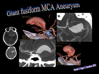
Giant Fusiform Medial Cerebral Artery Aneurysm
•Download as PPT, PDF•
1 like•523 views

49 years old woman that comes to E.R. with Midriais & Comatous State. Routine Brain CT shows Subrachnoid Hemorrhage. Willis angioCT MIP & VR depicts Giant Fusiform MCA Aneurysm.
Report
Share
Report
Share
Recommended
Tinnitus aus der Sicht des Allgemeinarztes
Fachvortrag von Dr. Andreas Müller anlässlich des Internationalen Tinnitus Symposiums für Ärzte und Therapeuten am 11.12.2010 in Regensburg, veranstaltet vom Tinnituszentrum Regensburg
Tinnitus -
eine psychotherapeutische Herausforderung am Beispiel des gestoer...
Tinnitus -
eine psychotherapeutische Herausforderung am Beispiel des gestoer...Tinnitus Research Initiative
Fachvortrag von Dr. Tatjana Crönlein anlässlich des Internationalen Tinnitus Symposiums für Ärzte und Therapeuten am 11.12.2010 in Regensburg, veranstaltet vom Tinnituszentrum RegensburgReabilitação através da Hidroterapia em paciente com AVE Isquêmico: Relato de...
O documento descreve um acidente vascular encefálico (AVE) isquêmico sofrido por um homem de 33 anos que resultou em hemiplegia do lado esquerdo do corpo. O documento também detalha os exercícios de fisioterapia realizados para fortalecimento muscular e reabilitação funcional do paciente.
Right varicocele. Ultrasound.
The medical document discusses three findings: a right varicocele, dilated veins within the right testis; rette testis dilatation, enlarged veins within the network of veins draining the testis; and an epididymal cyst, a fluid-filled sac within the epididymis, the coiled tubular structure that collects and transports sperm from the testis.
2nd meningiomas meet up
O documento discute vários casos de pacientes que sofreram recidivas de meningiomas ou desenvolveram múltiplos meningiomas. Detalha tratamentos como cirurgia e embolização para remover os tumores e controlar o crescimento de novos tumores. Também menciona um caso raro de múltipla meningiomatose.
Type 3 Frontal Cell
The document discusses two anatomical structures: Type 3 Frontal Cell and Agger nasi. Type 3 Frontal Cell refers to a specific type of frontal sinus cell located in the frontal bone above the nose. Agger nasi refers to a small bony projection located in the nasal cavity near the front of the nasal septum.
Hypoglossal Nerve Schwannoma
A patient presented with a hypoglossal nerve schwannoma located in the cisternal segment that was imaged with various MRI sequences including 3D CISS, T1 TSE, FLAIR, VIBE, post-gadolinium SPACE, and post-gadolinium to characterize the tumor and its relationship to surrounding structures.
Congenital Cholesteatoma of the Mastoid Bone on CT& MRI
Congenital cholesteatoma of the mastoid bone is a rare, non-epithelial cyst or tumor that exists in the mastoid bone at birth. Computed tomography (CT) and magnetic resonance imaging (MRI) are useful in evaluating congenital cholesteatoma and can help determine the extent of involvement of the mastoid bone and surrounding structures. Surgical removal is usually required to prevent complications like bone erosion and hearing loss.
Mantle B-Cell Lymphoma: Extradural & Foraminal Relapse
A patient was experiencing a relapse of mantle B-cell lymphoma that had spread outside the spinal cord into the extradural space and spinal foramina. Magnetic resonance imaging (MRI) and positron emission tomography-computed tomography (PET-CT) scans using fat saturated and non-fat saturated post-gadolinium T1 spin echo sequences were performed to evaluate the extent of the relapse.
Epilepsy in Preterm Neonate MRI
Epilepsy in Preterm Neonate MRI (2D TOF MIP, 3D MPRAGE, Diffusion, BLADE FLAIR; T1 TSE, TSTIR, T2 TSE & Tractography) . Temporary Cytotoxic Edema. Third Day of
Life. Total Recovery 2 weeks later.
Intracavernous Arachnoid Cyst MRI
An arachnoid cyst is a fluid-filled sac that develops within the arachnoid membrane layers that surround the brain or spinal cord. Magnetic resonance imaging (MRI) is often used to diagnose arachnoid cysts as it can clearly visualize the size, location, and other characteristics of the cyst. MRI is a useful non-invasive tool to evaluate arachnoid cysts and help determine if any treatment may be needed.
Choroidal Angioma on Image
Color doppler Ultrasound, PET-CT and MRI (TSTIR, Diffusion, pre & postcontrast T1 SE & T2 GE) to differentiate from Melanoma.
Virchow-Robin versus Lacunar Infarct CT & MRI
This document discusses the differences between a lacunar infarct and a Virchow-Robin space as seen on CT and MRI scans. A lacunar infarct is typically larger than 5mm, asymmetric, located in the superior two-thirds of the putamen, and not isointense with CSF. In contrast, a Virchow-Robin space is usually less than 5mm, symmetric, located in the inferior third of the putamen, and isointense with CSF. While these are the general differences, the document notes there was an asymmetric case that was unusual.
Alzheimer’s Disease Image Findings
Alzheimer's disease causes brain atrophy that can be seen on imaging tests. It initially affects the hippocampus and entorhinal cortex, then spreads to the posterior cingulum, medial temporal lobe, and insular cortex. Over time, atrophy progresses to the prefrontal and orbitofrontal cortices bilaterally in a symmetric and diffuse pattern.
Mild cognitive Impairment & Dementia MRI Protocol
I have decided to use this MRI protocol to study patients with clinical suspicion of Mild Cognitive Impairment & Dementia.
1.2 mm isotropic sagittal T1 3D MP-RAGE is centered on thalamus to be used for Voxel Based Morphometry, if necessary.
Axial T2 GE is used to discover small blood foci secondary to amyloid angiopathy.
DTI is used for VOIs on previously determined strategic areas to try to diagnose the type of dementia by image findings.
Voxel-Based Morphometry in Alzheimer's Disease 1
Example 1: Global Neuronal Volume Loss. Hipocampal, Entorhinal, Central & Postcentral Selective Neuronal Loss.
Ecchordosis Physaliphora
Ecchordosis Physaliphora. Cystic Clival Lesion. Notochord Remnant. CT & MRI.
Petrous Apex Mucocele
CT & MRI of Left Petrous Apex Mucocele. Sequences on MRI: CISS 3D, SE T1, B600 & ADC. Expansion to Carotid Canal & Posterior Fossa.
Multiple System Atrophy Cerebellar type
MRI of an 80 years old woman with piramidalism. Middle Cerebellar Peduncles & Anterior Pons Atrophy. Dp-T2 TSE, FLAIR & ADC Hyperintense Putaminal Ribbon. Decrease Naa/Cr & Cho/Cr ratios in pons & middle cerebellar peduncles on H1 MRS.
Pulmonary Embolism CT
I.V. Contrast CT. Pulmonary Embolism. 10 b Branch. Multicoloured Volume Rendering.
Surgicel MRI 2
Surgicel is a bioabsorbable topical haemostatic agent used to control bleeding in surgical procedures. How to differentiate its signal from Haemoglobin degradation products?
Note: There was an interpretative mistake first time uploaded: Blood signal was misinterpreted. I upload the correction. Sorry!.
Spinal tuberculous leptomeningitis & subdural empyema & lately intraspinal tu...
A patient presented with spinal tuberculous leptomeningitis and subdural empyema as seen on MRI, with leptomeningeal enhancement and lung cavitated nodules. The patient underwent posterior laminectomy for the spinal condition. Follow up MRIs one month and three weeks later showed progression of the leptomeningeal nodular enhancement and a new subdural empyema, indicating active intraspinal tuberculosis despite surgery.
Astrocitoma grado II intervenido en cuatro ocasiones
Una mujer de 39 años acudió a revisión tras haber sido intervenida en cuatro ocasiones de un astrocitoma grado II. Las imágenes por resonancia magnética mostraban hallazgos compatibles con la enfermedad en las secuencias FLAIR, TIRM y T1 con contraste.
MRI of Oxidized Cellulose (Surgicel)
Surgicel is a bioabsorbable topical haemostatic agent used to control bleeding in surgical procedures. How to differentiate its signal from Haemoglobin degradation products?.
Von Hipple Lindau
A 67-year-old man presented with retinal angiomas, cerebellum and spinal cord hemangioblastomas. Imaging showed an intraspinal cystic lesion with an enhancing mural nodule and an enhancing left hemicerebellum nodular tumor. He also had bilateral enhancing eye globe tumors, consistent with a diagnosis of Von Hippel-Lindau disease.
Inclusión of tooth No.13 CT
The document discusses including tooth number 13 in dental nomenclature. It mentions using volume rendering CT to visualize tooth number 13. The summary provides high-level information that tooth 13 is being discussed in the context of dental nomenclature and imaging techniques without including unnecessary details.
Stenon’s duct lithiasis
Stenon’s duct lithiasis. 43 years old man with a long history of parotiditis. Neck CT. Dilated proximal duct.
Subaracnoid haemorrhage evolution CT. MCA aneurysm.
Subaracnoid haemorrhage evolution CT. MCA aneurysm. Surgical clipage. Secondary hydrcephalus, new haemorrage and bone plastia.
Does Over-Masturbation Contribute to Chronic Prostatitis.pptx
In some case, your chronic prostatitis may be related to over-masturbation. Generally, natural medicine Diuretic and Anti-inflammatory Pill can help mee get a cure.
More Related Content
More from Ruth Martín Boizas
Mantle B-Cell Lymphoma: Extradural & Foraminal Relapse
A patient was experiencing a relapse of mantle B-cell lymphoma that had spread outside the spinal cord into the extradural space and spinal foramina. Magnetic resonance imaging (MRI) and positron emission tomography-computed tomography (PET-CT) scans using fat saturated and non-fat saturated post-gadolinium T1 spin echo sequences were performed to evaluate the extent of the relapse.
Epilepsy in Preterm Neonate MRI
Epilepsy in Preterm Neonate MRI (2D TOF MIP, 3D MPRAGE, Diffusion, BLADE FLAIR; T1 TSE, TSTIR, T2 TSE & Tractography) . Temporary Cytotoxic Edema. Third Day of
Life. Total Recovery 2 weeks later.
Intracavernous Arachnoid Cyst MRI
An arachnoid cyst is a fluid-filled sac that develops within the arachnoid membrane layers that surround the brain or spinal cord. Magnetic resonance imaging (MRI) is often used to diagnose arachnoid cysts as it can clearly visualize the size, location, and other characteristics of the cyst. MRI is a useful non-invasive tool to evaluate arachnoid cysts and help determine if any treatment may be needed.
Choroidal Angioma on Image
Color doppler Ultrasound, PET-CT and MRI (TSTIR, Diffusion, pre & postcontrast T1 SE & T2 GE) to differentiate from Melanoma.
Virchow-Robin versus Lacunar Infarct CT & MRI
This document discusses the differences between a lacunar infarct and a Virchow-Robin space as seen on CT and MRI scans. A lacunar infarct is typically larger than 5mm, asymmetric, located in the superior two-thirds of the putamen, and not isointense with CSF. In contrast, a Virchow-Robin space is usually less than 5mm, symmetric, located in the inferior third of the putamen, and isointense with CSF. While these are the general differences, the document notes there was an asymmetric case that was unusual.
Alzheimer’s Disease Image Findings
Alzheimer's disease causes brain atrophy that can be seen on imaging tests. It initially affects the hippocampus and entorhinal cortex, then spreads to the posterior cingulum, medial temporal lobe, and insular cortex. Over time, atrophy progresses to the prefrontal and orbitofrontal cortices bilaterally in a symmetric and diffuse pattern.
Mild cognitive Impairment & Dementia MRI Protocol
I have decided to use this MRI protocol to study patients with clinical suspicion of Mild Cognitive Impairment & Dementia.
1.2 mm isotropic sagittal T1 3D MP-RAGE is centered on thalamus to be used for Voxel Based Morphometry, if necessary.
Axial T2 GE is used to discover small blood foci secondary to amyloid angiopathy.
DTI is used for VOIs on previously determined strategic areas to try to diagnose the type of dementia by image findings.
Voxel-Based Morphometry in Alzheimer's Disease 1
Example 1: Global Neuronal Volume Loss. Hipocampal, Entorhinal, Central & Postcentral Selective Neuronal Loss.
Ecchordosis Physaliphora
Ecchordosis Physaliphora. Cystic Clival Lesion. Notochord Remnant. CT & MRI.
Petrous Apex Mucocele
CT & MRI of Left Petrous Apex Mucocele. Sequences on MRI: CISS 3D, SE T1, B600 & ADC. Expansion to Carotid Canal & Posterior Fossa.
Multiple System Atrophy Cerebellar type
MRI of an 80 years old woman with piramidalism. Middle Cerebellar Peduncles & Anterior Pons Atrophy. Dp-T2 TSE, FLAIR & ADC Hyperintense Putaminal Ribbon. Decrease Naa/Cr & Cho/Cr ratios in pons & middle cerebellar peduncles on H1 MRS.
Pulmonary Embolism CT
I.V. Contrast CT. Pulmonary Embolism. 10 b Branch. Multicoloured Volume Rendering.
Surgicel MRI 2
Surgicel is a bioabsorbable topical haemostatic agent used to control bleeding in surgical procedures. How to differentiate its signal from Haemoglobin degradation products?
Note: There was an interpretative mistake first time uploaded: Blood signal was misinterpreted. I upload the correction. Sorry!.
Spinal tuberculous leptomeningitis & subdural empyema & lately intraspinal tu...
A patient presented with spinal tuberculous leptomeningitis and subdural empyema as seen on MRI, with leptomeningeal enhancement and lung cavitated nodules. The patient underwent posterior laminectomy for the spinal condition. Follow up MRIs one month and three weeks later showed progression of the leptomeningeal nodular enhancement and a new subdural empyema, indicating active intraspinal tuberculosis despite surgery.
Astrocitoma grado II intervenido en cuatro ocasiones
Una mujer de 39 años acudió a revisión tras haber sido intervenida en cuatro ocasiones de un astrocitoma grado II. Las imágenes por resonancia magnética mostraban hallazgos compatibles con la enfermedad en las secuencias FLAIR, TIRM y T1 con contraste.
MRI of Oxidized Cellulose (Surgicel)
Surgicel is a bioabsorbable topical haemostatic agent used to control bleeding in surgical procedures. How to differentiate its signal from Haemoglobin degradation products?.
Von Hipple Lindau
A 67-year-old man presented with retinal angiomas, cerebellum and spinal cord hemangioblastomas. Imaging showed an intraspinal cystic lesion with an enhancing mural nodule and an enhancing left hemicerebellum nodular tumor. He also had bilateral enhancing eye globe tumors, consistent with a diagnosis of Von Hippel-Lindau disease.
Inclusión of tooth No.13 CT
The document discusses including tooth number 13 in dental nomenclature. It mentions using volume rendering CT to visualize tooth number 13. The summary provides high-level information that tooth 13 is being discussed in the context of dental nomenclature and imaging techniques without including unnecessary details.
Stenon’s duct lithiasis
Stenon’s duct lithiasis. 43 years old man with a long history of parotiditis. Neck CT. Dilated proximal duct.
Subaracnoid haemorrhage evolution CT. MCA aneurysm.
Subaracnoid haemorrhage evolution CT. MCA aneurysm. Surgical clipage. Secondary hydrcephalus, new haemorrage and bone plastia.
More from Ruth Martín Boizas (20)
Mantle B-Cell Lymphoma: Extradural & Foraminal Relapse
Mantle B-Cell Lymphoma: Extradural & Foraminal Relapse
Spinal tuberculous leptomeningitis & subdural empyema & lately intraspinal tu...
Spinal tuberculous leptomeningitis & subdural empyema & lately intraspinal tu...
Astrocitoma grado II intervenido en cuatro ocasiones
Astrocitoma grado II intervenido en cuatro ocasiones
Subaracnoid haemorrhage evolution CT. MCA aneurysm.
Subaracnoid haemorrhage evolution CT. MCA aneurysm.
Recently uploaded
Does Over-Masturbation Contribute to Chronic Prostatitis.pptx
In some case, your chronic prostatitis may be related to over-masturbation. Generally, natural medicine Diuretic and Anti-inflammatory Pill can help mee get a cure.
Local Advanced Lung Cancer: Artificial Intelligence, Synergetics, Complex Sys...
Overall life span (LS) was 1671.7±1721.6 days and cumulative 5YS reached 62.4%, 10 years – 50.4%, 20 years – 44.6%. 94 LCP lived more than 5 years without cancer (LS=2958.6±1723.6 days), 22 – more than 10 years (LS=5571±1841.8 days). 67 LCP died because of LC (LS=471.9±344 days). AT significantly improved 5YS (68% vs. 53.7%) (P=0.028 by log-rank test). Cox modeling displayed that 5YS of LCP significantly depended on: N0-N12, T3-4, blood cell circuit, cell ratio factors (ratio between cancer cells-CC and blood cells subpopulations), LC cell dynamics, recalcification time, heparin tolerance, prothrombin index, protein, AT, procedure type (P=0.000-0.031). Neural networks, genetic algorithm selection and bootstrap simulation revealed relationships between 5YS and N0-12 (rank=1), thrombocytes/CC (rank=2), segmented neutrophils/CC (3), eosinophils/CC (4), erythrocytes/CC (5), healthy cells/CC (6), lymphocytes/CC (7), stick neutrophils/CC (8), leucocytes/CC (9), monocytes/CC (10). Correct prediction of 5YS was 100% by neural networks computing (error=0.000; area under ROC curve=1.0).
Top Effective Soaps for Fungal Skin Infections in India
Swisschem Dermacare has mentioned the List of The Best Antifungal Soap In India 2022. All of these soaps are trusted by various Dermatology Experts.
The Nervous and Chemical Regulation of Respiration
These lecture slides, by Dr Sidra Arshad, offer a simplified look into the mechanisms involved in the regulation of respiration:
Learning objectives:
1. Describe the organisation of respiratory center
2. Describe the nervous control of inspiration and respiratory rhythm
3. Describe the functions of the dorsal and respiratory groups of neurons
4. Describe the influences of the Pneumotaxic and Apneustic centers
5. Explain the role of Hering-Breur inflation reflex in regulation of inspiration
6. Explain the role of central chemoreceptors in regulation of respiration
7. Explain the role of peripheral chemoreceptors in regulation of respiration
8. Explain the regulation of respiration during exercise
9. Integrate the respiratory regulatory mechanisms
10. Describe the Cheyne-Stokes breathing
Study Resources:
1. Chapter 42, Guyton and Hall Textbook of Medical Physiology, 14th edition
2. Chapter 36, Ganong’s Review of Medical Physiology, 26th edition
3. Chapter 13, Human Physiology by Lauralee Sherwood, 9th edition
Top-Vitamin-Supplement-Brands-in-India List
Swisschem Dermacare provides the Top 10 Vitamin Supplement Brands in India. To know more about us give us call at our official number
Integrating Ayurveda into Parkinson’s Management: A Holistic Approach
Explore the benefits of combining Ayurveda with conventional Parkinson's treatments. Learn how a holistic approach can manage symptoms, enhance well-being, and balance body energies. Discover the steps to safely integrate Ayurvedic practices into your Parkinson’s care plan, including expert guidance on diet, herbal remedies, and lifestyle modifications.
8 Surprising Reasons To Meditate 40 Minutes A Day That Can Change Your Life.pptx
8 Surprising Reasons To Meditate 40 Minutes A Day That Can Change Your Life.pptxHolistified Wellness
We’re talking about Vedic Meditation, a form of meditation that has been around for at least 5,000 years. Back then, the people who lived in the Indus Valley, now known as India and Pakistan, practised meditation as a fundamental part of daily life. This knowledge that has given us yoga and Ayurveda, was known as Veda, hence the name Vedic. And though there are some written records, the practice has been passed down verbally from generation to generation.Role of Mukta Pishti in the Management of Hyperthyroidism
Muktapishti is a traditional Ayurvedic preparation made from Shoditha Mukta (Purified Pearl), is believed to help regulate thyroid function and reduce symptoms of hyperthyroidism due to its cooling and balancing properties. Clinical evidence on its efficacy remains limited, necessitating further research to validate its therapeutic benefits.
Histololgy of Female Reproductive System.pptx
Dive into an in-depth exploration of the histological structure of female reproductive system with this comprehensive lecture. Presented by Dr. Ayesha Irfan, Assistant Professor of Anatomy, this presentation covers the Gross anatomy and functional histology of the female reproductive organs. Ideal for students, educators, and anyone interested in medical science, this lecture provides clear explanations, detailed diagrams, and valuable insights into female reproductive system. Enhance your knowledge and understanding of this essential aspect of human biology.
Hemodialysis: Chapter 4, Dialysate Circuit - Dr.Gawad
- Video recording of this lecture in English language: https://youtu.be/kqbnxVAZs-0
- Video recording of this lecture in Arabic language: https://youtu.be/SINlygW1Mpc
- Link to download the book free: https://nephrotube.blogspot.com/p/nephrotube-nephrology-books.html
- Link to NephroTube website: www.NephroTube.com
- Link to NephroTube social media accounts: https://nephrotube.blogspot.com/p/join-nephrotube-on-social-media.html
share - Lions, tigers, AI and health misinformation, oh my!.pptx
• Pitfalls and pivots needed to use AI effectively in public health
• Evidence-based strategies to address health misinformation effectively
• Building trust with communities online and offline
• Equipping health professionals to address questions, concerns and health misinformation
• Assessing risk and mitigating harm from adverse health narratives in communities, health workforce and health system
Recently uploaded (20)
Does Over-Masturbation Contribute to Chronic Prostatitis.pptx
Does Over-Masturbation Contribute to Chronic Prostatitis.pptx
Vestibulocochlear Nerve by Dr. Rabia Inam Gandapore.pptx
Vestibulocochlear Nerve by Dr. Rabia Inam Gandapore.pptx
Local Advanced Lung Cancer: Artificial Intelligence, Synergetics, Complex Sys...
Local Advanced Lung Cancer: Artificial Intelligence, Synergetics, Complex Sys...
Top Effective Soaps for Fungal Skin Infections in India
Top Effective Soaps for Fungal Skin Infections in India
The Nervous and Chemical Regulation of Respiration
The Nervous and Chemical Regulation of Respiration
Integrating Ayurveda into Parkinson’s Management: A Holistic Approach
Integrating Ayurveda into Parkinson’s Management: A Holistic Approach
8 Surprising Reasons To Meditate 40 Minutes A Day That Can Change Your Life.pptx
8 Surprising Reasons To Meditate 40 Minutes A Day That Can Change Your Life.pptx
Role of Mukta Pishti in the Management of Hyperthyroidism
Role of Mukta Pishti in the Management of Hyperthyroidism
Ear and its clinical correlations By Dr. Rabia Inam Gandapore.pptx
Ear and its clinical correlations By Dr. Rabia Inam Gandapore.pptx
Hemodialysis: Chapter 4, Dialysate Circuit - Dr.Gawad
Hemodialysis: Chapter 4, Dialysate Circuit - Dr.Gawad
share - Lions, tigers, AI and health misinformation, oh my!.pptx
share - Lions, tigers, AI and health misinformation, oh my!.pptx