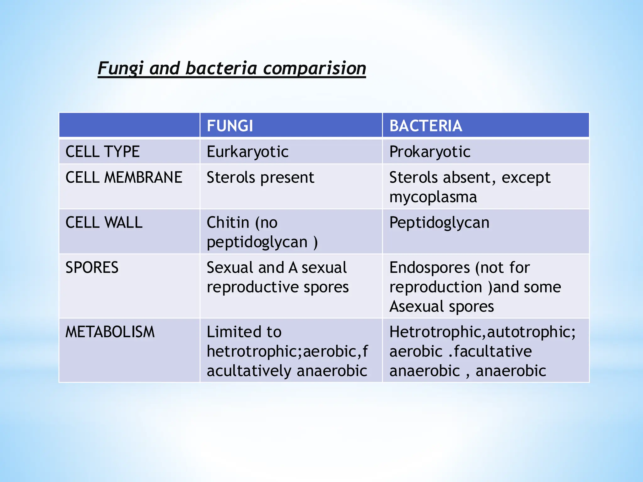
FUNGI BACTERIA
CELL TYPEEurkaryotic Prokaryotic
CELL MEMBRANE Sterols present Sterols absent, except
mycoplasma
CELL WALL Chitin (no
peptidoglycan )
Peptidoglycan
SPORES Sexual and A sexual
reproductive spores
Endospores (not for
reproduction )and some
Asexual spores
METABOLISM Limited to
hetrotrophic;aerobic,f
acultatively anaerobic
Hetrotrophic,autotrophic;
aerobic .facultative
anaerobic , anaerobic
Fungi and bacteria comparision
5.
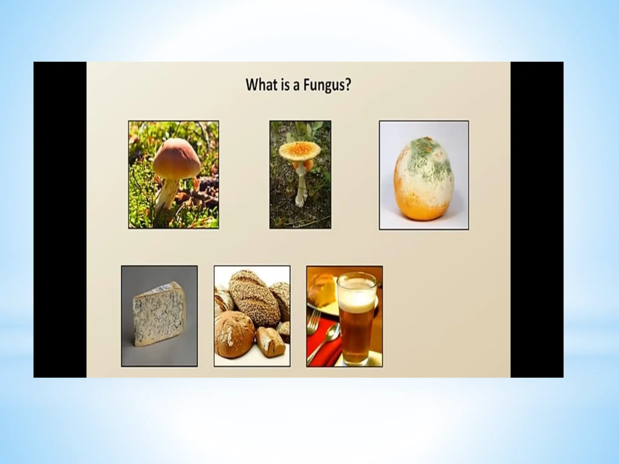
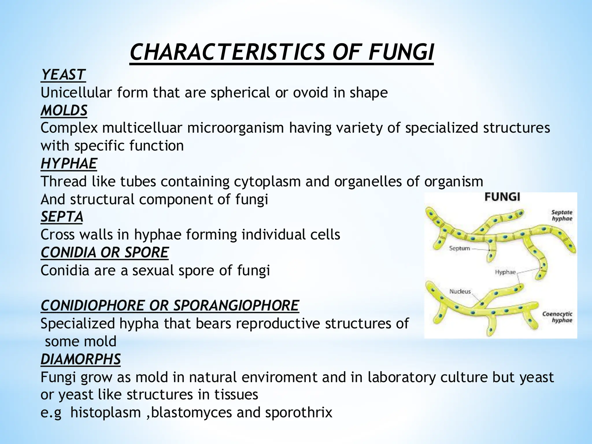
CHARACTERISTICS OF FUNGI
YEAST
Unicellularform that are spherical or ovoid in shape
MOLDS
Complex multicelluar microorganism having variety of specialized structures
with specific function
HYPHAE
Thread like tubes containing cytoplasm and organelles of organism
And structural component of fungi
SEPTA
Cross walls in hyphae forming individual cells
CONIDIA OR SPORE
Conidia are a sexual spore of fungi
CONIDIOPHORE OR SPORANGIOPHORE
Specialized hypha that bears reproductive structures of
some mold
DIAMORPHS
Fungi grow as mold in natural enviroment and in laboratory culture but yeast
or yeast like structures in tissues
e.g histoplasm ,blastomyces and sporothrix
7.
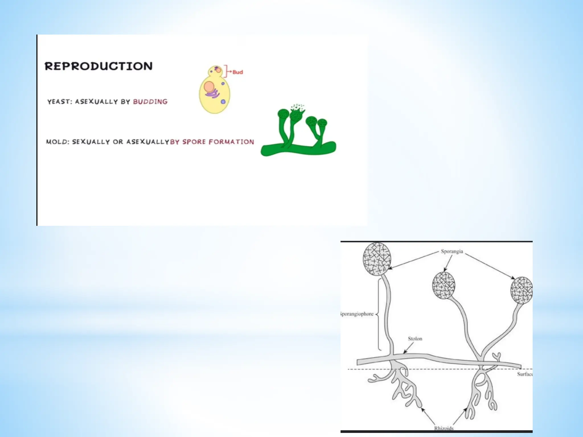
Rhizoids
Rhizoids are smallbranching hyphae that grow downwards from the
stolons that anchor the fungus .they release digestive enzymes and
absorb digested organic material
*Filamentous out growth
MYCELIUM (body of fungus )
Network of fungal threads or hyphae
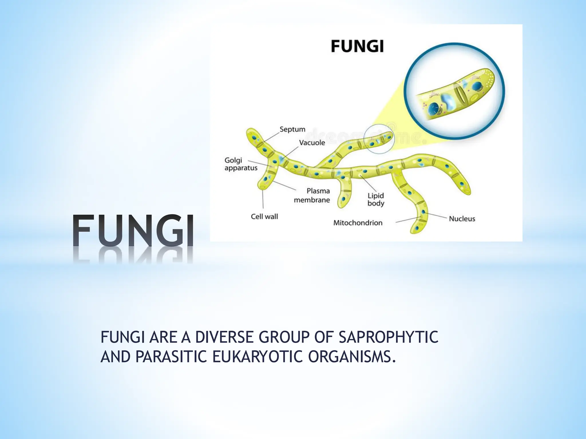
• Fungi are eukaryotic absorptive hetrotrophs
• They have kingdom mycota
• Virtually all organism are subject to fungal infection .of some
2000,000 fungal species ,only about 100 have pathogenic potential for
humans .
• Fungi is a member of a large group of eukaryotic organism that
included microorganism such as yeast and molds as well as more
familiar mushrooms
• There cell wall made up of chitin
• They are decomposers ,source of antibiotics
8.
Diffrential propertises fromplants
*Absorptive hetrotrophs
*Molecular +genetics
*Nuclear mitosis
*Haploid
Cell wall and cell membrane
The cell wall of fungi is made up of
✓Chitin
✓A polymer N-acetyl glucosamine
✓Fungal membrane contain ergosterol rather than
cholesterol
NUTRITIONAL ADAPTATIONS
*Chemohetrotrophs
*Absorptive in nature
9.
*Grow in enviromentof ph 5
*Most are resistant to osmotic pressure
*Grow on substances with a very low moisture
CLASSIFICATION OF FUNGI
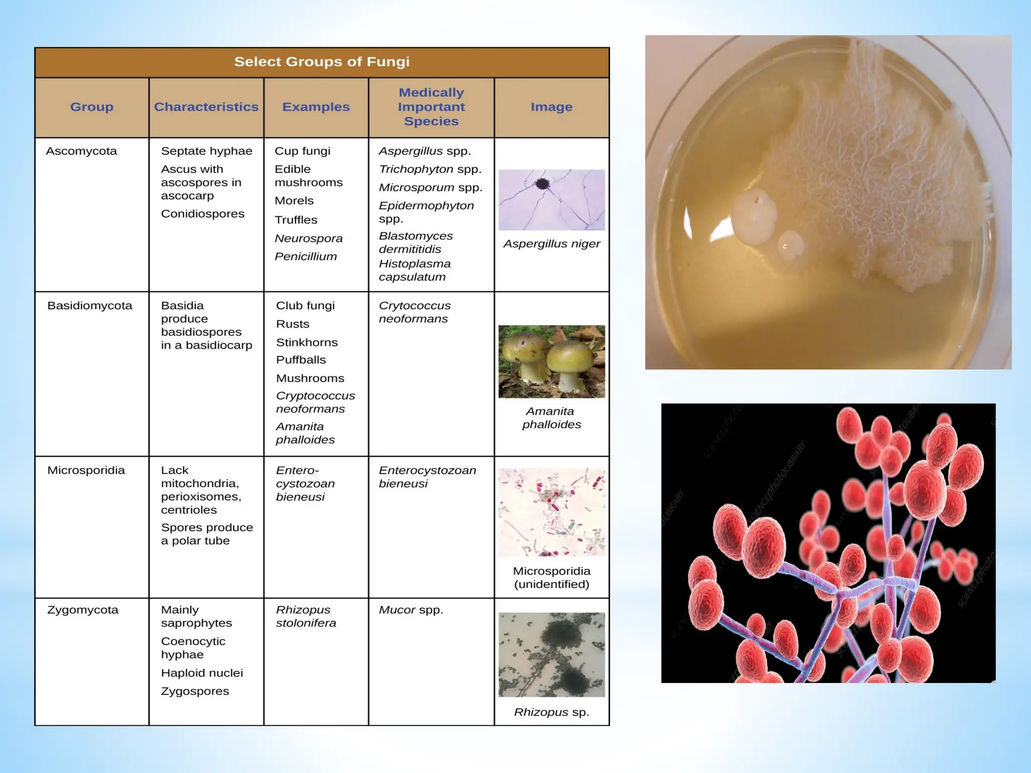
Fungi are classified as
❑ Zygomycota
❑ Basidiomycota
❑ Ascomycota
❑ Deuteromycota
1. Zygomycota
➢ The division zygomycota contains the fungi kown as zygomycetes .
➢ They have about 600 species
➢ Most live on decaying plants and animal matter in soil (few are
parasites of plants ,insects ,animals and human
➢ The hyphae of zygomycetes are coenocytic with many haploid nuclei
➢ Asexual spores are usually wind dispersed ,develop in sporangia at the
tip of hyphae
➢ Sexual reproduction produces tough thick walled zygotes called zygo
spores that can remain dormant when the enviroment is to harsh for
10.
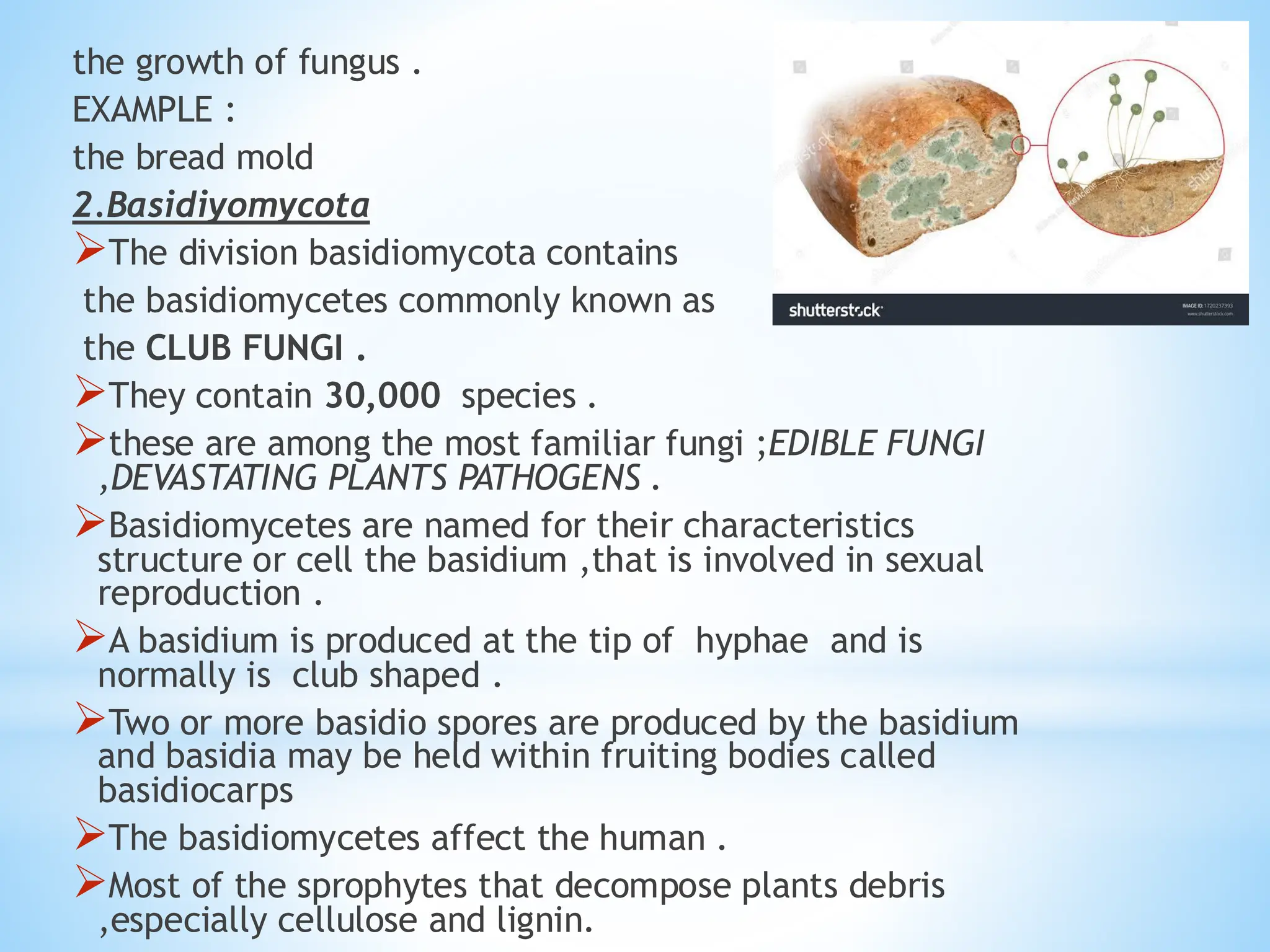
the growth offungus .
EXAMPLE :
the bread mold
2.Basidiyomycota
➢The division basidiomycota contains
the basidiomycetes commonly known as
the CLUB FUNGI .
➢They contain 30,000 species .
➢these are among the most familiar fungi ;EDIBLE FUNGI
,DEVASTATING PLANTS PATHOGENS .
➢Basidiomycetes are named for their characteristics
structure or cell the basidium ,that is involved in sexual
reproduction .
➢A basidium is produced at the tip of hyphae and is
normally is club shaped .
➢Two or more basidio spores are produced by the basidium
and basidia may be held within fruiting bodies called
basidiocarps
➢The basidiomycetes affect the human .
➢Most of the sprophytes that decompose plants debris
,especially cellulose and lignin.
11.
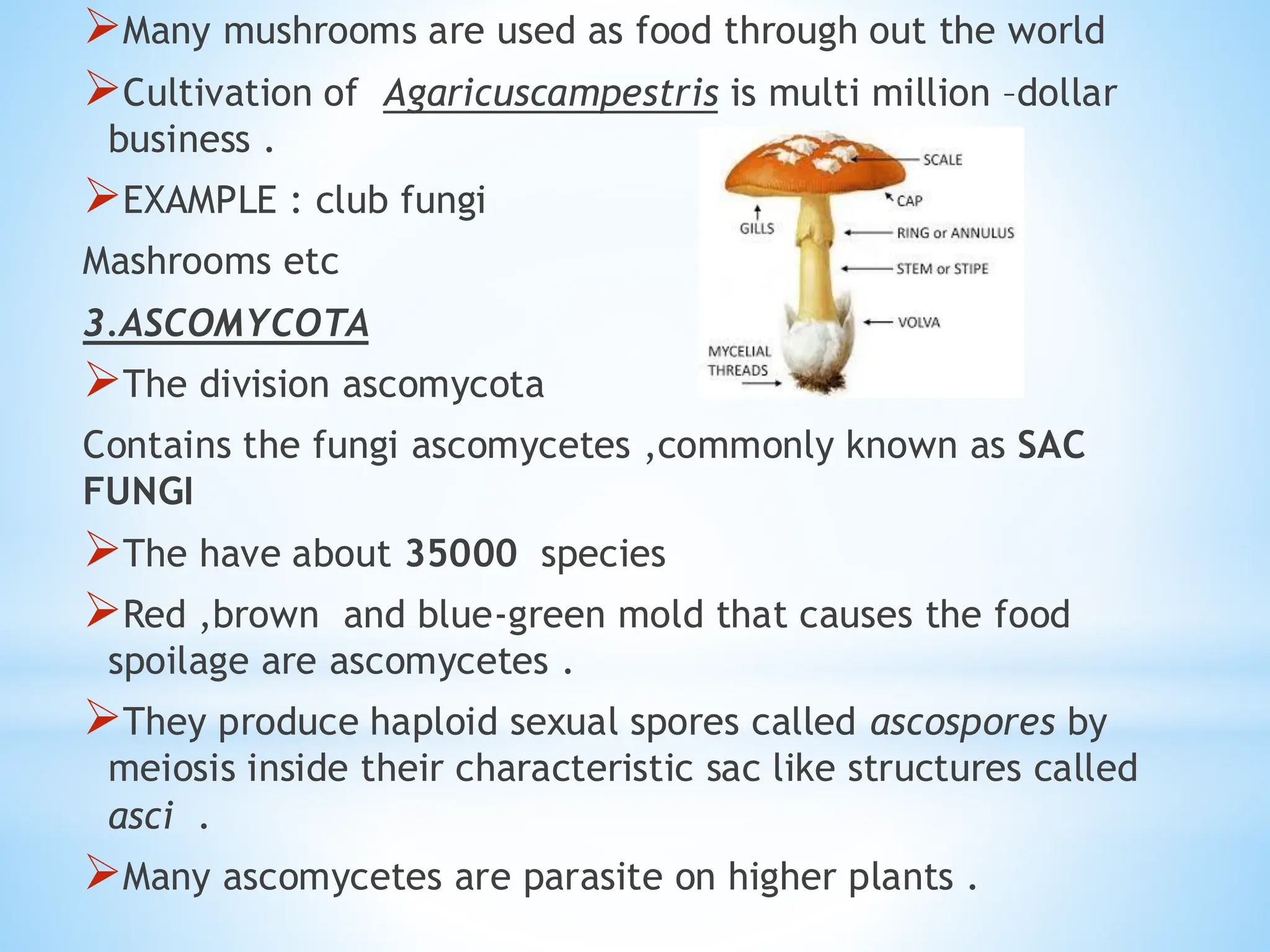
➢Many mushrooms areused as food through out the world
➢Cultivation of Agaricuscampestris is multi million –dollar
business .
➢EXAMPLE : club fungi
Mashrooms etc
3.ASCOMYCOTA
➢The division ascomycota
Contains the fungi ascomycetes ,commonly known as SAC
FUNGI
➢The have about 35000 species
➢Red ,brown and blue-green mold that causes the food
spoilage are ascomycetes .
➢They produce haploid sexual spores called ascospores by
meiosis inside their characteristic sac like structures called
asci .
➢Many ascomycetes are parasite on higher plants .
12.
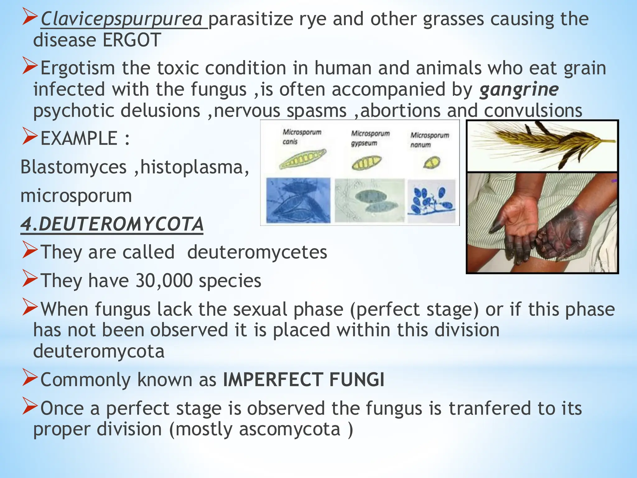
➢Clavicepspurpurea parasitize ryeand other grasses causing the
disease ERGOT
➢Ergotism the toxic condition in human and animals who eat grain
infected with the fungus ,is often accompanied by gangrine
psychotic delusions ,nervous spasms ,abortions and convulsions
➢EXAMPLE :
Blastomyces ,histoplasma,
microsporum
4.DEUTEROMYCOTA
➢They are called deuteromycetes
➢They have 30,000 species
➢When fungus lack the sexual phase (perfect stage) or if this phase
has not been observed it is placed within this division
deuteromycota
➢Commonly known as IMPERFECT FUNGI
➢Once a perfect stage is observed the fungus is tranfered to its
proper division (mostly ascomycota )
13.
➢Most fungi imperfectiare terrestia , with only few being reported
from fresh water and marine habitats
➢The majority are saprophytes or parasites of plants .a few are
parasitic over other fungi
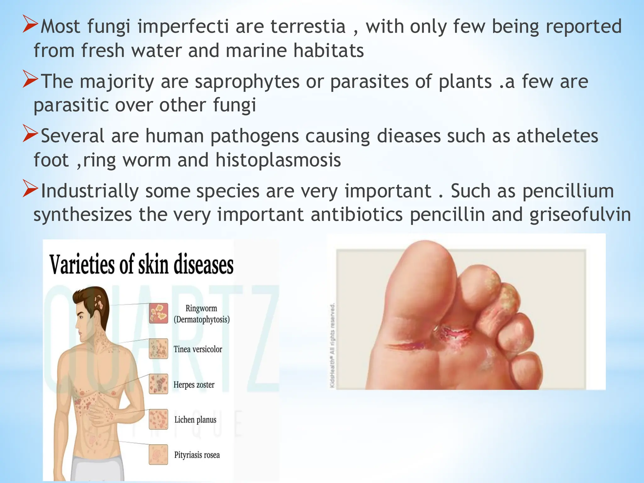
➢Several are human pathogens causing dieases such as atheletes
foot ,ring worm and histoplasmosis
➢Industrially some species are very important . Such as pencillium
synthesizes the very important antibiotics pencillin and griseofulvin
15.
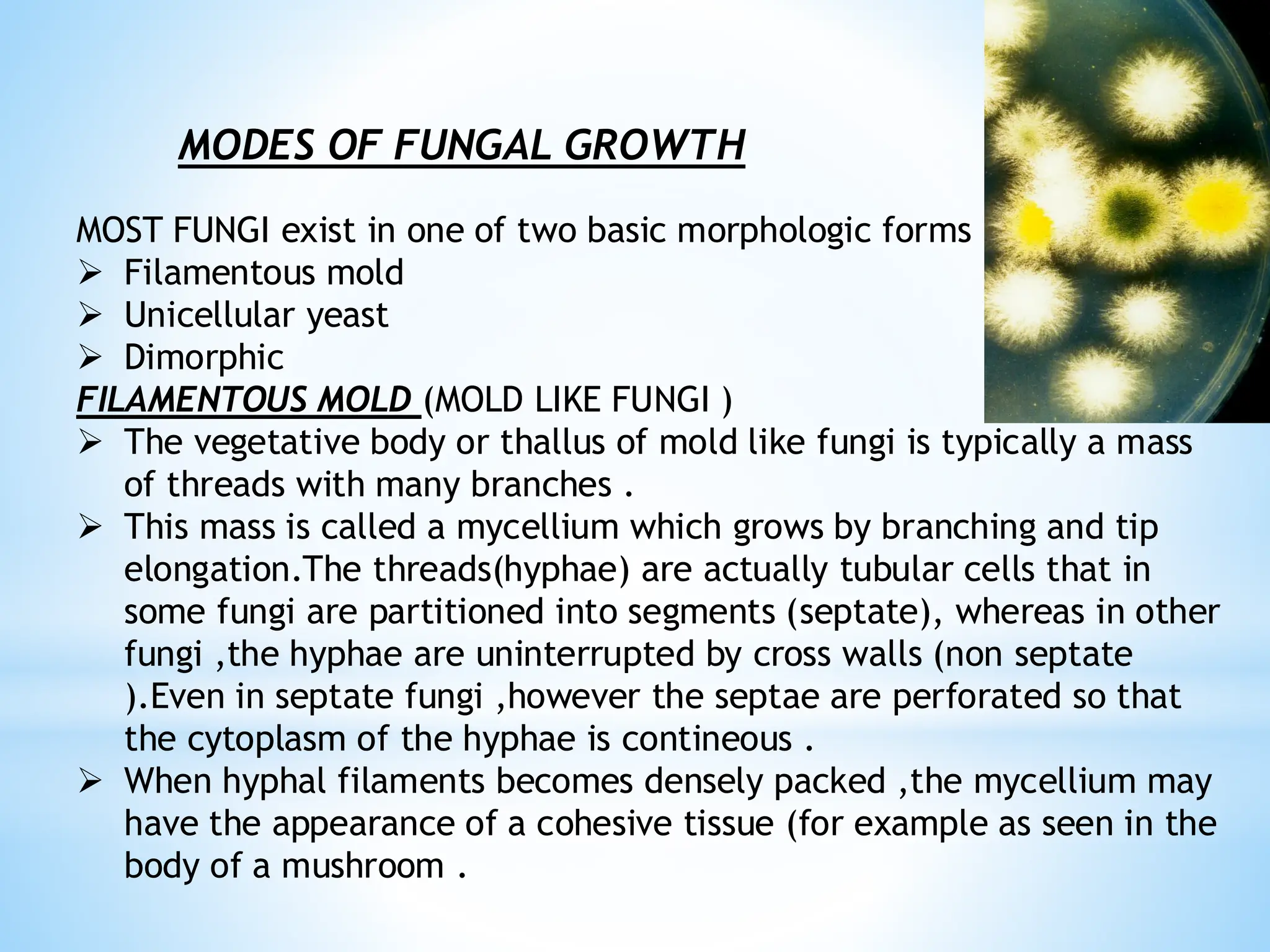
MODES OF FUNGALGROWTH
MOST FUNGI exist in one of two basic morphologic forms
➢ Filamentous mold
➢ Unicellular yeast
➢ Dimorphic
FILAMENTOUS MOLD (MOLD LIKE FUNGI )
➢ The vegetative body or thallus of mold like fungi is typically a mass
of threads with many branches .
➢ This mass is called a mycellium which grows by branching and tip
elongation.The threads(hyphae) are actually tubular cells that in
some fungi are partitioned into segments (septate), whereas in other
fungi ,the hyphae are uninterrupted by cross walls (non septate
).Even in septate fungi ,however the septae are perforated so that
the cytoplasm of the hyphae is contineous .
➢ When hyphal filaments becomes densely packed ,the mycellium may
have the appearance of a cohesive tissue (for example as seen in the
body of a mushroom .
16.
YEAST LIKE FUNGI
▪Thesefungi exsist as population of single,unconnected,spheroid
cells, not unlike many bacteria , although they are some ten times
larger than a typical bacterial cell .Yeast –like fungi generally
reproduce by budding .
DIMORPHIC FUNGI
▪Some fungal species , especially those that cause systemic mycoses
, are dimorphic ,being yeast- like in one enviroment and mold- like
in another. Conditions that can affect morphology include
temperature and carbon dioxide level .
▪Examples of dimorphic fungi includes blastomyces dermatiditis and
histoplasma capsulatum .
SPORULATION
▪The process of production of spores is called
sporulation.
▪Spores can generated either asexually or sexually .
17.
spore: It isa minute, simple propagating unit of the fungi,
functioning as a seed but differs from it in lacking a performed
embryo that serves in the reproduction of the same specie.
▪Spores varies in colour , size , number of cells and the way in
which they are born.
Types of spores:
There are two types of spores which are following :
a. Asexual sporulation
b.Sexual sporulation
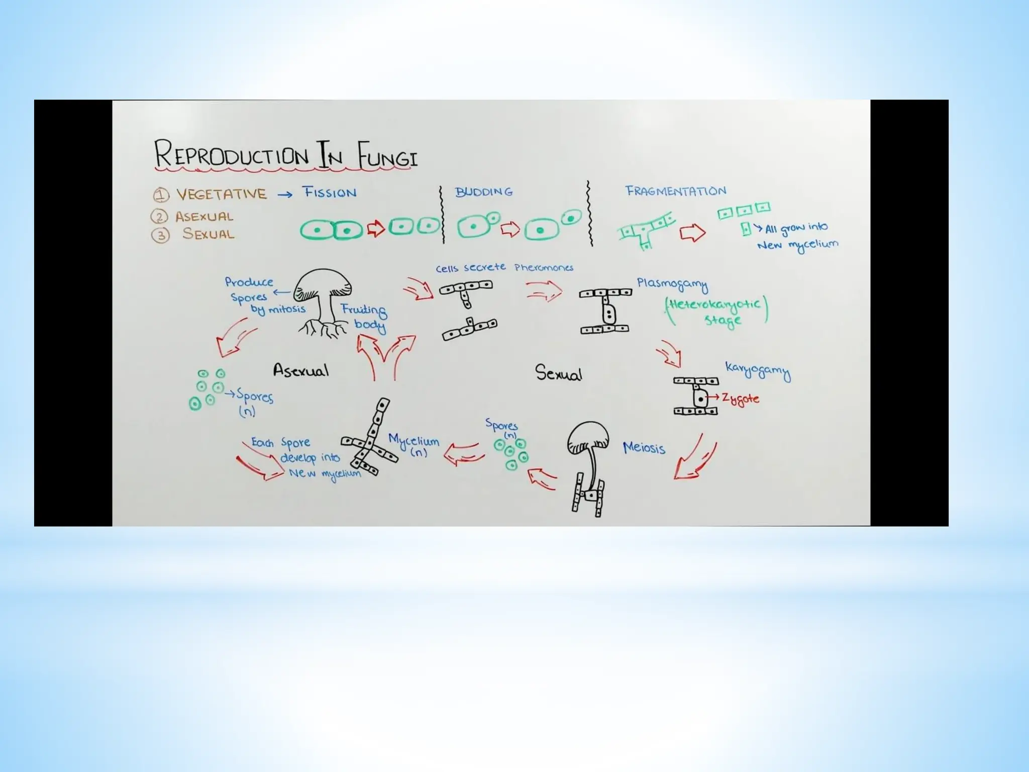
Asexual sporulation:
❑Asexual spores (conidia) are formed by mitosis in or on
specialized hyphae (conidiophores).
❑In fungi, asexual reproduction is more important for the
propagation of species.
18.
❑It is repeatedseveral times during the life span of a fungus
producing numerous asexual spores .
❑In fungi the following are the common methods of asexual
reproduction.
❑Asexual spores are formed after mitosis , hence also called
mitospores.
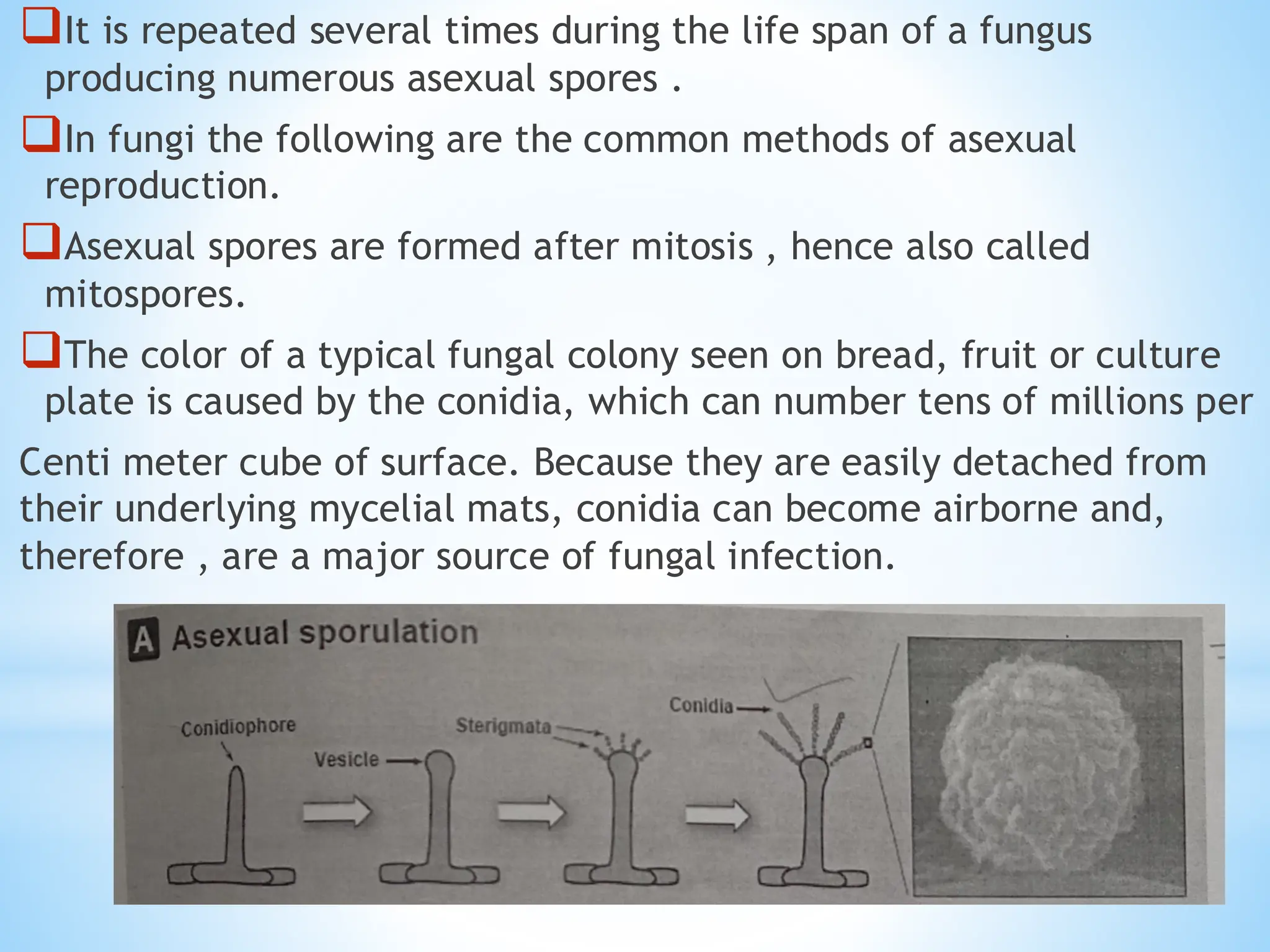
❑The color of a typical fungal colony seen on bread, fruit or culture
plate is caused by the conidia, which can number tens of millions per
Centi meter cube of surface. Because they are easily detached from
their underlying mycelial mats, conidia can become airborne and,
therefore , are a major source of fungal infection.
19.
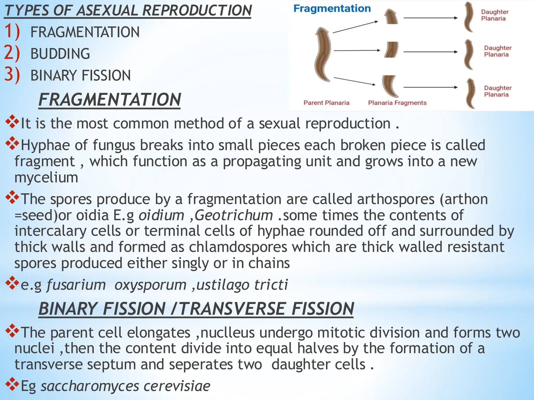
TYPES OF ASEXUALREPRODUCTION
1) FRAGMENTATION
2) BUDDING
3) BINARY FISSION
FRAGMENTATION
❖It is the most common method of a sexual reproduction .
❖Hyphae of fungus breaks into small pieces each broken piece is called
fragment , which function as a propagating unit and grows into a new
mycelium
❖The spores produce by a fragmentation are called arthospores (arthon
=seed)or oidia E.g oidium ,Geotrichum .some times the contents of
intercalary cells or terminal cells of hyphae rounded off and surrounded by
thick walls and formed as chlamdospores which are thick walled resistant
spores produced either singly or in chains
❖e.g fusarium oxysporum ,ustilago tricti
BINARY FISSION /TRANSVERSE FISSION
❖The parent cell elongates ,nuclleus undergo mitotic division and forms two
nuclei ,then the content divide into equal halves by the formation of a
transverse septum and seperates two daughter cells .
❖Eg saccharomyces cerevisiae
20.
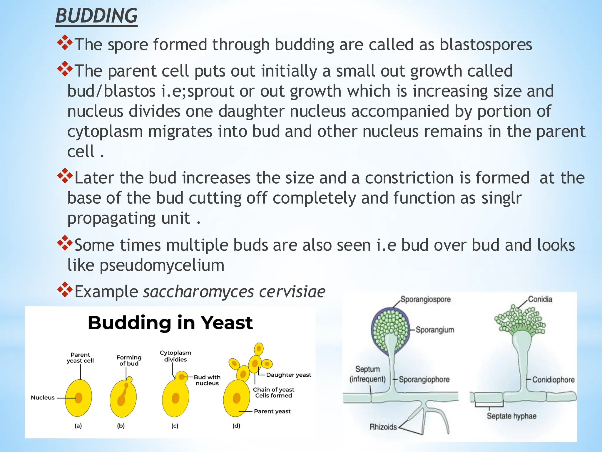
BUDDING
❖The spore formedthrough budding are called as blastospores
❖The parent cell puts out initially a small out growth called
bud/blastos i.e;sprout or out growth which is increasing size and
nucleus divides one daughter nucleus accompanied by portion of
cytoplasm migrates into bud and other nucleus remains in the parent
cell .
❖Later the bud increases the size and a constriction is formed at the
base of the bud cutting off completely and function as singlr
propagating unit .
❖Some times multiple buds are also seen i.e bud over bud and looks
like pseudomycelium
❖Example saccharomyces cervisiae
21.
SEXUAL REPRODUCTION
➢Sexual reproductioninvolves union of two compatible nuclie or
cells or organs or somatic cell or somatic hyphae for the formation
of new individuals
➢Sexual stage is perfect stage and technically called as telomorphic
stage
➢Sexual cycle occurs once in the life span of fungus
PHASES DURING THE SEXUAL REPRODUCTION
Plasmogamy
➢Union of two protoplast taking place as a result of the two nuclei
come together within the same cell
KARYOGAMY
➢Union of two sexually compatible nuclei brought together by
plasmogamy to form a diploid nucleus 2n zygote
MEIOSIS
➢This the reduction division . The number of chromosome is reduced
to haploid (n) i.e diploid nucleus result in haploid spores
23.
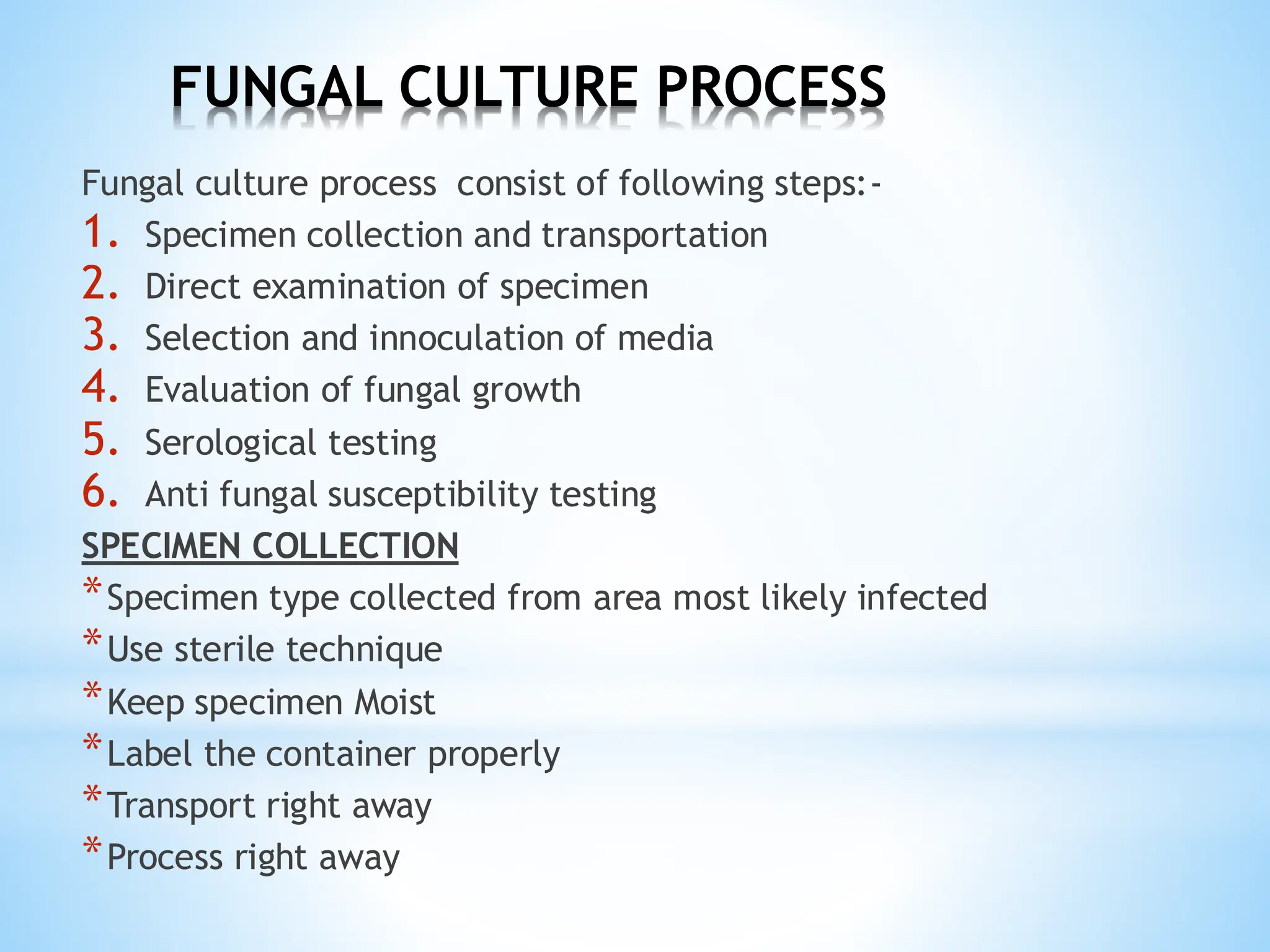
FUNGAL CULTURE PROCESS
Fungalculture process consist of following steps:-
1. Specimen collection and transportation
2. Direct examination of specimen
3. Selection and innoculation of media
4. Evaluation of fungal growth
5. Serological testing
6. Anti fungal susceptibility testing
SPECIMEN COLLECTION
*Specimen type collected from area most likely infected
*Use sterile technique
*Keep specimen Moist
*Label the container properly
*Transport right away
*Process right away
24.
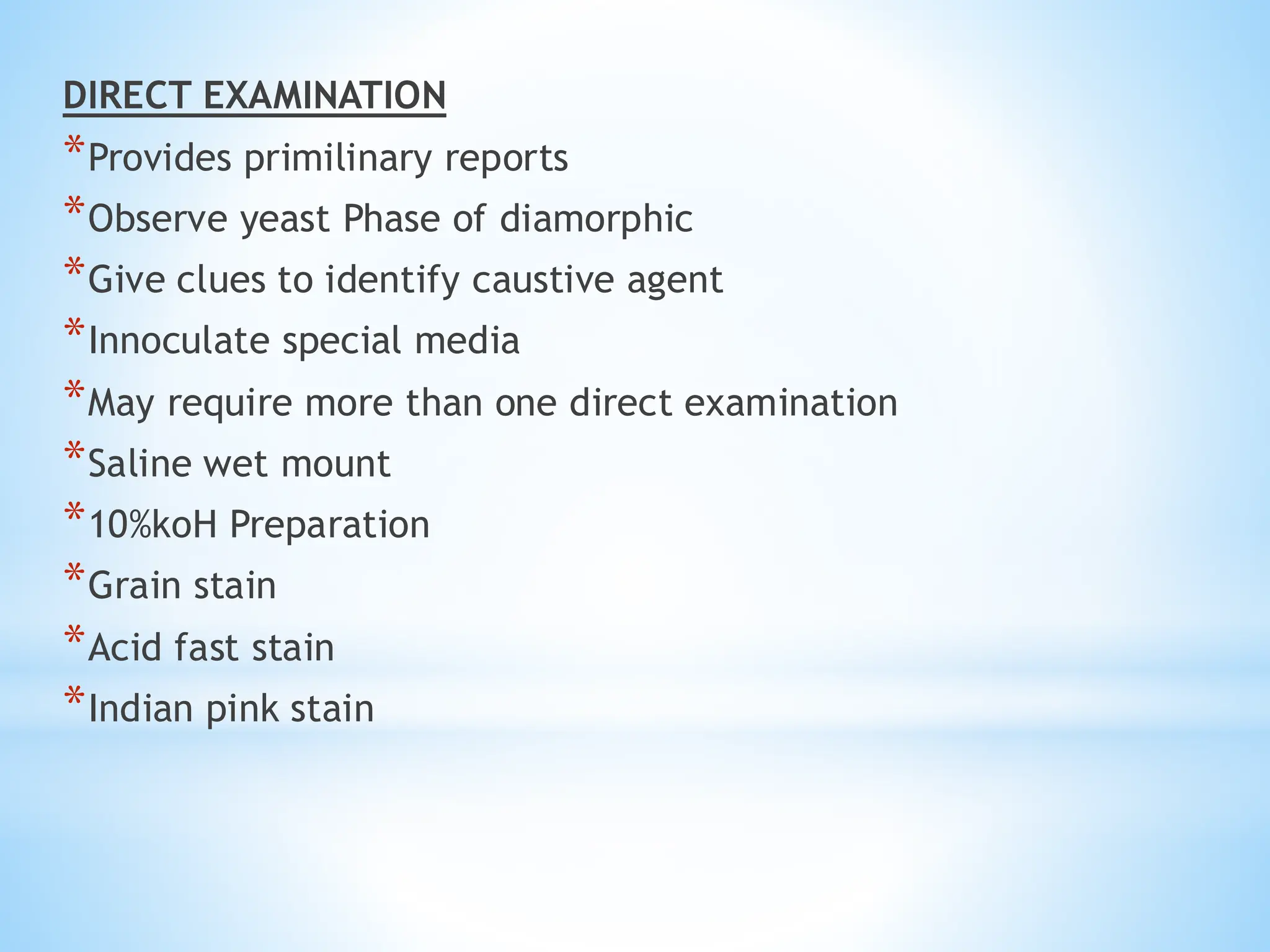
DIRECT EXAMINATION
*Provides primilinaryreports
*Observe yeast Phase of diamorphic
*Give clues to identify caustive agent
*Innoculate special media
*May require more than one direct examination
*Saline wet mount
*10%koH Preparation
*Grain stain
*Acid fast stain
*Indian pink stain
25.
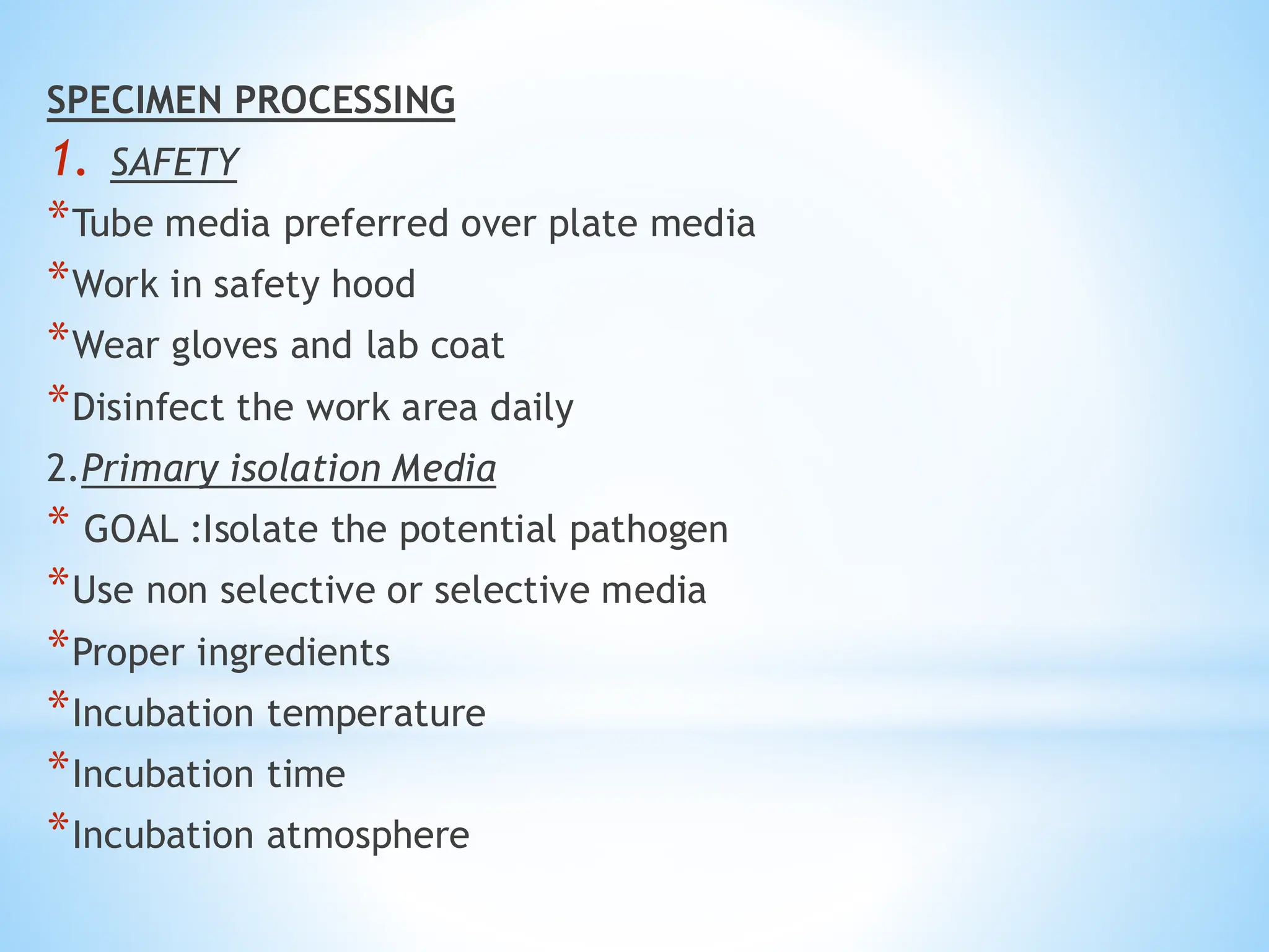
SPECIMEN PROCESSING
1. SAFETY
*Tubemedia preferred over plate media
*Work in safety hood
*Wear gloves and lab coat
*Disinfect the work area daily
2.Primary isolation Media
* GOAL :Isolate the potential pathogen
*Use non selective or selective media
*Proper ingredients
*Incubation temperature
*Incubation time
*Incubation atmosphere
26.
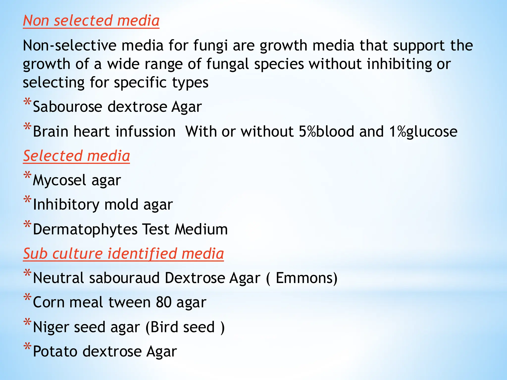
Non selected media
Non-selectivemedia for fungi are growth media that support the
growth of a wide range of fungal species without inhibiting or
selecting for specific types
*Sabourose dextrose Agar
*Brain heart infussion With or without 5%blood and 1%glucose
Selected media
*Mycosel agar
*Inhibitory mold agar
*Dermatophytes Test Medium
Sub culture identified media
*Neutral sabouraud Dextrose Agar ( Emmons)
*Corn meal tween 80 agar
*Niger seed agar (Bird seed )
*Potato dextrose Agar
27.
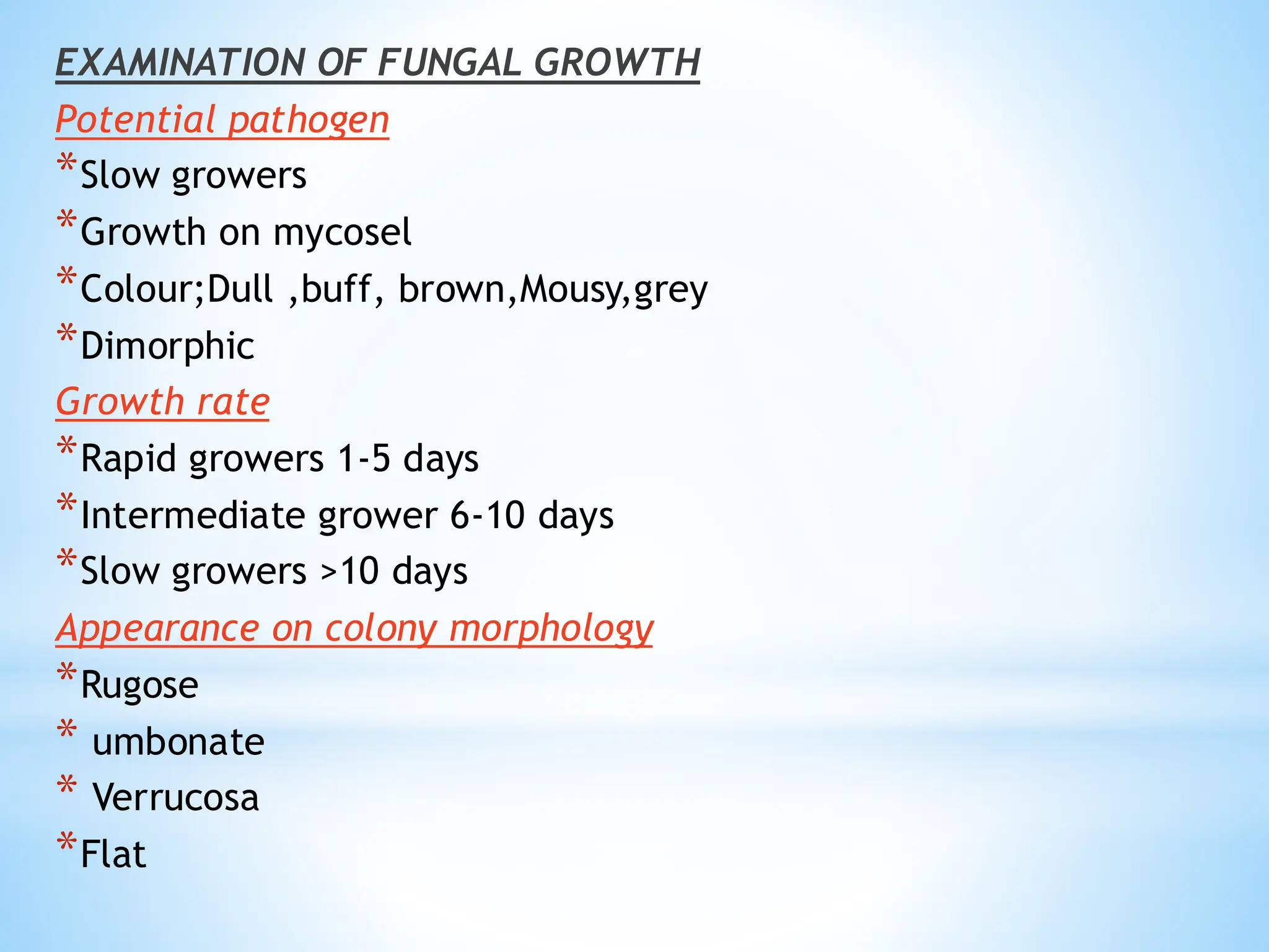
EXAMINATION OF FUNGALGROWTH
Potential pathogen
*Slow growers
*Growth on mycosel
*Colour;Dull ,buff, brown,Mousy,grey
*Dimorphic
Growth rate
*Rapid growers 1-5 days
*Intermediate grower 6-10 days
*Slow growers >10 days
Appearance on colony morphology
*Rugose
* umbonate
* Verrucosa
*Flat
28.
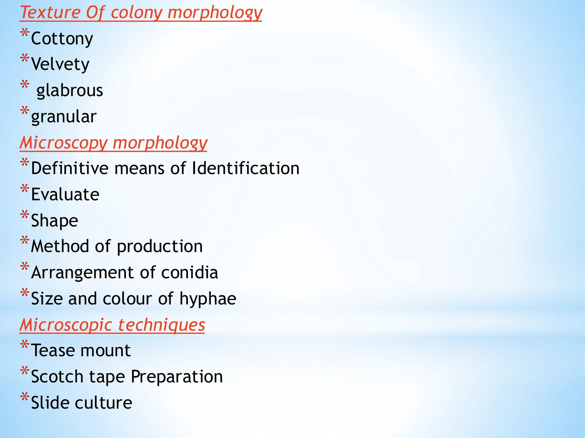
Texture Of colonymorphology
*Cottony
*Velvety
* glabrous
*granular
Microscopy morphology
*Definitive means of Identification
*Evaluate
*Shape
*Method of production
*Arrangement of conidia
*Size and colour of hyphae
Microscopic techniques
*Tease mount
*Scotch tape Preparation
*Slide culture
29.
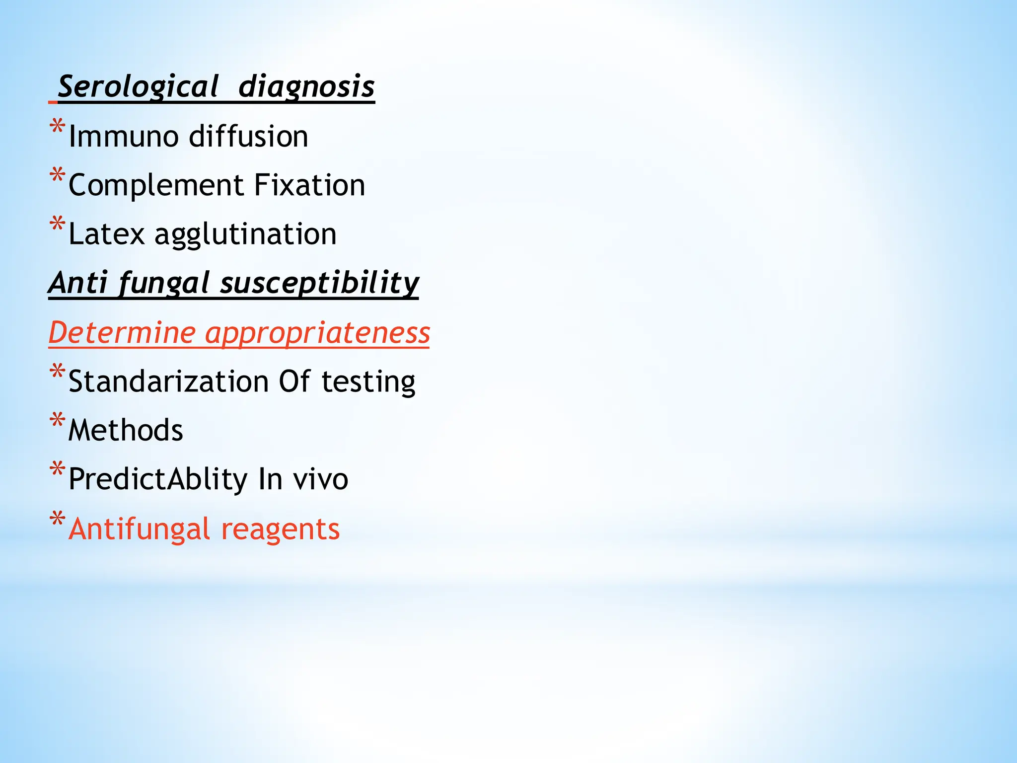
Serological diagnosis
*Immuno diffusion
*ComplementFixation
*Latex agglutination
Anti fungal susceptibility
Determine appropriateness
*Standarization Of testing
*Methods
*PredictAblity In vivo
*Antifungal reagents
30.
MYCOSES:
* “A diseasecaused by infection with a fungus is
called MYCOSES.”
Typesof Mycoses:-
There are following types of Mycoses,
• Superficial Mycoses
• Cutaneous Mycoses
• Subcutaneous Mycoses
• Systemic Mycoses due to primary pathogens
• Systemic Mycoses due to opportunistic pathogens
31.
1-Superficial mycoses
Superficial Mycosesare limited to the outermost layers of
the skin and hair.
* Do not elicit immune response
*No discomfort
*Cosmetics problems
*Limited to stratum corneum
Infections:-
• Pityriasis versicolor –affects skin
• Tinea nigra –affects skin
• Black piedra- affect hair
• White piedra – affects hair
32.
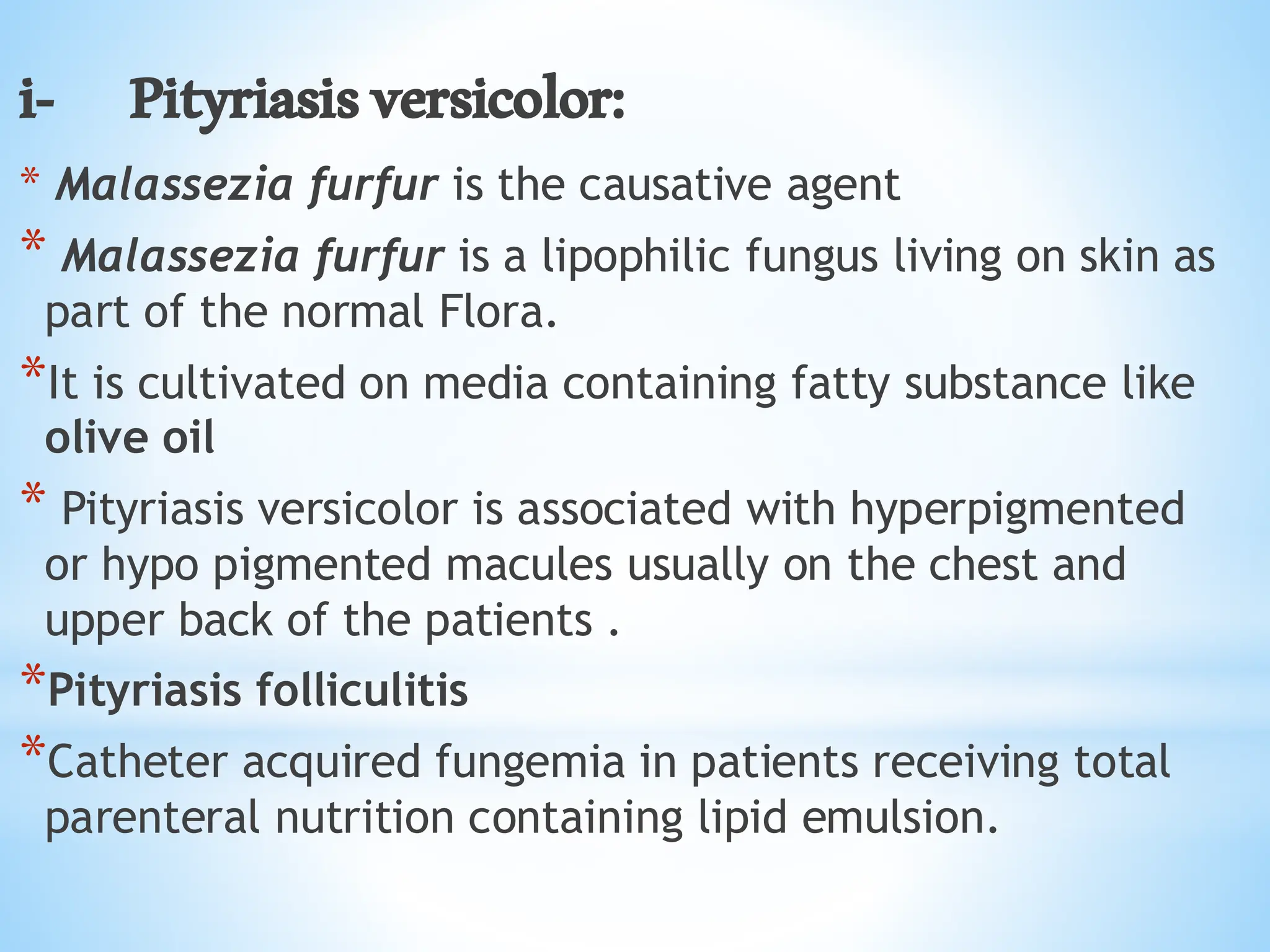
i- Pityriasisversicolor:
* Malasseziafurfur is the causative agent
* Malassezia furfur is a lipophilic fungus living on skin as
part of the normal Flora.
*It is cultivated on media containing fatty substance like
olive oil
* Pityriasis versicolor is associated with hyperpigmented
or hypo pigmented macules usually on the chest and
upper back of the patients .
*Pityriasis folliculitis
*Catheter acquired fungemia in patients receiving total
parenteral nutrition containing lipid emulsion.
33.
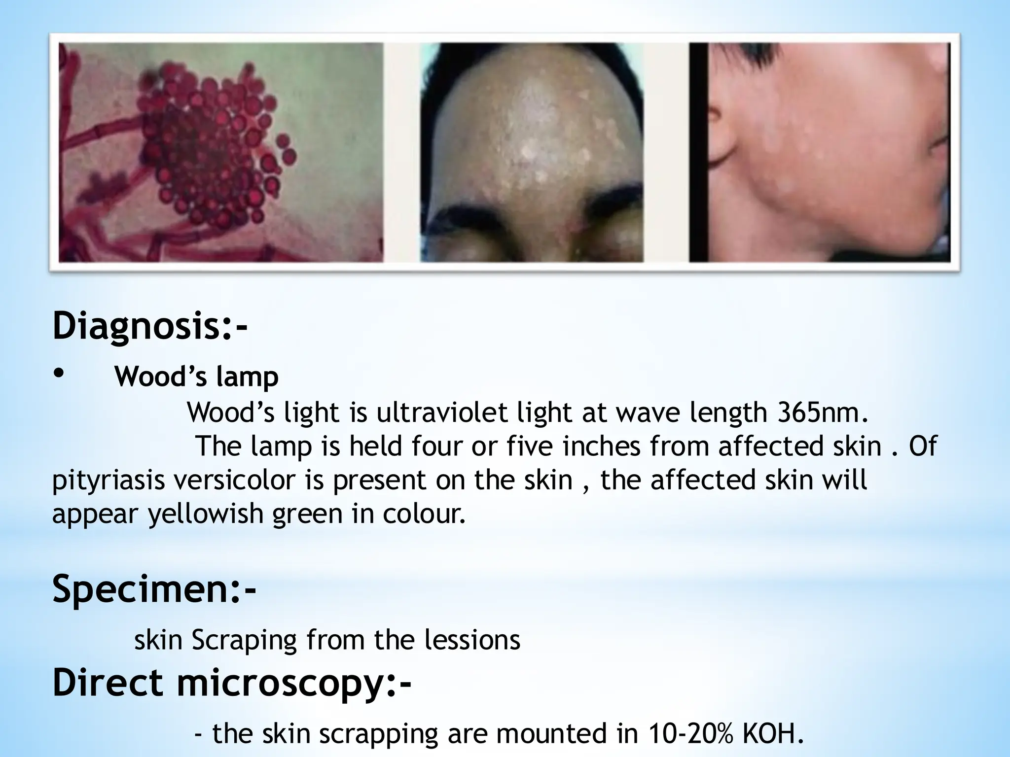
Diagnosis:-
• Wood’s lamp
Wood’slight is ultraviolet light at wave length 365nm.
The lamp is held four or five inches from affected skin . Of
pityriasis versicolor is present on the skin , the affected skin will
appear yellowish green in colour.
Specimen:-
skin Scraping from the lessions
Direct microscopy:-
- the skin scrapping are mounted in 10-20% KOH.
34.
ii- Tinea Nigra:-
* Causative agent: Exosphiala (Hortae) werneckii
. - It is a saprophytic fungus which occurs in the soil .
- it is a dematicaeous fungus (contains melanin in its
cell wall and appear dark in colour under the microscope).
* Specimen:
skin scrapping from the lessions.
- It will show spherical budding cells and short unbranched
angular septate hyphae(spaghetti and meatballs)
- These microscopic features are diagnostic for Malassezia
furfur and culture is not necessary.
Treatment:-
- Daily application of selenium sulfide
- Topical or oral azoles are very effective.
35.
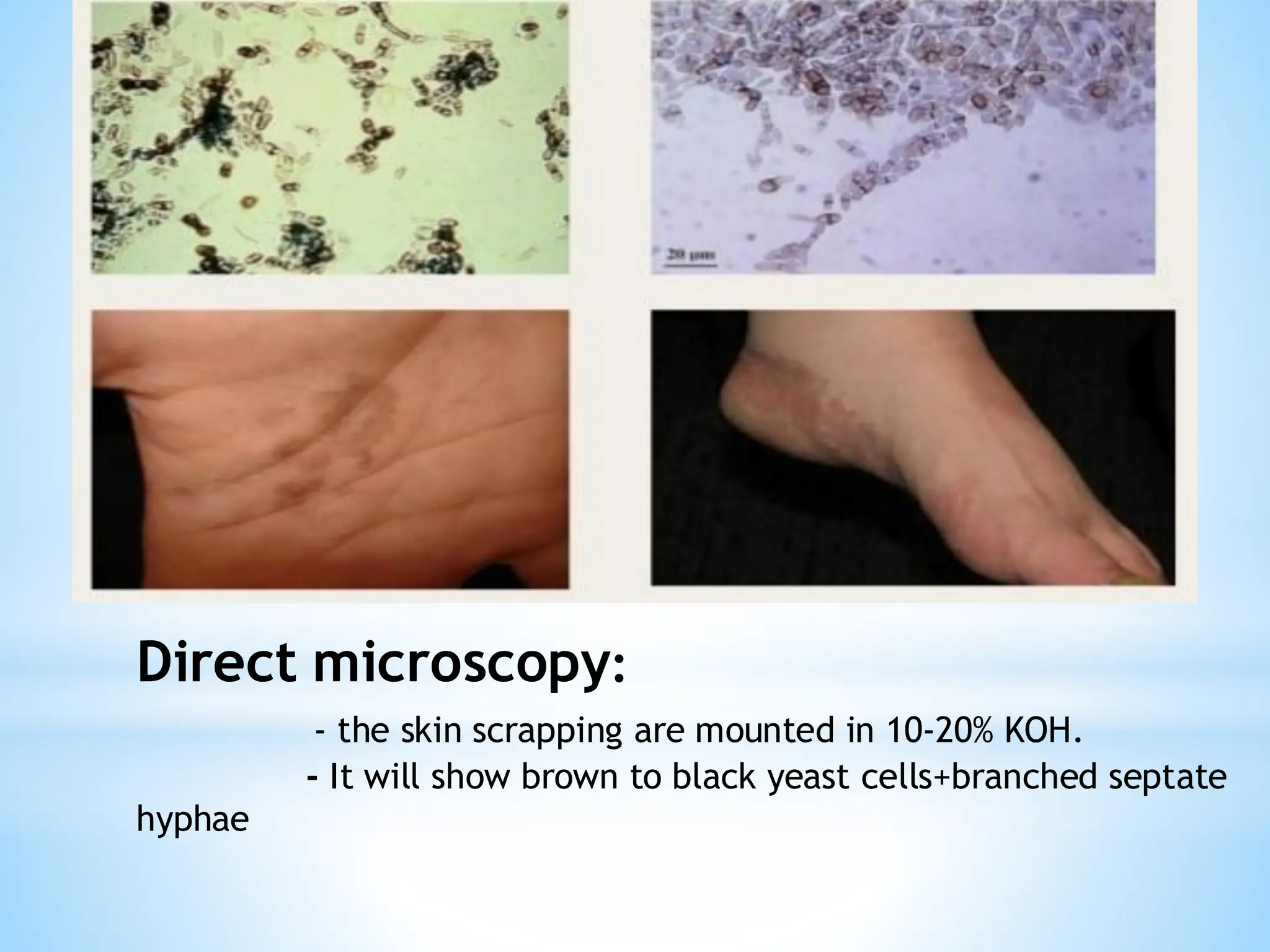
Direct microscopy:
- theskin scrapping are mounted in 10-20% KOH.
- It will show brown to black yeast cells+branched septate
hyphae
36.
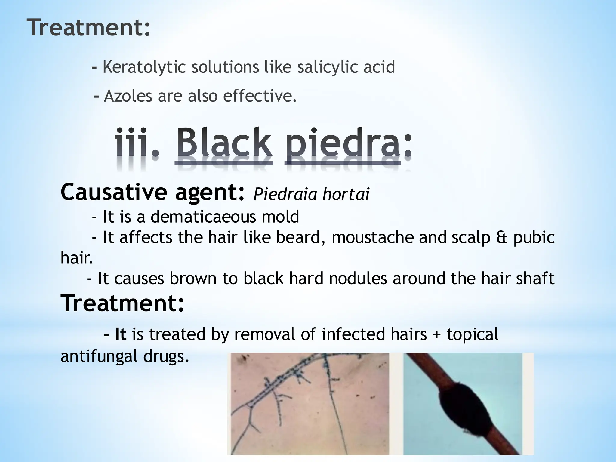
iii. Black piedra:
Treatment:
-Keratolytic solutions like salicylic acid
- Azoles are also effective.
Causative agent: Piedraia hortai
- It is a dematicaeous mold
- It affects the hair like beard, moustache and scalp & pubic
hair.
- It causes brown to black hard nodules around the hair shaft
Treatment:
- It is treated by removal of infected hairs + topical
antifungal drugs.
37.
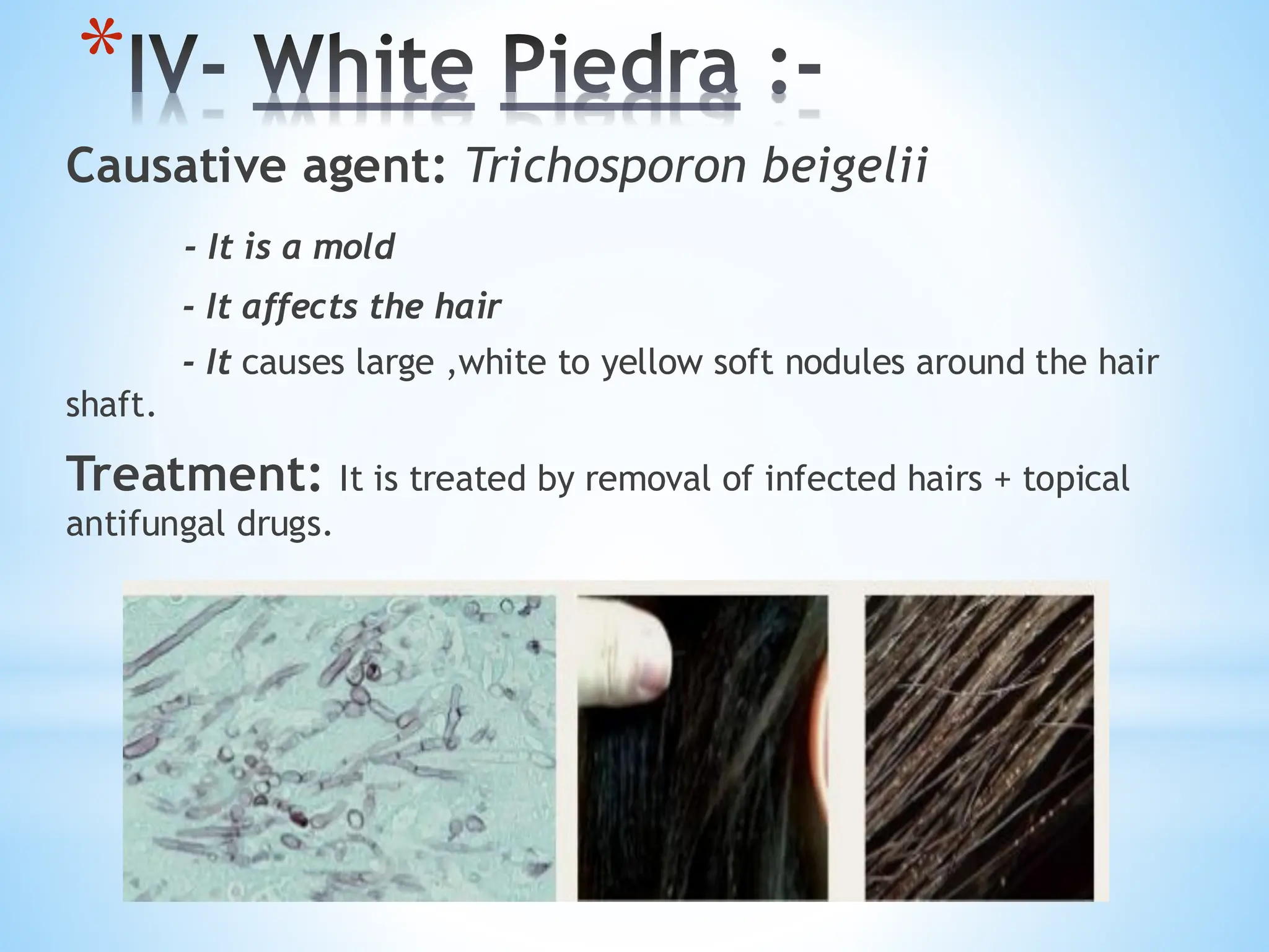
*IV- White Piedra:-
Causative agent: Trichosporon beigelii
- It is a mold
- It affects the hair
- It causes large ,white to yellow soft nodules around the hair
shaft.
Treatment: It is treated by removal of infected hairs + topical
antifungal drugs.
38.
2. Cutaneous Mycoses:-
CutaneousMycoses are fungal infections usually
confined to the outer layer of skin , hair , and nails , and do not invade
living tissues.
Also known as “ringworm” and tinea (latin”worm”) because of round
shape of lessions
Also called dermatophytes.
There are 3 genera :
* Trichophyton
*Microsporum
* Epidermophyton
Classification:
On the basis of ecology :
i- Anthropophilic
ii- Zoophilic
iii- Geophilic
39.
i- Trichophyton (19species)
* Anthropophilic:
- Associated with humans only . Person-to-perspn transmission through
contaminated objects (comb, hat)
- All three genera i.e, Trichophyton, Microsporum, Epidermophyton.
* Zoophilic:
- Associated with animals . Direct transmission to humans by close
contact with animals.
- Only two genera i.e, Trichophyton and Microsporum
* Geophilic:
- Usually found in soil. Transmitted to humans by direct exposure
- Microsporum
- The genus Trichophyton is characterized by the development of both
smooth-walled macro and microconidia.
- Causes infections of hair, skin , nails .
- Causative agents are :
T. rubrum
T. violaceum
T. mentagrophytes
40.
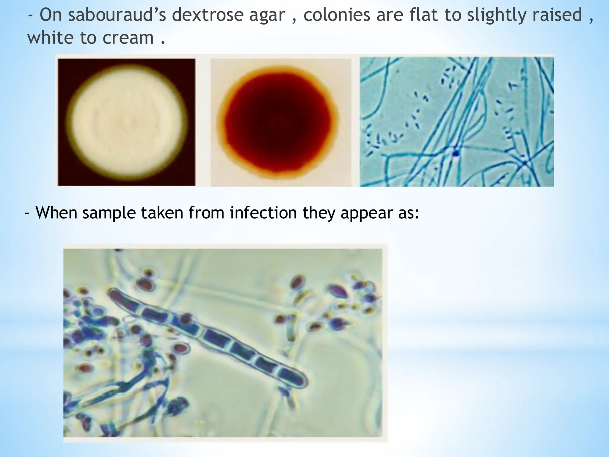
- On sabouraud’sdextrose agar , colonies are flat to slightly raised ,
white to cream .
- When sample taken from infection they appear as:
41.
ii- Microsporum (13species)
- Microsporum forms both macroconidia (large asexual
reproductive structures) and microconidia (smaller asexual
reproductive structures) on short conidiophores.
- Causes infection of skin and hair
- Causative agent are
M. audouinii
M. canis
M. gypseum
iii- Epidermophyton:-
- Causes infection in skin and nails
- Causative agent is
Epidermophyton floccosum
42.
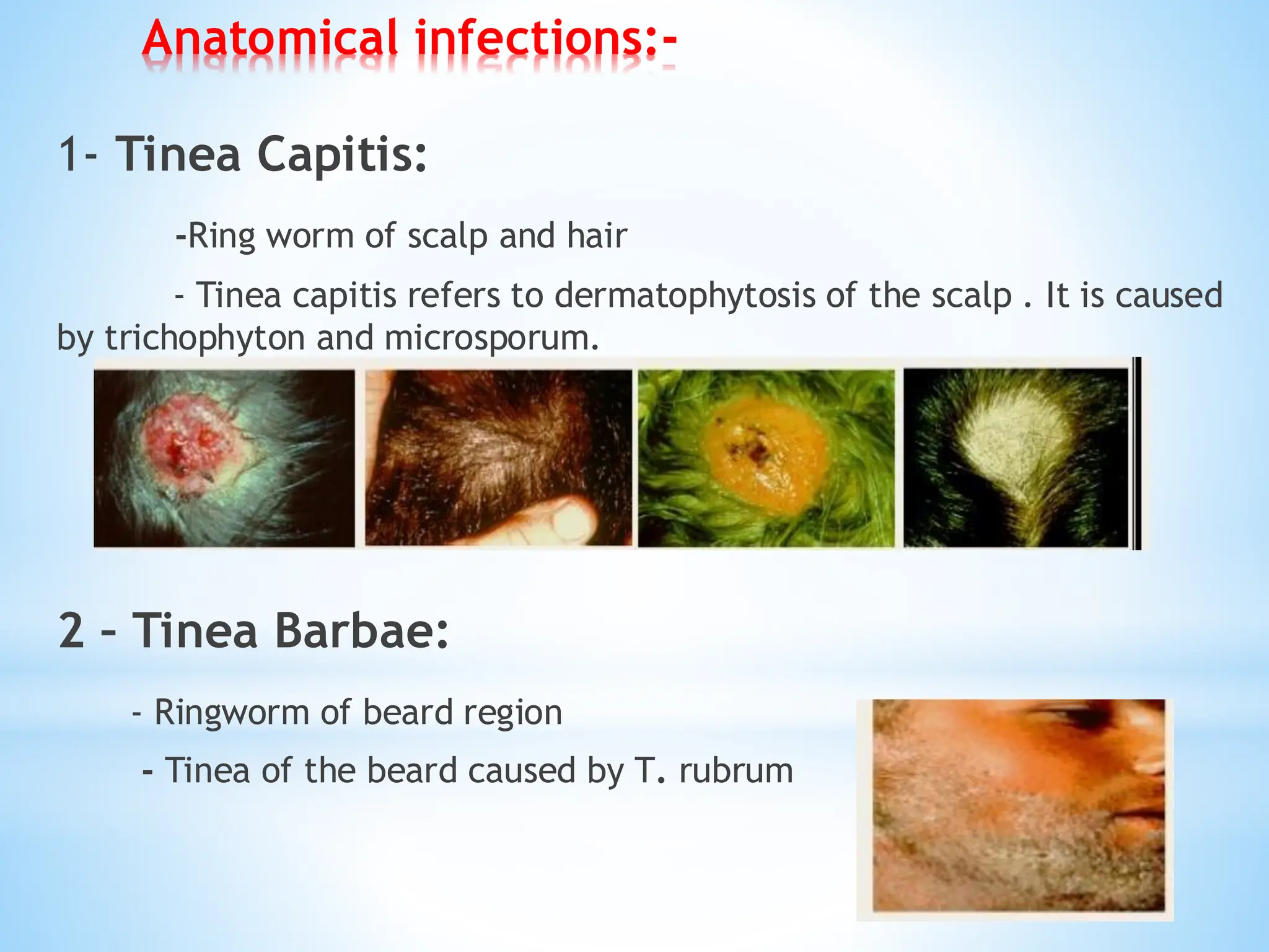
Anatomical infections:-
1- TineaCapitis:
-Ring worm of scalp and hair
- Tinea capitis refers to dermatophytosis of the scalp . It is caused
by trichophyton and microsporum.
2 – Tinea Barbae:
- Ringworm of beard region
- Tinea of the beard caused by T. rubrum
43.
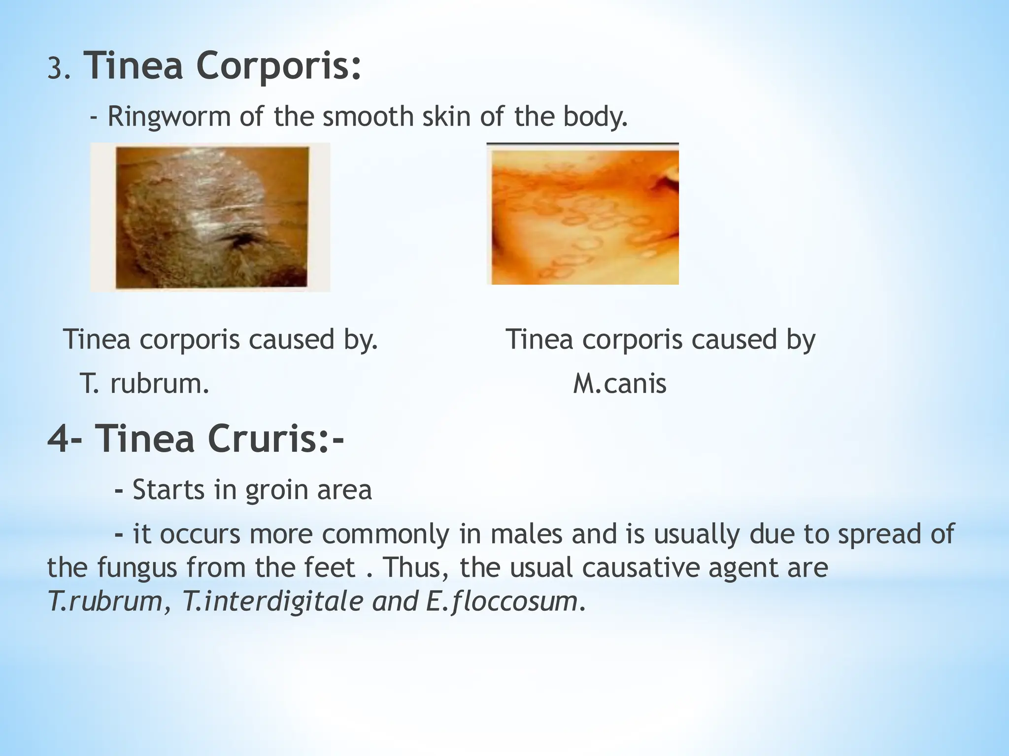
3. Tinea Corporis:
-Ringworm of the smooth skin of the body.
Tinea corporis caused by. Tinea corporis caused by
T. rubrum. M.canis
4- Tinea Cruris:-
- Starts in groin area
- it occurs more commonly in males and is usually due to spread of
the fungus from the feet . Thus, the usual causative agent are
T.rubrum, T.interdigitale and E.floccosum.
44.
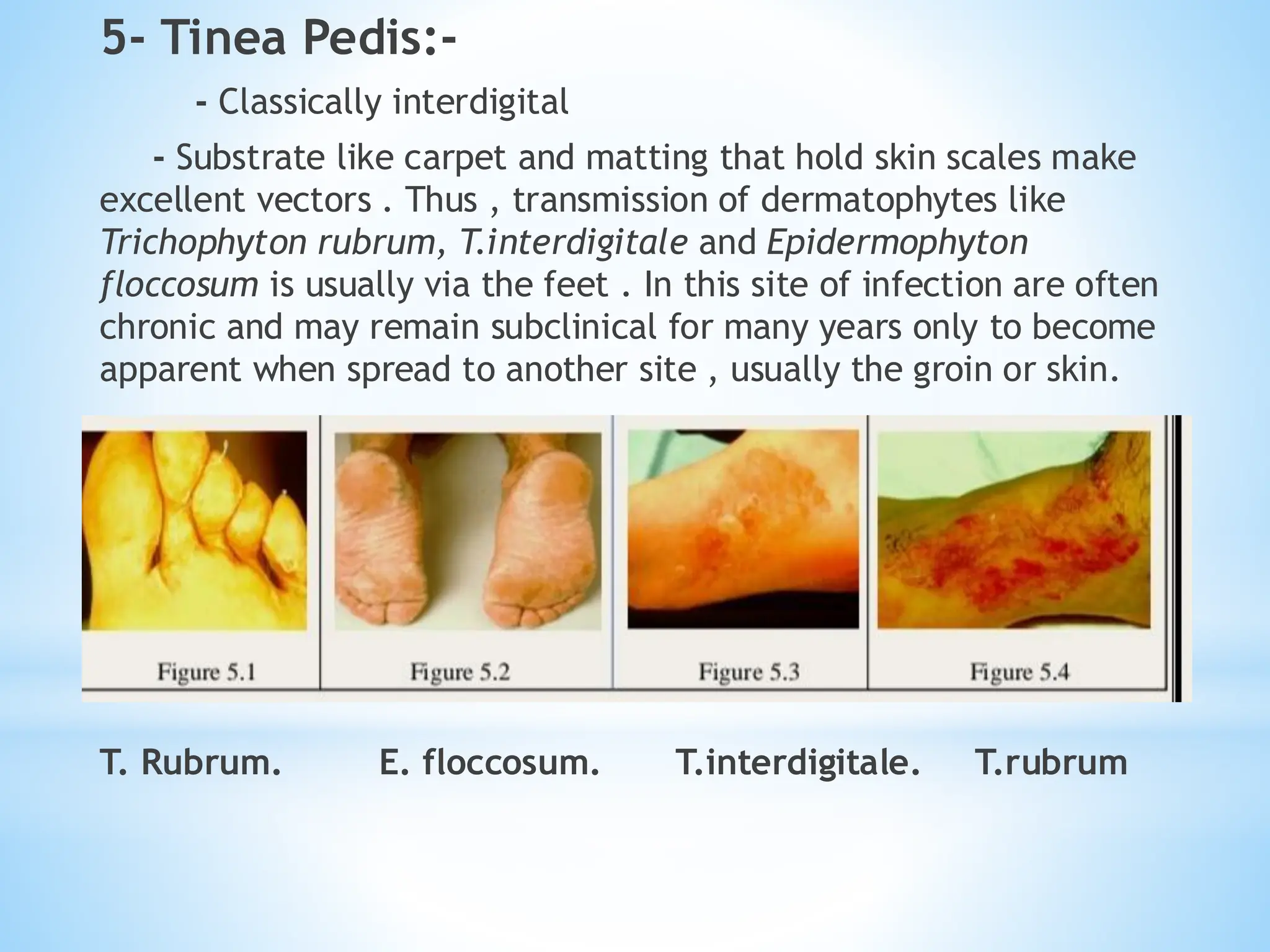
5- Tinea Pedis:-
-Classically interdigital
- Substrate like carpet and matting that hold skin scales make
excellent vectors . Thus , transmission of dermatophytes like
Trichophyton rubrum, T.interdigitale and Epidermophyton
floccosum is usually via the feet . In this site of infection are often
chronic and may remain subclinical for many years only to become
apparent when spread to another site , usually the groin or skin.
T. Rubrum. E. floccosum. T.interdigitale. T.rubrum
45.
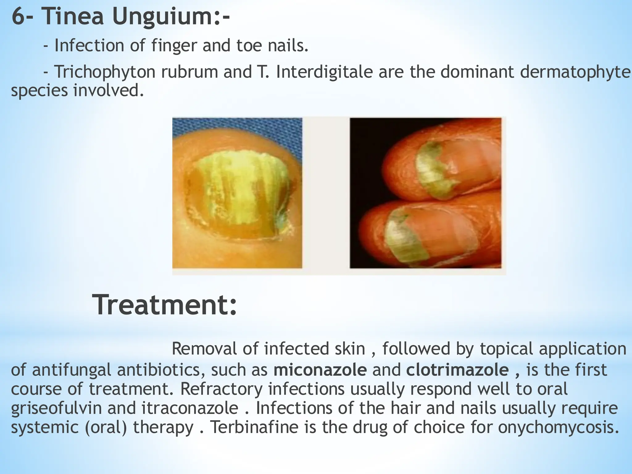
6- Tinea Unguium:-
-Infection of finger and toe nails.
- Trichophyton rubrum and T. Interdigitale are the dominant dermatophyte
species involved.
Treatment:
Removal of infected skin , followed by topical application
of antifungal antibiotics, such as miconazole and clotrimazole , is the first
course of treatment. Refractory infections usually respond well to oral
griseofulvin and itraconazole . Infections of the hair and nails usually require
systemic (oral) therapy . Terbinafine is the drug of choice for onychomycosis.
46.
3- Subcutaneous Mycoses:-
SubcutaneousMycoses involve the dermis , subcutaneous
tissue, muscle, and fascia.
These infections are chronic and can be initiated by piercing
trauma to the skin , which allows the fungi to enter.
These infections are difficult to treat and may require surgical
interventions.
There are following types of subcutaneous Mycoses:
* Sporotrichosis
*Chromoblastomycosis
* Mycetoma
*Zygomycosis
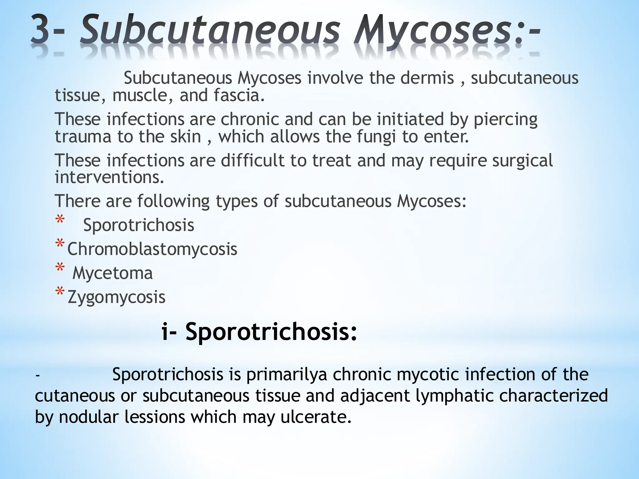
i- Sporotrichosis:
- Sporotrichosis is primarilya chronic mycotic infection of the
cutaneous or subcutaneous tissue and adjacent lymphatic characterized
by nodular lessions which may ulcerate.
47.
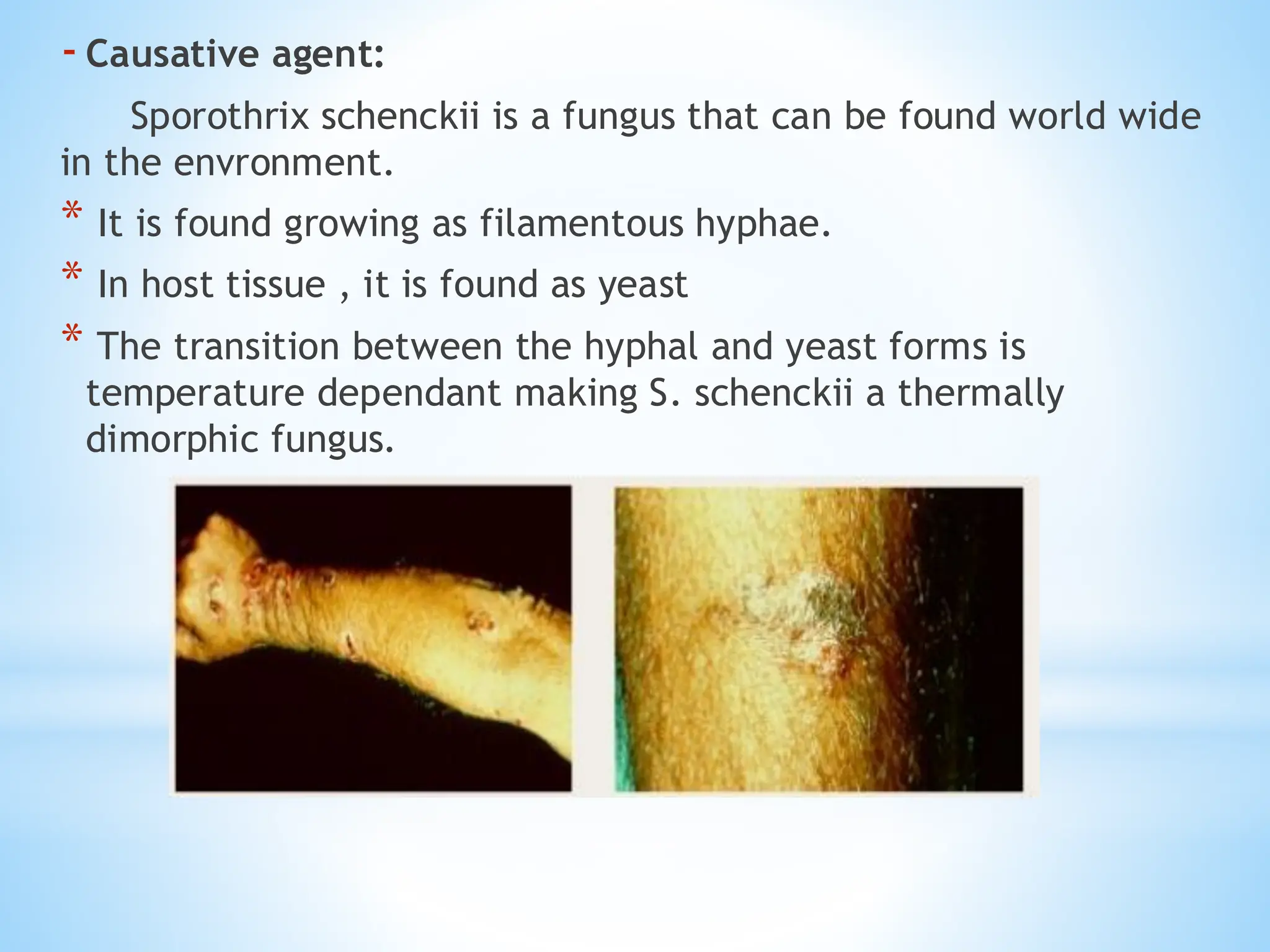
-Causative agent:
Sporothrix schenckiiis a fungus that can be found world wide
in the envronment.
* It is found growing as filamentous hyphae.
* In host tissue , it is found as yeast
* The transition between the hyphal and yeast forms is
temperature dependant making S. schenckii a thermally
dimorphic fungus.
48.
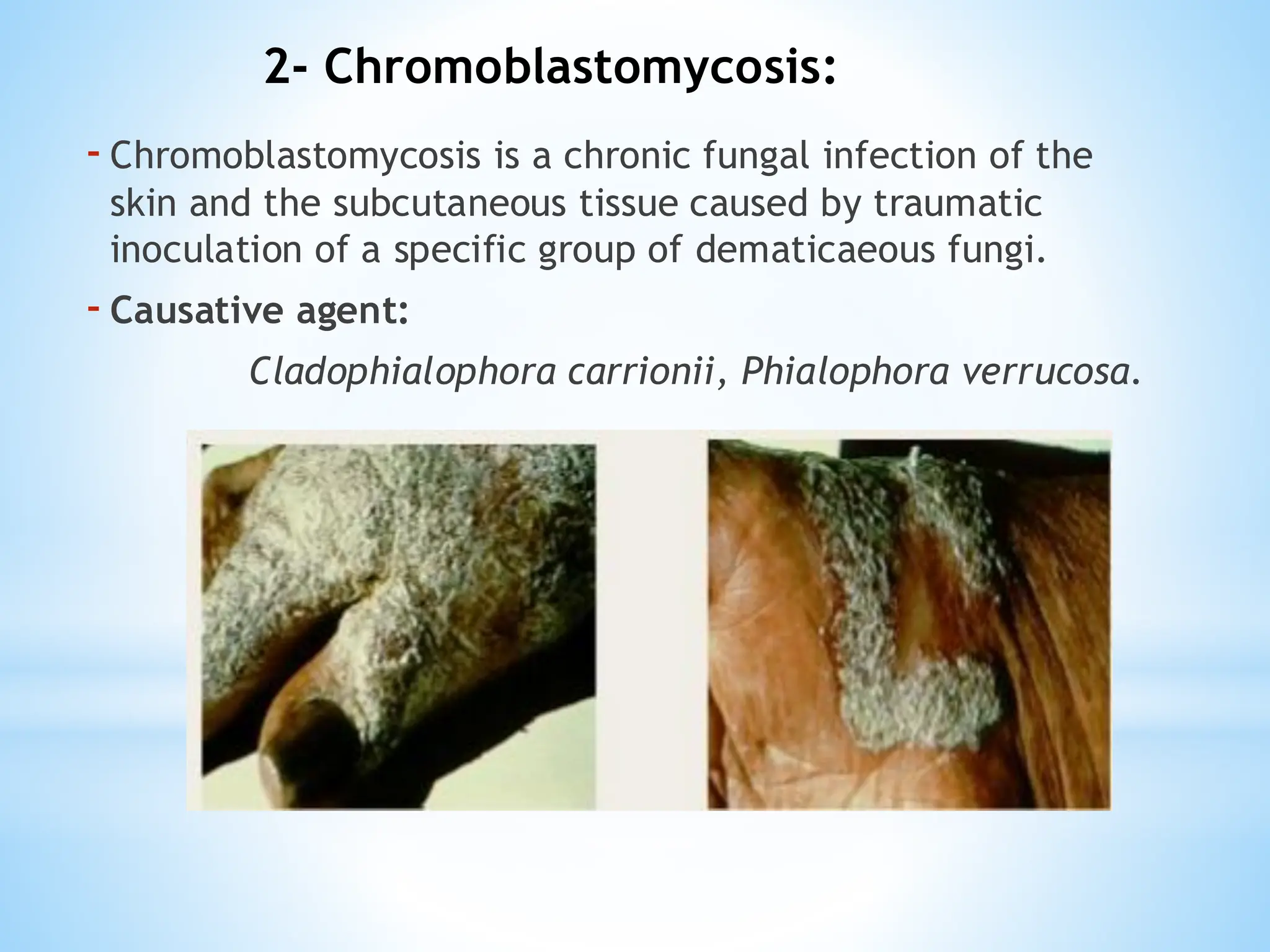
-Chromoblastomycosis is achronic fungal infection of the
skin and the subcutaneous tissue caused by traumatic
inoculation of a specific group of dematicaeous fungi.
-Causative agent:
Cladophialophora carrionii, Phialophora verrucosa.
2- Chromoblastomycosis: