Estradiol Partially Recapitulates Murine Pituitary Cell Cycle Response to Pre...
FGF21-FluoRatFinal
1. FGF-21 Activation of Hypothalamic Nuclei that Controls Metabolic and Cardiovascular Activity
Andrew Keens, Barbara Bernardo, Corey Petrella Matthew Zahner
Pfizer Worldwide Research & Development, Groton Laboratories, Groton, CT 06340
INTRODUCTION
Endogenous FGF21 binds to the suprachiasmatic nucleus of the hypothalamus
which effects blood glucose regulation and increases metabolism, leading to
weight loss. The hypothalamus plays a significant role in autonomic control of
both metabolic and cardiovascular activity. Several hypothalamic nuclei work
together to create appropriate circuitry to regulate food intake and energy
metabolism. One main hypothalamic nuclei involved in energy balance and food
intake is the paraventricular nucleus (PVN). In rats and several other species,
PVN lesion causes hyperphagia and obesity. Because FGF21 has been shown
to increase sympathetic metabolic activity it is suggested that the beneficial
metabolic effects of FGF21 are mediated by hypothalamic mechanisms. Based
on this we hypothesize that FGF21-mediated increased metabolism relies on an
intact PVN. To test this hypothesis we performed ibotenic acid lesion of the PVN
(or sham) and measured body weight, temperature, and food consumption for 4
weeks. Although PVN lesion has previously been shown to increase food intake
in rats fed standard chow, leading to obesity, it did not augment food intake in rats
fed a high fat diet. In this regard, all rats became hyperphagic and obese. We
speculate that this may be due to the high palatability of the chow causing
hyperphagia in sham lesion rats as well. Nevertheless because all rats became
obese we were unable to determine the involvement of PVN on feeding behavior.
Many central mechanisms involved in the regulation of metabolism and food
intake also involve central autonomic pathways involved in sympathetic control of
blood pressure. Sympathetic neurons in the PVN project to either the
cardiovascular regulation site in the rostroventrolateral medulla (RVLM) in the
brainstem medulla, the sympathetic preganglionic neurons in the
intermediolateral cell column of the spinal cord, or both. Sympathetic regulation
of metabolism is mediated by neurons in the raphe nucleus located in the
rostroventromedial medulla. Electrophysiological studies have demonstrated that
FGF21 activates sympathetic fibers that innervate intrascapular brown adipose
tissue which is involved in autonomic regulation of metabolism and body
temperature. Because several metabolically active signaling hormones, including
FGF21, increase sympathetic activity we hypothesize that FGF21 also activates
cardiovascular-related sympathetic neurons in the PVN. To test this hypothesis
we performed neuroanatomical assessment to differentiate cardiovascular-related
hypothalamic neurons from metabolically-related hypothalamic neurons. To do
this we injected two different fluorescent retrograde tracers into either the RVLM
which is involved in sympathetic cardiovascular activity, or the raphe which is
involved in sympathetic metabolic activity. Neurons that project to these regions in
the brainstem will take up the fluorescent microspheres and allow us to identify
the hypothalamic origin of those projections. We identified robust fluorescence by
both RVLM-projecting neurons which were mainly located in the PVN as well as
raphe-projecting neurons which were mainly located in the dorsomedial nucleus.
In order to determine the role of FGF21 in differential activation of presympathetic
cardiovascular vs. metabolically-related neurons we will treat rats with FGF21 and
assess the Fos activation of these fluorescent neurons in the PVN and
dorsomedial hypothalamic nucleus.
STUDY DESIGN; BEHAVIORAL AND NEUROANATOMICAL ASSESSMENT FOR CENTRAL AUTONOMIC
REGULATION OF ENERGY BALANCE
RESULTS
BEHAVIORAL ASSESSMENT PARAVENTRICULAR NUCLEUS LESION
A
B
B
SUMMARY
• PVN lesion did not augment increased obesity in high-fat diet fed rats.
• Future testing of the hypothalamic role in feeding and metabolism will be
conducted by using standard chow.
• Using fluoresce microscopy we identified robust co-localization of cardiovascular
(RVLM) and metabolic (raphe) hypothalamic neurons.
• We will use FGF21-induced pERK and FOS activation to better understand
differential activation of these pre-sympathetic cardiovascular and metabolically-
related neurons.
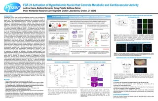
FLUORESCENCE MICROSCOPY IDENTIFICATION OF HYPOTHALAMIC
AUTONOMIC NUCLEI
Figure 3. Confocal images showing immunofluorescence identification of retrograde labeled
RVLM-projecting paraventricular nucleus neurons (red) and raphe-projecting dorsomedial
hypothalamic neurons (green). Parasagittal stereotaxic drawing with corresponding anterior-
posterior coronal sections (A). Photomicrographs of corresponding hypothalamic sections (B).
Tiled (10X), low (10X) and high (40X z-stack). Low magnification (10X) and high magnification
(40X), scale bars: 200 um in low and 50 um in high.
CO-LOCALIZATION OF RVLM & RAPHE-PROJECTING NEURONS
Figure 4. Confocal images showing co-localization of RVLM-projecting paraventricular nucleus
neurons (red) and raphe-projecting dorsomedial hypothalamic neurons (green)at 10X (A) 20X (B)
and 63X (C) showing RVLM-projecting and raphe-projecting neurons.
MICROINJECTION OF RETROGRADE TRACER INTO BRAINSTEM
Figure 5. Identification of microinjection site for fluospheres into the brainstem. Coronal
stereotaxic drawing at the level of the RVLM and raphe (A). Image of the frozen brainstem at the
corresponding level (B). Distribution of the injectant viewed under florescent illumination (C). Red
fluosphere was injected bilaterally into the RVLM (100nl) and yellow (green under fluorescence)
was injected into the raphe.
ACKNOWLEDGEMENTS
Thank you Timothy Coskran and James Finley for taking the time to help us take
and examine these images.
Figure 2. Coronal stereotaxic drawing at the level of the PVN (A).
Representative images of H&E stained coronal sections (at 2X
magnification) showing the PVN of either a sham or ibotenic acid (2 ng/
nl) lesioned rat (B). Note the two darkened regions indicated by the
arrows and the absence of PVN’s neurons below them. A 10X
magnification of the same tissue, seen to the right, with the PVN
outlined in white.
Figure 1. Lesion of the PVN did not augment obesity. Body weights (A),
food intake (B) and temperature (C) in rats with either PVN lesion
(n=15) or sham (n=15) were recorded weekly. All rats were acclimated
to and maintained on a high fat diet throughout the study. Data are
represented as mean ± SEM.
A
C
METHODS
• Male Sprague-Dawley rats maintained on high-fat diet.
• Bilateral PVN lesion using 100 nl injections of ibotenic acid (2 ng/nl) or sham.
• Body weights, body temperatures, and food consumption recorded weekly.
• In a separate group of rats, fluorescently labeled microspheres were injected
into the RVLM and the raphe of the brainstem medulla.
• Rats were euthanized for histological identification PVN lesion or of
hypothalamic RVLM and raphe projecting neurons.
• Brains were postfixed with 4% formaldehyde for 24 hours and cryoprotected
with 30% sucrose prior to sectioning.
• Histology was performed on 40 mm frozen sections with a sliding microtome.
• Sections were examined on a Zeiss Confocal microscope and tiled Z-stack
images were collected and processed for 3D reconstruction in Zen software
(Carl Zeiss, Inc).