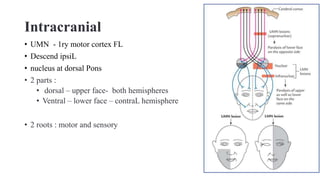
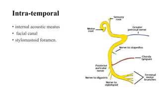
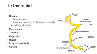
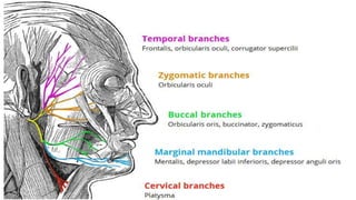
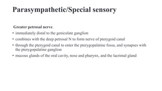
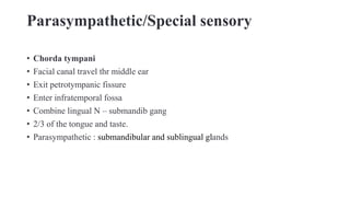
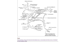
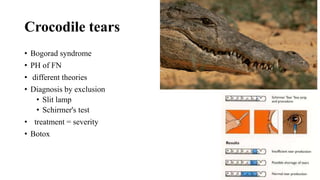
Facial nerve
Introduction
• VII
• second pharyngeal arch
• Motor:
• Sensory
• Special sensory
• Parasympathetic
Intracranial
• UMN - 1ry motor cortex FL
• Descend ipsiL
• nucleus at dorsal Pons
• 2 parts :
• dorsal – upper face- both hemispheres
• Ventral – lower face – contraL hemisphere
• 2 roots : motor and sensory
Intra-temporal
• internal acoustic meatus
• facial canal
• stylomastoid foramen.
Extracranial
• Branches :
• Post auricular :
• Branch to post belly of the digastric muscle
• stylohyoid muscle
• Parotid gland
• Temporal
• Zygomatic
• Buccal
• Marginal mandibular
• Cervical
Parasympathetic/Special sensory
Greater petrosal nerve.
• immediately distal to the geniculate ganglion
• combines with the deep petrosal N to form nerve of pterygoid canal
• through the pterygoid canal to enter the pterygopalatine fossa, and synapses with
the pterygopalatine ganglion
• mucous glands of the oral cavity, nose and pharynx, and the lacrimal gland
Parasympathetic/Special sensory
• Chorda tympani
• Facial canal travel thr middle ear
• Exit petrotympanic fissure
• Enter infratemporal fossa
• Combine lingual N – submandib gang
• 2/3 of the tongue and taste.
• Parasympathetic : submandibular and sublingual glands
Intracranial Injury
• Muscles of facial expression
• Chorda tympani – altered salivation and loss of taste on the ipsilateral 2/3 of the
tongue.
• Nerve to stapedius – ipsilateral hyperacusis.
• Greater petrosal nerve – ipsilateral reduced lacrimal fluid production.
Extracranial injury
• distal to the stylomastoid foramen.
• Only the motor function : muscles of facial expression.
Crocodile tears
• Bogorad syndrome
• PH of FN
• different theories
• Diagnosis by exclusion
• Slit lamp
• Schirmer's test
• treatment = severity
• Botox
Hyperacusis
• unusual tolerance
• Less data
• Tinnitus
• 1.5-hydroxytryptamine (5-HT) dysfunction
• 2.neurotransmitters
• 3.auditory efferent dysfunction
• Desensitization
facial nerve.pptx

facial nerve.pptx

  • 1.
  • 2.
    Introduction • VII • secondpharyngeal arch • Motor: • Sensory • Special sensory • Parasympathetic
  • 3.
    Intracranial • UMN -1ry motor cortex FL • Descend ipsiL • nucleus at dorsal Pons • 2 parts : • dorsal – upper face- both hemispheres • Ventral – lower face – contraL hemisphere • 2 roots : motor and sensory
  • 4.
    Intra-temporal • internal acousticmeatus • facial canal • stylomastoid foramen.
  • 5.
    Extracranial • Branches : •Post auricular : • Branch to post belly of the digastric muscle • stylohyoid muscle • Parotid gland • Temporal • Zygomatic • Buccal • Marginal mandibular • Cervical
  • 7.
    Parasympathetic/Special sensory Greater petrosalnerve. • immediately distal to the geniculate ganglion • combines with the deep petrosal N to form nerve of pterygoid canal • through the pterygoid canal to enter the pterygopalatine fossa, and synapses with the pterygopalatine ganglion • mucous glands of the oral cavity, nose and pharynx, and the lacrimal gland
  • 8.
    Parasympathetic/Special sensory • Chordatympani • Facial canal travel thr middle ear • Exit petrotympanic fissure • Enter infratemporal fossa • Combine lingual N – submandib gang • 2/3 of the tongue and taste. • Parasympathetic : submandibular and sublingual glands
  • 9.
    Intracranial Injury • Musclesof facial expression • Chorda tympani – altered salivation and loss of taste on the ipsilateral 2/3 of the tongue. • Nerve to stapedius – ipsilateral hyperacusis. • Greater petrosal nerve – ipsilateral reduced lacrimal fluid production.
  • 10.
    Extracranial injury • distalto the stylomastoid foramen. • Only the motor function : muscles of facial expression.
  • 12.
    Crocodile tears • Bogoradsyndrome • PH of FN • different theories • Diagnosis by exclusion • Slit lamp • Schirmer's test • treatment = severity • Botox
  • 13.
    Hyperacusis • unusual tolerance •Less data • Tinnitus • 1.5-hydroxytryptamine (5-HT) dysfunction • 2.neurotransmitters • 3.auditory efferent dysfunction • Desensitization

Editor's Notes

  • #4 It has two roots; a large motor, and a smaller sensory root (sometimes known as the intermediate nerve).
  • #5 roots travel into internal acoustic meatus, of the temporal bone. Then leave it and enter into the facial canal.  3 events happen : Roots unite geniculate ganglion 3 branches as follow Greater petrosal nerve – parasympathetic fibres to mucous and lacrimal gland. Nerve to stapedius – motor fibres to stapedius muscle of the middle ear. Chorda tympani – special sensory fibres to the anterior 2/3 tongue and parasympathetic fibres to the submandibular and sublingual glands. Nerve exits the facial canal (and the cranium) via the stylomastoid foramen.
  • #6  Post auricular  post belly of the occipitofrontalis and the auricular muscles and contributes cutaneous sensation from the skin covering the mastoid process and parts of the auricle.
  • #13  Theories : 1.Regenerating salivary nerve fibers undergo are misdirected to ultimately innervate the lacrimal gland instead of the submandibular gland 2. had underlying lateral rectus palsy 3. formation of an artificial synapse at the injury site
  • #14 5-HT does appear to have a role in modulating auditory gain and the determination of significance of sound