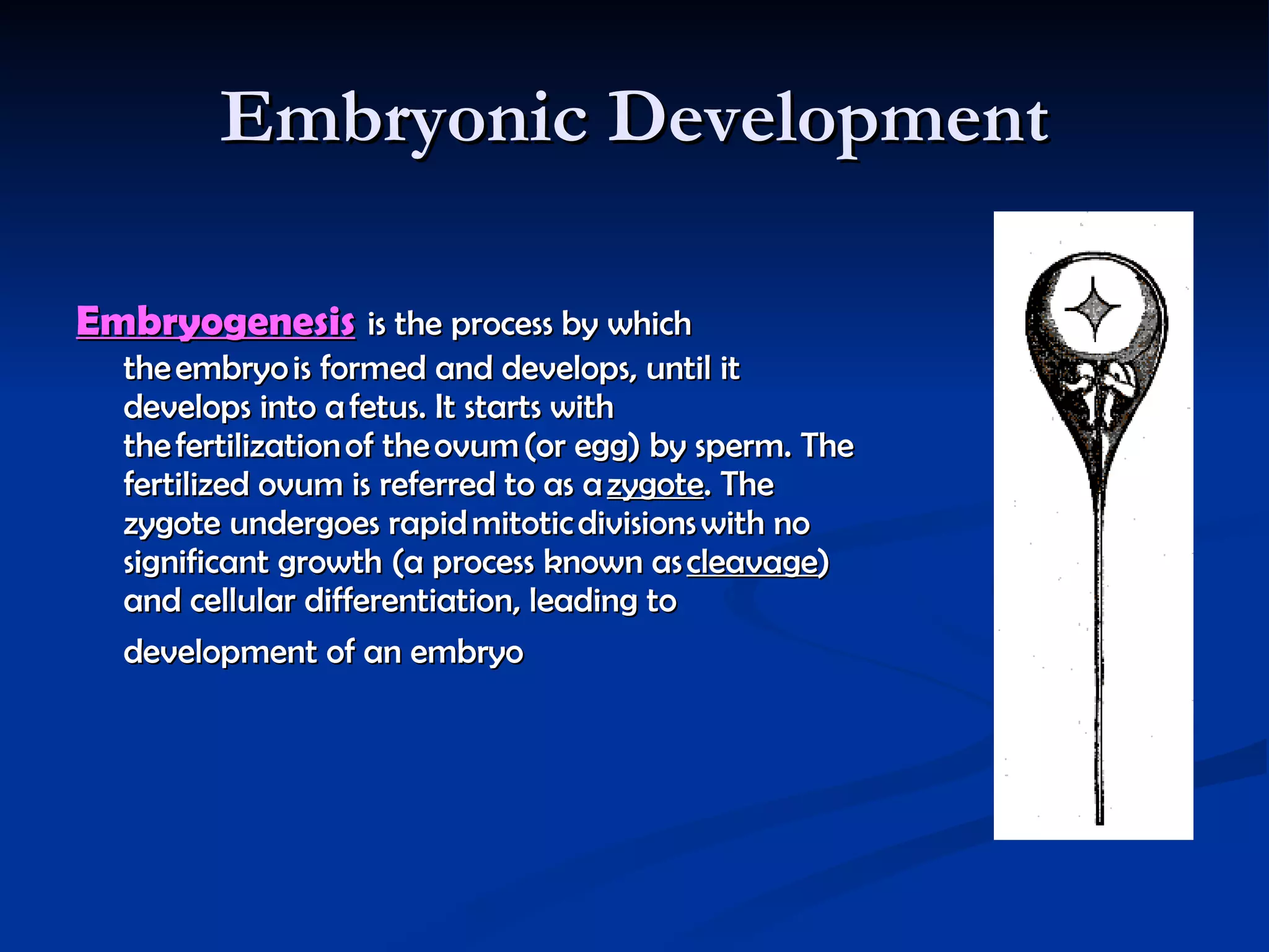
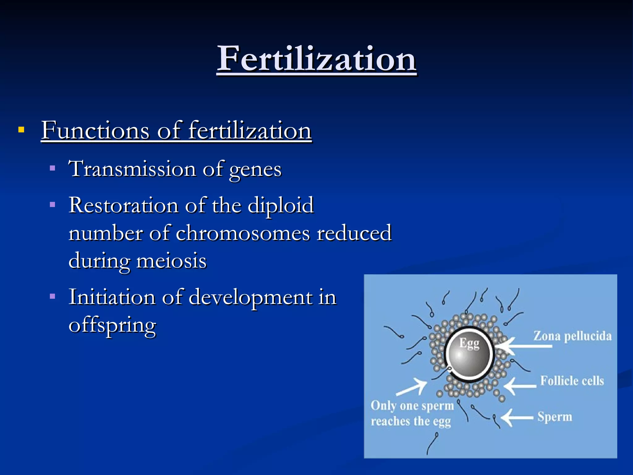
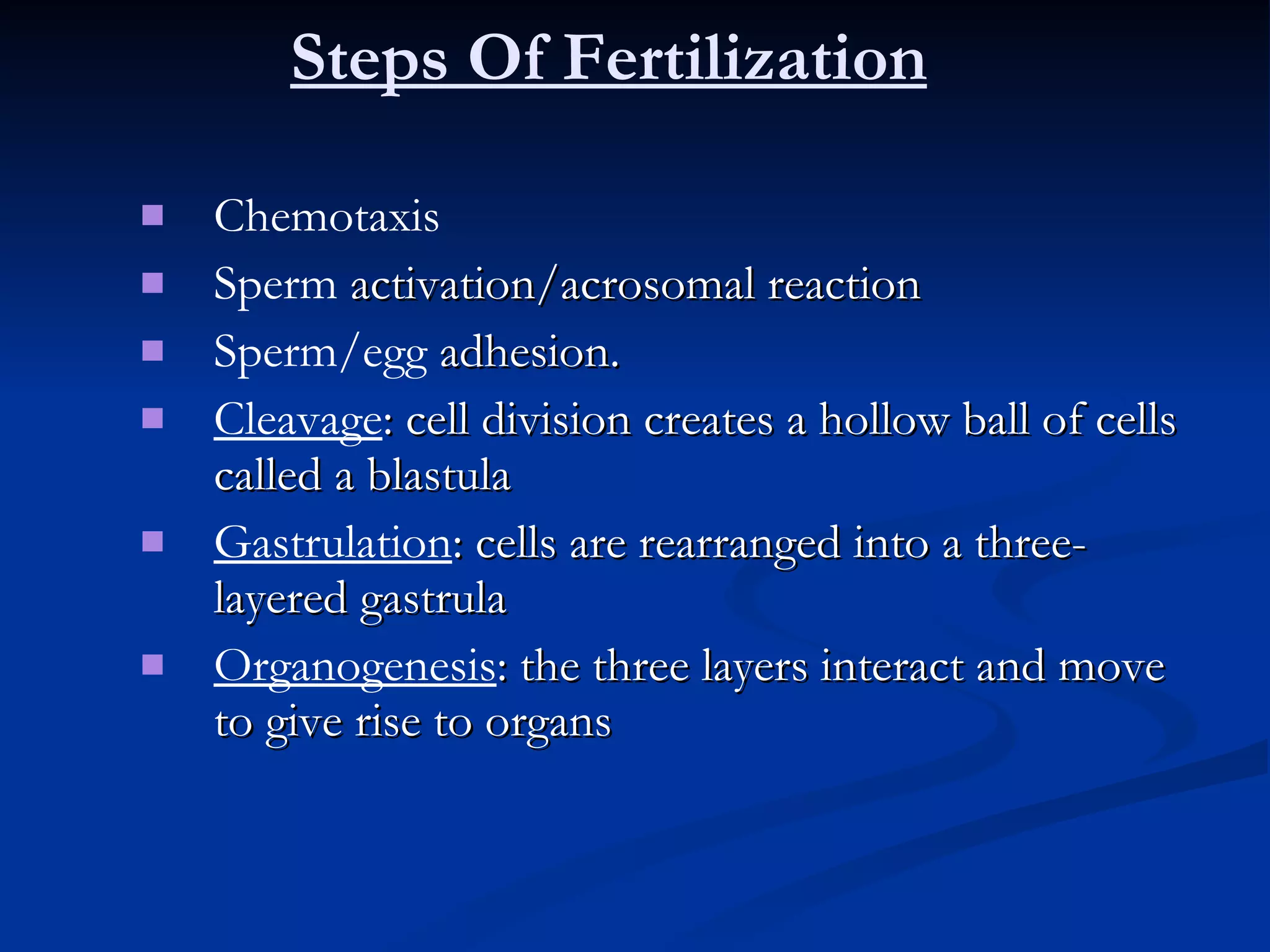
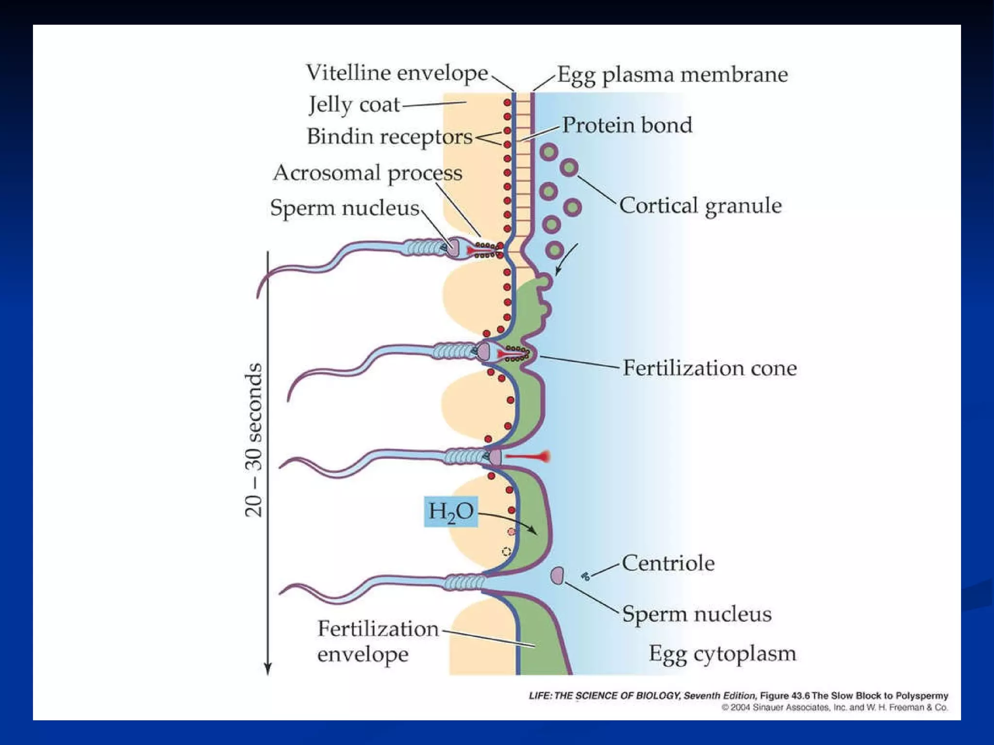
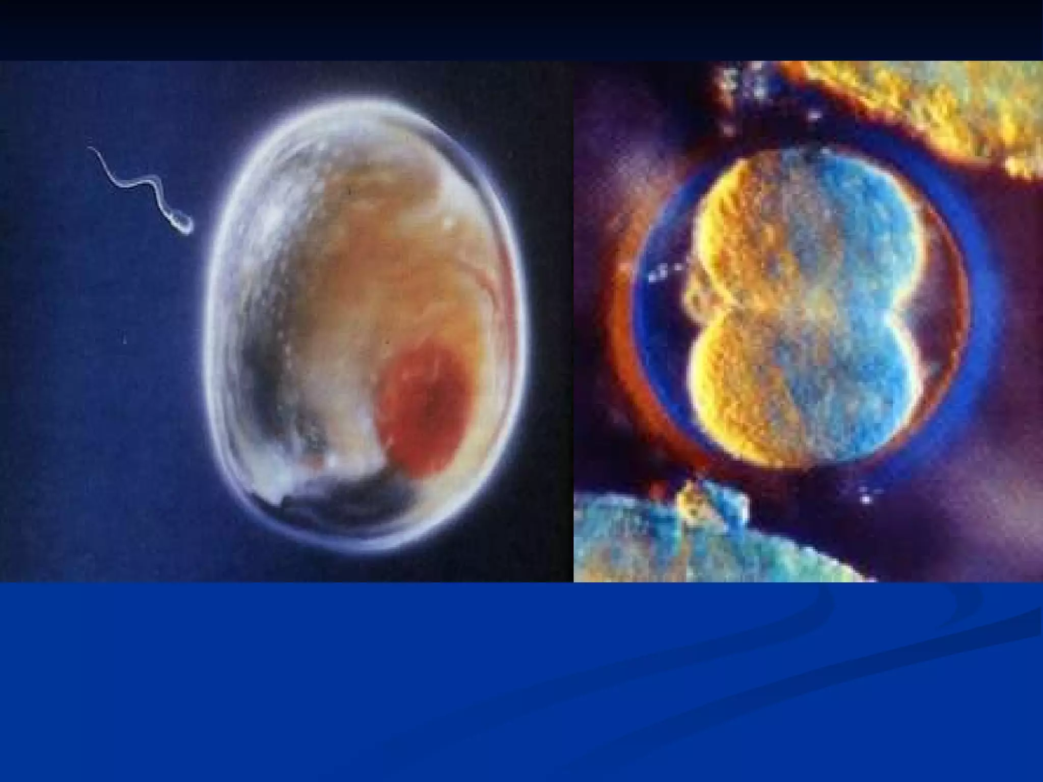
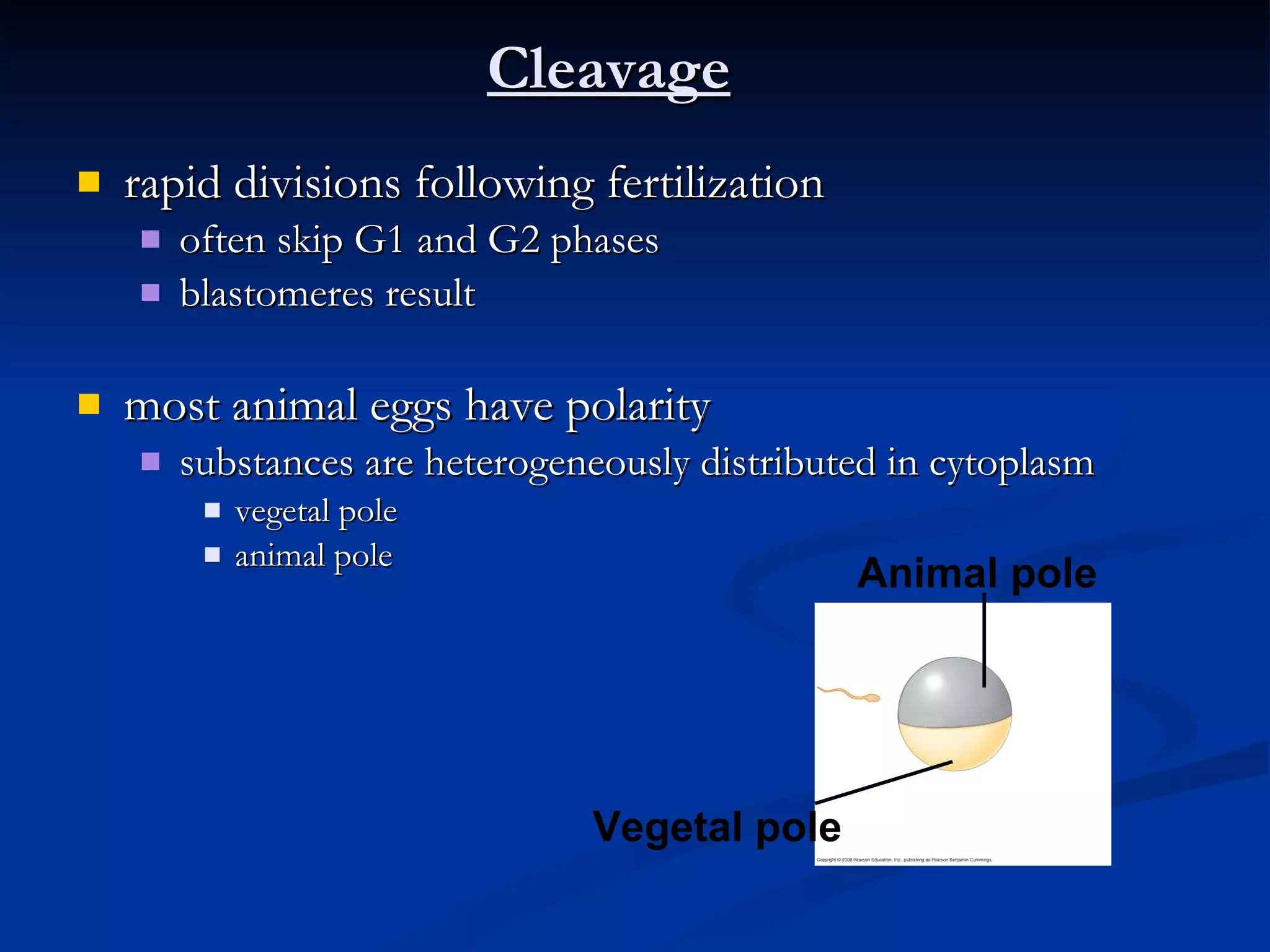
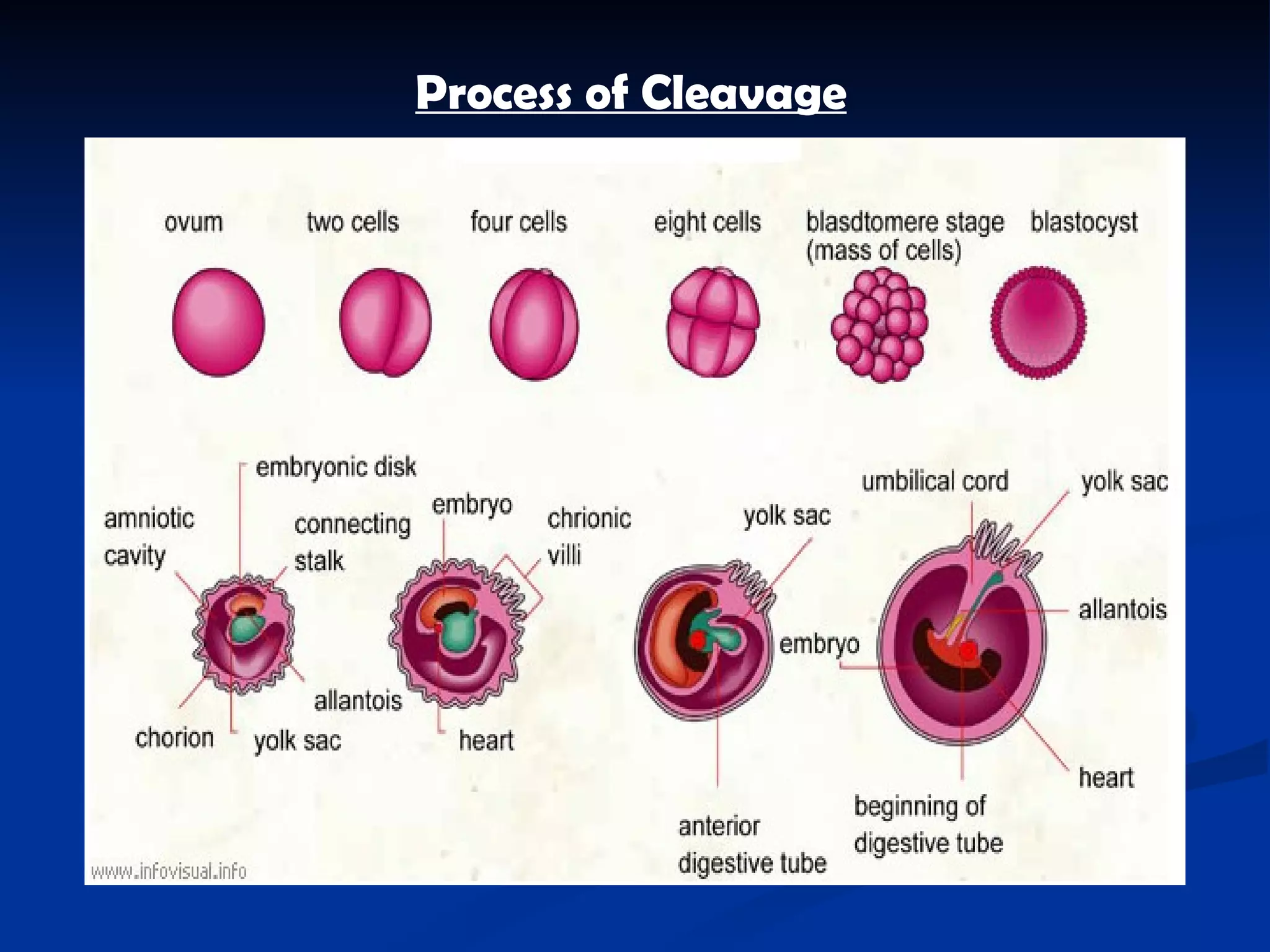
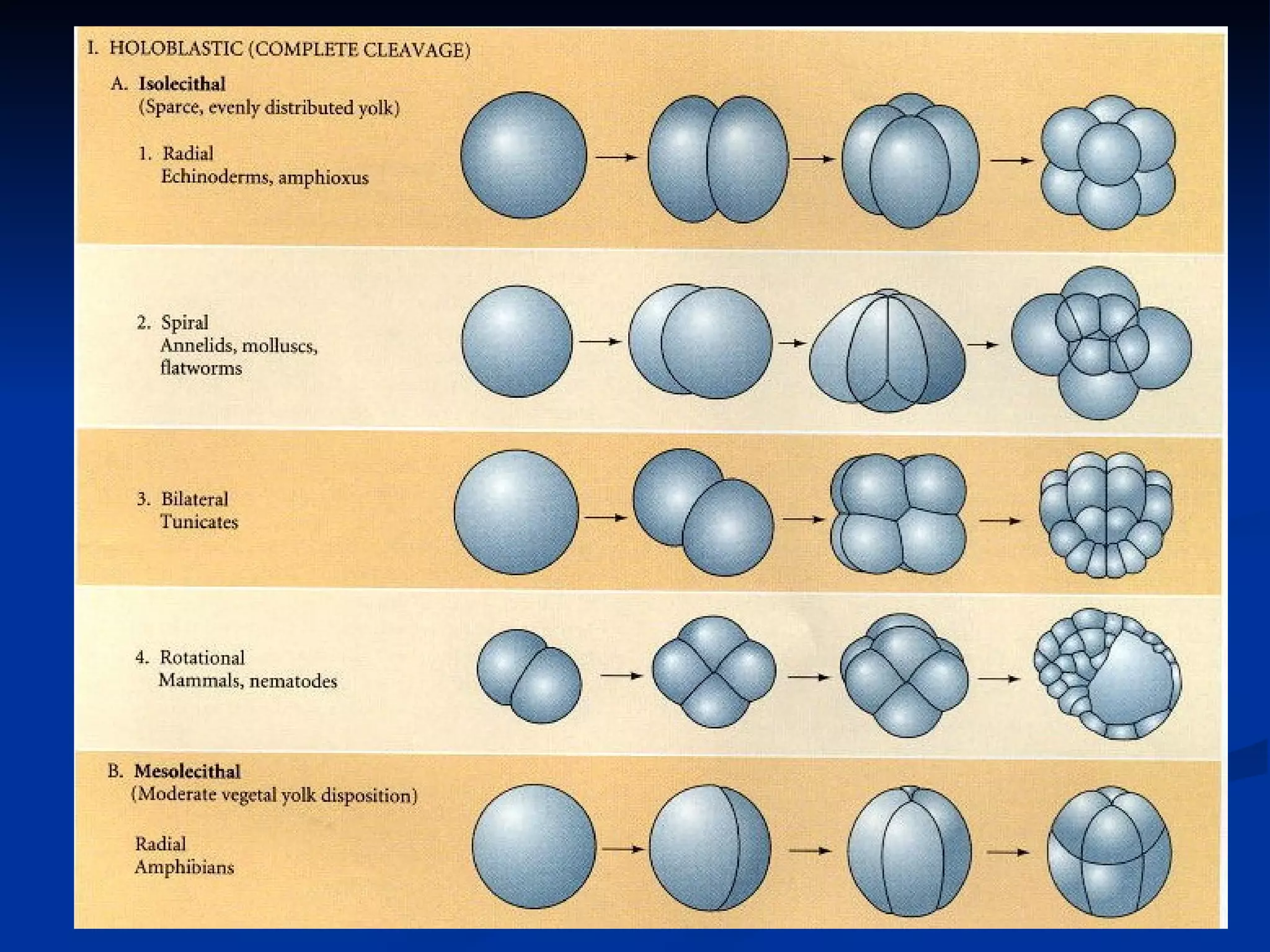
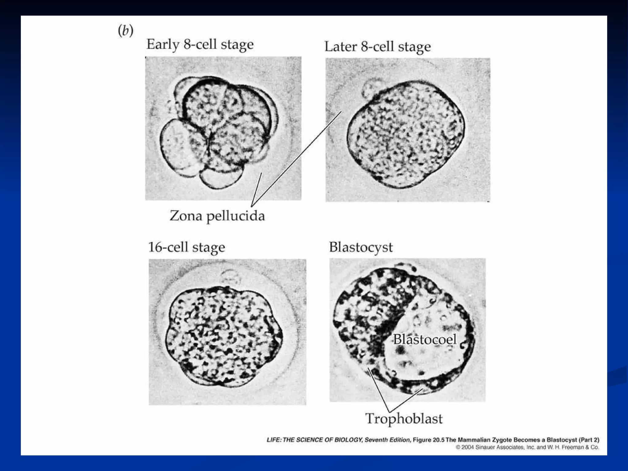
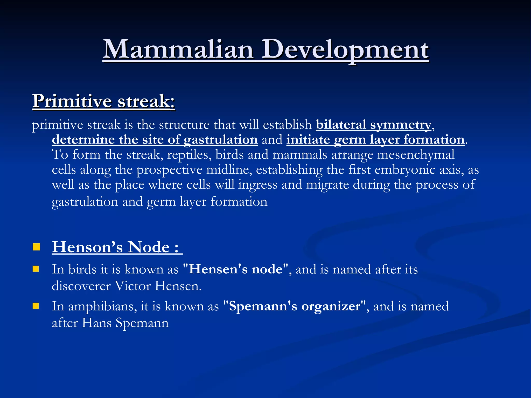
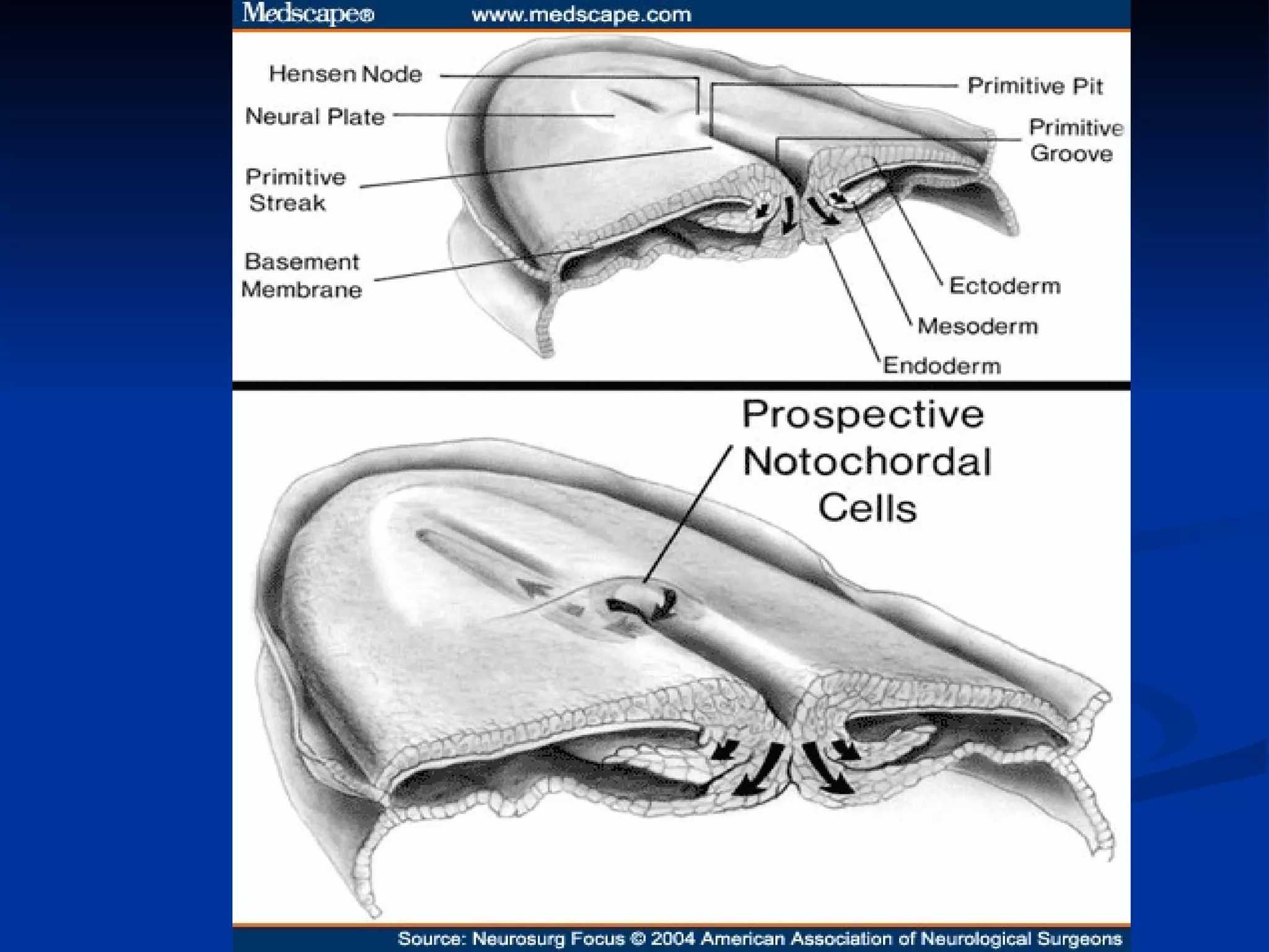
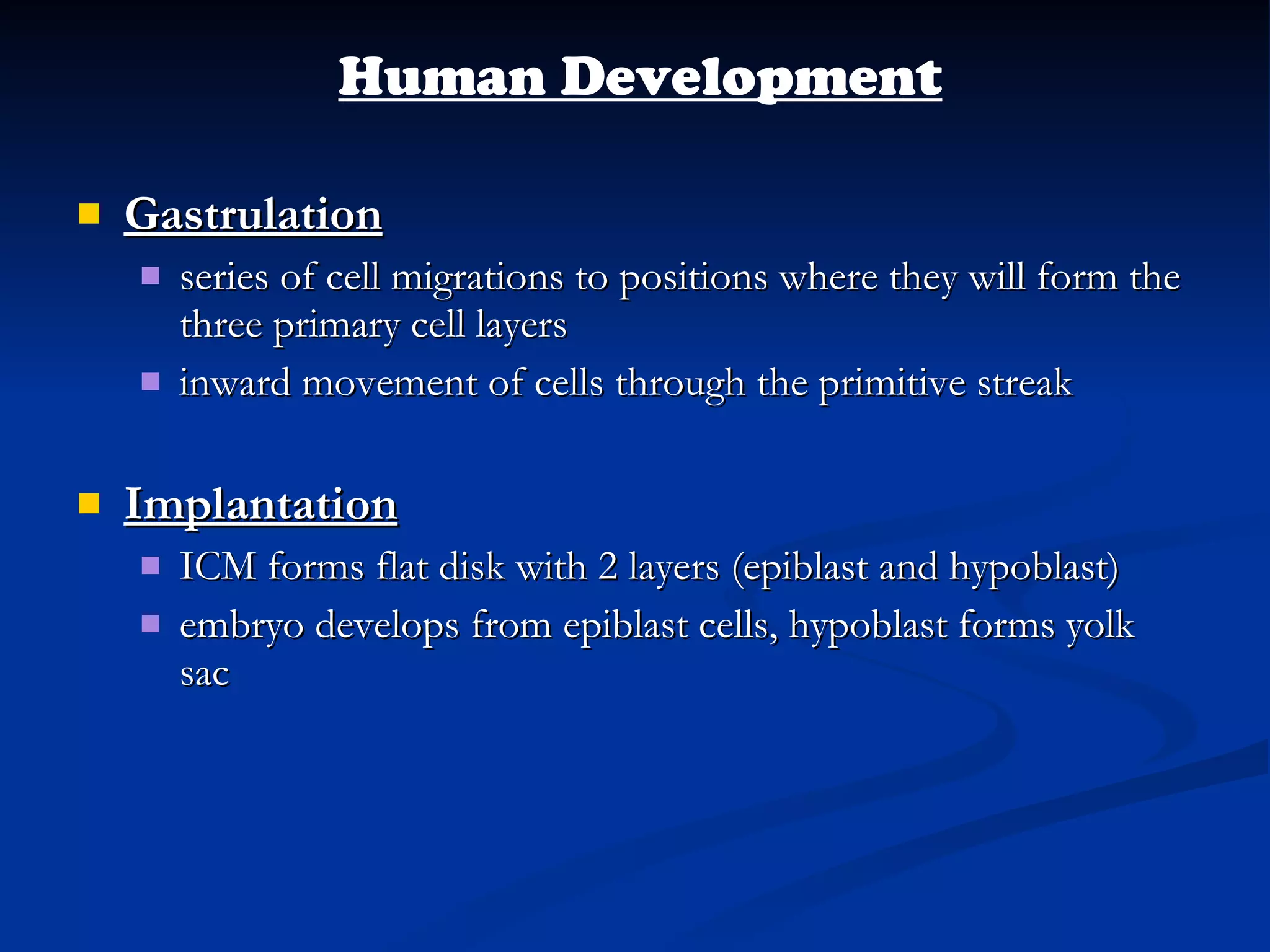
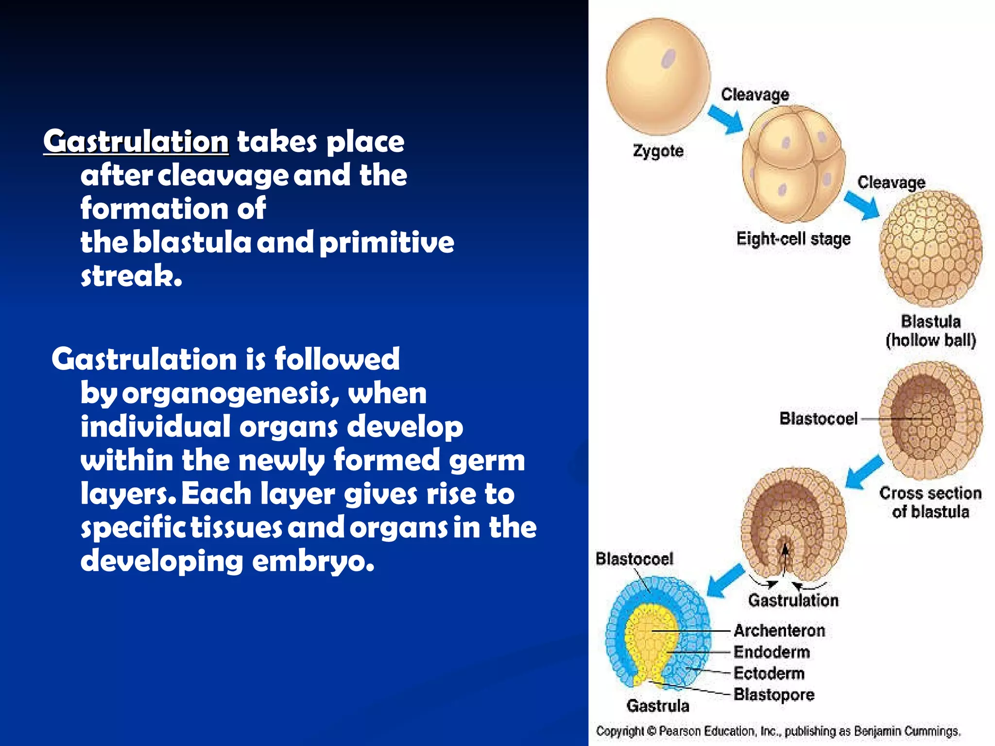
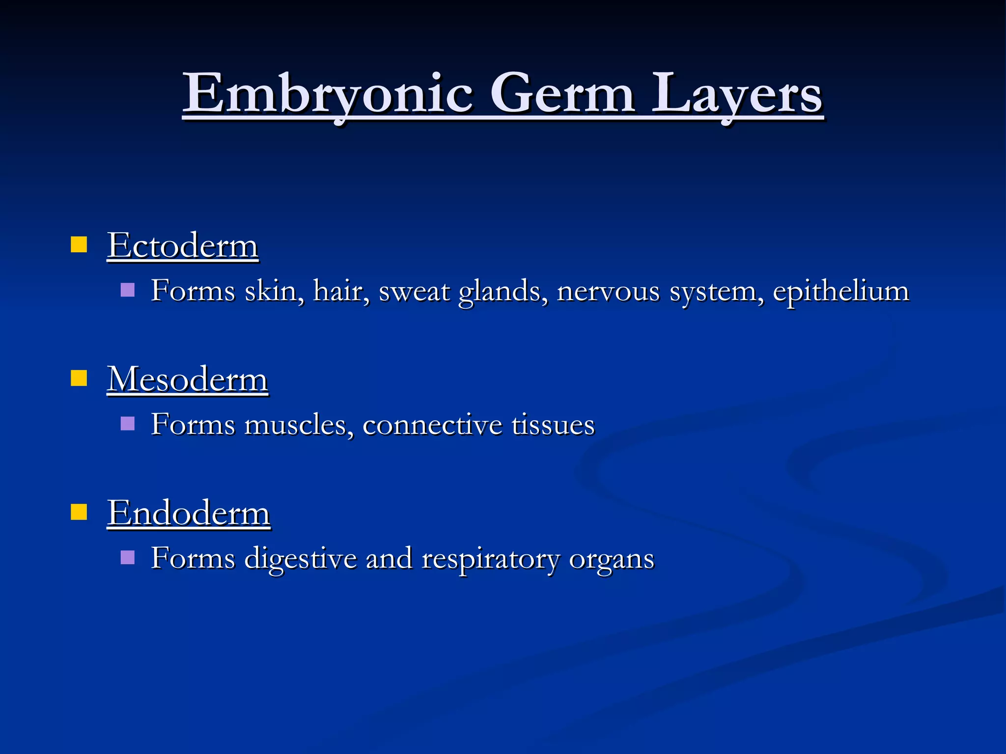
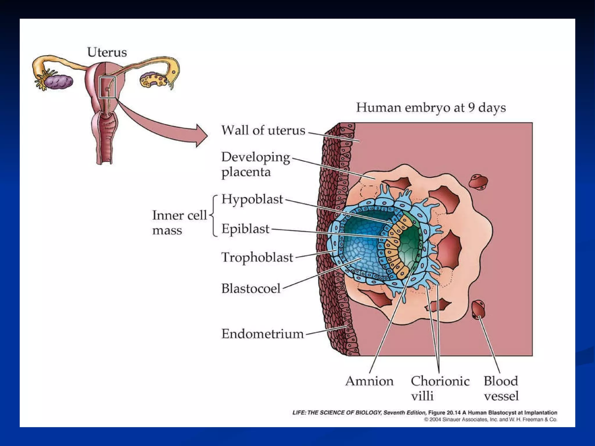
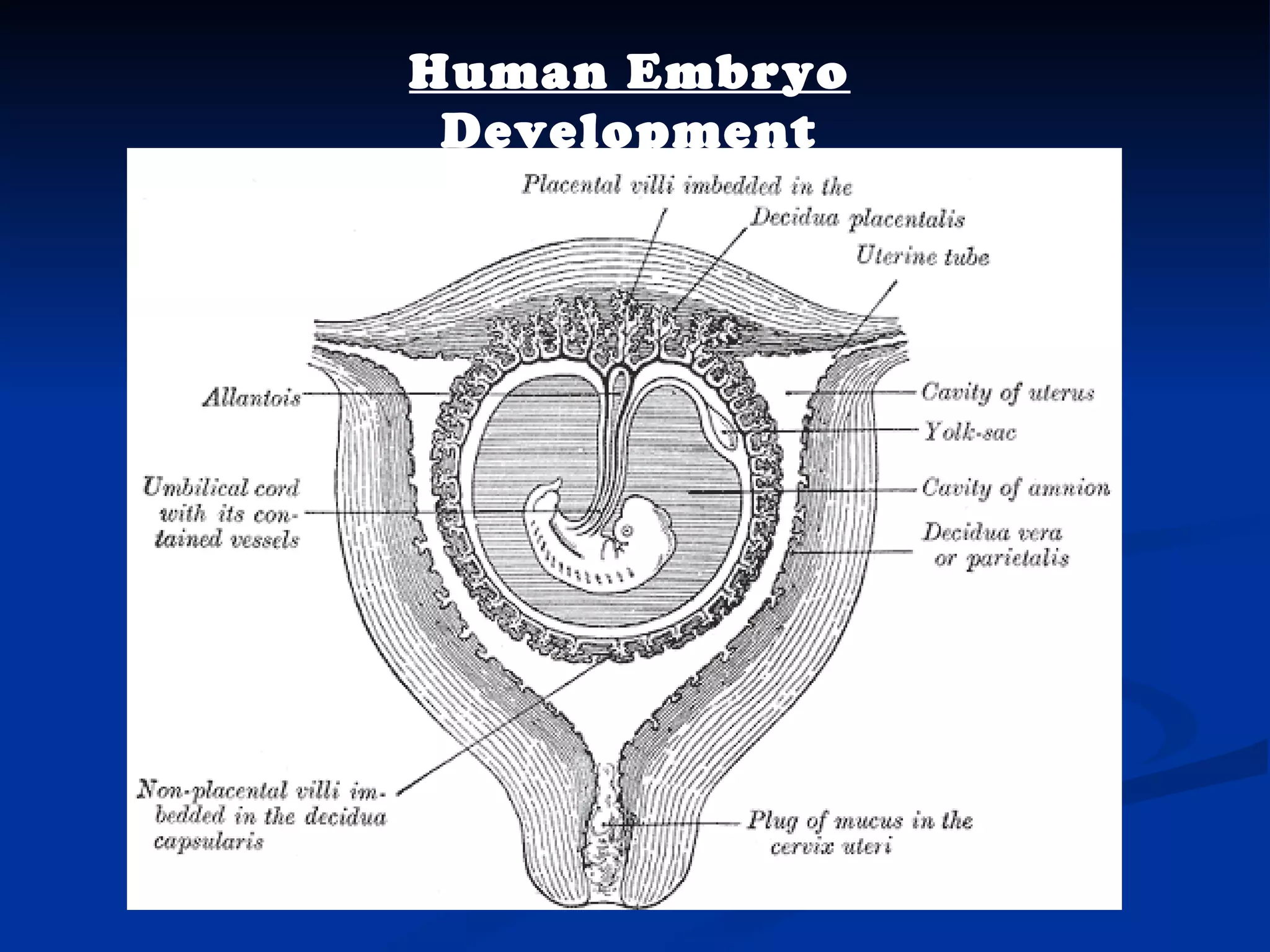
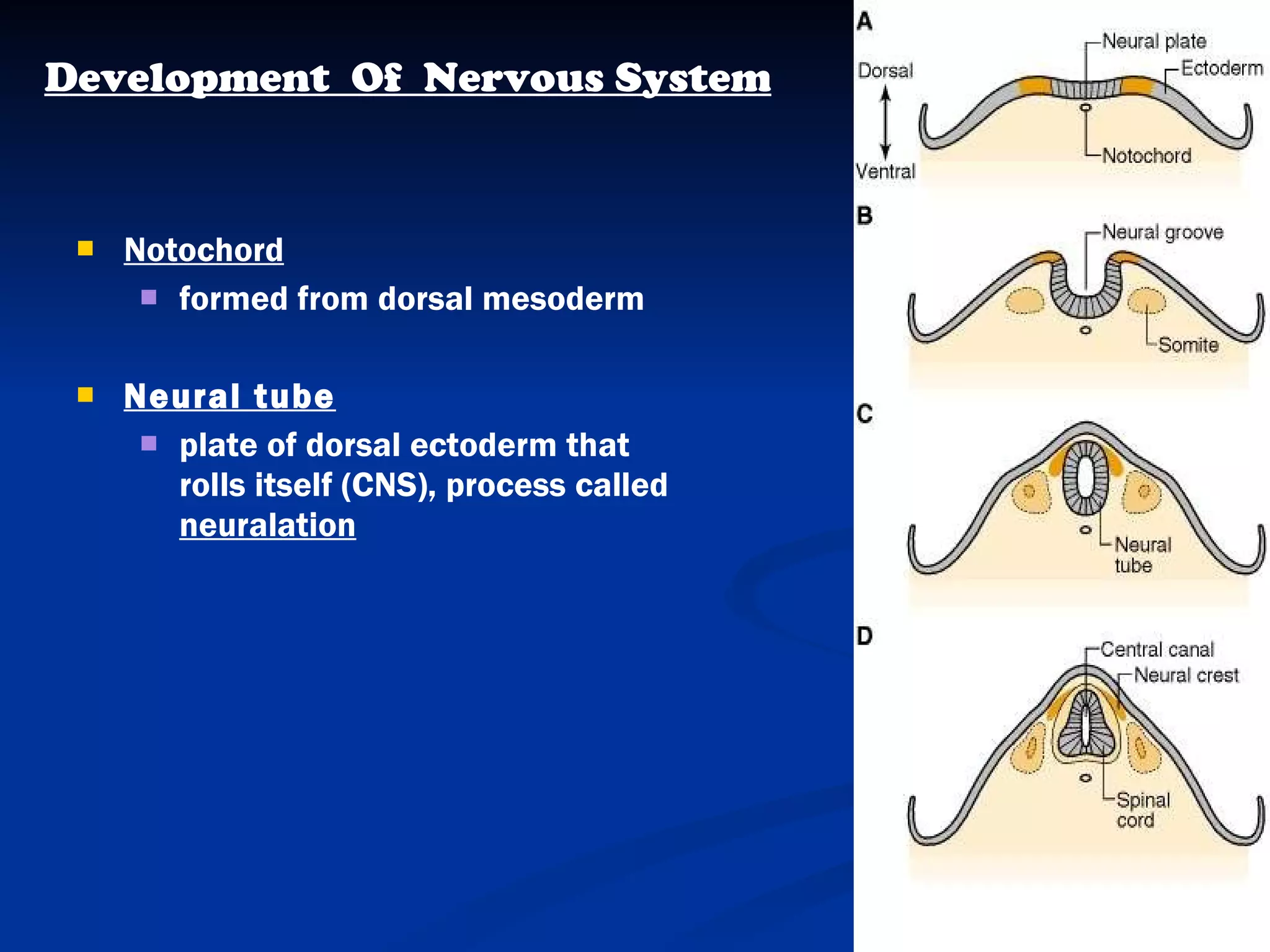
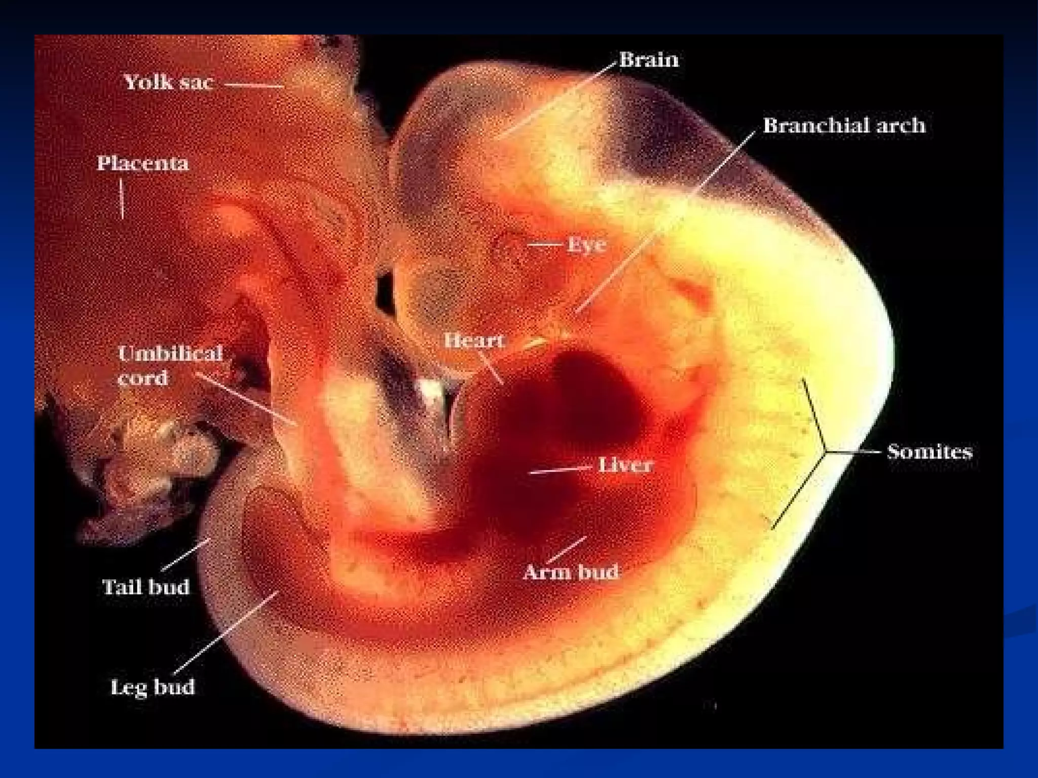
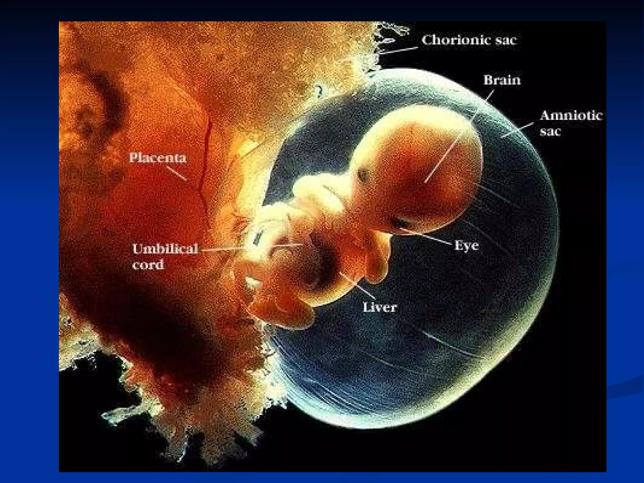
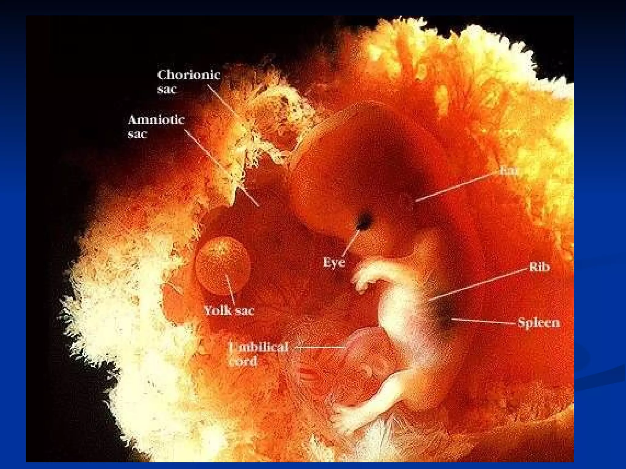
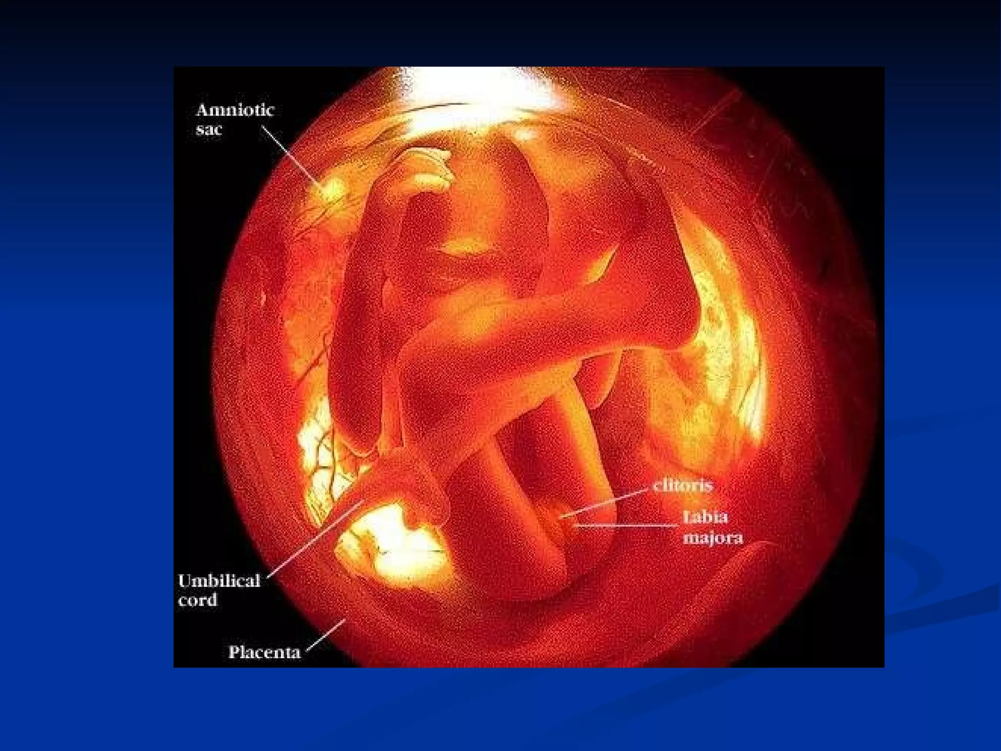
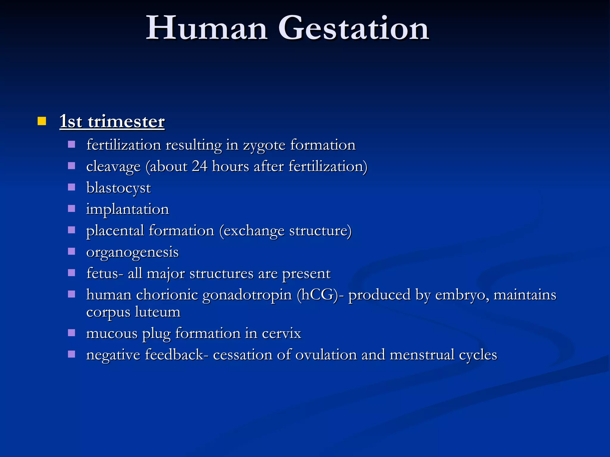
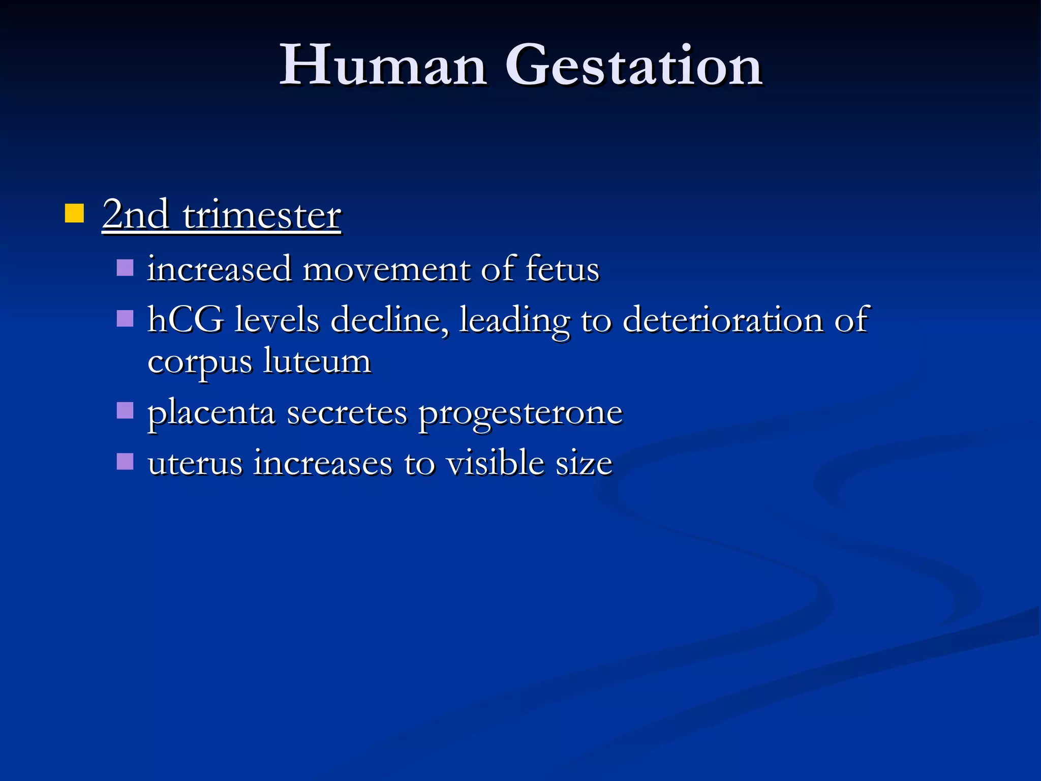
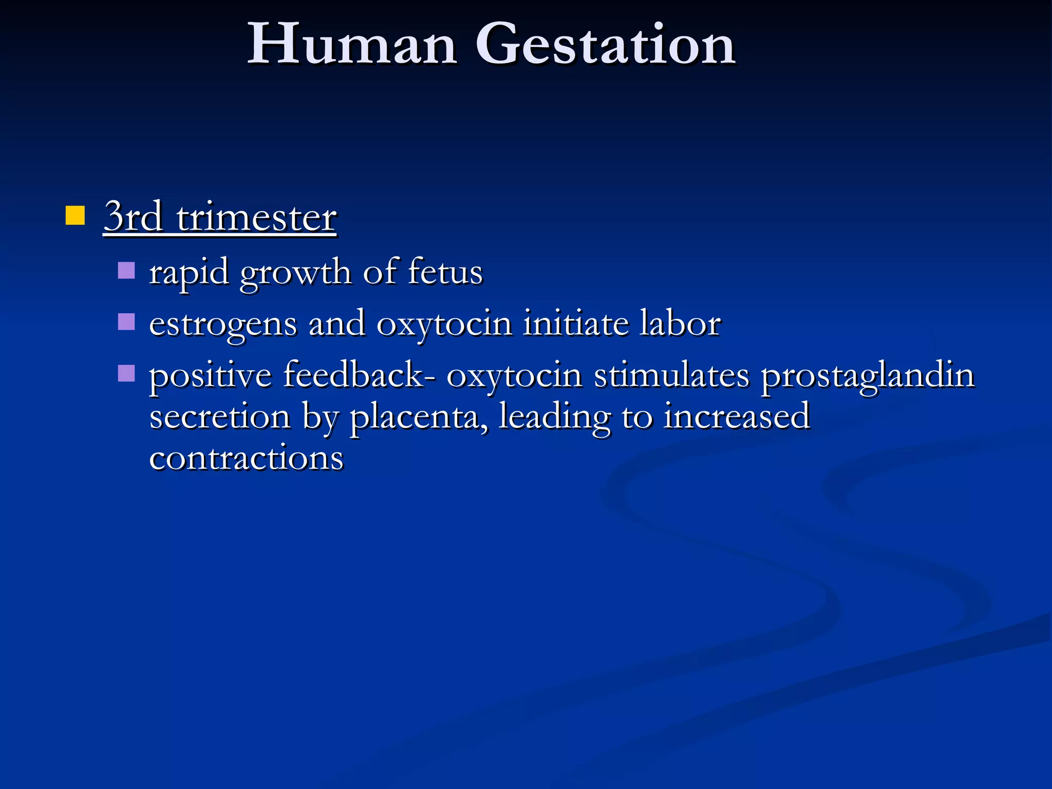
Embryonic Development Submitted by : Ananya Choudhary BSc (Hons) Medical Biotech Sem-6 BMB/08/101
Embryonic Development Embryogenesis   is the process by which the embryo is formed and develops, until it develops into a fetus. It starts with the fertilization of the ovum (or egg) by sperm. The fertilized ovum is referred to as a  zygote . The zygote undergoes rapid mitotic divisions with no significant growth (a process known as  cleavage ) and cellular differentiation, leading to development of an embryo
Fertilization Functions of fertilization Transmission of genes  Restoration of the diploid number of chromosomes reduced during meiosis Initiation of development in offspring
Steps Of Fertilization Chemotaxis Sperm  activation/acrosomal reaction Sperm/egg  adhesion. Cleavage : cell division creates a hollow ball of cells called a blastula Gastrulation : cells are rearranged into a three-layered gastrula Organogenesis : the three layers interact and move to give rise to organs
 
Fertilization activates the egg, initiating metabolic processes acrosomal reaction sperm are activated  acrosomal process sperm and egg membranes fuse ion channels open, allowing Na +  to flow in fast block to polyspermy cortical reaction egg’s ER releases Ca 2+  into the cytosol at site of sperm entry
Fertilization slow block to polyspermy Ca 2+  causes cortical granules underneath the plasma membrane to fuse Mucopolysaccharides draw water into the space, swelling it Vitelline layer becomes the fertilization membrane


Cleavage rapid divisions following fertilization often skip G1 and G2 phases blastomeres result most animal eggs have polarity substances are heterogeneously distributed in cytoplasm vegetal pole animal pole Animal   pole Vegetal pole
Process of Cleavage


Mammalian Development Primitive streak :   primitive streak is the structure that will establish  bilateral symmetry ,  determine the site of gastrulation  and  initiate germ layer formation . To form the streak, reptiles, birds and mammals arrange mesenchymal cells along the prospective midline, establishing the first embryonic axis, as well as the place where cells will ingress and migrate during the process of gastrulation and germ layer formation    Henson’s Node :  In birds it is known as " Hensen's node ", and is named after its discoverer Victor Hensen. In amphibians, it is known as " Spemann's organizer ", and is named after Hans Spemann
 
Gastrulation series of cell migrations to positions where they will form the three primary cell layers inward movement of cells through the primitive streak Implantation ICM forms flat disk with 2 layers (epiblast and hypoblast) embryo develops from epiblast cells, hypoblast forms yolk sac Human Development
Gastrulation   takes place after cleavage and the formation of the blastula and primitive streak. Gastrulation is followed by organogenesis, when individual organs develop within the newly formed germ layers. Each layer gives rise to specific tissues and organs in the developing embryo.
Embryonic Germ Layers Ectoderm Forms skin, hair, sweat glands, nervous system, epithelium Mesoderm Forms muscles, connective tissues Endoderm Forms digestive and respiratory organs
 
Mammalian Development Allantois - outpocketing of embryo’s gut, incorporated into umbilical cord, forms blood vessels of umbilical cord This sac-like structure is primarily involved in  nutrition and excretion , and is webbed with blood vessels. The function of the allantois is to  collect liquid waste from the embryo , as well as to exchange gases used by the embryo.
Human Embryo Development
Notochord formed from dorsal mesoderm Neural tube plate of dorsal ectoderm that rolls itself (CNS), process called  neuralation Development  Of  Nervous System
Organogenesis Somites serially arranged strips of lateral mesoderm become vertebrae and muscles of axial skeleton Neural crest   created when neural tube pinches from ectoderm cells migrate and form teeth, bones, PNS, medulla of adrenal gland


Human Gestation 1st trimester fertilization resulting in zygote formation cleavage (about 24 hours after fertilization) blastocyst implantation placental formation (exchange structure) organogenesis fetus- all major structures are present  human chorionic gonadotropin (hCG)- produced by embryo, maintains corpus luteum mucous plug formation in cervix negative feedback- cessation of ovulation and menstrual cycles
Human Gestation 2nd trimester increased movement of fetus hCG levels decline, leading to deterioration of corpus luteum placenta secretes progesterone uterus increases to visible size
Human Gestation 3rd trimester rapid growth of fetus estrogens and oxytocin initiate labor positive feedback- oxytocin stimulates prostaglandin secretion by placenta, leading to increased contractions

Embryonic Development

  • 1.
    Embryonic Development Submittedby : Ananya Choudhary BSc (Hons) Medical Biotech Sem-6 BMB/08/101
  • 2.
    Embryonic Development Embryogenesis  is the process by which the embryo is formed and develops, until it develops into a fetus. It starts with the fertilization of the ovum (or egg) by sperm. The fertilized ovum is referred to as a  zygote . The zygote undergoes rapid mitotic divisions with no significant growth (a process known as  cleavage ) and cellular differentiation, leading to development of an embryo
  • 3.
    Fertilization Functions offertilization Transmission of genes Restoration of the diploid number of chromosomes reduced during meiosis Initiation of development in offspring
  • 4.
    Steps Of FertilizationChemotaxis Sperm activation/acrosomal reaction Sperm/egg adhesion. Cleavage : cell division creates a hollow ball of cells called a blastula Gastrulation : cells are rearranged into a three-layered gastrula Organogenesis : the three layers interact and move to give rise to organs
  • 5.
  • 6.
    Fertilization activates theegg, initiating metabolic processes acrosomal reaction sperm are activated acrosomal process sperm and egg membranes fuse ion channels open, allowing Na + to flow in fast block to polyspermy cortical reaction egg’s ER releases Ca 2+ into the cytosol at site of sperm entry
  • 7.
    Fertilization slow blockto polyspermy Ca 2+ causes cortical granules underneath the plasma membrane to fuse Mucopolysaccharides draw water into the space, swelling it Vitelline layer becomes the fertilization membrane
  • 8.
  • 9.
  • 10.
  • 11.
    Cleavage rapid divisionsfollowing fertilization often skip G1 and G2 phases blastomeres result most animal eggs have polarity substances are heterogeneously distributed in cytoplasm vegetal pole animal pole Animal pole Vegetal pole
  • 12.
  • 13.
  • 14.
  • 15.
  • 16.
    Mammalian Development Primitivestreak : primitive streak is the structure that will establish  bilateral symmetry , determine the site of gastrulation  and initiate germ layer formation . To form the streak, reptiles, birds and mammals arrange mesenchymal cells along the prospective midline, establishing the first embryonic axis, as well as the place where cells will ingress and migrate during the process of gastrulation and germ layer formation   Henson’s Node : In birds it is known as " Hensen's node ", and is named after its discoverer Victor Hensen. In amphibians, it is known as " Spemann's organizer ", and is named after Hans Spemann
  • 17.
  • 18.
    Gastrulation series ofcell migrations to positions where they will form the three primary cell layers inward movement of cells through the primitive streak Implantation ICM forms flat disk with 2 layers (epiblast and hypoblast) embryo develops from epiblast cells, hypoblast forms yolk sac Human Development
  • 19.
    Gastrulation takes place after cleavage and the formation of the blastula and primitive streak. Gastrulation is followed by organogenesis, when individual organs develop within the newly formed germ layers. Each layer gives rise to specific tissues and organs in the developing embryo.
  • 20.
    Embryonic Germ LayersEctoderm Forms skin, hair, sweat glands, nervous system, epithelium Mesoderm Forms muscles, connective tissues Endoderm Forms digestive and respiratory organs
  • 21.
  • 22.
    Mammalian Development Allantois- outpocketing of embryo’s gut, incorporated into umbilical cord, forms blood vessels of umbilical cord This sac-like structure is primarily involved in  nutrition and excretion , and is webbed with blood vessels. The function of the allantois is to collect liquid waste from the embryo , as well as to exchange gases used by the embryo.
  • 23.
  • 24.
    Notochord formed fromdorsal mesoderm Neural tube plate of dorsal ectoderm that rolls itself (CNS), process called neuralation Development Of Nervous System
  • 25.
    Organogenesis Somites seriallyarranged strips of lateral mesoderm become vertebrae and muscles of axial skeleton Neural crest created when neural tube pinches from ectoderm cells migrate and form teeth, bones, PNS, medulla of adrenal gland
  • 26.
  • 27.
  • 28.
  • 29.
  • 30.
  • 31.
  • 32.
    Human Gestation 1sttrimester fertilization resulting in zygote formation cleavage (about 24 hours after fertilization) blastocyst implantation placental formation (exchange structure) organogenesis fetus- all major structures are present human chorionic gonadotropin (hCG)- produced by embryo, maintains corpus luteum mucous plug formation in cervix negative feedback- cessation of ovulation and menstrual cycles
  • 33.
    Human Gestation 2ndtrimester increased movement of fetus hCG levels decline, leading to deterioration of corpus luteum placenta secretes progesterone uterus increases to visible size
  • 34.
    Human Gestation 3rdtrimester rapid growth of fetus estrogens and oxytocin initiate labor positive feedback- oxytocin stimulates prostaglandin secretion by placenta, leading to increased contractions