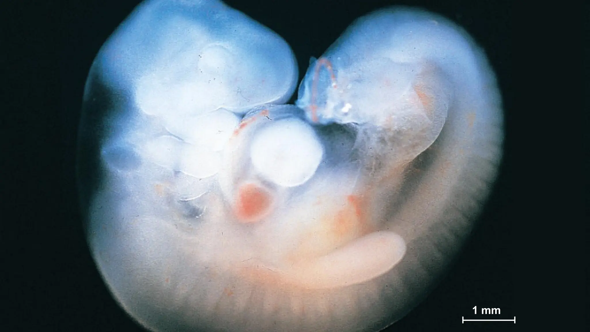
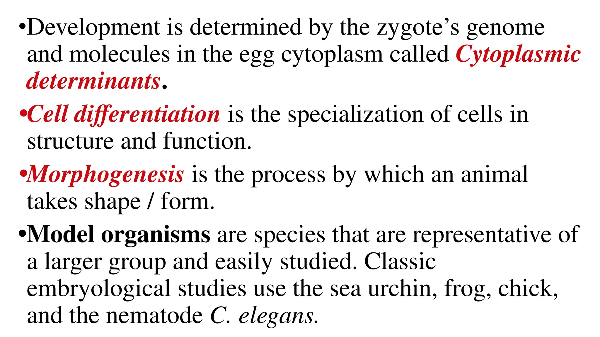
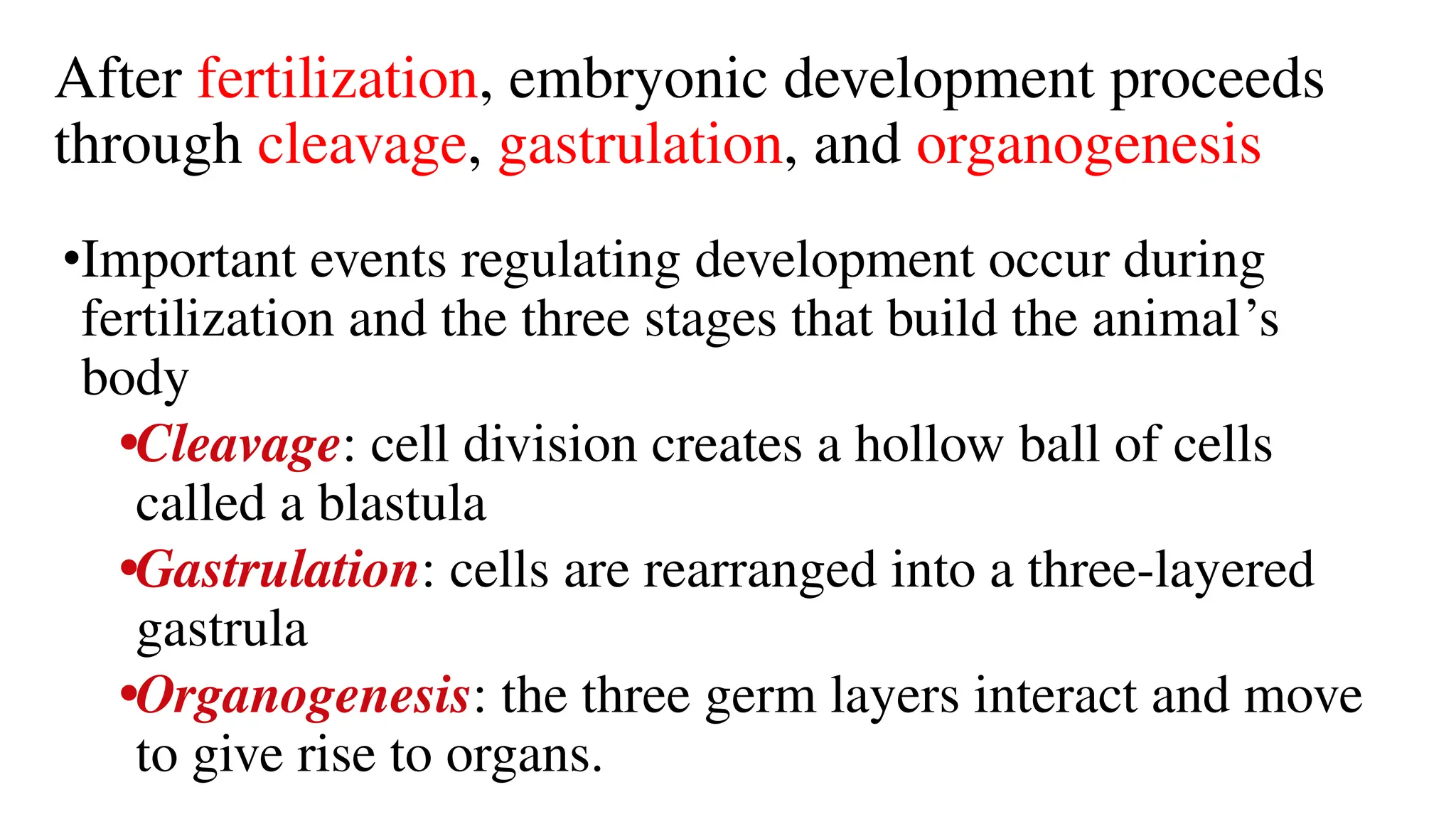
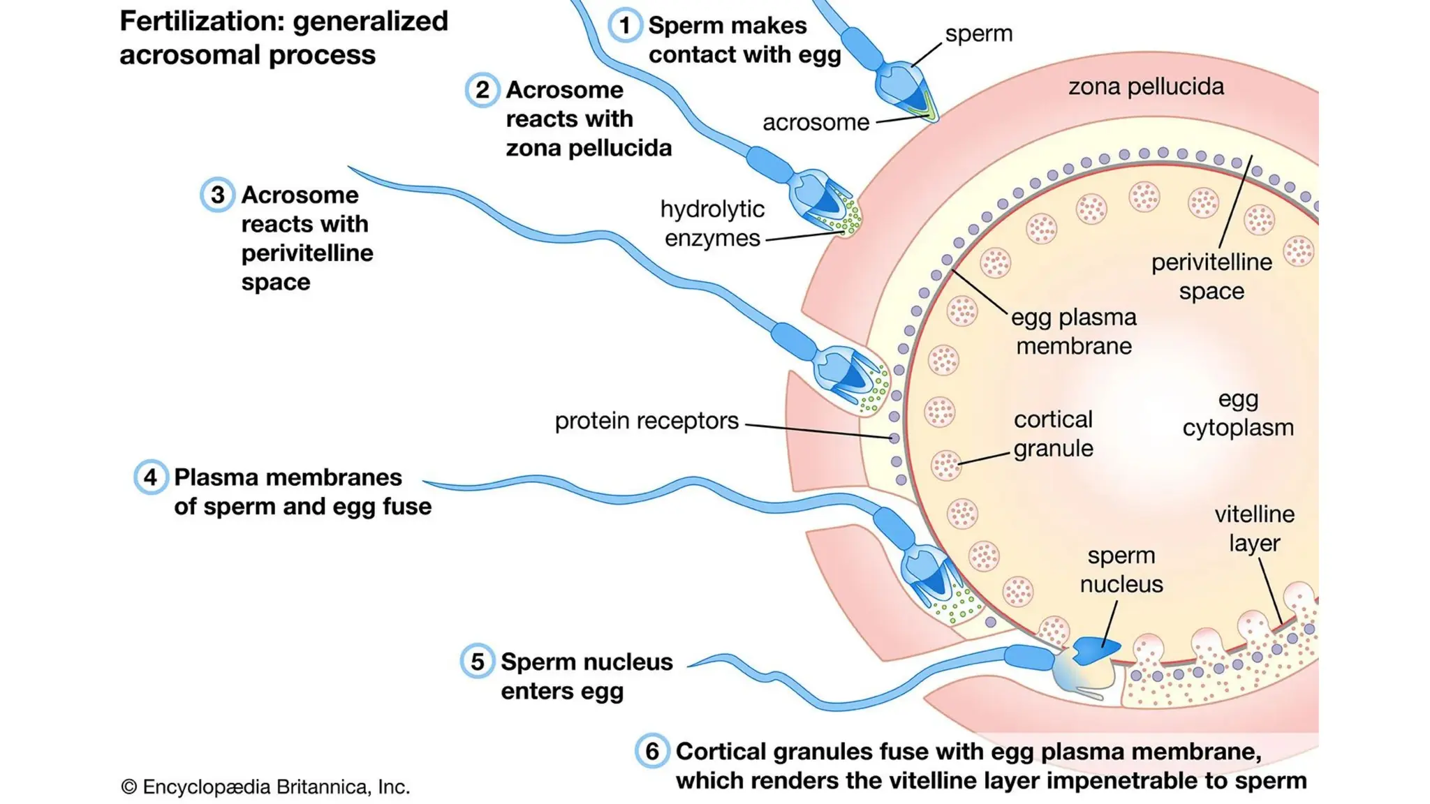
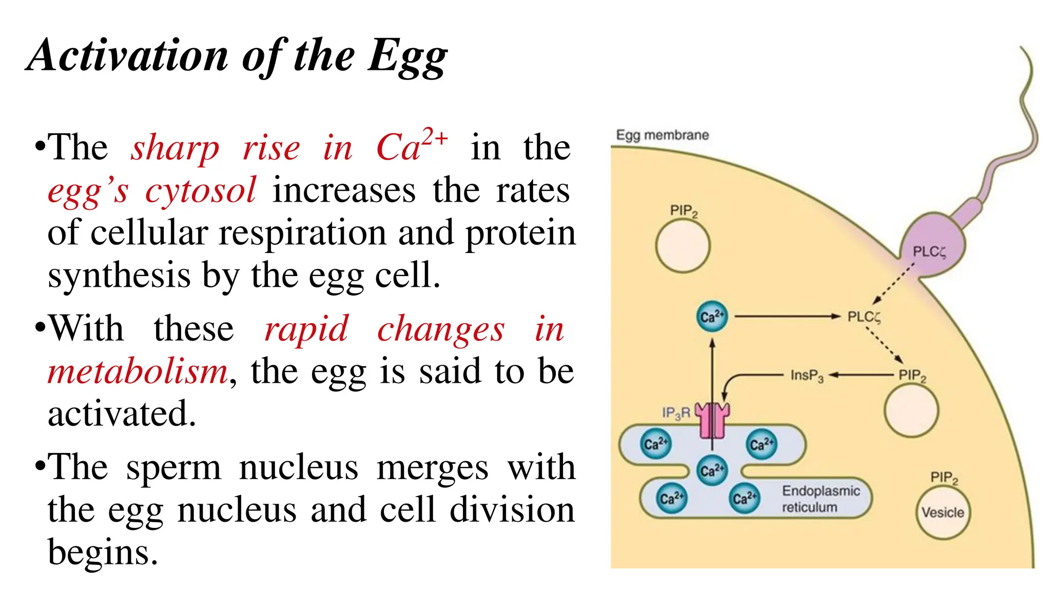
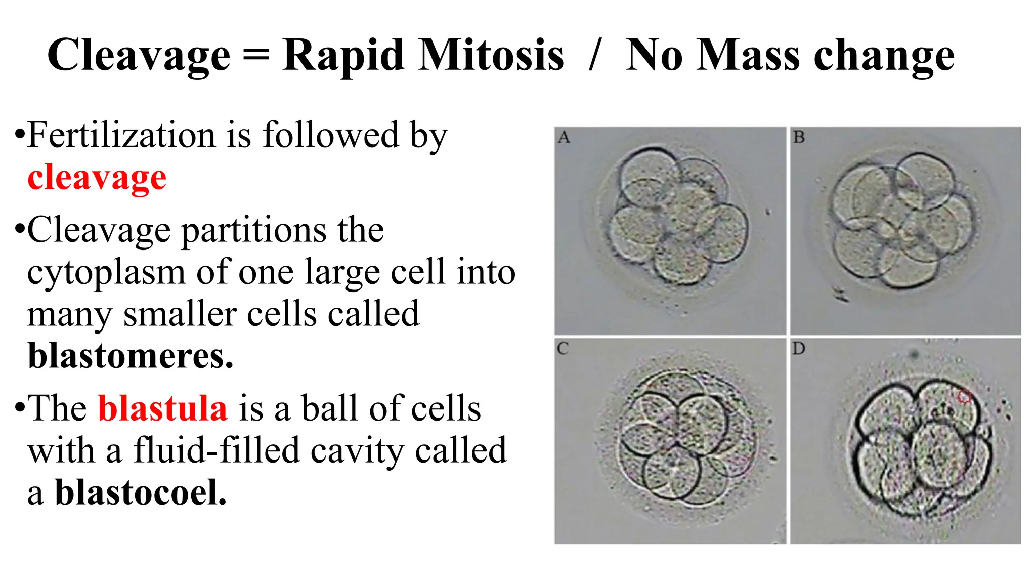
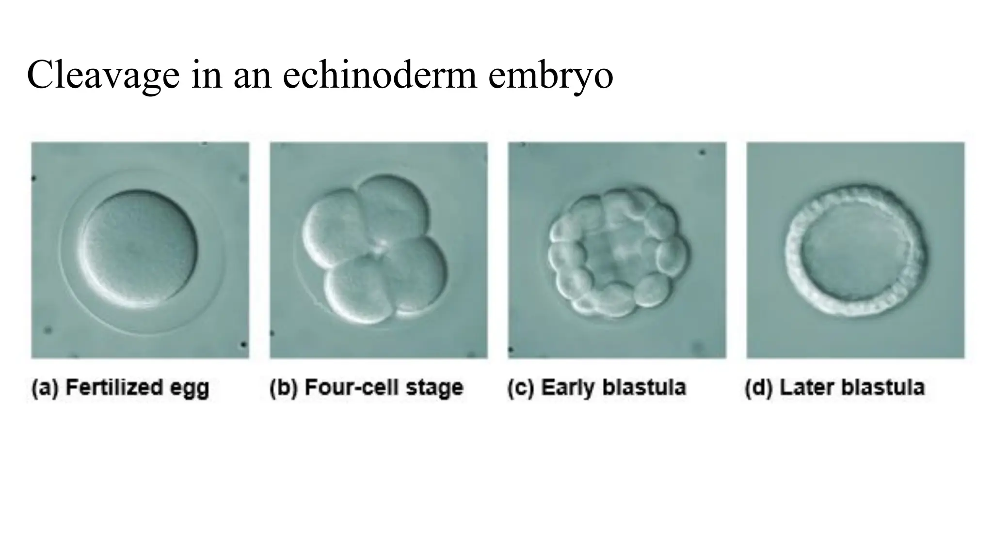
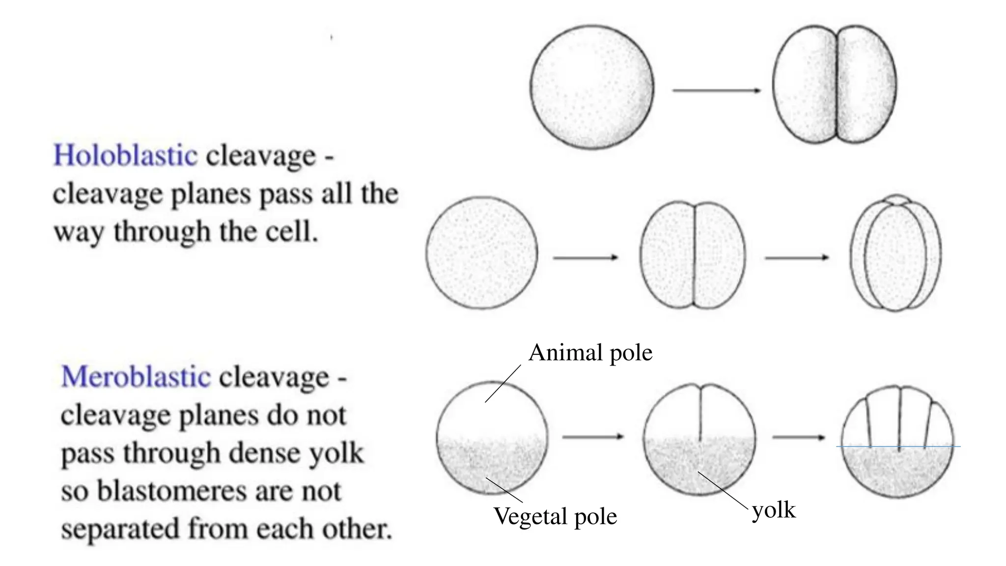
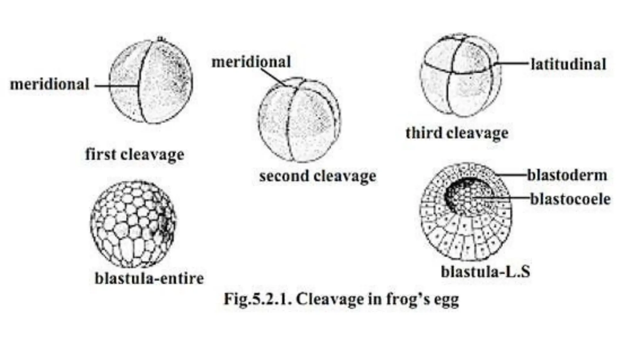
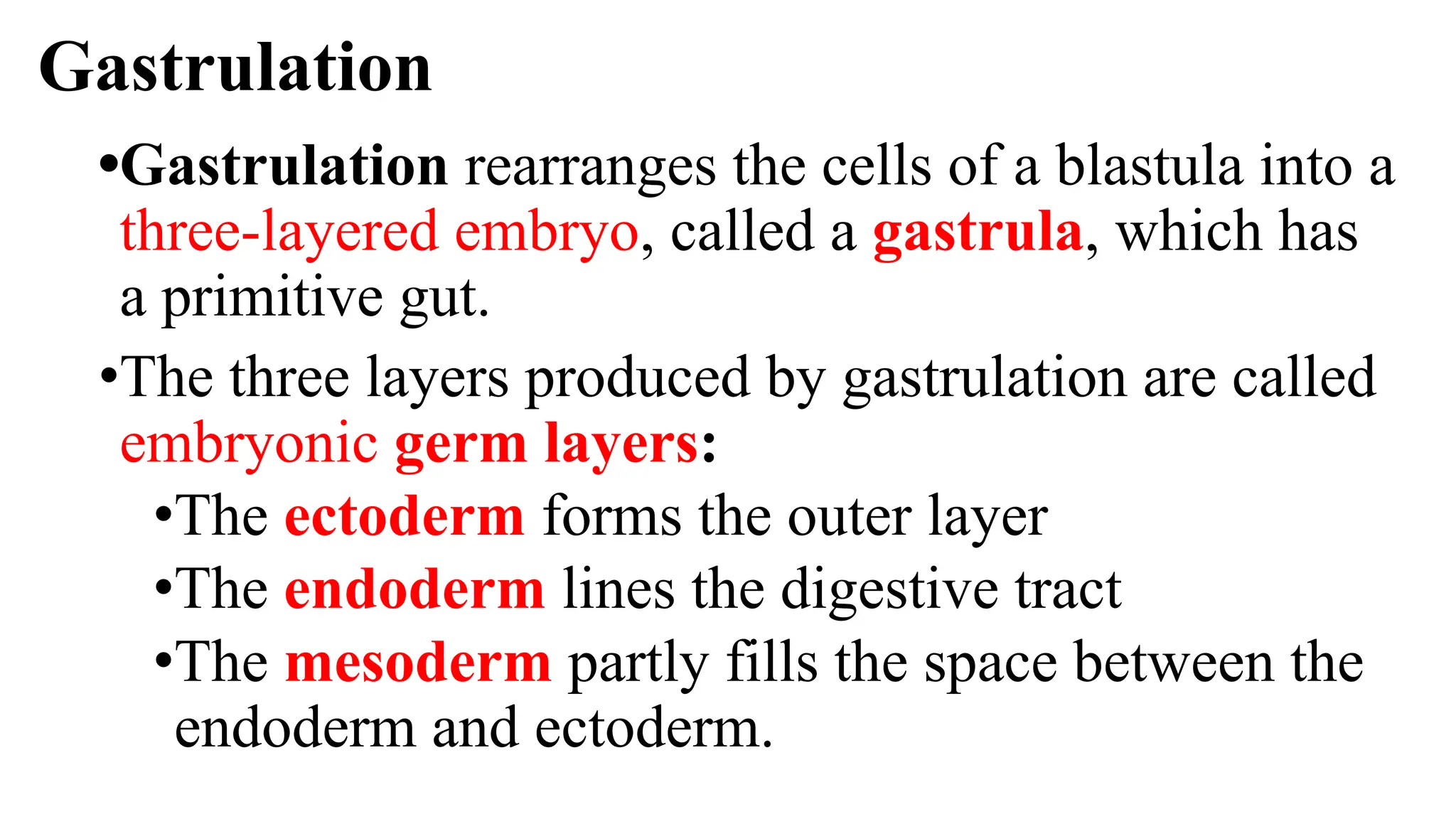
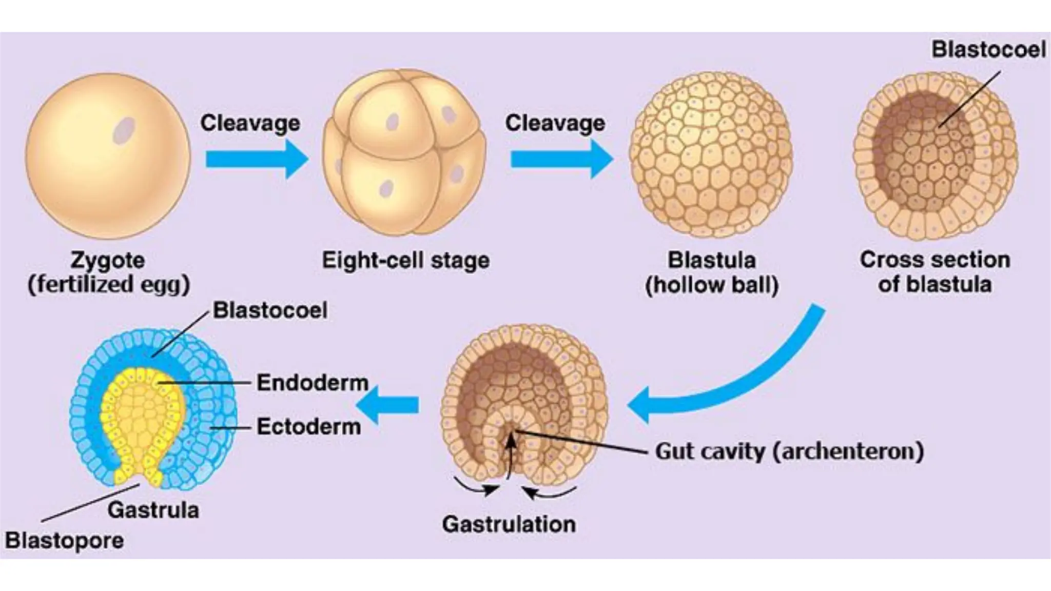
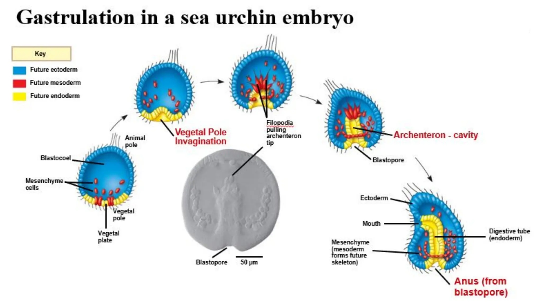
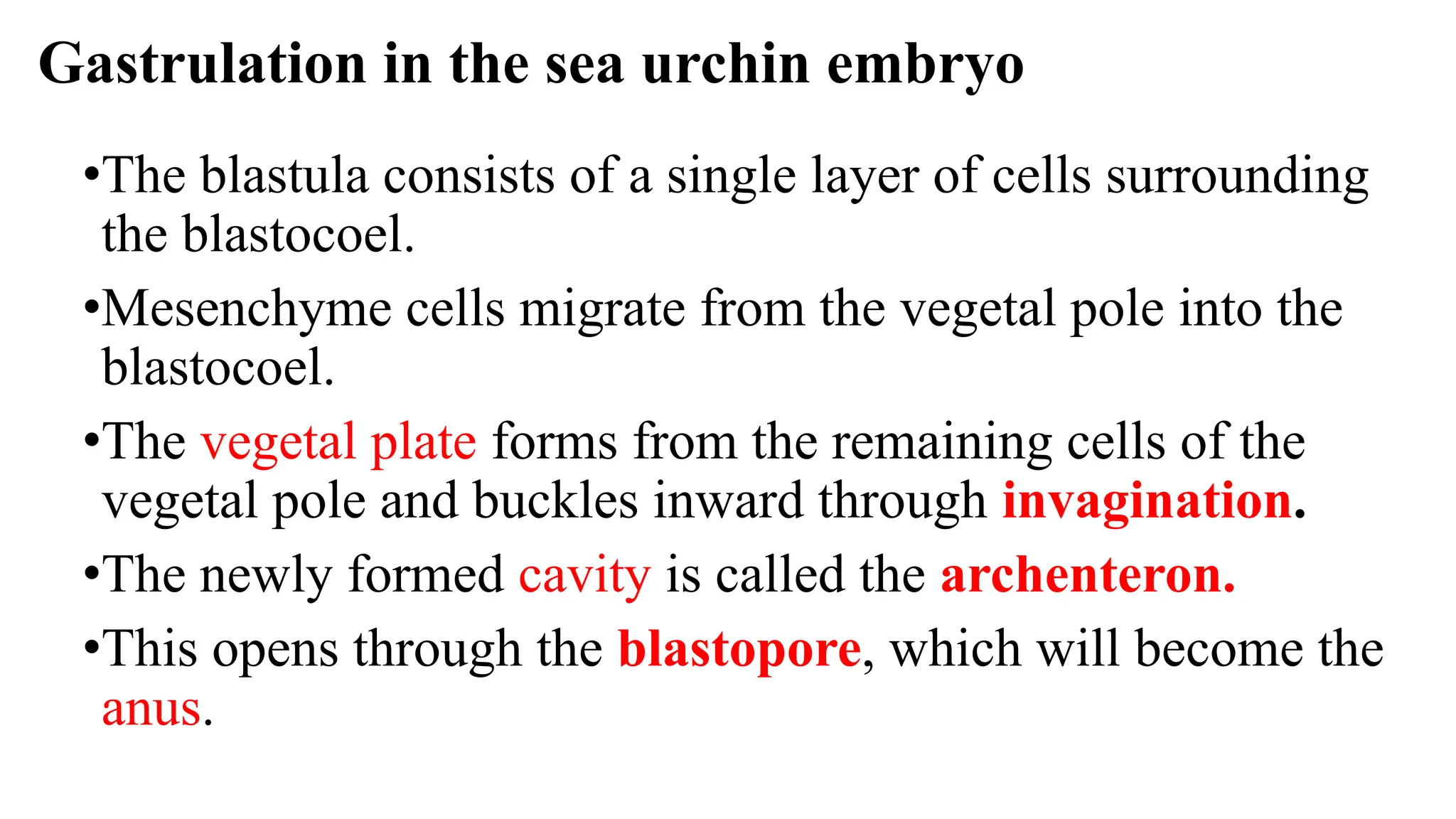
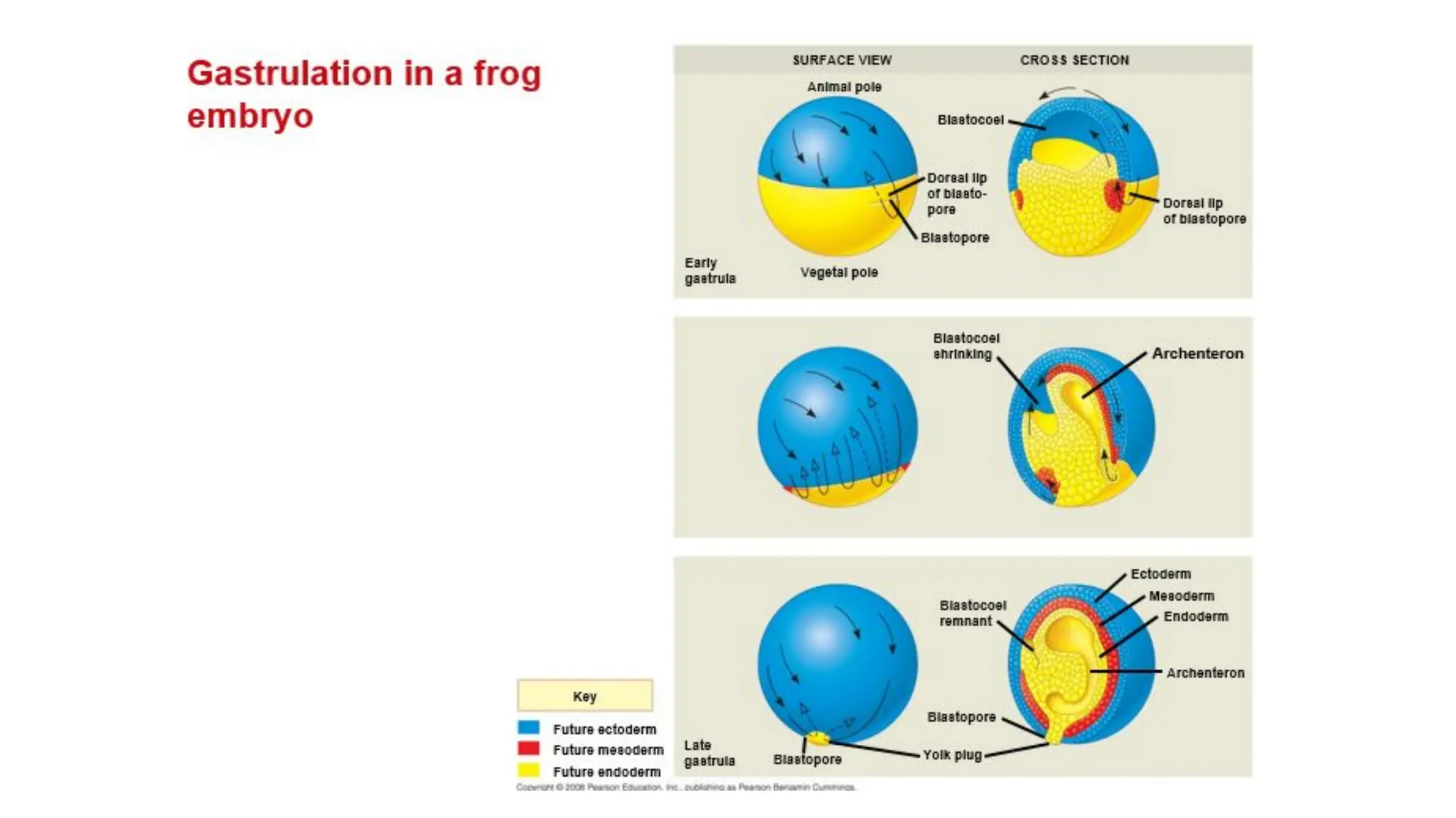
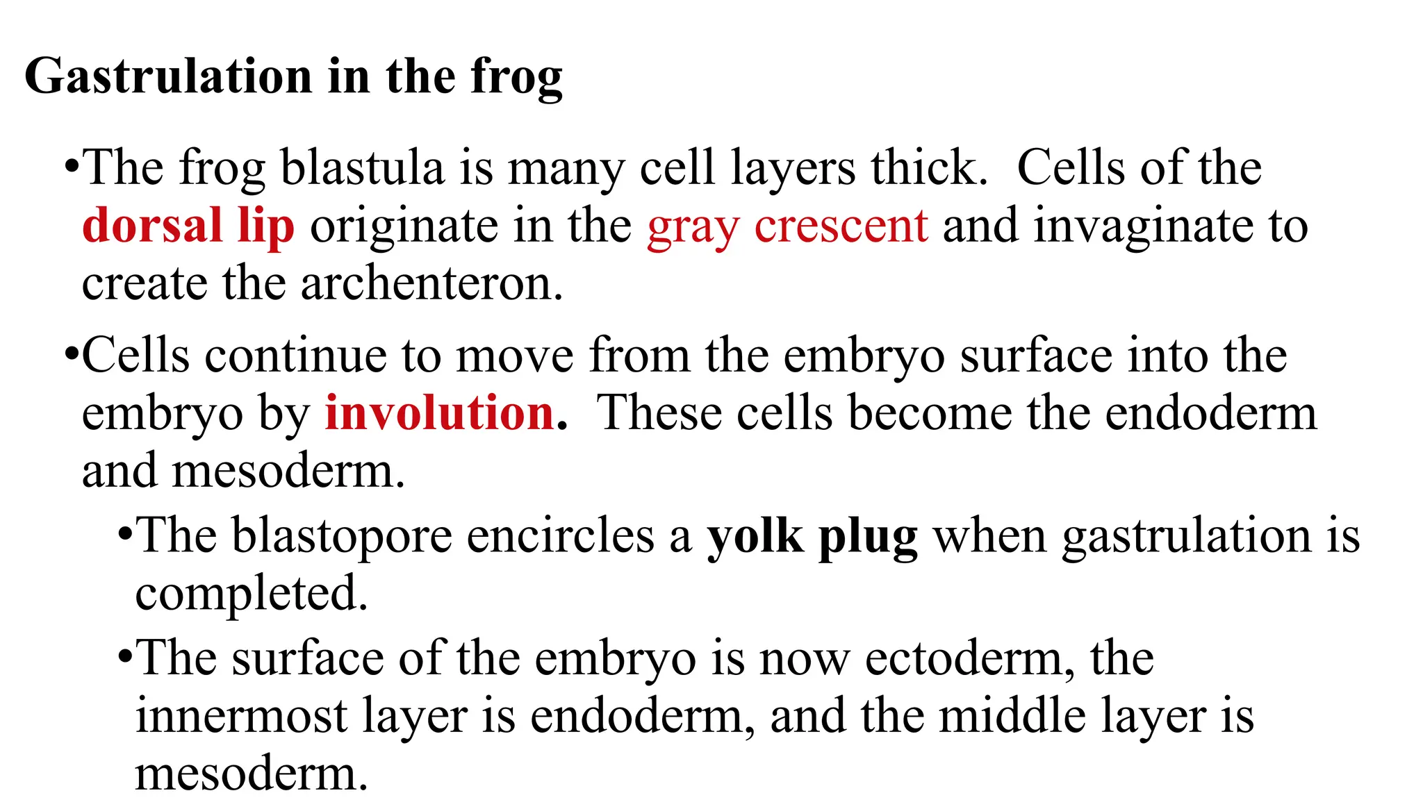
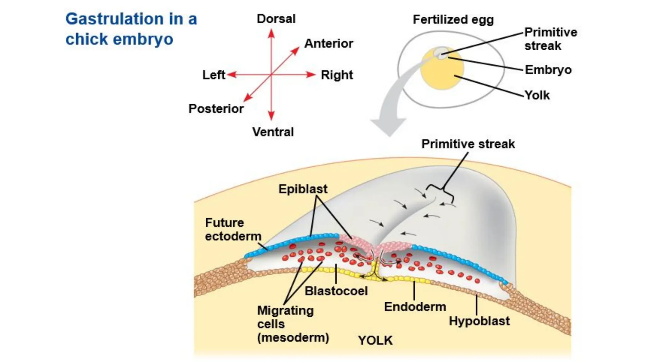
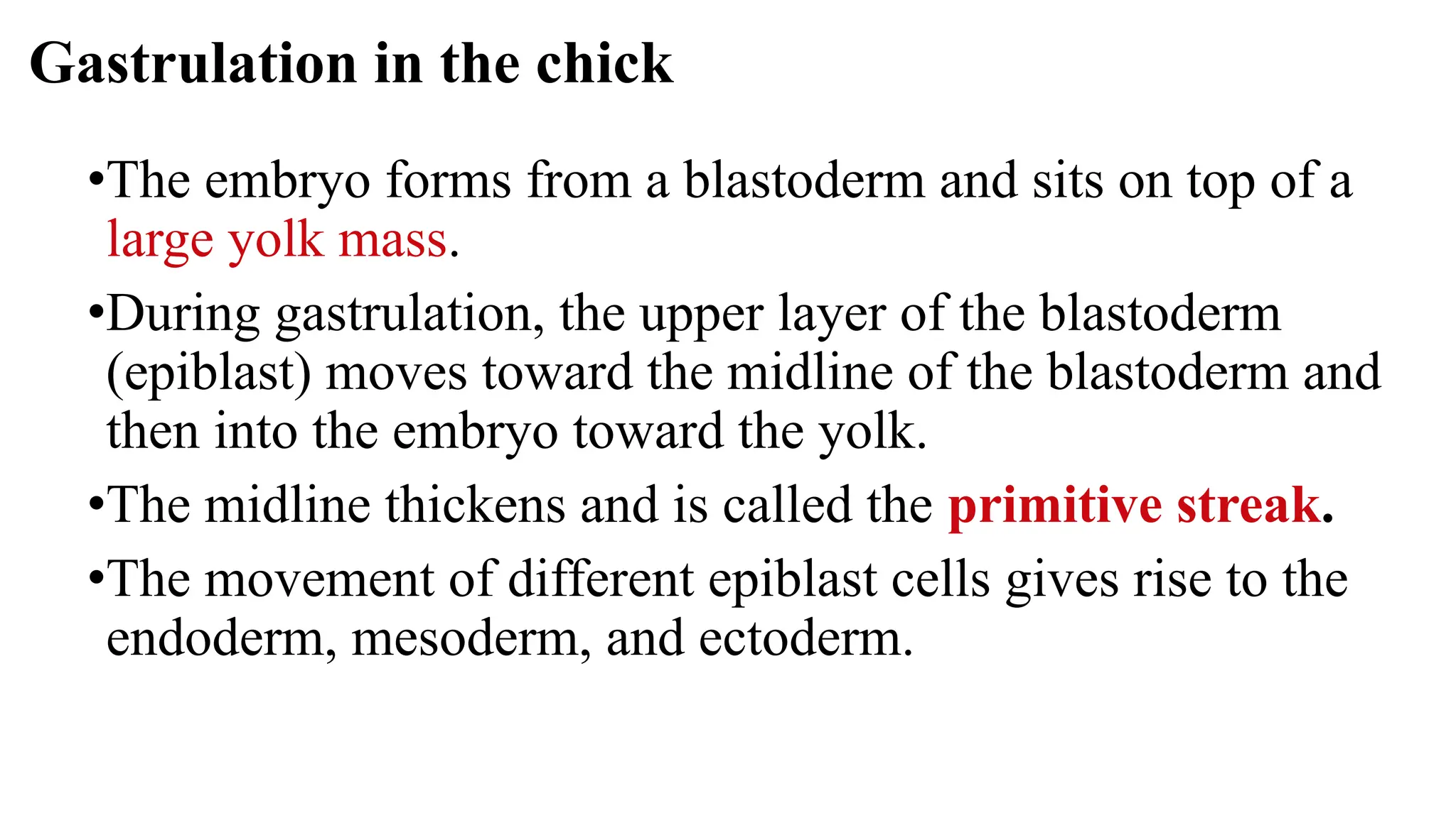
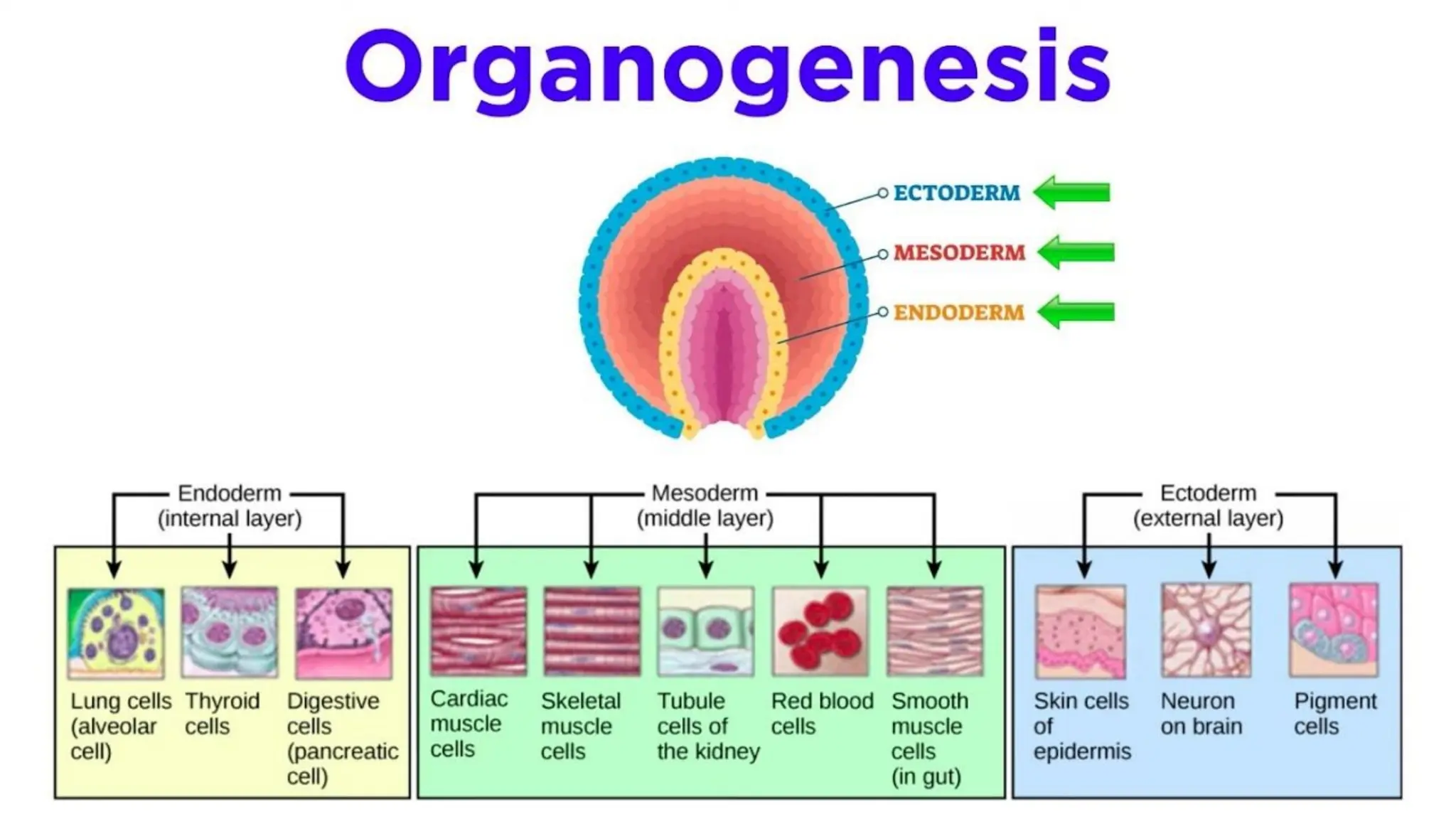
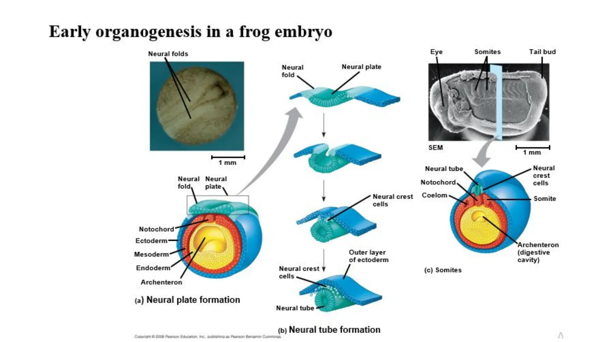
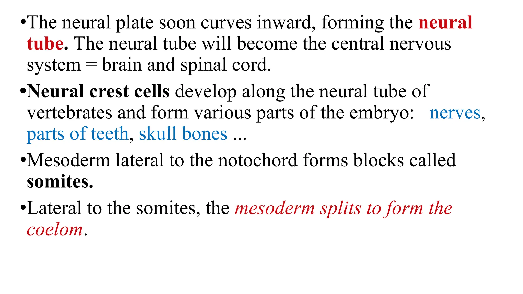
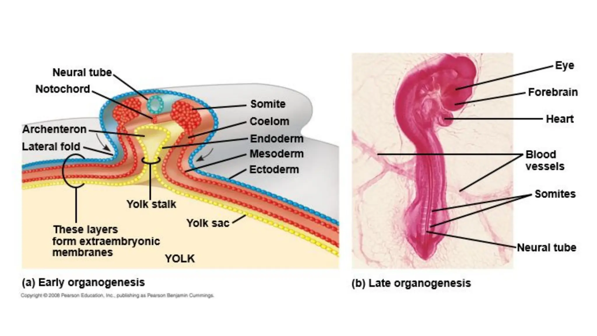
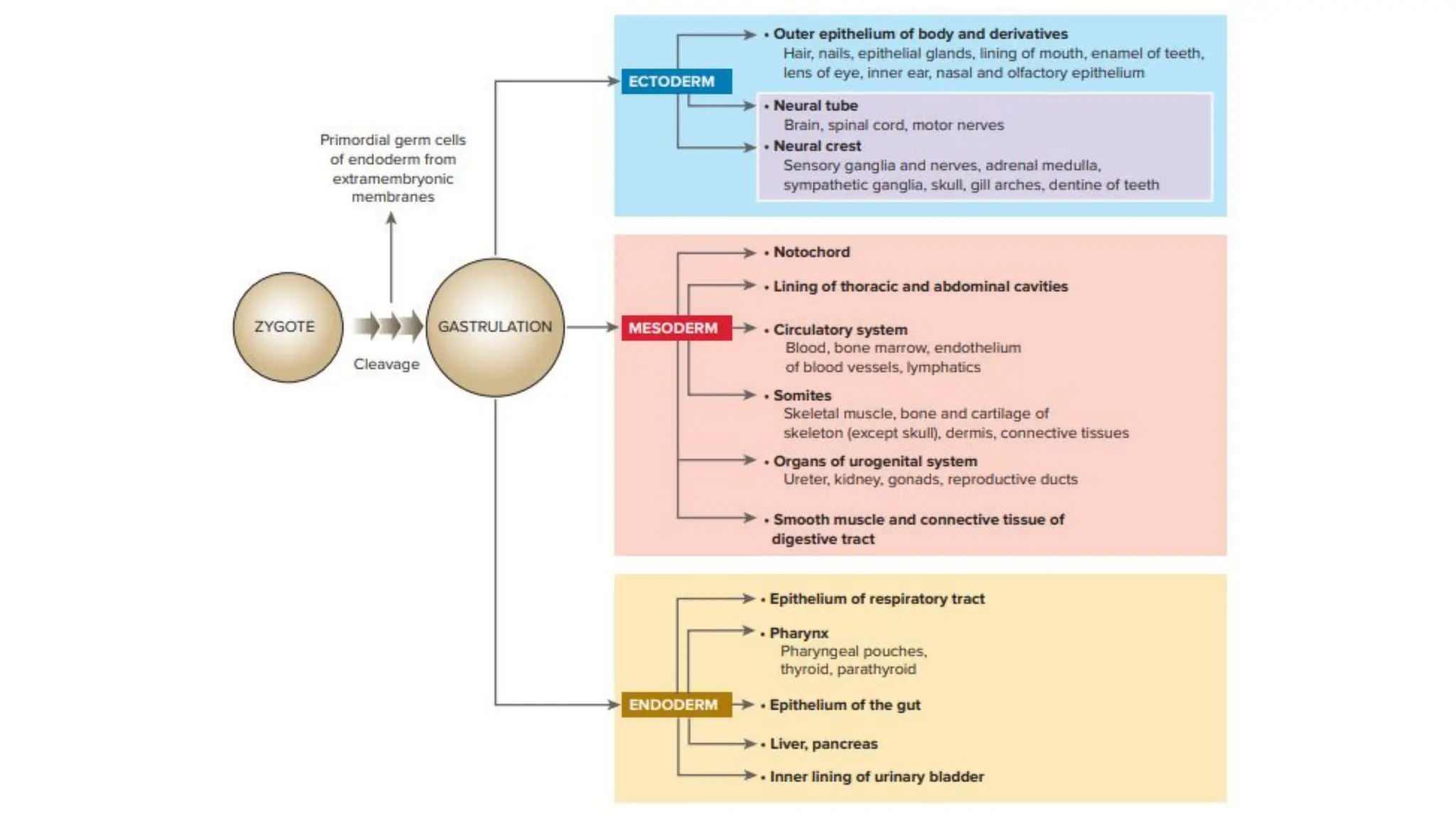
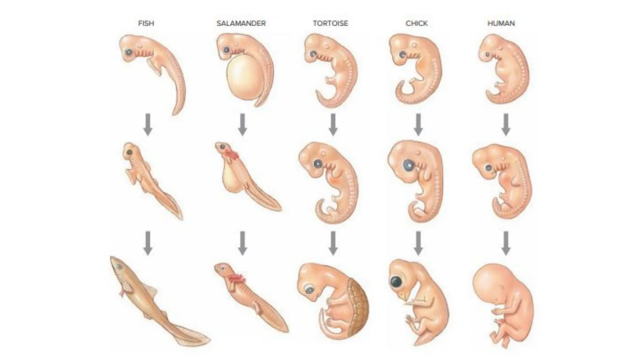
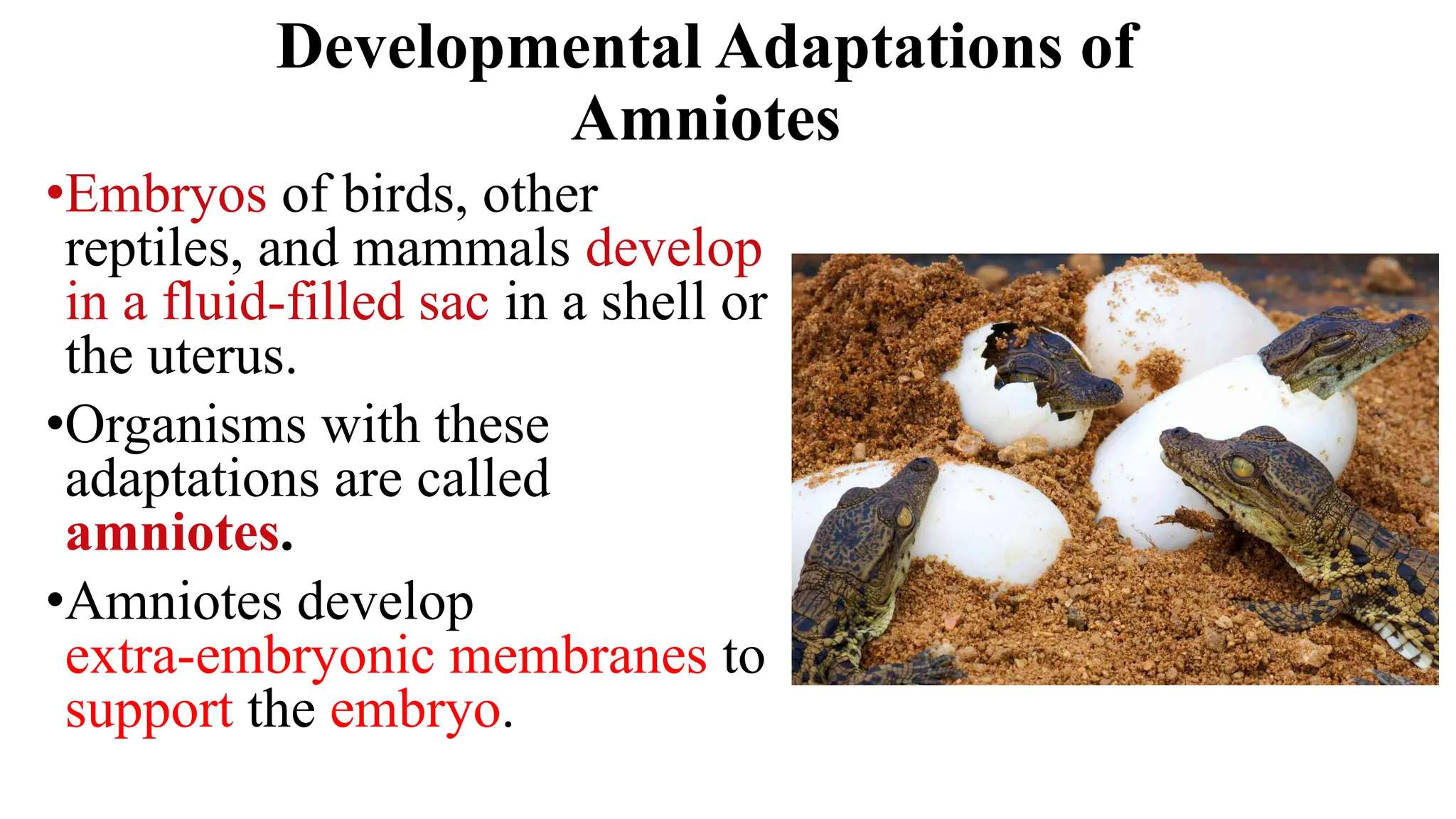
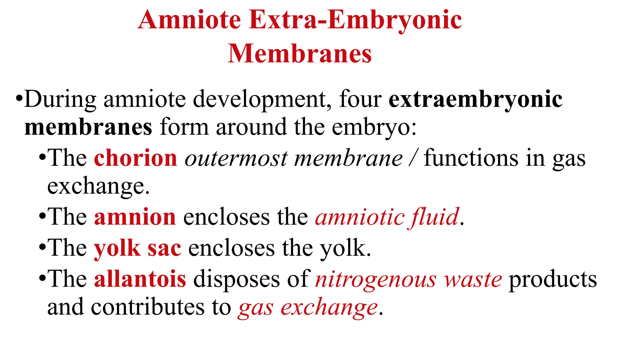
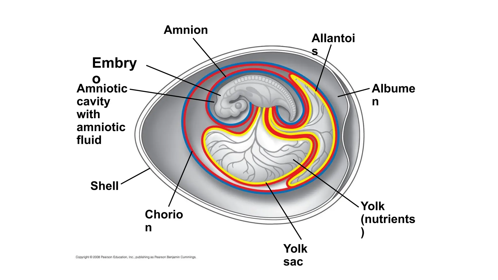
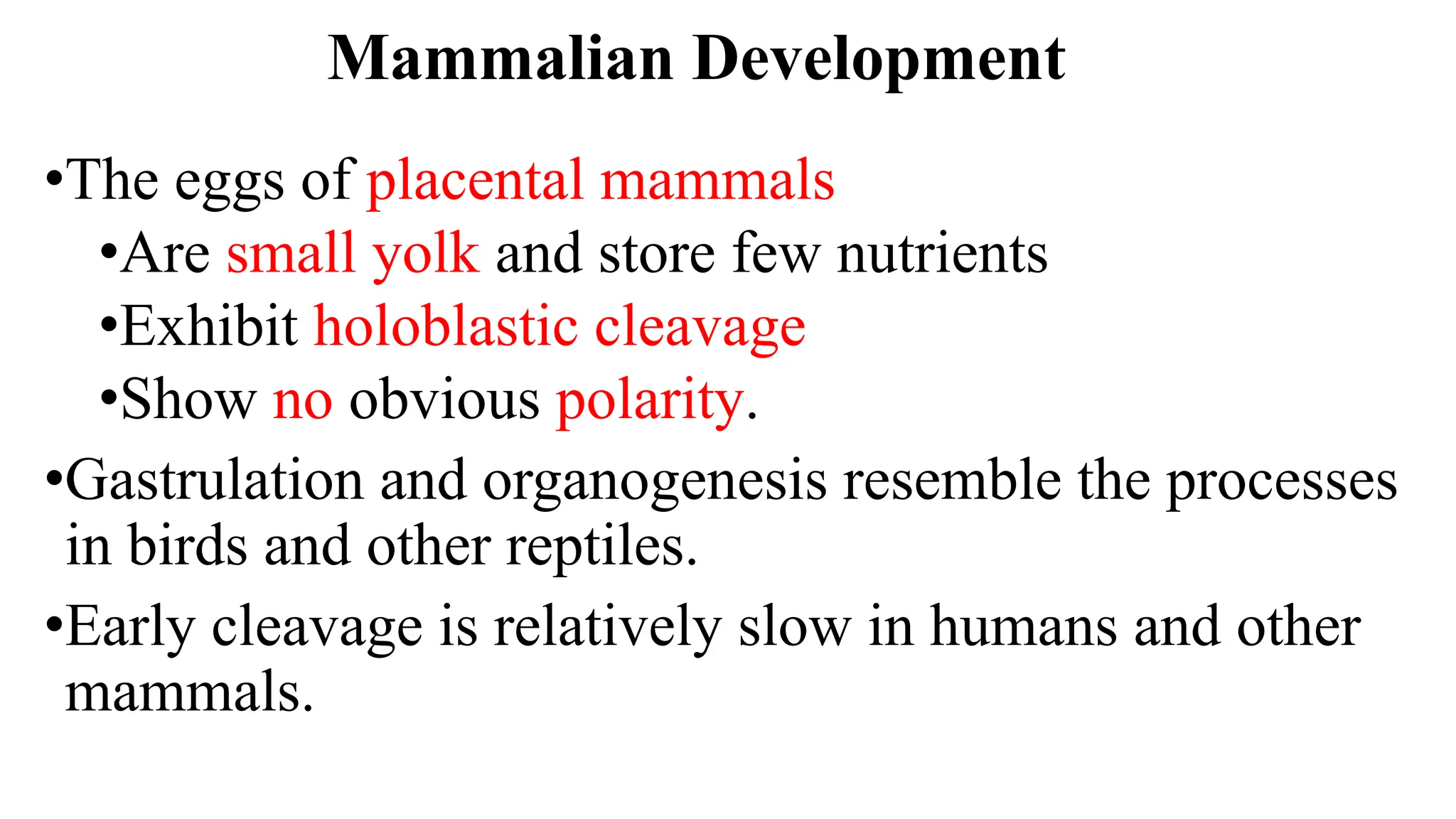
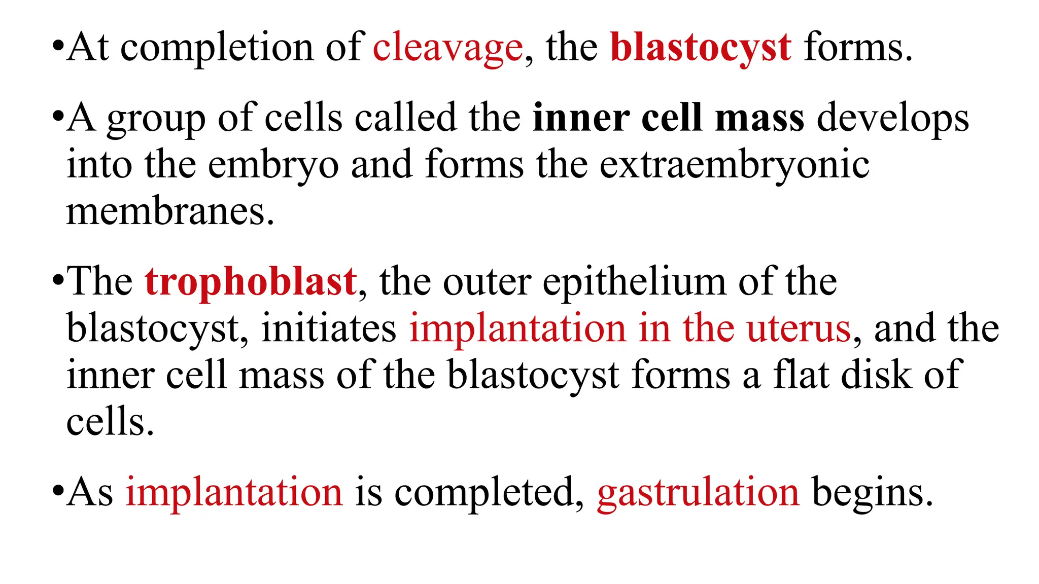
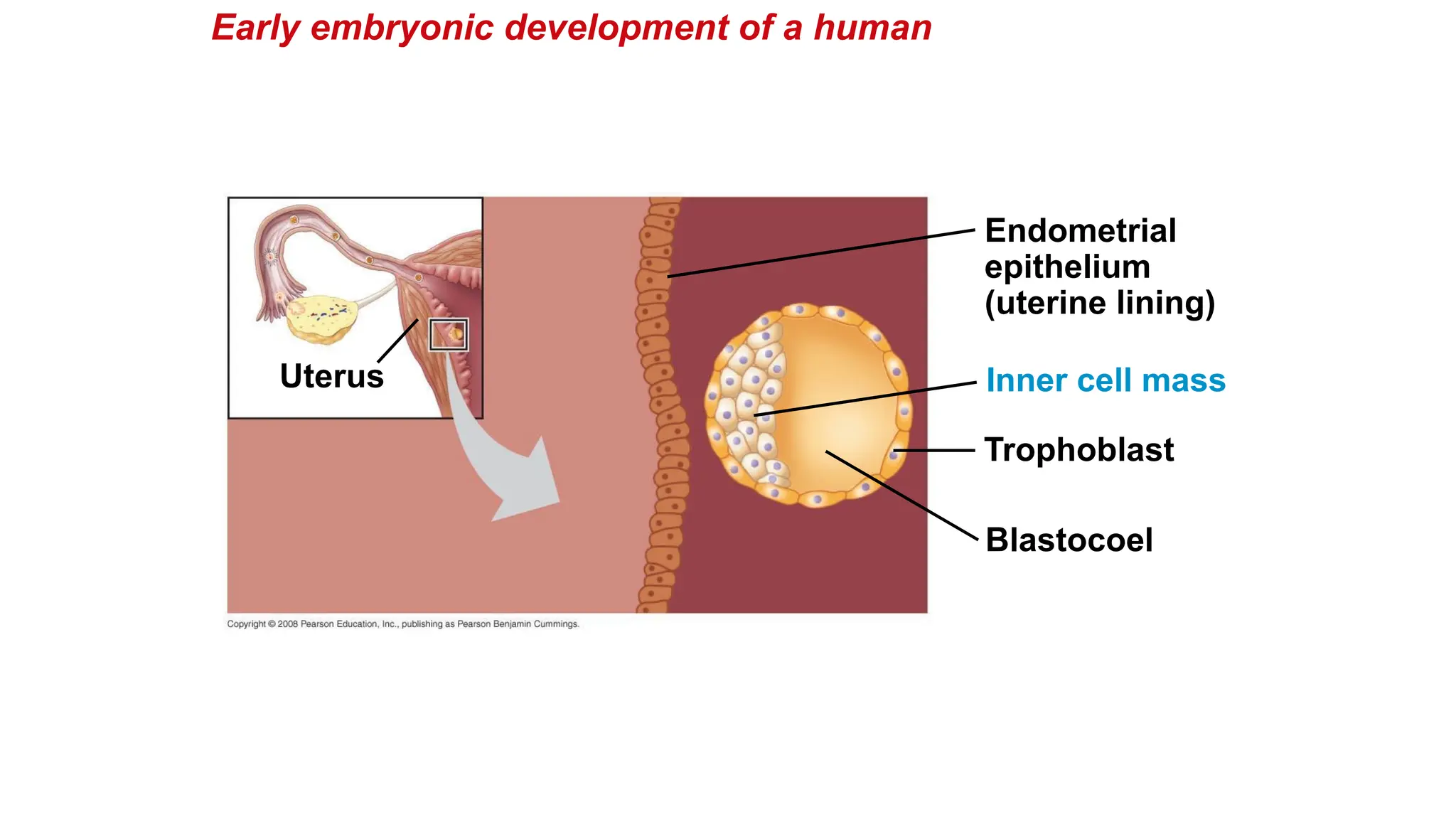
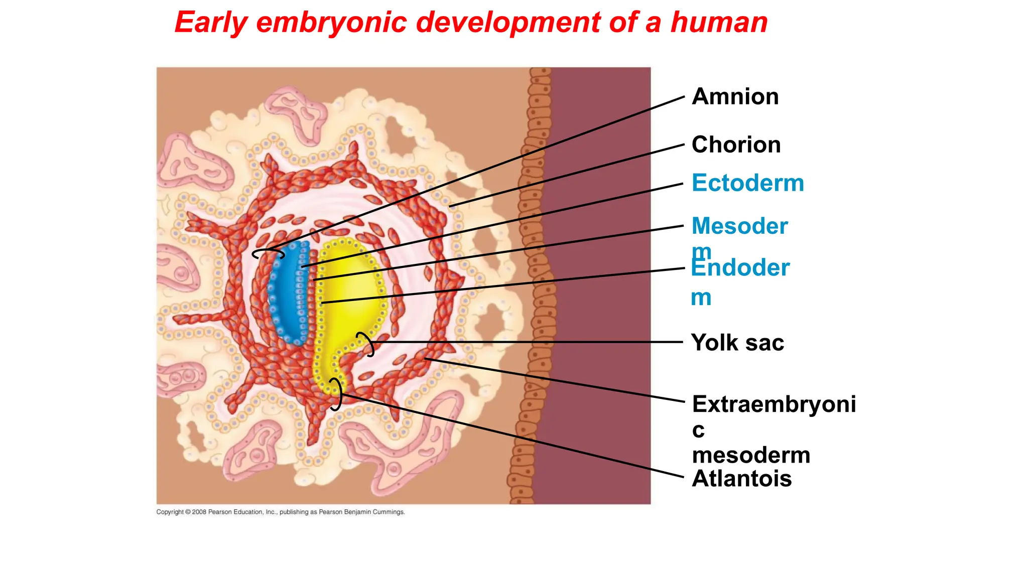
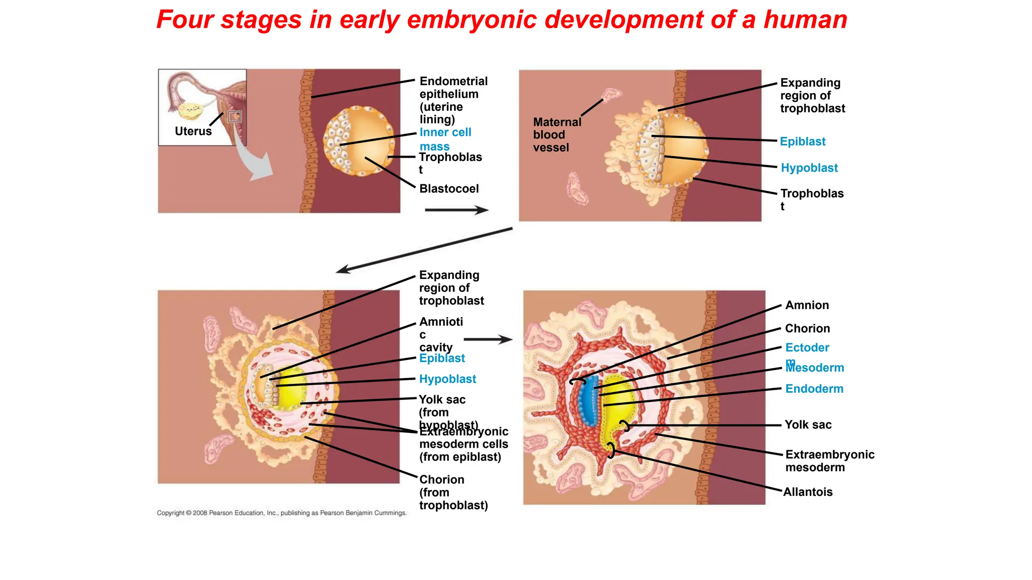
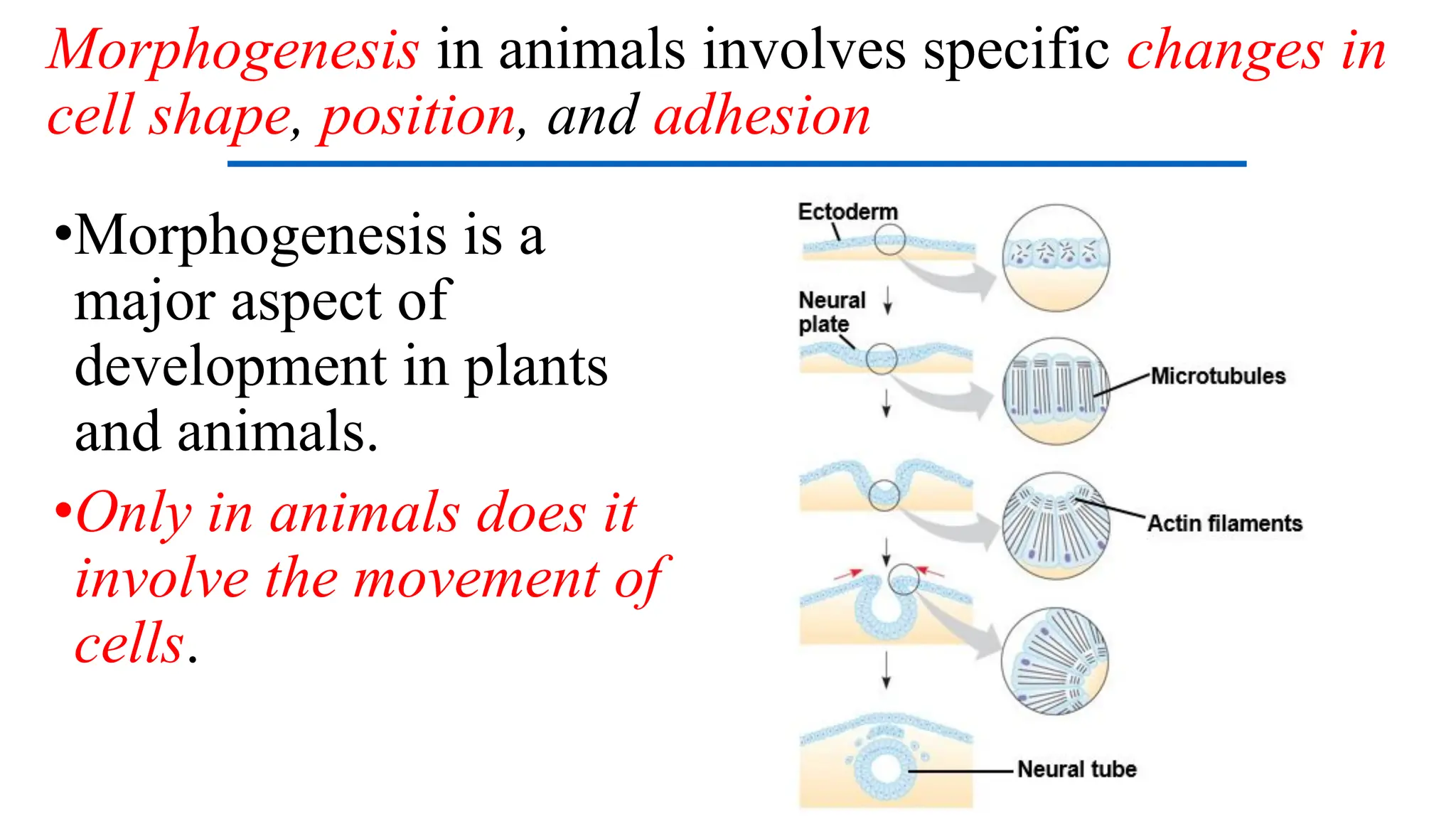
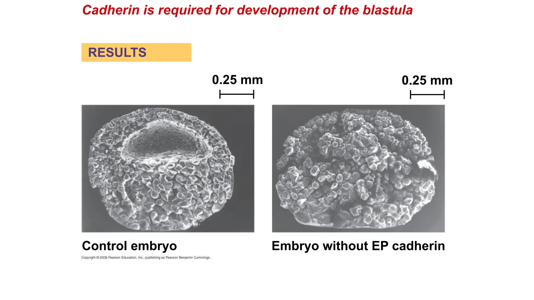
ANIMAL DEVELOPMENT
BIO102 General Zoology
1 mm
•Development is determined by the zygote’s genome
and molecules in the egg cytoplasm called Cytoplasmic
determinants.
•Cell differentiation is the specialization of cells in
structure and function.
•Morphogenesis is the process by which an animal
takes shape / form.
•Model organisms are species that are representative of
a larger group and easily studied. Classic
embryological studies use the sea urchin, frog, chick,
and the nematode C. elegans.
After fertilization, embryonic development proceeds
through cleavage, gastrulation, and organogenesis
•Important events regulating development occur during
fertilization and the three stages that build the animal’s
body
•Cleavage: cell division creates a hollow ball of cells
called a blastula
•Gastrulation: cells are rearranged into a three-layered
gastrula
•Organogenesis: the three germ layers interact and move
to give rise to organs.
The Acrosomal Reaction
•The acrosomal reaction is triggered when the
sperm meets the egg.
•The acrosome at the tip of the sperm releases
hydrolytic enzymes that digest material
surrounding the egg.
•Gamete contact and/or fusion depolarizes the egg
cell membrane and sets up a fast block to
polyspermy.
The Cortical Reaction
•Fusion of egg and sperm also initiates the cortical
reaction:
•This reaction induces a rise in Ca2+
that stimulates
cortical granules to release their contents outside the
egg.
•These changes cause formation of a fertilization
envelope that functions as a slow block to polyspermy.
Activation of the Egg
•The sharp rise in Ca2+
in the
egg’s cytosol increases the rates
of cellular respiration and protein
synthesis by the egg cell.
•With these rapid changes in
metabolism, the egg is said to be
activated.
•The sperm nucleus merges with
the egg nucleus and cell division
begins.
Cleavage = Rapid Mitosis / No Mass change
•Fertilization is followed by
cleavage
•Cleavage partitions the
cytoplasm of one large cell into
many smaller cells called
blastomeres.
•The blastula is a ball of cells
with a fluid-filled cavity called
a blastocoel.
Cleavage in an echinoderm embryo
yolk
Animal pole
Vegetal pole
Gastrulation
•Gastrulation rearranges the cells of a blastula into a
three-layered embryo, called a gastrula, which has
a primitive gut.
•The three layers produced by gastrulation are called
embryonic germ layers:
•The ectoderm forms the outer layer
•The endoderm lines the digestive tract
•The mesoderm partly fills the space between the
endoderm and ectoderm.
•The blastula consists of a single layer of cells surrounding
the blastocoel.
•Mesenchyme cells migrate from the vegetal pole into the
blastocoel.
•The vegetal plate forms from the remaining cells of the
vegetal pole and buckles inward through invagination.
•The newly formed cavity is called the archenteron.
•This opens through the blastopore, which will become the
anus.
Gastrulation in the sea urchin embryo
•The frog blastula is many cell layers thick. Cells of the
dorsal lip originate in the gray crescent and invaginate to
create the archenteron.
•Cells continue to move from the embryo surface into the
embryo by involution. These cells become the endoderm
and mesoderm.
•The blastopore encircles a yolk plug when gastrulation is
completed.
•The surface of the embryo is now ectoderm, the
innermost layer is endoderm, and the middle layer is
mesoderm.
Gastrulation in the frog
•The embryo forms from a blastoderm and sits on top of a
large yolk mass.
•During gastrulation, the upper layer of the blastoderm
(epiblast) moves toward the midline of the blastoderm and
then into the embryo toward the yolk.
•The midline thickens and is called the primitive streak.
•The movement of different epiblast cells gives rise to the
endoderm, mesoderm, and ectoderm.
Gastrulation in the chick
•The neural plate soon curves inward, forming the neural
tube. The neural tube will become the central nervous
system = brain and spinal cord.
•Neural crest cells develop along the neural tube of
vertebrates and form various parts of the embryo: nerves,
parts of teeth, skull bones ...
•Mesoderm lateral to the notochord forms blocks called
somites.
•Lateral to the somites, the mesoderm splits to form the
coelom.
Developmental Adaptations of
Amniotes
•Embryos of birds, other
reptiles, and mammals develop
in a fluid-filled sac in a shell or
the uterus.
•Organisms with these
adaptations are called
amniotes.
•Amniotes develop
extra-embryonic membranes to
support the embryo.
•During amniote development, four extraembryonic
membranes form around the embryo:
•The chorion outermost membrane / functions in gas
exchange.
•The amnion encloses the amniotic fluid.
•The yolk sac encloses the yolk.
•The allantois disposes of nitrogenous waste products
and contributes to gas exchange.
Amniote Extra-Embryonic
Membranes
Embry
o
Amnion
Amniotic
cavity
with
amniotic
fluid
Shell
Chorio
n
Yolk
sac
Yolk
(nutrients
)
Allantoi
s
Albume
n
Mammalian Development
•The eggs of placental mammals
•Are small yolk and store few nutrients
•Exhibit holoblastic cleavage
•Show no obvious polarity.
•Gastrulation and organogenesis resemble the processes
in birds and other reptiles.
•Early cleavage is relatively slow in humans and other
mammals.
•At completion of cleavage, the blastocyst forms.
•A group of cells called the inner cell mass develops
into the embryo and forms the extraembryonic
membranes.
•The trophoblast, the outer epithelium of the
blastocyst, initiates implantation in the uterus, and the
inner cell mass of the blastocyst forms a flat disk of
cells.
•As implantation is completed, gastrulation begins.
Early embryonic development of a human
Blastocoel
Trophoblast
Uterus
Endometrial
epithelium
(uterine lining)
Inner cell mass
Early embryonic development of a human
Trophobl
ast
Hypoblast
Matern
al
blood
vessel
Expanding
region of
trophoblast
Epiblast
•The epiblast cells invaginate through a primitive streak to
form mesoderm and endoderm.
•The placenta is formed from the trophoblast, mesodermal
cells from the epiblast, and adjacent endometrial tissue.
•The placenta allows for the exchange of materials between
the mother and embryo.
•By the end of gastrulation, the embryonic germ layers have
formed. The extraembryonic membranes in mammals are
homologous to those of birds and other reptiles and develop
in a similar way.
Early embryonic development of a human
Yolk sac (from
hypoblast)
Hypoblast
Expandin
g
region of
trophobl
ast
Amniot
ic
cavity
Epiblast
Extraembryo
nic
mesoderm
cells
(from epiblast)
Chorion
(from
trophoblast)
Early embryonic development of a human
Yolk sac
Mesoder
m
Amnion
Chorion
Ectoderm
Extraembryoni
c
mesoderm
Atlantois
Endoder
m
Four stages in early embryonic development of a human
Yolk sac
Mesoderm
Amnion
Chorion
Ectoder
m
Extraembryonic
mesoderm
Trophoblas
t
Endoderm
Hypoblast
Expanding
region of
trophoblast
Epiblast
Maternal
blood
vessel
Allantois
Trophoblas
t
Hypoblast
Endometrial
epithelium
(uterine
lining)
Inner cell
mass
Blastocoel
Uterus
Epiblast
Amnioti
c
cavity
Expanding
region of
trophoblast
Yolk sac
(from
hypoblast)
Chorion
(from
trophoblast)
Extraembryonic
mesoderm cells
(from epiblast)
Morphogenesis in animals involves specific changes in
cell shape, position, and adhesion
•Morphogenesis is a
major aspect of
development in plants
and animals.
•Only in animals does it
involve the movement of
cells.
•The cytoskeleton also drives cell migration, or cell
crawling, the active movement of cells.
•In gastrulation, tissue invagination is caused by
changes in cell shape and migration.
•Cell crawling is involved in convergent
extension, a morphogenetic movement in which
cells of a tissue become narrower and longer.
Cadherin is required for development of the blastula
Control embryo Embryo without EP cadherin
0.25 mm 0.25 mm
RESULTS
END

Zoology lecture ANIMAL-DEVELOPMENT pdf ppt

  • 1.
  • 3.
  • 4.
    •Development is determinedby the zygote’s genome and molecules in the egg cytoplasm called Cytoplasmic determinants. •Cell differentiation is the specialization of cells in structure and function. •Morphogenesis is the process by which an animal takes shape / form. •Model organisms are species that are representative of a larger group and easily studied. Classic embryological studies use the sea urchin, frog, chick, and the nematode C. elegans.
  • 5.
    After fertilization, embryonicdevelopment proceeds through cleavage, gastrulation, and organogenesis •Important events regulating development occur during fertilization and the three stages that build the animal’s body •Cleavage: cell division creates a hollow ball of cells called a blastula •Gastrulation: cells are rearranged into a three-layered gastrula •Organogenesis: the three germ layers interact and move to give rise to organs.
  • 7.
    The Acrosomal Reaction •Theacrosomal reaction is triggered when the sperm meets the egg. •The acrosome at the tip of the sperm releases hydrolytic enzymes that digest material surrounding the egg. •Gamete contact and/or fusion depolarizes the egg cell membrane and sets up a fast block to polyspermy.
  • 9.
    The Cortical Reaction •Fusionof egg and sperm also initiates the cortical reaction: •This reaction induces a rise in Ca2+ that stimulates cortical granules to release their contents outside the egg. •These changes cause formation of a fertilization envelope that functions as a slow block to polyspermy.
  • 10.
    Activation of theEgg •The sharp rise in Ca2+ in the egg’s cytosol increases the rates of cellular respiration and protein synthesis by the egg cell. •With these rapid changes in metabolism, the egg is said to be activated. •The sperm nucleus merges with the egg nucleus and cell division begins.
  • 11.
    Cleavage = RapidMitosis / No Mass change •Fertilization is followed by cleavage •Cleavage partitions the cytoplasm of one large cell into many smaller cells called blastomeres. •The blastula is a ball of cells with a fluid-filled cavity called a blastocoel.
  • 12.
    Cleavage in anechinoderm embryo
  • 13.
  • 15.
    Gastrulation •Gastrulation rearranges thecells of a blastula into a three-layered embryo, called a gastrula, which has a primitive gut. •The three layers produced by gastrulation are called embryonic germ layers: •The ectoderm forms the outer layer •The endoderm lines the digestive tract •The mesoderm partly fills the space between the endoderm and ectoderm.
  • 18.
    •The blastula consistsof a single layer of cells surrounding the blastocoel. •Mesenchyme cells migrate from the vegetal pole into the blastocoel. •The vegetal plate forms from the remaining cells of the vegetal pole and buckles inward through invagination. •The newly formed cavity is called the archenteron. •This opens through the blastopore, which will become the anus. Gastrulation in the sea urchin embryo
  • 20.
    •The frog blastulais many cell layers thick. Cells of the dorsal lip originate in the gray crescent and invaginate to create the archenteron. •Cells continue to move from the embryo surface into the embryo by involution. These cells become the endoderm and mesoderm. •The blastopore encircles a yolk plug when gastrulation is completed. •The surface of the embryo is now ectoderm, the innermost layer is endoderm, and the middle layer is mesoderm. Gastrulation in the frog
  • 22.
    •The embryo formsfrom a blastoderm and sits on top of a large yolk mass. •During gastrulation, the upper layer of the blastoderm (epiblast) moves toward the midline of the blastoderm and then into the embryo toward the yolk. •The midline thickens and is called the primitive streak. •The movement of different epiblast cells gives rise to the endoderm, mesoderm, and ectoderm. Gastrulation in the chick
  • 25.
    •The neural platesoon curves inward, forming the neural tube. The neural tube will become the central nervous system = brain and spinal cord. •Neural crest cells develop along the neural tube of vertebrates and form various parts of the embryo: nerves, parts of teeth, skull bones ... •Mesoderm lateral to the notochord forms blocks called somites. •Lateral to the somites, the mesoderm splits to form the coelom.
  • 29.
    Developmental Adaptations of Amniotes •Embryosof birds, other reptiles, and mammals develop in a fluid-filled sac in a shell or the uterus. •Organisms with these adaptations are called amniotes. •Amniotes develop extra-embryonic membranes to support the embryo.
  • 30.
    •During amniote development,four extraembryonic membranes form around the embryo: •The chorion outermost membrane / functions in gas exchange. •The amnion encloses the amniotic fluid. •The yolk sac encloses the yolk. •The allantois disposes of nitrogenous waste products and contributes to gas exchange. Amniote Extra-Embryonic Membranes
  • 31.
  • 32.
    Mammalian Development •The eggsof placental mammals •Are small yolk and store few nutrients •Exhibit holoblastic cleavage •Show no obvious polarity. •Gastrulation and organogenesis resemble the processes in birds and other reptiles. •Early cleavage is relatively slow in humans and other mammals.
  • 33.
    •At completion ofcleavage, the blastocyst forms. •A group of cells called the inner cell mass develops into the embryo and forms the extraembryonic membranes. •The trophoblast, the outer epithelium of the blastocyst, initiates implantation in the uterus, and the inner cell mass of the blastocyst forms a flat disk of cells. •As implantation is completed, gastrulation begins.
  • 34.
    Early embryonic developmentof a human Blastocoel Trophoblast Uterus Endometrial epithelium (uterine lining) Inner cell mass
  • 35.
    Early embryonic developmentof a human Trophobl ast Hypoblast Matern al blood vessel Expanding region of trophoblast Epiblast
  • 36.
    •The epiblast cellsinvaginate through a primitive streak to form mesoderm and endoderm. •The placenta is formed from the trophoblast, mesodermal cells from the epiblast, and adjacent endometrial tissue. •The placenta allows for the exchange of materials between the mother and embryo. •By the end of gastrulation, the embryonic germ layers have formed. The extraembryonic membranes in mammals are homologous to those of birds and other reptiles and develop in a similar way.
  • 37.
    Early embryonic developmentof a human Yolk sac (from hypoblast) Hypoblast Expandin g region of trophobl ast Amniot ic cavity Epiblast Extraembryo nic mesoderm cells (from epiblast) Chorion (from trophoblast)
  • 38.
    Early embryonic developmentof a human Yolk sac Mesoder m Amnion Chorion Ectoderm Extraembryoni c mesoderm Atlantois Endoder m
  • 39.
    Four stages inearly embryonic development of a human Yolk sac Mesoderm Amnion Chorion Ectoder m Extraembryonic mesoderm Trophoblas t Endoderm Hypoblast Expanding region of trophoblast Epiblast Maternal blood vessel Allantois Trophoblas t Hypoblast Endometrial epithelium (uterine lining) Inner cell mass Blastocoel Uterus Epiblast Amnioti c cavity Expanding region of trophoblast Yolk sac (from hypoblast) Chorion (from trophoblast) Extraembryonic mesoderm cells (from epiblast)
  • 40.
    Morphogenesis in animalsinvolves specific changes in cell shape, position, and adhesion •Morphogenesis is a major aspect of development in plants and animals. •Only in animals does it involve the movement of cells.
  • 41.
    •The cytoskeleton alsodrives cell migration, or cell crawling, the active movement of cells. •In gastrulation, tissue invagination is caused by changes in cell shape and migration. •Cell crawling is involved in convergent extension, a morphogenetic movement in which cells of a tissue become narrower and longer.
  • 42.
    Cadherin is requiredfor development of the blastula Control embryo Embryo without EP cadherin 0.25 mm 0.25 mm RESULTS
  • 43.