Dr. masciotra a clinical case of renal angiomyolipoma
•
1 like•291 views

A 50-year-old woman undergoing an abdominal ultrasound for breast cancer staging was found to have a renal mass. Computer tomography and magnetic resonance imaging confirmed features consistent with renal angiomyolipoma. Biopsy of the mass showed the classic microscopic pathology of angiomyolipoma, with myoid cells and mature fat tissue surrounding large vessels. Elastography found the mean stiffness of the angiomyolipoma to be similar to renal cortex but less than the renal medulla.
Report
Share
Report
Share
Download to read offline

Recommended
Coherencia : una eina per a la millora educativa
la coherència com a eina per a la millora dels centres educatius
Emad hamed.insulin resistance idf
This document discusses insulin resistance (IR) and its relationship to various medical conditions. It begins by defining IR and explaining common methods to assess IR, such as HOMA-IR and QUICKI. It then discusses the epidemiology of IR and its role in conditions like type 1 diabetes, metabolic syndrome, non-alcoholic fatty liver disease, obesity, hypertension, polycystic ovarian syndrome, and others. Management strategies for IR are also reviewed. The document provides an overview of the importance of recognizing and addressing IR in clinical practice.
Abbas orabi.translating evidence
Translating evidence into patients’ benefits
[1] Sulfonylureas like glimepiride stimulate insulin secretion from the pancreas by closing ATP-sensitive potassium channels, increasing intracellular calcium levels, and promoting the fusion of insulin granules. [2] In addition to stimulating insulin secretion, glimepiride may also increase insulin sensitivity and glucose uptake in muscle and fat cells through increased translocation of GLUT4 transporters. [3] By both secreting insulin from pancreatic beta cells and improving insulin resistance, glimepiride provides glycemic control for patients with type 2 diabetes through its unique dual mode of action.
Dr. masciotra case of the day n.1 - breast cancer
A 50-year-old woman felt a slightly painful nodule in her left breast. Comparison of mammograms from 1 year and 2 months ago showed an increase in density and volume of the nodule. Ultrasound, MRI and shear wave elastography of the breast and axilla revealed a 3 cm cancer close to the pectoral fascia and metastatic involvement of at least two axillary lymph nodes. High breast density can obscure cancers on mammography. Multiple modalities including ultrasound and MRI may be needed for screening in dense breasts. Shear wave elastography provides information on size and stiffness of cancers and lymph nodes beyond conventional ultrasound.
ثانيا : العدالة الاجتماعية
عيش .. حرية .. عدالة اجتماعية.. كرامة انسانية
خواطر رمضانية .. قرآنية .. ثورية
ثانيا : العدالة الاجتماعية فى القرآن
Dr. masciotra swe in a selection of primitive and secondary lymphnodes mali...
This is a collection of clinical cases of primary and secondary lymphodes malignancies with analysis of the information that Shear Wave Elastography can add to diagnostic workup.
Abbas orabi.translating evidence
Glimepiride is an oral anti-hyperglycemic medication from the sulfonylurea class. It works by stimulating the pancreas to secrete more insulin and by increasing insulin sensitivity in muscle and fat cells. Glimepiride has been shown to effectively lower blood glucose levels with a lower risk of hypoglycemia compared to other sulfonylureas like glibenclamide. Studies also indicate Glimepiride may provide cardiovascular benefits such as reducing inflammatory markers and not blocking the protective effects of ischemic preconditioning like glibenclamide. When used in combination with metformin, Glimepiride is associated with lower mortality than other sulfonylureas.
Hypertension & diabetes
This document discusses the management of hypertension in patients with diabetes. It begins by outlining the magnitude of the problem globally, noting that diabetes currently affects 246 million people worldwide and is expected to affect 380 million by 2025. It then discusses the strong relationship between diabetes and hypertension, and the cardiovascular burden associated with the coexistence of the two conditions. The renin angiotensin system and its role in hypertension is explained. The document recommends the use of ACE inhibitors in the management of diabetic hypertension due to evidence from clinical trials demonstrating their efficacy in reducing cardiovascular events and mortality compared to other agents. It also discusses the benefits of ACE inhibitors versus angiotensin receptor blockers.
Edwin gale.cost effective diabetes treatment
This document discusses effective and cost-effective treatment for diabetes. It summarizes the key points made in the document in 3 sentences:
Intensified glucose control alone offers limited benefits for type 2 diabetes patients, as it provides only weak protection against cardiovascular outcomes and has not been shown to improve life expectancy. Combined treatment of all cardiovascular risk factors can significantly improve outcomes. The best approach is to consider a patient's individual prognosis, risks, and preferences when determining the appropriate treatment goals and therapies.
Ivon daskalova.diabetes and cancer
This document discusses the link between diabetes and increased cancer risk. Several factors contribute to this relationship, including aging, obesity, diet, physical activity, and treatments that can increase insulin and insulin-like growth factor levels. The document reviews evidence that diabetes treatments like metformin may be protective against cancer, while insulin and sulfonylureas may promote tumor growth. Maintaining a healthy lifestyle and using diabetes treatments cautiously can help reduce cancer risk.
Ibrahim elebrashy.insulin therapy
This document discusses challenges with insulin therapy for primary care and provides solutions. It notes that insulin reduces A1C more than oral agents, with insulin reducing A1C by at least 2.5%. The ADA-EASD consensus recommends starting insulin first for patients with an A1C over 10%, fasting blood sugar over 250 mg/dl, or random blood sugars consistently over 300 mg/dl. Treating fasting hyperglycemia with basal insulin can lower overall 24-hour plasma glucose levels. Insulin glargine reduces risks of nocturnal hypoglycemia compared to NPH insulin. Studies also found patients were able to remain on insulin glargine plus oral drugs for an extended period without needing intensified treatment.
أئمة التنوير
This document contains a series of page breaks without any other text or information. It does not provide any meaningful content that can be summarized.
Ttt of diabetes (bnf may 2012)
This document provides information on drugs used to treat diabetes mellitus. It discusses different types of insulin, including short-acting, intermediate-acting and long-acting insulins. It describes the roles of insulin in regulating carbohydrate, fat and protein metabolism. It also discusses the management of diabetes with insulin, including recommended insulin regimens and factors that can affect insulin requirements such as hepatic or renal impairment, pregnancy, and breastfeeding.
Adel abdel aziz.cgc 2
Glycaemic targets are often not met with current diabetes treatments. While treatment aims to lower blood glucose, it can lead to weight gain and hypoglycemia. Achieving comprehensive glycaemic control requires addressing both fasting and post-prandial glucose to reduce symptoms, complications, and improve quality of life. The risk of severe hypoglycemia increases with longer duration of insulin treatment. Early and sustained glycemic control can reduce long-term complications like heart attacks and eye/kidney/nerve damage.
Egyptalum integrated diabetes center profile
The document describes the EgyptAlum Integrated Diabetes Center (EIDC), including its vision, mission, services, team members, and quality policies. The EIDC aims to combat diabetes in Upper Egypt through awareness, early detection, standard care, and affordable services. It provides various diabetes clinics and education. The center implements quality assurance and 5S protocols based on International Diabetes Federation guidelines.
Dr. masciotra sonoelastography and us in the diagnosis of small thyroid pap...
Three small thyroid papillary cancers were detected in two asymptomatic women. Shear wave elastography proved reliable in identifying stiff nodules, correctly characterizing the single benign nodule as soft. While papillary cancers are usually stiff, follicular cancers can be soft. Small nodules can be cancerous, while large nodules may be benign. Ultrasonography offers multiple parameters including stiffness, standard deviation, and elasticity ratios, providing a multiparametric approach rather than relying on a single mode.
Shear waves elastography movies compresso
The document discusses shear wave propagation in different mediums like isotropic phantoms, the liver, and brain. It shows images from ultrasound and MR elastography experiments visualizing the propagation of shear waves through these tissues at different driving frequencies, as well as the resulting elastograms estimating tissue stiffness.
Dr. masciotra shear wave elastography and more in a clinical case of multip...
This is the presentation of a clinical case of Multiple Myeloma with a thorough discussion on many aspects of the disease and on applications of Shear Wave Elastography.
Dr. masciotra new emerging tools in us technology in a case of thyroid fol...
A 51-year-old woman underwent ultrasound examination of her thyroid which revealed two nodules - a mixed 25mm nodule in the right lobe and a solid 6mm nodule in the left lobe. Various ultrasound techniques were used to analyze the nodules, including color Doppler, power Doppler, and shear wave elastography. While TIRADS analysis only slightly differed between the nodules, shear wave elastography showed the right nodule to be significantly stiffer, correctly indicating it was a follicular carcinoma based on areas of capsular invasion. Advanced ultrasound tools like shear wave elastography provide important additional information beyond traditional ultrasound in evaluating thyroid nodules.
Dr masciotra clinical case of intrahepatic cholelithiasis
This document describes a case of intrahepatic cholelithiasis (gallstones within the liver) in a 29-year-old woman who had no symptoms. Ultrasound images showed multiple small (<1mm) hyperechoic foci distributed along the intrahepatic biliary tree in segments 6 and 7, consistent with lipid deposits. The distribution and appearance indicated it was not caused by air in the bile ducts. In young women, this condition can be caused by low phospholipid content in the bile due to genetic mutations affecting bile acid transport.
Dr. masciotra breast swe clinical cases
Ultrasound gives today more informations both in morphology (through speed of sound optimisation and 3D acquisition) and in mechanical properties evaluations (it gives mean, maximum and minimum stiffness as well standard deviation in the ROI). Furthermore it's possible to draw freehand ROI, very useful to differentiate the tumor and the stiff surrounding stroma also in the C plane with 3D SWE. Here some clinical cases are presented.
Dr. masciotra speed of sound role in us studies
In this presentation dr. Masciotra shows how the speed of sound selection in US imaging does change the informations of US data, both qualitative (morphology, echogenicity, spatial and contrast resolution) and quantitative (density of vessels, stiffness map) of the tissues examined.
In questa presentazione il dr. Masciotra analizza sulle immagini di casi clinici gli effetti della scelta della velocità del suono sulla qualità delle informazioni dei dati ecografici.
Come si può vedere il parametro della velocità del suono condiziona in maniera sensibile le informazioni sia qualitative (morfologia, ecostruttura, risoluzione spaziale e di contrasto) che quantitative (densità dei vasi e proprietà meccaniche come l'elasticità) dei tessuti esaminati.
Swe on breast cancer in dense breast with considerations on tnm staging
This case shows how dense breast can hinder cancer at mammography and the important role of US and SWE elastography in situation like this.
SWE could be the most reliable tool for the measurement of the 'T' parameter of TNM staging of breast cancer, especially if performed with 3D probe.
Carotid and vertebral arteries cd, pd, ultrafast doppler, cimt and pulsed wav...
Carotid and vertebral arteries cd, pd, ultrafast doppler, cimt and pulsed wav...antonio pio masciotra
Ultrasound is currently the only mean to determine non-invasively:
•the elastic properties of the arterial wall material (Young's elastic modulus)
•the relationship between intima-media thickness (IMT) and elastic properties or the influence of inward or outward remodeling on arterial distensibility.
Here you find also a demonstration of UltraFast doppler technology in the study of the carotids and vertebral arteries.
UltraFast doppler acquisition lasts only a few seconds and then in postprocessing it is possible to do spectral analysis in up to 3 different vessels (or 3 different sites in the same vessel).
It automatically identifies the frames with :
1) Maximum velocities
2) Mean velocities
3) Peak sistolic velocity.
It's very fast and reliable.D. antonio pio masciotra breast cancer seen on chest ct
1) A 74-year-old woman underwent an unenhanced CT scan of the chest for cough and dyspnea that incidentally found a 3 cm solid mass in her right breast. Further imaging with mammography, ultrasound, and shear wave elastography provided details of the mass's irregular margins, infiltrating growth, and stiff mechanical properties.
2) A follow-up CT scan with contrast found the mass had a contrast enhancement pattern between Type 1 and Type 2 curves on MR, which is not highly indicative of malignancy based on kinetics. Density increments were also relatively low.
3) The CT scan also incidentally found endometrial cancer, and total body CT was done for staging. Incidental breast
Dr. masciotra liver shear wave elastography clinical cases
The document discusses liver shear wave elastography and presents several clinical cases. It provides a table comparing the liver fibrosis classification using liver biopsy and Metavir scores to the corresponding ranges measured by shear wave elastography in kilopascals and meters/second. Several images show shear wave elastography measurements of livers with conditions like steatosis, cirrhosis, and hepatocellular carcinoma. The document demonstrates using shear wave elastography to evaluate liver fibrosis in patients with hepatitis C at different stages of disease.
Pavia 2013 dr. masciotra sonoelastography of the testis
This is the slideshow of the presentation held at 3d International Meeting on Sono-Elastography in Pavia on Oct. 1st 2013 concerning both elastography of the testis and general considerations on elastography.
Queste sono le slides presentate al Terzo Meeting Internazionale di Sonoelastografia tenutosi a Pavia il 01/10/2013.
Vengono trattati sia l'elastografia del testicolo che gli aspetti più generali dell'elastografia con le sue prospettive di sviluppo.
Sono elastography meeting pavia 2011 may 29-30 - presentation of dr. masciotra
This presentation was held by dr. Antonio Pio Masciotra in 2011 at Pavia's Elastography Meeting in the round table
Masciotra pavia 2012 clinical cases
This document discusses the use of breast and thyroid sonoelastography and shear wave elastography in evaluating various clinical cases. It presents case studies demonstrating how elastography can help characterize nodules, cysts, lymph nodes and tissue changes after radiation therapy. Quantitative shear wave elastography provides stiffness measurements that can help differentiate benign from malignant lesions and identify tissue abnormalities. The goal of elastography is to correctly quantify tissue elasticity and identify elasticity cut-off values to aid the diagnostic evaluation of diffuse and focal diseases.
Breast elastography
This presentation was held by dr. Antonio Pio Masciotra - italian radiologist - on Novembre 2012 at Prato.
It concerns about neoplastic tissue's elasticity and breast elastography.
More Related Content
Viewers also liked
Edwin gale.cost effective diabetes treatment
This document discusses effective and cost-effective treatment for diabetes. It summarizes the key points made in the document in 3 sentences:
Intensified glucose control alone offers limited benefits for type 2 diabetes patients, as it provides only weak protection against cardiovascular outcomes and has not been shown to improve life expectancy. Combined treatment of all cardiovascular risk factors can significantly improve outcomes. The best approach is to consider a patient's individual prognosis, risks, and preferences when determining the appropriate treatment goals and therapies.
Ivon daskalova.diabetes and cancer
This document discusses the link between diabetes and increased cancer risk. Several factors contribute to this relationship, including aging, obesity, diet, physical activity, and treatments that can increase insulin and insulin-like growth factor levels. The document reviews evidence that diabetes treatments like metformin may be protective against cancer, while insulin and sulfonylureas may promote tumor growth. Maintaining a healthy lifestyle and using diabetes treatments cautiously can help reduce cancer risk.
Ibrahim elebrashy.insulin therapy
This document discusses challenges with insulin therapy for primary care and provides solutions. It notes that insulin reduces A1C more than oral agents, with insulin reducing A1C by at least 2.5%. The ADA-EASD consensus recommends starting insulin first for patients with an A1C over 10%, fasting blood sugar over 250 mg/dl, or random blood sugars consistently over 300 mg/dl. Treating fasting hyperglycemia with basal insulin can lower overall 24-hour plasma glucose levels. Insulin glargine reduces risks of nocturnal hypoglycemia compared to NPH insulin. Studies also found patients were able to remain on insulin glargine plus oral drugs for an extended period without needing intensified treatment.
أئمة التنوير
This document contains a series of page breaks without any other text or information. It does not provide any meaningful content that can be summarized.
Ttt of diabetes (bnf may 2012)
This document provides information on drugs used to treat diabetes mellitus. It discusses different types of insulin, including short-acting, intermediate-acting and long-acting insulins. It describes the roles of insulin in regulating carbohydrate, fat and protein metabolism. It also discusses the management of diabetes with insulin, including recommended insulin regimens and factors that can affect insulin requirements such as hepatic or renal impairment, pregnancy, and breastfeeding.
Adel abdel aziz.cgc 2
Glycaemic targets are often not met with current diabetes treatments. While treatment aims to lower blood glucose, it can lead to weight gain and hypoglycemia. Achieving comprehensive glycaemic control requires addressing both fasting and post-prandial glucose to reduce symptoms, complications, and improve quality of life. The risk of severe hypoglycemia increases with longer duration of insulin treatment. Early and sustained glycemic control can reduce long-term complications like heart attacks and eye/kidney/nerve damage.
Egyptalum integrated diabetes center profile
The document describes the EgyptAlum Integrated Diabetes Center (EIDC), including its vision, mission, services, team members, and quality policies. The EIDC aims to combat diabetes in Upper Egypt through awareness, early detection, standard care, and affordable services. It provides various diabetes clinics and education. The center implements quality assurance and 5S protocols based on International Diabetes Federation guidelines.
Dr. masciotra sonoelastography and us in the diagnosis of small thyroid pap...
Three small thyroid papillary cancers were detected in two asymptomatic women. Shear wave elastography proved reliable in identifying stiff nodules, correctly characterizing the single benign nodule as soft. While papillary cancers are usually stiff, follicular cancers can be soft. Small nodules can be cancerous, while large nodules may be benign. Ultrasonography offers multiple parameters including stiffness, standard deviation, and elasticity ratios, providing a multiparametric approach rather than relying on a single mode.
Shear waves elastography movies compresso
The document discusses shear wave propagation in different mediums like isotropic phantoms, the liver, and brain. It shows images from ultrasound and MR elastography experiments visualizing the propagation of shear waves through these tissues at different driving frequencies, as well as the resulting elastograms estimating tissue stiffness.
Dr. masciotra shear wave elastography and more in a clinical case of multip...
This is the presentation of a clinical case of Multiple Myeloma with a thorough discussion on many aspects of the disease and on applications of Shear Wave Elastography.
Dr. masciotra new emerging tools in us technology in a case of thyroid fol...
A 51-year-old woman underwent ultrasound examination of her thyroid which revealed two nodules - a mixed 25mm nodule in the right lobe and a solid 6mm nodule in the left lobe. Various ultrasound techniques were used to analyze the nodules, including color Doppler, power Doppler, and shear wave elastography. While TIRADS analysis only slightly differed between the nodules, shear wave elastography showed the right nodule to be significantly stiffer, correctly indicating it was a follicular carcinoma based on areas of capsular invasion. Advanced ultrasound tools like shear wave elastography provide important additional information beyond traditional ultrasound in evaluating thyroid nodules.
Viewers also liked (11)
Dr. masciotra sonoelastography and us in the diagnosis of small thyroid pap...
Dr. masciotra sonoelastography and us in the diagnosis of small thyroid pap...
Dr. masciotra shear wave elastography and more in a clinical case of multip...
Dr. masciotra shear wave elastography and more in a clinical case of multip...
Dr. masciotra new emerging tools in us technology in a case of thyroid fol...
Dr. masciotra new emerging tools in us technology in a case of thyroid fol...
More from antonio pio masciotra
Dr masciotra clinical case of intrahepatic cholelithiasis
This document describes a case of intrahepatic cholelithiasis (gallstones within the liver) in a 29-year-old woman who had no symptoms. Ultrasound images showed multiple small (<1mm) hyperechoic foci distributed along the intrahepatic biliary tree in segments 6 and 7, consistent with lipid deposits. The distribution and appearance indicated it was not caused by air in the bile ducts. In young women, this condition can be caused by low phospholipid content in the bile due to genetic mutations affecting bile acid transport.
Dr. masciotra breast swe clinical cases
Ultrasound gives today more informations both in morphology (through speed of sound optimisation and 3D acquisition) and in mechanical properties evaluations (it gives mean, maximum and minimum stiffness as well standard deviation in the ROI). Furthermore it's possible to draw freehand ROI, very useful to differentiate the tumor and the stiff surrounding stroma also in the C plane with 3D SWE. Here some clinical cases are presented.
Dr. masciotra speed of sound role in us studies
In this presentation dr. Masciotra shows how the speed of sound selection in US imaging does change the informations of US data, both qualitative (morphology, echogenicity, spatial and contrast resolution) and quantitative (density of vessels, stiffness map) of the tissues examined.
In questa presentazione il dr. Masciotra analizza sulle immagini di casi clinici gli effetti della scelta della velocità del suono sulla qualità delle informazioni dei dati ecografici.
Come si può vedere il parametro della velocità del suono condiziona in maniera sensibile le informazioni sia qualitative (morfologia, ecostruttura, risoluzione spaziale e di contrasto) che quantitative (densità dei vasi e proprietà meccaniche come l'elasticità) dei tessuti esaminati.
Swe on breast cancer in dense breast with considerations on tnm staging
This case shows how dense breast can hinder cancer at mammography and the important role of US and SWE elastography in situation like this.
SWE could be the most reliable tool for the measurement of the 'T' parameter of TNM staging of breast cancer, especially if performed with 3D probe.
Carotid and vertebral arteries cd, pd, ultrafast doppler, cimt and pulsed wav...
Carotid and vertebral arteries cd, pd, ultrafast doppler, cimt and pulsed wav...antonio pio masciotra
Ultrasound is currently the only mean to determine non-invasively:
•the elastic properties of the arterial wall material (Young's elastic modulus)
•the relationship between intima-media thickness (IMT) and elastic properties or the influence of inward or outward remodeling on arterial distensibility.
Here you find also a demonstration of UltraFast doppler technology in the study of the carotids and vertebral arteries.
UltraFast doppler acquisition lasts only a few seconds and then in postprocessing it is possible to do spectral analysis in up to 3 different vessels (or 3 different sites in the same vessel).
It automatically identifies the frames with :
1) Maximum velocities
2) Mean velocities
3) Peak sistolic velocity.
It's very fast and reliable.D. antonio pio masciotra breast cancer seen on chest ct
1) A 74-year-old woman underwent an unenhanced CT scan of the chest for cough and dyspnea that incidentally found a 3 cm solid mass in her right breast. Further imaging with mammography, ultrasound, and shear wave elastography provided details of the mass's irregular margins, infiltrating growth, and stiff mechanical properties.
2) A follow-up CT scan with contrast found the mass had a contrast enhancement pattern between Type 1 and Type 2 curves on MR, which is not highly indicative of malignancy based on kinetics. Density increments were also relatively low.
3) The CT scan also incidentally found endometrial cancer, and total body CT was done for staging. Incidental breast
Dr. masciotra liver shear wave elastography clinical cases
The document discusses liver shear wave elastography and presents several clinical cases. It provides a table comparing the liver fibrosis classification using liver biopsy and Metavir scores to the corresponding ranges measured by shear wave elastography in kilopascals and meters/second. Several images show shear wave elastography measurements of livers with conditions like steatosis, cirrhosis, and hepatocellular carcinoma. The document demonstrates using shear wave elastography to evaluate liver fibrosis in patients with hepatitis C at different stages of disease.
Pavia 2013 dr. masciotra sonoelastography of the testis
This is the slideshow of the presentation held at 3d International Meeting on Sono-Elastography in Pavia on Oct. 1st 2013 concerning both elastography of the testis and general considerations on elastography.
Queste sono le slides presentate al Terzo Meeting Internazionale di Sonoelastografia tenutosi a Pavia il 01/10/2013.
Vengono trattati sia l'elastografia del testicolo che gli aspetti più generali dell'elastografia con le sue prospettive di sviluppo.
Sono elastography meeting pavia 2011 may 29-30 - presentation of dr. masciotra
This presentation was held by dr. Antonio Pio Masciotra in 2011 at Pavia's Elastography Meeting in the round table
Masciotra pavia 2012 clinical cases
This document discusses the use of breast and thyroid sonoelastography and shear wave elastography in evaluating various clinical cases. It presents case studies demonstrating how elastography can help characterize nodules, cysts, lymph nodes and tissue changes after radiation therapy. Quantitative shear wave elastography provides stiffness measurements that can help differentiate benign from malignant lesions and identify tissue abnormalities. The goal of elastography is to correctly quantify tissue elasticity and identify elasticity cut-off values to aid the diagnostic evaluation of diffuse and focal diseases.
Breast elastography
This presentation was held by dr. Antonio Pio Masciotra - italian radiologist - on Novembre 2012 at Prato.
It concerns about neoplastic tissue's elasticity and breast elastography.
Masciotra prato 2012 compresso
The document discusses the transition from morphological imaging to physiopathological imaging. It notes that in ancient Egypt, hard masses in the body were linked to disease, and in Hippocratic medicine, palpation was an essential part of examination. It states that in the 21st century, remote palpation through elastographic imaging is becoming a reality. The document also provides examples of elasticity imaging techniques for breast lesions and lymph nodes and discusses aims of quantifying tissue elasticity and identifying disease states.
More from antonio pio masciotra (12)
Dr masciotra clinical case of intrahepatic cholelithiasis
Dr masciotra clinical case of intrahepatic cholelithiasis
Swe on breast cancer in dense breast with considerations on tnm staging
Swe on breast cancer in dense breast with considerations on tnm staging
Carotid and vertebral arteries cd, pd, ultrafast doppler, cimt and pulsed wav...
Carotid and vertebral arteries cd, pd, ultrafast doppler, cimt and pulsed wav...
D. antonio pio masciotra breast cancer seen on chest ct
D. antonio pio masciotra breast cancer seen on chest ct
Dr. masciotra liver shear wave elastography clinical cases
Dr. masciotra liver shear wave elastography clinical cases
Pavia 2013 dr. masciotra sonoelastography of the testis
Pavia 2013 dr. masciotra sonoelastography of the testis
Sono elastography meeting pavia 2011 may 29-30 - presentation of dr. masciotra
Sono elastography meeting pavia 2011 may 29-30 - presentation of dr. masciotra
Recently uploaded
Data-Driven Dispensing- Rise of AI in Pharmacies.pdf
Imagine AI making your pharmacy experience smoother, safer, and more personalized.
Psychedelic Retreat Portugal - Escape to Lighthouse Retreats for an unforgett...
Our aim is to organise conscious gatherings and retreats for open and inquisitive minds and souls, with and without the assistance of sacred plants.
Innovative Minds France's Most Impactful Healthcare Leaders.pdf
This edition features a handful of Innovative Minds: France's Most Impactful Healthcare Leaders that are leading us into a better future.
Digital Health in India_Health Informatics Trained Manpower _DrDevTaneja_15.0...
Digital India will need a big trained army of Health Informatics educated & trained manpower in India.
Presently, generalist IT manpower does most of the work in the healthcare industry in India. Academic Health Informatics education is not readily available at school & health university level or IT education institutions in India.
We look into the evolution of health informatics and its applications in the healthcare industry.
HIMMS TIGER resources are available to assist Health Informatics education.
Indian Health universities, IT Education institutions, and the healthcare industry must proactively collaborate to start health informatics courses on a big scale. An advocacy push from various stakeholders is also needed for this goal.
Health informatics has huge employment potential and provides a big business opportunity for the healthcare industry. A big pool of trained health informatics manpower can lead to product & service innovations on a global scale in India.
Hypertension and it's role of physiotherapy in it.
This particular slides consist of- what is hypertension,what are it's causes and it's effect on body, risk factors, symptoms,complications, diagnosis and role of physiotherapy in it.
This slide is very helpful for physiotherapy students and also for other medical and healthcare students.
Here is summary of hypertension -
Hypertension, also known as high blood pressure, is a serious medical condition that occurs when blood pressure in the body's arteries is consistently too high. Blood pressure is the force of blood pushing against the walls of blood vessels as the heart pumps it. Hypertension can increase the risk of heart disease, brain disease, kidney disease, and premature death.
FACIAL NERVE
The facial nerve, also known as cranial nerve VII, is one of the 12 cranial nerves originating from the brain. It's a mixed nerve, meaning it contains both sensory and motor fibres, and it plays a crucial role in controlling various facial muscles, as well as conveying sensory information from the taste buds on the anterior two-thirds of the tongue.
Hypotension and role of physiotherapy in it
This particular slides consist of- what is hypotension,what are it's causes and it's effect on body, risk factors, symptoms,complications, diagnosis and role of physiotherapy in it.
This slide is very helpful for physiotherapy students and also for other medical and healthcare students.
Here is the summary of hypotension:
Hypotension, or low blood pressure, is when the pressure of blood circulating in the body is lower than normal or expected. It's only a problem if it negatively impacts the body and causes symptoms. Normal blood pressure is usually between 90/60 mmHg and 120/80 mmHg, but pressures below 90/60 are generally considered hypotensive.
Monopoly PCD Pharma Franchise in Tripura
Our company incorporates various drug formulations covering pharma tablets, syrups, capsules, gels, sachets, ointments, creams, injectables.
Mental Health and Physical Wellbeing.pdf
Mental Health and well-being Presentation. Exploring innovative approaches and strategies for enhancing mental well-being. Discover cutting-edge research, effective strategies, and practical methods for fostering mental well-being.
U Part Wigs_ A Natural Look with Minimal Effort Jokerwigs.in.pdf
Joker Wigs has been a one-stop-shop for hair products for over 26 years. We provide high-quality hair wigs, hair extensions, hair toppers, hair patch, and more for both men and women.
nurs fpx 4050 assessment 4 final care coordination plan.pdf
nurs fpx 4050 assessment 4 final care coordination plan.pdf
Sexual Disorders.gender identity disorderspptx
Gender identity disorder, paraphilias , sexual dysfunction
Pneumothorax and role of Physiotherapy in it.
This particular slides consist of- what is Pneumothorax,what are it's causes and it's effect on body, risk factors, symptoms,complications, diagnosis and role of physiotherapy in it.
This slide is very helpful for physiotherapy students and also for other medical and healthcare students.
Here is a summary of Pneumothorax:
Pneumothorax, also known as a collapsed lung, is a condition that occurs when air leaks into the space between the lung and chest wall. This air buildup puts pressure on the lung, preventing it from expanding fully when you breathe. A pneumothorax can cause a complete or partial collapse of the lung.
一比一原版(UoA毕业证)昆士兰科技大学毕业证如何办理
UoA毕业证学历书【微信95270640】办文凭{昆士兰科技大学毕业证}Q微Q微信95270640UoA毕业证书成绩单/学历认证UoA Diploma未毕业、挂科怎么办?+QQ微信:Q微信95270640-大学Offer(申请大学)、成绩单(申请考研)、语言证书、在读证明、使馆公证、办真实留信网认证、真实大使馆认证、学历认证
办理国外昆士兰科技大学毕业证书 #成绩单改成绩 #教育部学历学位认证 #毕业证认证 #留服认证 #使馆认证(留学回国人员证明) #(证)等
真实教育部认证教育部存档中国教育部留学服务中心认证(即教育部留服认证)网站100%可查.
真实使馆认证(即留学人员回国证明)使馆存档可通过大使馆查询确认.
留信网认证国家专业人才认证中心颁发入库证书留信网永久存档可查.
昆士兰科技大学昆士兰科技大学毕业证文凭证书毕业证 #成绩单等全套材料从防伪到印刷从水印到钢印烫金跟学校原版100%相同.
国际留学归国服务中心:实体公司注册经营行业标杆精益求精!
国外毕业证学位证成绩单办理流程:
1客户提供办理昆士兰科技大学昆士兰科技大学毕业证文凭证书信息:姓名生日专业学位毕业时间等(如信息不确定可以咨询顾问:我们有专业老师帮你查询);
2开始安排制作昆士兰科技大学毕业证成绩单电子图;
3昆士兰科技大学毕业证成绩单电子版做好以后发送给您确认;
4昆士兰科技大学毕业证成绩单电子版您确认信息无误之后安排制作成品;
5昆士兰科技大学成品做好拍照或者视频给您确认;
6快递给客户(国内顺丰国外DHLUPS等快递邮寄昆士兰科技大学昆士兰科技大学毕业证文凭证书)。闷不乐的样子父亲特意带山娃去找三楼房东家的儿子小伍玩小伍比山娃小一岁虎头虎脑的很霸气父亲让山娃跟小伍去夏令营听课山娃很高兴夏令营就设在附近一所小学山娃发现那所小学比自己的学校更大更美操场上还铺有塑胶跑道呢里面很多小朋友一班一班的快快乐乐原来城里娃都藏这儿来了怪不得平时见不到他们山娃恍然大悟起来吹拉弹唱琴棋书画山娃都不懂却什么都想学山娃怨自己太笨什么都不会斟酌再三山娃终于选定了学美术当听说每月要交感
Emotional and Behavioural Problems in Children - Counselling and Family Thera...
A proprietary approach developed by bringing together the best of learning theories from Psychology, design principles from the world of visualization, and pedagogical methods from over a decade of training experience, that enables you to: Learn better, faster!
Recently uploaded (20)
Data-Driven Dispensing- Rise of AI in Pharmacies.pdf
Data-Driven Dispensing- Rise of AI in Pharmacies.pdf
Psychedelic Retreat Portugal - Escape to Lighthouse Retreats for an unforgett...
Psychedelic Retreat Portugal - Escape to Lighthouse Retreats for an unforgett...
Innovative Minds France's Most Impactful Healthcare Leaders.pdf
Innovative Minds France's Most Impactful Healthcare Leaders.pdf
Digital Health in India_Health Informatics Trained Manpower _DrDevTaneja_15.0...
Digital Health in India_Health Informatics Trained Manpower _DrDevTaneja_15.0...
Hypertension and it's role of physiotherapy in it.
Hypertension and it's role of physiotherapy in it.
chatgptfornlp-230314021506-2f03f614.pdf. 21506-2f03f614.pdf
chatgptfornlp-230314021506-2f03f614.pdf. 21506-2f03f614.pdf
U Part Wigs_ A Natural Look with Minimal Effort Jokerwigs.in.pdf
U Part Wigs_ A Natural Look with Minimal Effort Jokerwigs.in.pdf
nurs fpx 4050 assessment 4 final care coordination plan.pdf
nurs fpx 4050 assessment 4 final care coordination plan.pdf
Emotional and Behavioural Problems in Children - Counselling and Family Thera...
Emotional and Behavioural Problems in Children - Counselling and Family Thera...
Dr. masciotra a clinical case of renal angiomyolipoma
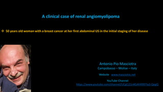
- 1. Antonio Pio Masciotra Campobasso – Molise – Italy Website www.masciotra.net YouTube Channel https://www.youtube.com/channel/UCgCj21nKGAhR997Ia3-QegQ A clinical case of renal angiomyolipoma 50 years old woman with a breast cancer at her first abdominal US in the initial staging of her disease
- 5. Renal Angiomyolipoma Feature AML Cortex Medulla Mean stiffness (kPa) 25.7 17.0 27.9 Ratio AML/Cortex 1.5 Ratio AML/Medulla 0.9
- 6. CT an MRI usual features of angiomyolipoma
- 8. Myoid cells with clear cytoplasm spinning off of large vessels in a background of mature fat: the classic microscopic features of angiomyolipoma.