Got it 👍 — here’s a structured overview of disorders of the external ear (useful for ENT study notes or exams):
---
Disorders of the External Ear
1. Congenital Disorders
Microtia – small or malformed pinna.
Anotia – complete absence of pinna.
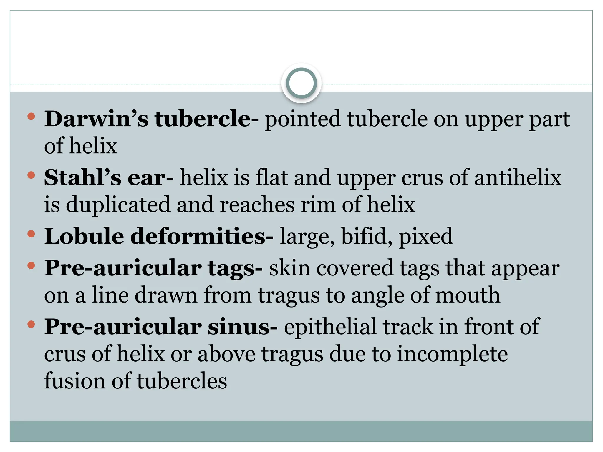
Preauricular sinus / tags – embryological remnant near pinna.
Atresia of external auditory canal – absent or narrowed ear canal → conductive hearing loss.
---
2. Trauma
Auricular hematoma – collection of blood between cartilage and skin → if untreated → “cauliflower ear.”
Lacerations / burns / frostbite – may lead to deformity.
---
3. Inflammatory Disorders
Otitis externa (swimmer’s ear) – infection of ear canal.
Causes: bacterial (Pseudomonas), fungal (Aspergillus, Candida).
Symptoms: ear pain, itching, discharge, swelling.
Furunculosis – staphylococcal abscess of hair follicle in outer canal.
Perichondritis – infection of auricular cartilage (often after trauma/piercing).
Eczema / psoriasis / allergic dermatitis – itching, scaling, redness.
---
4. Foreign Bodies
Common in children: beads, seeds, insects.
Symptoms: pain, discharge, hearing loss.
---
5. Tumors
Benign: exostosis, osteoma, papilloma, hemangioma.
Malignant: squamous cell carcinoma (most common), basal cell carcinoma, melanoma.
---
6. Miscellaneous
Cerumen impaction – wax accumulation → hearing loss, tinnitus.
Keloid – after ear piercing.
Actinic keratosis – precancerous lesion on pinna from sun exposure.
---
Key Points
Disorders may involve pinna, external auditory canal, or skin/cartilage.
Symptoms commonly include ear pain, discharge, itching, swelling, or hearing loss.
Treatment depends on cause: antibiotics/antifungals, removal of foreign body, drainage, surgery for tumors or congenital anomalies.
---
👉 Do you want me to make this into a short tabular format (disease → features → management) for quick revision?
You’re asking again about Cholesteatoma 🙂
Here’s a clear, exam-ready short note for you:
---
Cholesteatoma
Definition
An abnormal, non-cancerous collection of keratinizing squamous epithelium in the middle ear and mastoid that enlarges and destroys bone.
---
Types
1. Congenital – epithelial remnants behind intact tympanic membrane, usually in children.
2. Acquired – more common, due to:
Retraction pocket (from Eustachian tube dysfunction)
Migration of epithelium through a perforation
Metaplasia (rare theory)
---
Clinical Features
Persistent, foul-smelling scanty discharge (otorrhea)
Conductive hearing loss
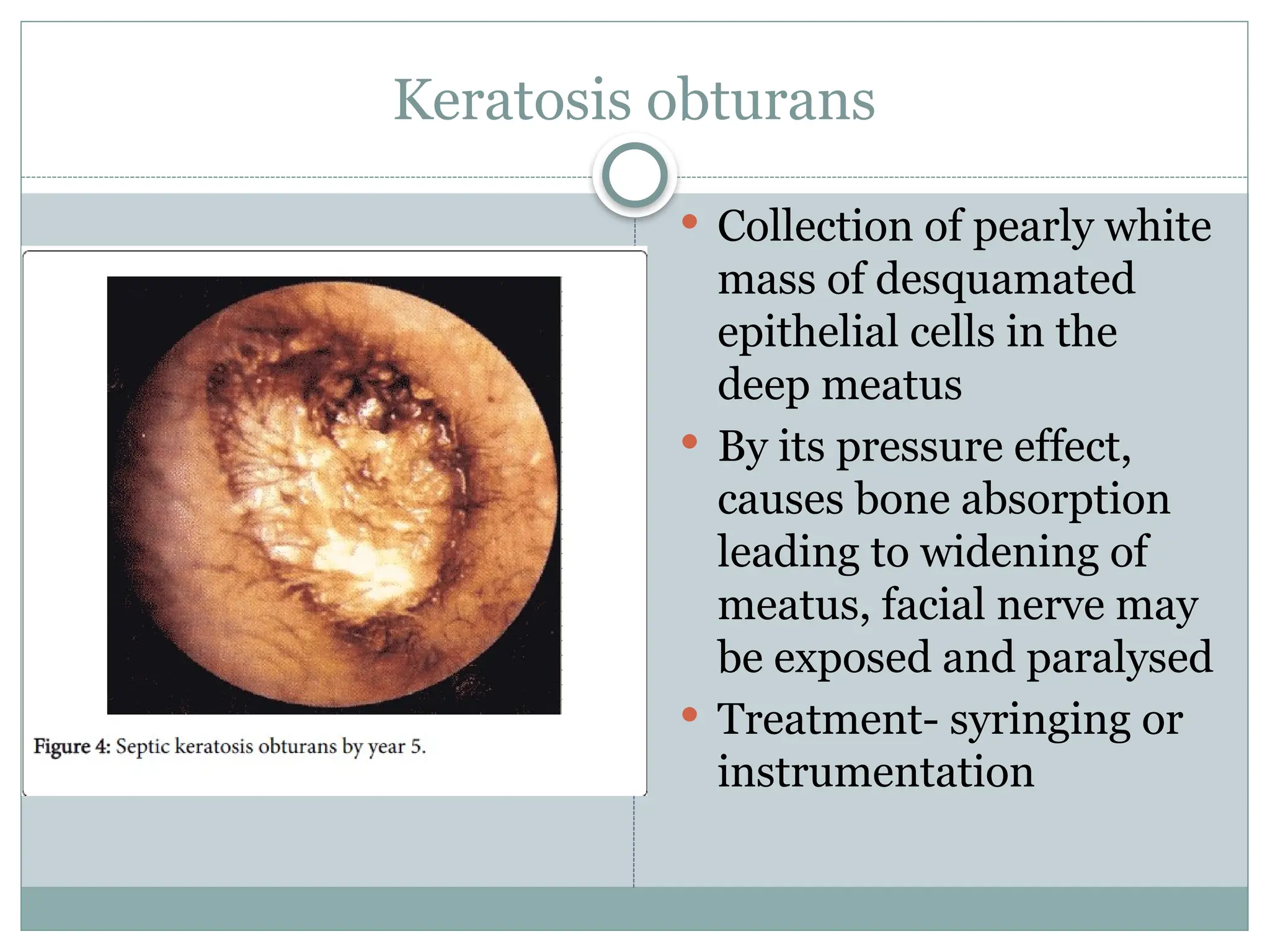
Retraction pocket or pearly white mass on otoscopy
Complications:
Ossicular erosion → severe hearing loss
Facial nerve palsy
Labyrinthine fistula → vertigo
Intracranial: meningitis, brain abscess, lateral sinus thrombosis
---
Diagnosis
Otoscopy: pearly white debris / retraction pocket
Audiometry: conductive hearing loss
HRCT temporal bone: extent, ossicle status, complications
---
Treatment
Surgical (definitive):
Modified radical mas