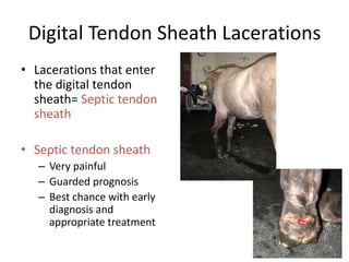
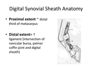
Digital Tendon Sheath
     Lacerations
Digital Tendon Sheath Lacerations
• Lacerations that enter
  the digital tendon
  sheath= Septic tendon
  sheath

• Septic tendon sheath
   – Very painful
   – Guarded prognosis
   – Best chance with early
     diagnosis and
     appropriate treatment
How Do I Know if a Laceration Enters the Digital
            Tendon Sheath ????
• How Do I Know if a Laceration Enters the
  Digital Tendon Sheath ????
  – Knowledge of anatomy- index of suspicion – (link to
     anatomy slides and saggital section video)

  – Checking for communication between the
    laceration and digital tendon sheath by fluid
    distension (link to video of checking for synovial structure involvement)
Digital Synovial Sheath Anatomy
• Thin walled structure
  that encompasses
  the majority of the
  SDFT and the entire
  DDFT.
Digital Synovial Sheath Anatomy
• Proximal extent ~ distal
  third of metacarpus


• Distal extent= T
  ligament (intersection of
  navicular bursa, palmar
  coffin joint and digital
  sheath)
Digital Synovial Sheath Anatomy
• Synovial structure- similar to joints and bursae
• Outer fibrous layer and inner synovial layer.


                             Image from Moyer et al Equine Joint Injection and Regional Anesthesia
Digital Synovial Sheath Function
• Decreases build up of friction and heat as
  tendons change direction around the palmar
  aspect of the fetlock and glide along side each
  other

Digital tendon sheath lacerations

  • 1.
  • 2.
    Digital Tendon SheathLacerations • Lacerations that enter the digital tendon sheath= Septic tendon sheath • Septic tendon sheath – Very painful – Guarded prognosis – Best chance with early diagnosis and appropriate treatment
  • 3.
    How Do IKnow if a Laceration Enters the Digital Tendon Sheath ????
  • 4.
    • How DoI Know if a Laceration Enters the Digital Tendon Sheath ???? – Knowledge of anatomy- index of suspicion – (link to anatomy slides and saggital section video) – Checking for communication between the laceration and digital tendon sheath by fluid distension (link to video of checking for synovial structure involvement)
  • 5.
    Digital Synovial SheathAnatomy • Thin walled structure that encompasses the majority of the SDFT and the entire DDFT.
  • 6.
    Digital Synovial SheathAnatomy • Proximal extent ~ distal third of metacarpus • Distal extent= T ligament (intersection of navicular bursa, palmar coffin joint and digital sheath)
  • 7.
    Digital Synovial SheathAnatomy • Synovial structure- similar to joints and bursae • Outer fibrous layer and inner synovial layer. Image from Moyer et al Equine Joint Injection and Regional Anesthesia
  • 8.
    Digital Synovial SheathFunction • Decreases build up of friction and heat as tendons change direction around the palmar aspect of the fetlock and glide along side each other