
Recommended
More Related Content
Similar to DIAGNOSTIC APPROACH AND MANAGEMENT OF ACUTE KIDNEY INJURY - Copy.pptx
Similar to DIAGNOSTIC APPROACH AND MANAGEMENT OF ACUTE KIDNEY INJURY - Copy.pptx (20)
Recently uploaded
Recently uploaded (20)
DIAGNOSTIC APPROACH AND MANAGEMENT OF ACUTE KIDNEY INJURY - Copy.pptx
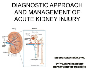
- 1. DIAGNOSTIC APPROACH AND MANAGEMENT OF ACUTE KIDNEY INJURY DR SUBHAYAN BATABYAL 2ND YEAR PG RESIDENT DEPARTMENT OF MEDICINE
- 2. ACUTE KIDNEY INJURY(AKI) is ABRUPT impairment of filtration and excretory function of kidneys over days to weeks (usually with in 7 days) resulting in retention of nitrogenous waste products with in blood. AKI is not a single entity rather it is a heterogenous group of disease that share common characteristics like reduction of URINE VOLUME and increase in serum CREATININE and UREA.
- 3. AKI is diagnosed if ONE of the following criteria is met: Increase in serum creatinine (SCr) at least 0.3mg/dl within 48hours . A 50% increase in baseline SCr with in 7 days . Urine output less than 0.5mg/kg /hr for atleast 6hours .
- 4. EPIDEMIOLOGY • AKI comprises of 5-7 percent of all hospital admission and 30 percent of ICU admission. • Large studies have shown that increase in serum creatinine by 0.3mg/dl may increase mortality by FOUR folds . • Mortality rate of about 50 percent is prevalent among ICU admitted patients despite improvement in medical infrastructure.
- 5. COMMUNITY ACQUIRED AKI HOSPITAL ACQUIRED AKI ICU ACQUIRED AKI INCIDENCE LOW <1% MODERATE(2-20%) HIGH (20-60%) CAUSE SINGLE SINGLE/MULTIPLE MULTIFACTORIAL MORTALITY RATE 15% 15-40% 30-50% RISK FACTORS CHF DRUG ADVERSE EFFECT URINARY TRACT OBSTRUCTION MALIGNANCY SEPSIS SURGERY NEPHROTOXIC DRUG REACTION VOLUME DEPLETION DURING SURGICAL PROCEDURE MODS NEPHROTOXIC DRUG REACTION SEPTIC SHOCK
- 6. TOP FIVE CAUSES OF AKI IN INDIA • DIARRHEAL DISEASES • SEPSIS • ACUTE FALCIPARUM MALARIA • DRUG INDUCED • HOSPITAL ACQUIRED
- 8. CLASSIFICATION OF AKI • RIFLE CRITERIA: RISK- INURY - FAILURE LOSS OF KIDNEY FUNCTION- END STAGE RENAL DISEASE • AKIN CRITERIA :ACUTE KIDNEY INJURY NETWORK • KDIGO CRITERIA :KIDNEY DISEASE IMPROVING GLOBAL OUTCOME
- 10. AKIN CRITERIA
- 11. KDIGO CRITERIA
- 12. PATHOPHYSIOLOGY • AKI is broadly classified into 3 categories : • PRERENAL • RENAL or INTRINSIC • POSTRENAL
- 15. INTRINSIC AKI GLOMERULAR TUBULAR AND INTERSTITIUM VASCULAR ACUTE GLOMERULO NEPHRITIS ISCHEMIA SEPSIS /INFECTON NEPHROTOXINS VASCULITIS RENAL ARTERY STENOSIS ,B/L RENAL VEIN THROMBOSIS OPERATIVE ARTERIAL CROSS CLAMPING MALIGNANT HYPERTENSION TTP-HUS POST OPERATIVE SIRS BURNS PANCREATITIS Due to microvascular and endothelial Damage by leucocytes and ROS Exogenous: Contrast agents Aminoglycoside Cisplatin Amphotericin B PPI NSAIDS Endogenous Rhabdomyolosis, hemolysis, myeloma, intratubular crystals ACUTE INTERSTITIAL NEPHRITIS : Drug HSN :Antibiotic uses ,Diuretics , NSAIDS ,Anticonvulsant drugs ,Allopurinol Infective: Bacterial(staph strepto,)viral ( CMV EBV),Tubercular,fungal(candida)
- 17. POST RENAL AKI • BLADDER OUTLET OBSTRUCTION(BPH,Neurogenic bladder anticholingeric drug blood clots calculi urethral strictures ) • BILATERAL PELVIURETERAL OBSTRUCTION(neoplasia retropetritoneal fibrosis abscess ) • SOLITARY FUNCTION KIDNEY
- 18. PATHOPHYSIOLOGY OF PRERENAL AND INTRINSIC AKI
- 20. CLINICAL PRESENTATIONS OF AKI DECREASED URINE OUTPUT NAUSEA /VOMITING/ DIARRHEA/ ANOREXIA WEIGHT GAIN PERIPHERAL EDEMA ALTERED MENTAL STATUS FATIGUE DYSPNEA PRURITIS
- 21. DIAGNOSTIC ALGORITHM TO AKI HISTORY TAKING AND PHYSICAL EVALUATION URINE FINDINGS BLOOD PARAMETERS RENAL FAILURE INDICES RADIOLOGICAL FINDINGS NOVEL BIOMARKERS RENAL BIOPSY
- 22. HISTORY AND PHYSICAL FINDINGS H/O of vomiting diarrhea Use of medications like NSAIDS, ACEI ,ARB and their doses H/O of prostatic disease, Nephrolithiasis , pelvic malignancy H/O of Autoimmune disease like SLE H/O of Pregnancy PHYSICAL SIGNS : Orthostatic hypotension, tachycardia , reduced JVP, decreased skin turgour ,dry tongue indicative of PRERENAL AZOTEMIA Supra pubic pain hints to Bladder outlet obstruction Atheroembolic disease leads to livedo reticularis and other emboli signs in leg Fever arthalgia and rashes may accompany in Allergic Interstitial Nephritis Palpable purpura raises the possibility of any vasculitis Tense abdomen may be considered as COMPARTMENT SYNDROME Signs of Limb ischemia opts for Rhabdomylosis
- 23. URINE ANALYSIS URINE ANALYSIS gives good interpretation towards cause of AKI but lacks sensitivity and specificity. Oliguria (urine output <400ml in 24 hrs denotes poor prognosis in AKI . Anuria is uncommon in AKI but usually associated with complete Urinary tract obstruction ,profound septic shock, severe ischemic necrosis ,vasculitis .
- 24. • CAUSES OF NORMAL URINARY FINDINGS OR FEW HYALINE CAST : 1) PRERENAL AZOTEMIA 2) POST RENAL AKI 3) TTP/HUS 4) PREGLOMEERULAR VASCULITIS 5) SCLERODERMA CRISIS 6) ATHERO EMBOLISM
- 25. ABNORMAL URINE FINDINGS RBC CAST WBC CAST RENAL TUBULAR CELLS / PIGMENT CAST GRANULAR CASTS EOSINOPHILS CRYSTALURIA GLOMERULO NEPHRITIS (GN) VASCULITIS MALIGNANT HYPERTNSION THROMBOTIC MICRO ANGIOPATHY PYELO NEPHRITIS INTERSTITIAL NEPHRITIS ALLOGRAFT REJECTION TUBULO INTERSTITIAL NEPHRITIS MYO – GLOBINURIA HEMO - GLOBINURIAA ATN GLOMERULO NEPHRITIS (GN) VASCULITIS ALLEGIC INTERSTTIAL NEPHRITIS GN PYELO NEPHRITIS CYSTITIS URIC ACID NEPHROPATHY CALCIUM GLUCONATE INGESTION DRUGS (SULFADIAZINE ,ACYCLOVIR , INDINAVIR )
- 26. BLOOD INVESTIGATIONS COMPLETE BLOOD COUNT PERIPHERAL BLOOD SMEAR RENAL FUNCTION TEST SERUM ELECTROLYTES SERUM URIC ACID /PHOSPHATES CPK /LDH SERUM ELECTROPHORESIS ANA/ANCA/ANTI GBM /ANTI PHOSPHOLIPASE A2 Antibodies COMPLEMENTS
- 27. BLOOD PARAMETERS ELEVATED serum CREATININE above baseline Associated findings In different clinical settings: LOW Haemoglobin(Hb) PERIPHERAL EOSINOPHILIA THROMBOCYTOPENIA HYPERKALEMIA HEPERPHOSPHATEMIA HYPERURICEMIA HYPOCALCEMIA ALTERED ANION GAP FREE LIGHT CHAINS IN SERUM ELECTROPHORESIS
- 28. RENAL FAILURE INDICES Several indices have been used to distinguish prerenal azotemia from intrinsic AKI . The FeNa (fractional excretion of filtered sodium load ) is one such indicator which justify the ability of kidneys to carry out proper tubular reabsorption . It has high sensitivity but low specificity.
- 33. • IN CKD kidneys are shruken except for Diabetic nephropathy ,HIV associated nephropathy ,Infiltrative diseases . • Kidneys are normal sized in AKI . CONTRAST AGENTS SHOULD BE USUALLY AVOIDED IN SEVERE AKI LIKE IODINATED AGENTS AND TYPE 1 GADOLINIUM BASED AGENTS.
- 34. NOVEL BIOMARKERS BUN and SCr are functional markers of glomerular filtration but not indicative of Tissue injury . BUN and Scr are slow to rise after AKI as a result accurate diagnosis gets belated . Several Protein Biomarkers have been discovered by research work and are used to confirm AKI faster .
- 35. NOVEL BIOMARKERS
- 36. APPROACH TO AKI TYPE HISTORY FEATURES PRERENAL AZOTEMIA H/O of poor fluid intake, fluid loss, diarrhea Vomiting , NSAIDS ,ARB ,ACEI Decreased circulatory volume (cirrhosis, heart failure ) BUN/Cr>20% FeNa<1% HYALINE CAST URINE OSMOLALITY >500 URINE SGPECIFIC GRAVITY >1.08 SEPSIS a/w AKI Septic shock or overt infection POSITIVE CULTURE FROM SITES ,URINE SEDIMENT GRANULAR CAST ,TUBULO EPITHELIAL CAST FeNa>1% ISHEMIA a/wAKI Systemic hypotension a/w sepsis CKD ,Old age URINE SEDIMENT GRANULAR CAST ,TUBULO EPITHELIAL CAST FeNa>1%
- 37. TYPE HISTORY FEATURES RHABDOMYLOSIS TRAUMATIC CRUSH INJURIES SEIZURE IMMOBILIZATION ELEVTED MYOGLOBIN, CREATINE KINASE, URIC ACID ,URINE HEME POSITIVE BUT FEW RBC HEMOLYSIS TRANSFUTION REACTION ANAEMIA ,ELEVATED LDH LOW HAPTOGLOBIN TUMOUR LYSIS SYNDROME RECENT CHEMO THERAPY HYPERKALEMIA HYPERPHOAPHATEMIA HYPOCALCEMIA NORMAL TO MARGINALLY INCREASED URIC ACID AND CREATINE KINASE MULTIPLE MYELOMA AGE >65 BONE PAIN CONSTITUTIONAL SYMPTOMS MONOCLONAL M SPIKE IN ELECTROPHORESIS LOW ANION GAP ANAEMIA
- 38. TYPE HISTORY FEATURES CONTRAST NEPHROPATHY EXPOSURE TO IODINATED COMPOUNDS RISE IN SCr WITH IN 2-3 DAYS PEAK IN 3-5 DAYS RECOVERY IN 7 DAYS ANTIBIOTICS : AMINOGLYCOSIDE,CISPLATIN ,VANCOMYCIN ,TENOFOVIR ETHELYNE GLYCOL MELAMINE SUGGESTIVE DRUG HISTORY URINE SEDIMENT GRANULAR CAST ,TUBULO EPITHELIAL CAST FeNa>1%
- 39. TYPE HISTORY FEATURES GLOMERULO NEPHRITIS, VASCULITIS SUGGESTIVE SYMPTOMS ANA,ANCA AGBM POSITIVE KIDNEY BIOPSY SUGGESTIVE INTERSTITIAL NEPHRITIS LEIGONELLA INFECTION TUBULOINTERSTITIAL NEPHRITIS- UVEITIS STERILE PYURIA ,EOSINOPHILURIA NONOLIGURIC TTP/HUS RECENT DIARRHEA , NEUROLOGICAL ANOMALIES ,CALCINEURIN INHIBITORS , PREGNANCY POST PARTUM SCHISTOCYTES IN PBS ELEVATED LDH ,ANAEMIA THROMBOCYTOPENIA TYPICAL HUS IS DIARRHEAL DISEASE DUE TO SHIGA TOXIN ATYPICAL HUS IS DUE TO ACQUIRED COMPLEMENT DYSREGULATION ADAMTS13 ACTIVITY REQUIRED
- 40. TYPE HISTORY FEATURES POST RENAL AKI H/O KIDNEY STONES PROSTRATE DISEASE OBSTRUCTED BLADDER RETROPERITONEAL PELVIC NEOPLASM MAY BE ASSOCIATED WITH PYURIA ,HEMATURIA USG ,CT SUGGESTIVE
- 41. INDICATION OF RENAL BIOPSY ARF of unknown etiology Suspicion of glomerulonephritis,systemic diseases (eg. Vasculitis )or AIN (acute interstitial nephritis). ATN not recovering after 4-6 weeks of dialysis with no more recurrent insults
- 42. COMPLICATIONS OF AKI HYPERVOLEMIA /HYPOVOLEMIA HYPONATREMIA HYPERKALEMIA ACIDOSIS HYPERPHOSPHATEMIA HYPOCALCEMIA BLEEDING INFECTIONS CARDIAC COMPLICATIONS(arrythmias,pericarditis,pericardial effusion ) MALNUTRITION
- 43. MANAGEMENT OF AKI MEDICAL MANAGEMENT DIALYSIS TREATMENT OF COMPLICATIONS
- 44. CONSERVATIVE MANAGEMENT • COMPLETE FLUID INTAKE AND OUTPUT • DAILY WEIGHT RECORD • INTRAVASCULAR VOLUME ASSESSMENT CLINICALLY DAILY. CV line for measure CVP • SERUM Na /K /Ur/Cr /Calcium/Phosphate
- 45. : PRENAL AKI MANAGEMENT a) Correct volume depletion When Prerenal AKI is due to deficits in ECF volume FLUID THERAPY is indicated. In severe hemorrhage PRBC is indicated . BUFFERED CRYSTALLOIDS SOLUTIONS (RINGER LACTATE ,PLASMALYTE,HARTMAN SOLUTION) In Severe Hypovolemia with hypochloremia 0.9% NORMAL SALINE is used.
- 46. FLUID CHALLENGE In young patients 500-1000ml of bolus should be given .In old patients 250 ml bolus given irrespective of cardiac status. ASSESS VOLUME STATUS If EUVOLEMIA achieved SERUM ELECTROLYTES are monitored. Total fluid requirment =Total output of kidney and GI tract in previuos 24hr with additional 500- 1000ml for inacessible losses.
- 47. b)Ineffective arterial blood volume with edema:
- 48. In case of Cardiac failure : DIURETIC AGENTS in combinatins with Digitalis therapy may increase Cardiac output and improve renal perfusion and lessen azotemia .CARDIAC AFTER LOAD REDUCERS like ACEI, nitrates ,hydralazine may improve the outcome.
- 49. 2.). Cirrhosis and Hepato renal failure: Fluid management in individulals with cirrhosis & ascites AKI Is challeging due to difficulty in assessing intravascular volume status. Administration of IV fluids in wrong pattern may lead to worsening of ascites and pulmonary compromise.
- 50. • ALBUMIN may prevent AKI in those treated with antibiotics for sponteneous bacterial peritonitis. • Definiive treatment for HEPATORENAL FAILURE Is LIVER TRANSPLANTATION. • Bridge therapy include terlipressin(vasopressin analogue) combination therapy with octreotide (somatostatin analogue)and midodrine(alfa adrenergic analogue)and noradrenaline along with albumin (1g/kgbody wt max to 100g)
- 51. MONITORING OF THERAPY • Assessment of JUGULARVENOUS PRESSURE ORTHOSTATIC CHANGES IN BLOOD PRESSURE AND PULSE. • ULTRASOUND GUIDED MEASUREMENT OF DIAMETER OF IVC WITH RESPIRATORY CHANGES is most reliable. • Presence of basal crepts and third heart sound indicates vigorous fluid administration with resultant pulmonary congesion.
- 52. • The patients in whom vigorous resusitation is required and cardiovascular tolerance to sudden fluid challenges ,monitoring should be done with CENTRAL VENOUS CATHETER. • It is a satisfactory guide to measure CVP which ranges between 8-12 cm of water. • In volume depleted states CVP ranges low to zero. • A CVP rise more than 5 cm of water suggests cardiac failure & indication to stop fluid transfusion.
- 53. MANAGEMENT OF INTRINSIC AKI There is no specific treatment of ATN. First of all VOLUME STATUS of patient is assessed. If VOLUME DEFICIT :it should be corrected with adequate fluid therapy. If EUVOLEMIC :Fluid challenge & Diuretic challenge. If VOLUME OVERLOAD :Diuretic challenge to convert oliguric AKI to nonoliguric AKI.
- 54. DIURETIC CHALLENGE: may be given in oliguric patients with volume overlod. Initially iv furosemide 40-120 mg given as bolus if urine output improves then continous infusion 20mg/hr should be given. Diuretics only convert oliguric renal failure to non oliguric renal failure but not improves renal complications like hyperkalemia.
- 55. RENAL DOSE OF DOPAMINE: DOPAMINE is a selective renal vasodilator acting in a dose range of 1-3 microgram /kg/per minute. In several studies it had been shown risk benefit ratio of dopamine is high leading to very rare use in ARF unless cardiovascular compromise is evident. Complications like BOWEL ISCHEMIA & ARRYTHMIAS may occur.
- 56. C.)AVOIDANCE OF NEPHROTOXIC DRUG: Potential nephrotoxic drugs like NSAIDS ,ACEI CYCLOSPORINE ,TACROLIMUS CONTRAST AGENTS AMPHOTERICIN B,AMINOGLYCOSIDE should not be used as they can agravate ARF. D.)ADJUSTMENT OF DRUG DOSES: Drug doses are adjusted on CREATININE CLEARANCE not on serum creatinine.
- 57. 3.) AKI DUE TO GLOMERULONEPHRITIS /VASCULITIS: May respond to immunosuppressants or plasmapheresis. 4.)ALLERGIC INTERSTITIAL NEPHRITIS :Tapering dose of glucocorticoids are used. 5.)AKI DUE TO SCLERODERMA: Should be treated with ACEI
- 58. SUPPORTIVE MANAGEMENT OF INTRINIC AKI INTRAVASCULAR VOLUME OVERLOAD: Restriction of salt<1g/day and water(<1Lt) DIURETICS :Usually furosemide as bolus or infusion .If no response opt for ULTRAFILTRATIION. HYPERKALEMIA : • Restriction of dietary potassium • Discontinue potassium sparing diuretics • CALCIUM GLUCONATE 10ml10% over 3mins • GLUCOSE +INSULIN • INHALED B2 AGONIST • K+ BINDING RESINS • SODIUM BICARBONATE
- 59. METABOLIC ACIDOSIS : • RESTRICTION OF DIETARY PROTEIN • SODIUM BICARBONATE • DIALYSIS HYPERPHOSPHATEMIA: • RESTRICTION OF DIETARY PHOSPHATE • PHOSPHATE BINDING AGENTS (CALCIUM CARBONATE /SEVELAMER) HYPOCALCEMIA: • CALCIUM CARBONATE (IV) HYPONATREMIA : RESTRICTION OF ORAL AND IV FLUIDS HYPERMAGNESEMIA: • DISCONTINUE MAGNESIUM CONTAINING ANTIACIDS NUTRITION : • RESTRICTION OF DIETARY PROTEIN • 0.8 G/KG/DAY UPTO 1.5 G/KG/DAY AND 25-30KCAL /DAY BY ENTERAL ROUTE
- 60. MANAGEMENT OF POST RENAL AKI • The site of obstruction defines the treatment approch .Transuretheral or suprapubic bladder catheterization is needed for urethral strictures bladder neck obstruction . • Ureteric obstruction is treated by percutaneous nephrostomy or ureteral stent .
- 62. MODES OF DIALYSIS • INTERMITTENT HEMODIALYSIS:Standard • INTERMITTENT PERITONEAL DIALYSIS: If hemodialysis not available. • CONTINUOUS RENAL REPLACEMENT THERAPY: 1.hemodyanamic instability 2.active bleeding
- 63. KEY PARAMETERS FOR MONITORING PATIENTS WITH AKI
- 64. CAUSES OF DIURETIC RESISTANCE IN PATIENTS OF AKI
- 65. REFERENCES • HARRISONS PRINCIPLES OF INTERNAL MEDICINE 21st EDITION • JAYPEE MANUAL OF NEPHROLOGY 2nd EDITION • BRENNER &RECTORS THE KIDNEY