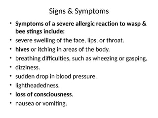
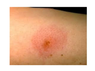
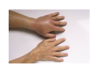
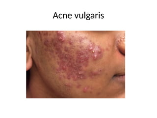
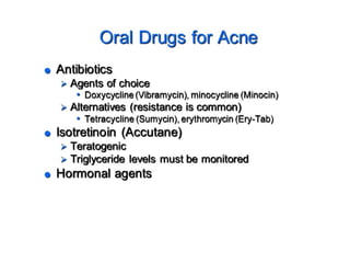
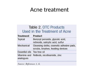
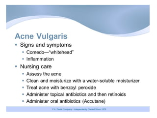
Signs & Symptoms
•Symptoms of a severe allergic reaction to wasp &
bee stings include:
• severe swelling of the face, lips, or throat.
• hives or itching in areas of the body.
• breathing difficulties, such as wheezing or gasping.
• dizziness.
• sudden drop in blood pressure.
• lightheadedness.
• loss of consciousness.
• nausea or vomiting.
22.
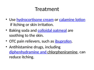
Treatment
• Use hydrocortisonecream or calamine lotion
if itching or skin irritation.
• Baking soda and colloidal oatmeal are
soothing to the skin.
• OTC pain relievers, such as ibuprofen.
• Antihistamine drugs, including
diphenhydramine and chlorpheniramine, can
reduce itching.
23.
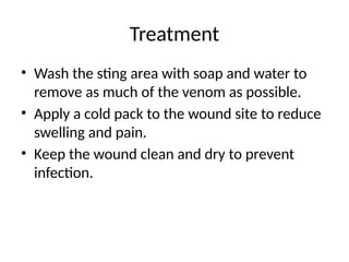
Treatment
• Wash thesting area with soap and water to
remove as much of the venom as possible.
• Apply a cold pack to the wound site to reduce
swelling and pain.
• Keep the wound clean and dry to prevent
infection.
24.
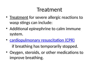
Treatment
• Treatment forsevere allergic reactions to
wasp stings can include:
• Additional epinephrine to calm immune
system.
• cardiopulmonary resuscitation (CPR)
if breathing has temporarily stopped.
• Oxygen, steroids, or other medications to
improve breathing.
25.
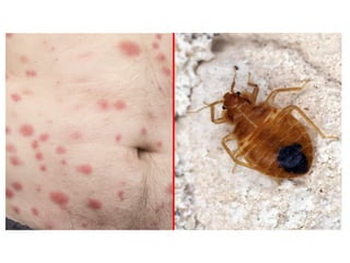
Bed Bugs
• Bedbugs are small parasitic insects that feed on
human blood.
• Bed bugs use a small tube-like structure called a
proboscis to pierce the skin and drink a person’s
blood.
• The pests are most active when humans are
asleep, during the night and early morning.
• Bed bugs can bite anywhere on the body where
there is skin. Typically, bites tend to occur on
areas exposed during sleeping.
27.
Symptoms
• In mostcases symptoms occur more or less immediately
after the bite, but they can develop or progress over the
following days as well.
• Without further irritation, symptoms typically resolve
after a week or so.
• Almost all bed bug bites will produce some degree of
discomfort, typically itchiness and inflammation.
• A burning painful sensation.
• Papular eruptions or areas of skin with raised or flat
patches that may be inflamed.
• Other symptoms: Increased likelihood of infection, Sleep
deprivation, Decreased wellbeing.
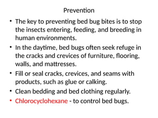
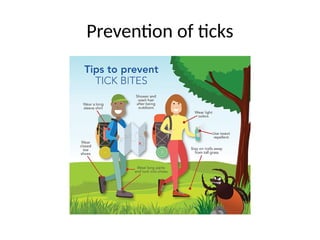
Prevention
• The keyto preventing bed bug bites is to stop
the insects entering, feeding, and breeding in
human environments.
• In the daytime, bed bugs often seek refuge in
the cracks and crevices of furniture, flooring,
walls, and mattresses.
• Fill or seal cracks, crevices, and seams with
products, such as glue or calking.
• Clean bedding and bed clothing regularly.
• Chlorocyclohexane - to control bed bugs.
30.
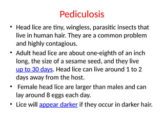
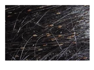
Pediculosis
• Head liceare tiny, wingless, parasitic insects that
live in human hair. They are a common problem
and highly contagious.
• Adult head lice are about one-eighth of an inch
long, the size of a sesame seed, and they live
up to 30 days. Head lice can live around 1 to 2
days away from the host.
• Female head lice are larger than males and can
lay around 8 eggs each day.
• Lice will appear darker if they occur in darker hair.
33.
Symptoms
• Itching isthe most common symptom of an
infestation.
• Other symptoms may include:
• tickling or a sensation of something moving in the
hair
• irritability and difficulty sleeping
• sores on the head from scratching
• swollen lymph nodes, or glands
• pink eye
34.
Treatment
• Combing wethair with a fine-toothed nit comb may
remove lice and some nits
• Comb the entire head from the scalp to the end of the
hair at least twice during a session.
• The process should be repeated every 3 to 4 days for at
least 2 weeks after no more lice are found.
• permethrin cream (1%)
• pyrethrin-based product
• malathion lotion (0.5%)
• benzyl alcohol lotion (5%)
35.
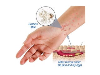
Scabies
• Scabies isa skin infestation caused by a mite
known as the Sarcoptes scabiei.
• Untreated, these microscopic mites can live on
skin for months.
• They reproduce on the surface of skin and
then burrow into it and lay eggs.
• This causes formation of itchy, red rash on
the skin.
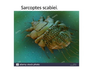
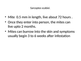
Sarcoptes scabiei.
• Mite0.5 mm in length, live about 72 hours .
• Once they enter into person, the mites can
live upto 2 months.
• Mites can burrow into the skin and symptoms
usually begin 3 to 6 weeks after infestation
38.
Scabies symptoms
• Afterthe initial exposure to scabies, it can take
up to six weeks for symptoms to appear. The
symptoms usually develop more quickly in
people who’ve had scabies before.
• The hallmark symptoms of scabies include a rash
and intense itching that gets worse at night.
• Continuous scratching of the infected area can
create sores that become infected.
• If this occurs, additional treatment with
antibiotics for the skin infection may be
recommended.
39.
•It’s a highlycontagious condition that
can easily be passed from one person
to another through direct skin contact
•scabies isn’t a sexually transmitted dis
ease.
•The infestation of mites may also be
transmitted through infested clothing
or bedding.
42.
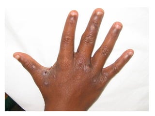
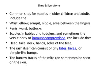
Signs & Symptoms
•Common sites for scabies in older children and adults
include the:
• Wrist, elbow, armpit, nipple, area between the fingers
• Penis, waist, buttocks
• Scabies in babies and toddlers, and sometimes the
very elderly or immunocompromised, can include the:
• Head, face, neck, hands, soles of the feet.
• The rash itself can consist of tiny bites, hives, or
pimple-like bumps.
• The burrow tracks of the mite can sometimes be seen
on the skin.
43.
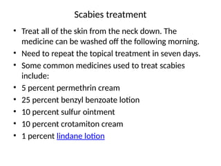
Scabies treatment
• Treatall of the skin from the neck down. The
medicine can be washed off the following morning.
• Need to repeat the topical treatment in seven days.
• Some common medicines used to treat scabies
include:
• 5 percent permethrin cream
• 25 percent benzyl benzoate lotion
• 10 percent sulfur ointment
• 10 percent crotamiton cream
• 1 percent lindane lotion
44.
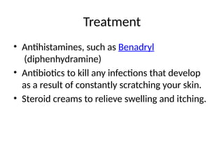
Treatment
• Antihistamines, suchas Benadryl
(diphenhydramine)
• Antibiotics to kill any infections that develop
as a result of constantly scratching your skin.
• Steroid creams to relieve swelling and itching.
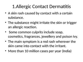
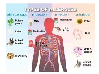
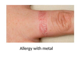
1.Allergic Contact Dermatitis
•A skin rash caused by contact with a certain
substance.
• The substance might irritate the skin or trigger
an allergic reaction.
• Some common culprits include soap,
cosmetics, fragrances, jewellery and poison ivy.
• The main symptom is a red rash wherever the
skin came into contact with the irritant.
• More than 10 million cases per year (India)
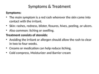
Symptoms & Treatment
Symptoms:
•The main symptom is a red rash wherever the skin came into
contact with the irritant.
• Skin: rashes, redness, blister, fissures, hives, peeling, or ulcers.
• Also common: itching or swelling.
Treatment consists of steroids:
• Avoiding the irritant or allergen should allow the rash to clear
in two to four weeks.
• Creams or medication can help reduce itching.
• Cold compress, Moisturizer and Barrier cream
56.
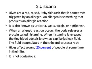
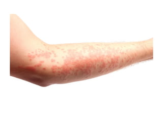
2.Urticaria
• Hives area red, raised, itchy skin rash that is sometimes
triggered by an allergen. An allergen is something that
produces an allergic reaction.
• It is also known as urticaria, welts, weals, or nettle rash.
• When an allergic reaction occurs, the body releases a
protein called histamine. When histamine is released,
the tiny blood vessels known as capillaries leak fluid.
The fluid accumulates in the skin and causes a rash.
• Hives affect around 20 percent of people at some time
in their life.
• It is not contagious.
58.
Examples of knowntriggers include:
• Medications, including some antibiotics and non-steroidal anti-inflammatory drugs
(NSAIDs), such as aspirin and ACE inhibitors, used for high blood pressure
• Foods, such nuts, shellfish, food additives, eggs, strawberries, and wheat products
• Infections, including influenza, the common cold, glandular fever, and hepatitis B
• Bacterial infections, including urinary tract infections and strep throat
• intestinal parasites
• extreme temperatures or changes in temperature
• high body temperature
• pet dander from dogs, cats, horses, and so on
• dust mites
• cockroaches and cockroach waste
• latex
• pollen
• some plants, including nettles, poison ivy, and poison oak
• insect bites and stings
• some chemicals
• chronic illness, such as thyroid disease or lupus
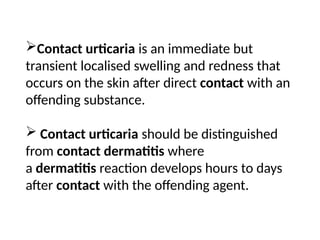
Contact urticaria isan immediate but
transient localised swelling and redness that
occurs on the skin after direct contact with an
offending substance.
Contact urticaria should be distinguished
from contact dermatitis where
a dermatitis reaction develops hours to days
after contact with the offending agent.
61.
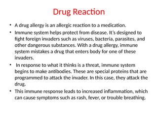
Drug Reaction
• Adrug allergy is an allergic reaction to a medication.
• Immune system helps protect from disease. It’s designed to
fight foreign invaders such as viruses, bacteria, parasites, and
other dangerous substances. With a drug allergy, immune
system mistakes a drug that enters body for one of these
invaders.
• In response to what it thinks is a threat, immune system
begins to make antibodies. These are special proteins that are
programmed to attack the invader. In this case, they attack the
drug.
• This immune response leads to increased inflammation, which
can cause symptoms such as rash, fever, or trouble breathing.
63.
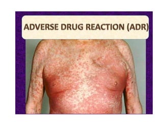
Symptoms
• The symptomsof a drug allergy may be so mild that you
hardly notice them. You might experience nothing more
than a slight rash.
• A severe drug allergy, however, can be life-threatening. It
could cause anaphylaxis.
• Anaphylaxis is a sudden, life-threatening, whole-body
reaction to a drug or other allergen.
• An anaphylactic reaction could occur minutes after take
the drug. In some cases, it could happen within 12 hours
of taking the drug.
• Symptoms can include: Irregular heartbeat, trouble
breathing, swelling, unconsciousness.
64.
Different drugs havedifferent effects on people.
•Certain drugs do tend to cause more allergic reactions
than others.
•These include:
Antibiotics such as penicillin and Sulfa antibiotics such
as sulfamethoxazole-trimethoprim
Aspirin.
Nonsteroidal anti-inflammatory medications, such as
ibuprofen.
Anticonvulsants such as carbamazepine and
lamotrigine
Chemotherapy drugs such as paclitaxel, docetaxel, and
procarbazine.
65.
Treatment
• Antihistamines
• Corticosteroids
•Bronchodilators
• If anyone know that they’re allergic to any drug,
take the following steps:
• Consider carrying a card or wearing a bracelet or
necklace that identifies their drug allergy. In an
emergency, this information could save their life.
66.
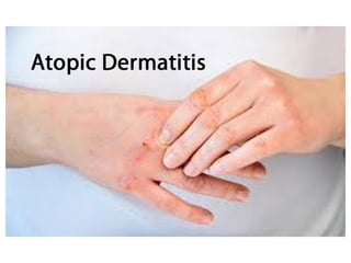
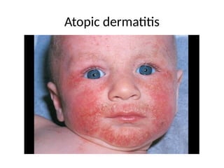
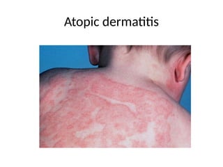
Atopic Dermatitis
• Atopicdermatitis (eczema) is a condition that
makes skin red and itchy. It's common in children
but can occur at any age. Atopic dermatitis is long
lasting (chronic) and tends to flare periodically.
• Known triggers for atopic dermatitis include
exposure to allergens such as pollen, pet dander
or peanuts, or by stress, dry skin and infection.
• Skin irritants such as some fabrics, soaps and
household cleaners may also trigger an atopic
dermatitis flare.
67.
Symptoms
Atopic dermatitis (eczema)signs and symptoms vary widely
from person to person and include:
• Dry skin
• Itching, which may be severe, especially at night
• Red to brownish-gray patches, especially on the hands, feet,
ankles, wrists, neck, upper chest, eyelids, inside the bend of
the elbows and knees,
• And in infants- the face and scalp
• Small, raised bumps, which may leak fluid and crust over
when scratched
• Thickened, cracked, scaly skin
• Raw, sensitive, swollen skin from scratching
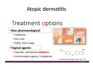
Treatment
Treatment of atopicdermatitis
• Rehydrating the skin with emollients like
petroleum jelly and the cautious use of topical
steroids to reduce inflammation and itching.
• Oral antihistamines may be helpful in breaking
the "itch-scratch" cycle.
• Since secondary infections can aggravate the
rash, oral antibiotics may also be occasionally
indicated.
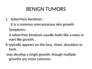
BENIGN TUMORS
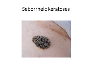
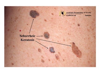
1. SeborrheicKeratosis :
it is a common noncancerous skin growth.
Symptoms :
A seborrheic keratosis usually looks like a waxy or
wart like growth.
It typically appears on the face, chest, shoulders or
back.
may develop a single growth, though multiple
growths are more common.
102.
symptoms
• Ranges incolor from light tan to brown or
black
• Is round or oval shaped
• Is flat or slightly raised with a scaly surface
• Ranges in size from very small to more than 1
inch (2.5 centimeters) across
• May itch
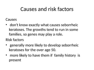
Causes and riskfactors
Causes
• don't know exactly what causes seborrheic
keratoses. The growths tend to run in some
families, so genes may play a role.
Risk factors
• generally more likely to develop seborrheic
keratoses for the over age 50.
• more likely to have them if family history is
present
106.
treatment
• Many growthsdevelop over a short time.
• The growths get irritated or bleed when
clothing rubs against them, these growths
need to be removed surgically.
• If any suspicious changes in the skin, such as
sores or growths that grow rapidly, bleed and
don't heal, these could be signs of skin cancer.
107.
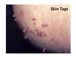
2.Arochordons (skin tags)
•A common skin growth in which a short, narrow
stalk sticks out.
• Skin tags are usually harmless and painless.
• The main symptom is a growth on the skin, often on
the neck, upper chest, underarms and eyelids. They
may become irritated from rubbing against clothing.
• Most skin tags don't require treatment. If a skin tag
is irritated or its appearance is bothersome, a doctor
can remove it.
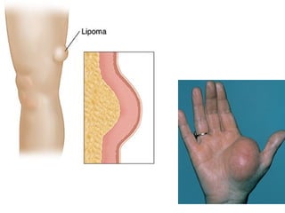
3.lipoma
• A lipomais a lump under the skin that occurs due to an
overgrowth of fat cells.
Lipomas can occur anywhere on the body where fat cells
are present, but they tend to appear on the shoulders,
chest, trunk, neck, thighs, and armpits. In less common
cases, they may also form in internal organs, bones, or
muscles.
• Lipomas feel soft and may move slightly under the skin
when people press down on them. They usually grow
slowly over a period of months or years and typically reach
a size of around 2–3 centimeters (cm). Occasionally, people
have giant lipomas, which can grow to more than 10 cm.
110.
causes
• do notfully understand what causes a lipoma.
• Some people inherit a faulty gene from their parents that can cause
one or more lipomas.
• Lipomas can occur more frequently in people with specific medical
conditions, such as:
• Gardner’s syndrome
• Cowden syndrome
• Madelung’s disease
• adiposis dolorosa
Other risk factors for developing a lipoma may include:
• obesity
• high cholesterol
• diabetes
• liver disease
• glucose intolerance
111.
symptoms
• A personwith a lipoma will typically feel a
soft, oval-shaped lump just beneath the skin.
• Lipomas are usually painless unless they affect
joints, organs, nerves, or blood vessels.
• In most cases, they do not cause other
symptoms.
112.
management
• People shouldalways tell their doctor if they
notice changes in a lipoma or if more lumps
appear. These changes might involve the lipoma:
• increasing in size or suddenly growing very quickly
• being painful
• becoming red or hot
• turning into a hard or immovable lump
• causing visible changes in the overlying skin
113.
diagnosis
• biopsy, wherethe doctor will remove a small
sample of cells from the lump and examine
the tissue under a microscope to look for signs
of cancer
• ultrasound scan
• MRI scan
• CT scan
114.
• Lipomas areusually harmless, so most people do not need to
have surgery to remove them.
People may want to remove a lipoma that:
• is cancerous
• is large or growing quickly
• causes bothersome symptoms, such as pain and discomfort
• interferes with normal body functions
• causes distress for cosmetic reasons
• the doctor is unable to confirm is a lipoma rather than
another type of tumor
116.
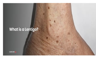
4. lentigo
In olderage, brown or black spots appear on the
skin.
These spots are especially common on sun-
exposed areas like face and the backs of hands.
They’re called lentigines, or liver spots. It’s called
lentigo because the spots can resemble lentils in
color.
These are increased number of normal
melanocytes in basal layer of epidermis.
117.
causes
• Exposure toUV radiation can cause lentigo. more likely
to get this condition when the people,…
• have fair skin
• have been exposed to the sun a lot, or have had several
sunburns
• tan indoors
• have had phototherapy or radiation therapy
• In other cases, an inherited syndrome can cause
lentigines.
• People of all ages and both genders can get lentigines.
118.
Symptoms
• Lentigo causesflat spots to appear on the
body. These spots are usually tan, brown, or
black in color. They may have rounded or
uneven edges.
• Lentigines can appear on different areas of the
body, depending on their cause. They don’t
itch or cause other symptoms.
119.
management
• Lentigines arenot typically a cause of medical concern, so
they don’t need to be treated. However, some may choose
to lighten or remove lentigines for aesthetic reasons.
• To lighten or remove lentigines, your dermatologist might
recommend one of these treatments:
• medicines such as bleaching creams containing
hydroquinone or retinoids (tretinoin)
• chemical peels
• laser or intense pulse light therapy to destroy melanocytes
• freezing (cryotherapy) to destroy melanocytes
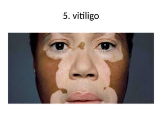
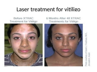
Vitiligo is acondition in which the skin
loses its pigment cells (melanocytes).
This can result in discolored patches in
different areas of the body, including
the skin, hair and mucous membranes.
123.
Causes
• We don'tknow exactly why this happens.
• It might be an autoimmune condition, where your body's defenses turn on
your own cells instead of attacking invading germs.
• Although vitiligo affects all races equally, it's more noticeable in dark-skinned
people.
• In most cases, it develops early in life, between ages 10 and 30. It will almost
always show up before age 40.
• Vitiligo may run in families.
• Autoimmune diseases, such as autoimmune thyroid disease (Hashimoto's
thyroiditis) or type 1 diabetes, can also raise it.
124.
Symptoms
• lose ofpigment quickly on several areas of skin. After
the white patches appear, they may stay the same for
a while, but later on, they might get bigger. may
have cycles of pigment loss and stability.
• Vitiligo commonly affects body folds (such as armpits),
places that have been injured in the past, and areas
exposed to sun, around moles, or around body
openings. It can also affect eyelids and hair.
• It's rare for pigment to return once the white patches
have developed.
125.
Diagnosis and Treatment
•doctor can usually make a diagnosis of vitiligo
by looking at skin during a physical exam.
• There's no known way to prevent or cure the
condition. But , can be improved the
appearance of affected skin with cosmetics
and corticosteroid creams.
• doctor can also try re-pigmenting the white
skin using UV light therapy or lightening the
skin in unaffected areas, or a skin graft.
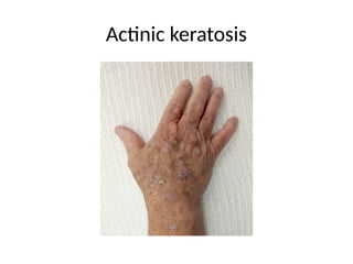
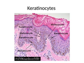
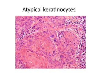
6 . Actinickeratosis( pre malignant)
• An actinic keratosis is a rough, scaly patch on the
skin that develops from years of exposure to the
sun.
• It's most commonly found on the face, lips, ears,
back of the hands, forearms, scalp or neck.
• It is an epidermal lesion characterized by
agregates of atypical, pleomorphic keratinocytes
at basal layer and extends upwards.
128.
• Also knownas a solar keratosis, an actinic
keratosis enlarges slowly and usually causes
no signs or symptoms other than a patch or
small spot on the skin. These patches take
years to develop, usually first appearing in
people over 40
symptoms
• The signsand symptoms of an actinic keratosis include:
• Rough, dry or scaly patch of skin, usually less than 1
inch (2.5 centimeters) in diameter
• Flat to slightly raised patch or bump on the top layer of
skin
• In some cases, a hard, wart like surface
• Color as varied as pink, red or brown
• Itching or burning in the affected area
131.
complications
• If treatedearly, almost all actinic keratoses can
be cleared up or removed before they develop
into skin cancer.
• If left untreated, some of these spots may
progress to squamous cell carcinoma — a type
of cancer that usually isn't life-threatening if
detected and treated early.
132.
• Prevention ofactinic keratoses is important.
• Sun safety is necessary to help prevent development
and recurrence of actinic keratosis patches and spots.
• Take these steps to protect the skin from the sun:
• Limit your time in the sun. Especially avoid time in
the sun between 10 a.m. and 2 p.m
• Use sunscreen on all exposed skin, and use lip balm
with sunscreen on your lips. Apply sunscreen 15
minutes before sun exposure and reapply it every two
hours — or more often if you're swimming or
perspiring.
133.
• Cover up.For extra protection from the sun,
wear tightly woven clothing that covers your
arms and legs. Also wear a broad-brimmed
hat
• Avoid tanning beds. The UV exposure from a
tanning bed can cause just as much skin
damage as a tan acquired from the sun.
• Examine your skin regularly, looking for the
development of new skin growths or changes
in existing moles, freckles, bumps and
birthmarks.
Difference of akand sk
• The main difference of these two conditions is
that actinic keratosis has the potential of
becoming cancerous. Seborrheic keratosis is
not known to develop into skin cancer.
137.
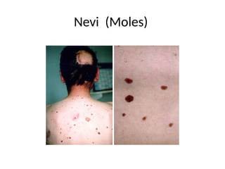
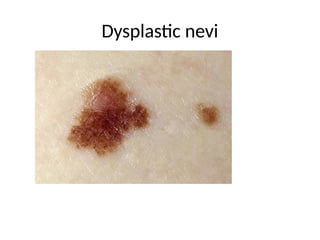
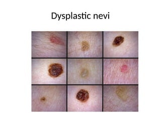
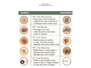
7 . Dysplasticnevi (pre malignant)
• Dysplastic nevi are moles that are larger and irregular
in shape than the average mole.
• They tend to have uneven color with dark brown
centers and lighter, uneven edges.
• These moles tend to be hereditary .
• People with dysplastic nevi may have more than 100
moles and have a greater chance of developing
melanoma, a serious and concerning form of skin
cancer.
• If the mole is found to be cancerous, a dermatolgist will
need to completely excise the mole and then close the
wound.
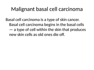
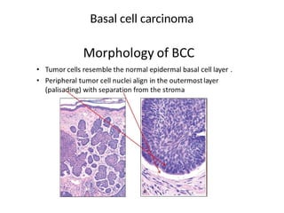
Malignant basal cellcarcinoma
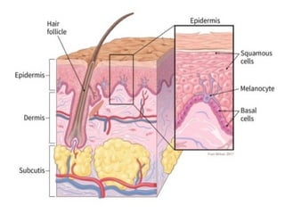
Basal cell carcinoma is a type of skin cancer.
Basal cell carcinoma begins in the basal cells
— a type of cell within the skin that produces
new skin cells as old ones die off.
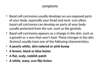
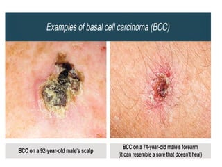
symptoms
• Basal cellcarcinoma usually develops on sun-exposed parts
of your body, especially your head and neck. Less often,
basal cell carcinoma can develop on parts of your body
usually protected from the sun, such as the genitals.
• Basal cell carcinoma appears as a change in the skin, such as
a growth or a sore that won't heal. These changes in the skin
(lesions) usually have one of the following characteristics:
• A pearly white, skin-colored or pink bump
• A brown, black or blue lesion
• A flat, scaly, reddish patch
• A white, waxy, scar-like lesion
145.
causes
• Basal cellcarcinoma occurs when one of the skin's basal cells
develops a mutation in its DNA.
• Basal cells are found at the bottom of the epidermis , the
outermost layer of skin. Basal cells produce new skin cells. As
new skin cells are produced, they push older cells toward the
skin's surface, where the old cells die and are sloughed off.
• The process of creating new skin cells is controlled by a basal
cell's DNA. The DNA contains the instructions that tell a cell
what to do.
• The mutation tells the basal cell to multiply rapidly and
continue growing when it would normally die. Eventually the
accumulating abnormal cells may form a cancerous tumor -
the lesion that appears on the skin.
146.
Risk factors
• Chronicsun exposure. A lot of time spent in the sun
• Radiation therapy.
• Fair skin.
• Increasing age.
• A personal or family history of skin cancer
• Immune-suppressing drugs
• Exposure to arsenic
• Inherited syndromes that cause skin cancer
147.
prevention
• To reducerisk of basal cell carcinoma :
• Avoid the sun during the middle of the day.
• Wear sunscreen year-round
• Wear protective clothing
• Avoid tanning beds
• Check your skin regularly and report changes to
your doctor.
148.
Diagnosis, Treatment
• Historyand general exam
• Skin sample for testing –biopsy
• The goal of treatment for basal cell carcinoma is to remove the
cancer completely
• Surgery
• Basal cell carcinoma is most often treated with surgery to remove
all of the cancer and some of the healthy tissue around it.
• Cryosurgery might be considered for treating small and thin basal
cell carcinomas when surgery isn't an option.
• Radiation therapy is sometimes used after surgery when there is
an increased risk that the cancer will return. It might also be used
when surgery isn't an option.
• Treatment for cancer that spreads- Chemotherapy.
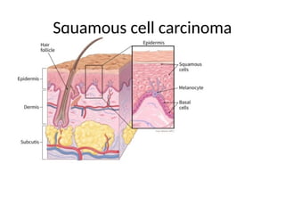
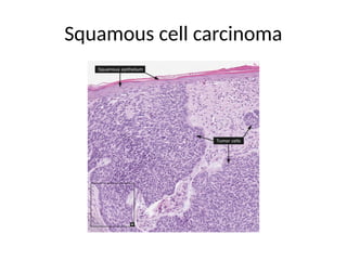
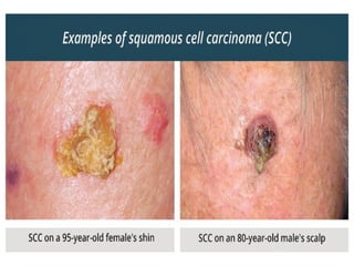
SIGNS AND SYMPTOMS
•Signs and symptoms of squamous cell carcinoma
of the skin include:
• A firm, red nodule
• A flat sore with a scaly crust
• A new sore or raised area on an old scar or ulcer
• A rough, scaly patch on your lip that may evolve
to an open sore
• A red sore or rough patch inside your mouth
• A red, raised patch or wartlike sore on or in the
anus or on your genitals
153.
• Squamous cellcarcinoma of the skin is usually not life-
threatening, though it can be aggressive. Untreated, squamous
cell carcinoma of the skin can grow large or spread to other
parts of your body, causing serious complications.
Causes
• Squamous cell carcinoma of the skin occurs when the flat, thin
squamous cells in the middle and outer layers of your skin
develop changes (mutations) in their DNA.
• Risk factors-same as BCC
Complications
• Untreated squamous cell carcinoma of the skin can destroy
nearby healthy tissue, spread to the lymph nodes or other
organs, and may be fatal.
PREVENTIVE measures – same as BCC
Treatment – same as BCC
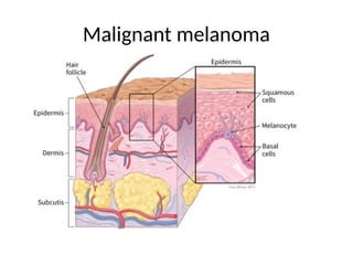
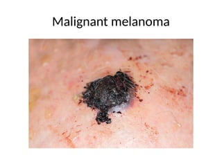
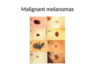
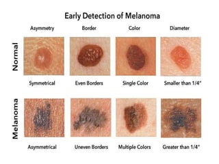
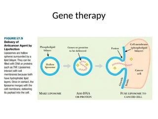
Malignant melanoma
• Themost serious type of skin cancer.
• Melanoma occurs when the pigment-producing
cells that give colour to the skin become
cancerous.
• Symptoms might include a new, unusual growth
or a change in an existing mole. Melanomas can
occur anywhere on the body.
• Treatment may involve surgery, radiation,
medication or in some cases, chemotherapy.
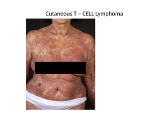
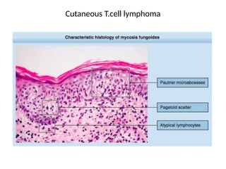
Cutaneous T –cell Lymphoma
• Cutaneous T-cell lymphoma (CTCL) is a rare type of cancer
that begins in white blood cells called T cells (T
lymphocytes).
• These cells normally help your body's germ-fighting
immune system. In cutaneous T-cell lymphoma, the T cells
develop abnormalities that make them attack the skin.
• Several types of cutaneous T-cell lymphoma exist. The
most common type is mycosis fungoides.
• Sezary syndrome is a less common type that causes skin
redness over the entire body.
• mycosis fungoides, progress slowly and others are more
aggressive.
162.
.
Signs and symptomsof cutaneous T-cell lymphoma include
• Round patches of skin that may be raised or scaly and might be
itchy
• Patches of skin that appear lighter in color than surrounding
skin
• Enlarged lymph nodes , and hair loss
• Thickening of the skin on the palms of the hands and soles of
the feet
• A rash-like skin redness over the entire body that is intensely
itchy.
Causes
• The exact cause of cutaneous T-cell lymphoma isn't known.
• In general, cancer begins when cells develop changes
(mutations) in their DNA
163.
• Diagnosis –history, physical exam, biopsy.
Treatment options may include:
• Skin creams and ointments, Chemotherapy can be applied
to the skin to attack cancer cells.
• Light therapy (phototherapy). Phototherapy involves
exposing the skin to wavelengths of light, such as ultraviolet
B or ultraviolet A
• Radiation therapy.
• Medications. steroid drugs and interferon. Chemotherapy
medicines attack quickly growing cells
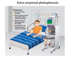
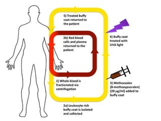
• Exposing blood cells to light. A procedure called
extracorporeal photopheresis involves taking a medicine
that makes your cells more sensitive to light
• Bone marrow transplant.