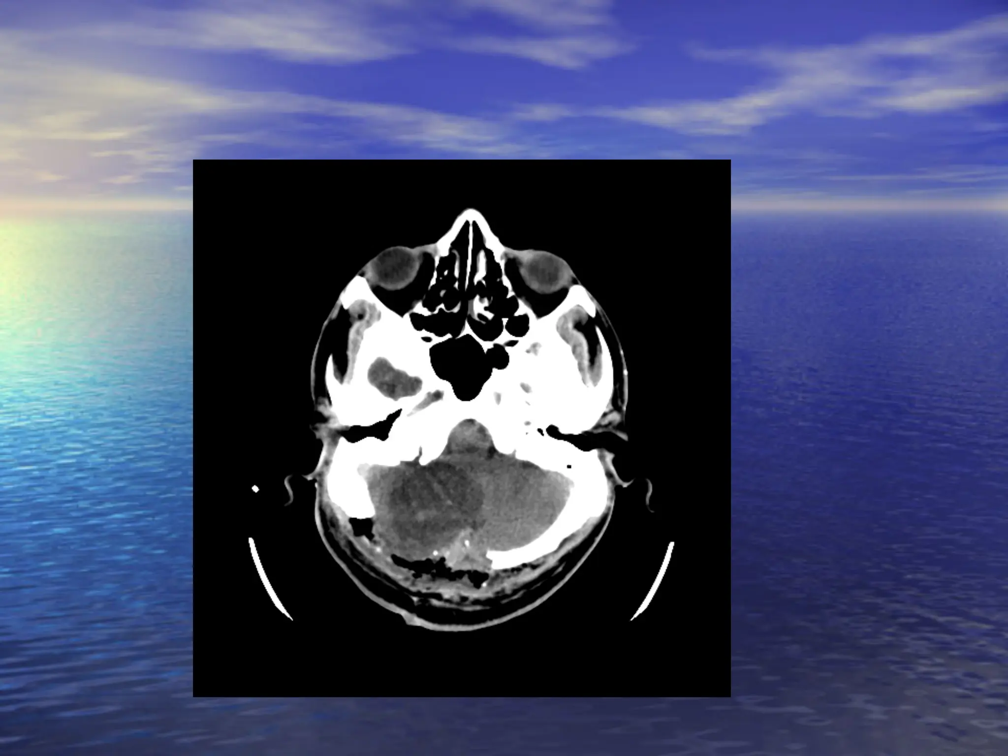
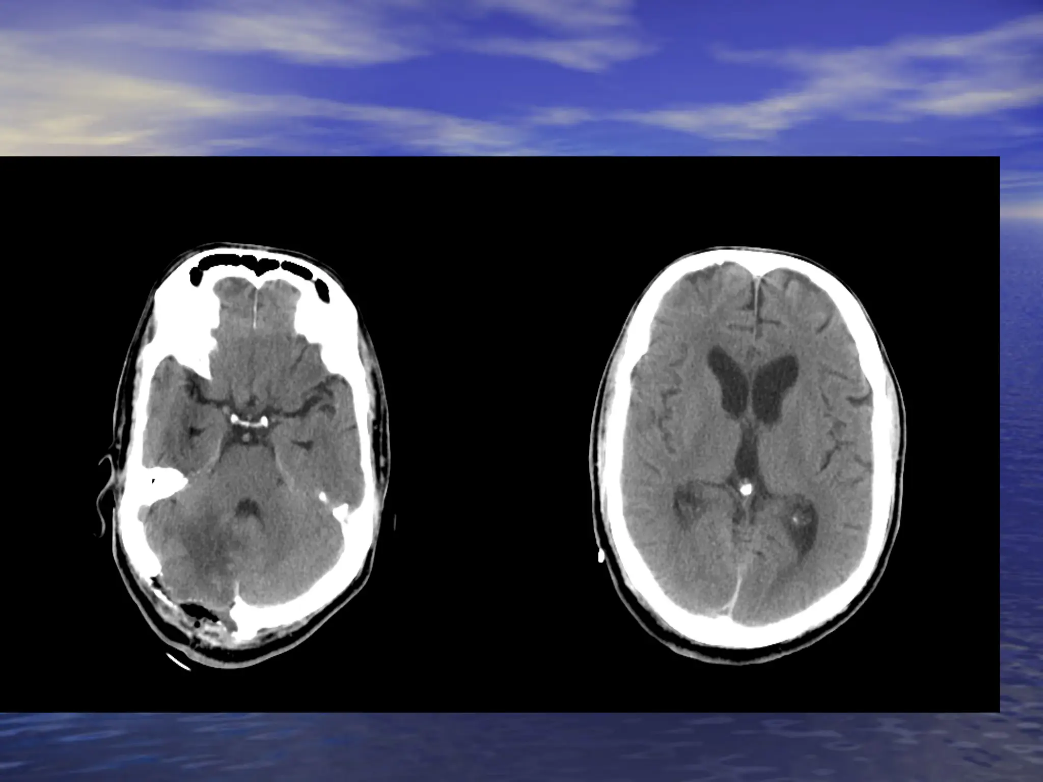
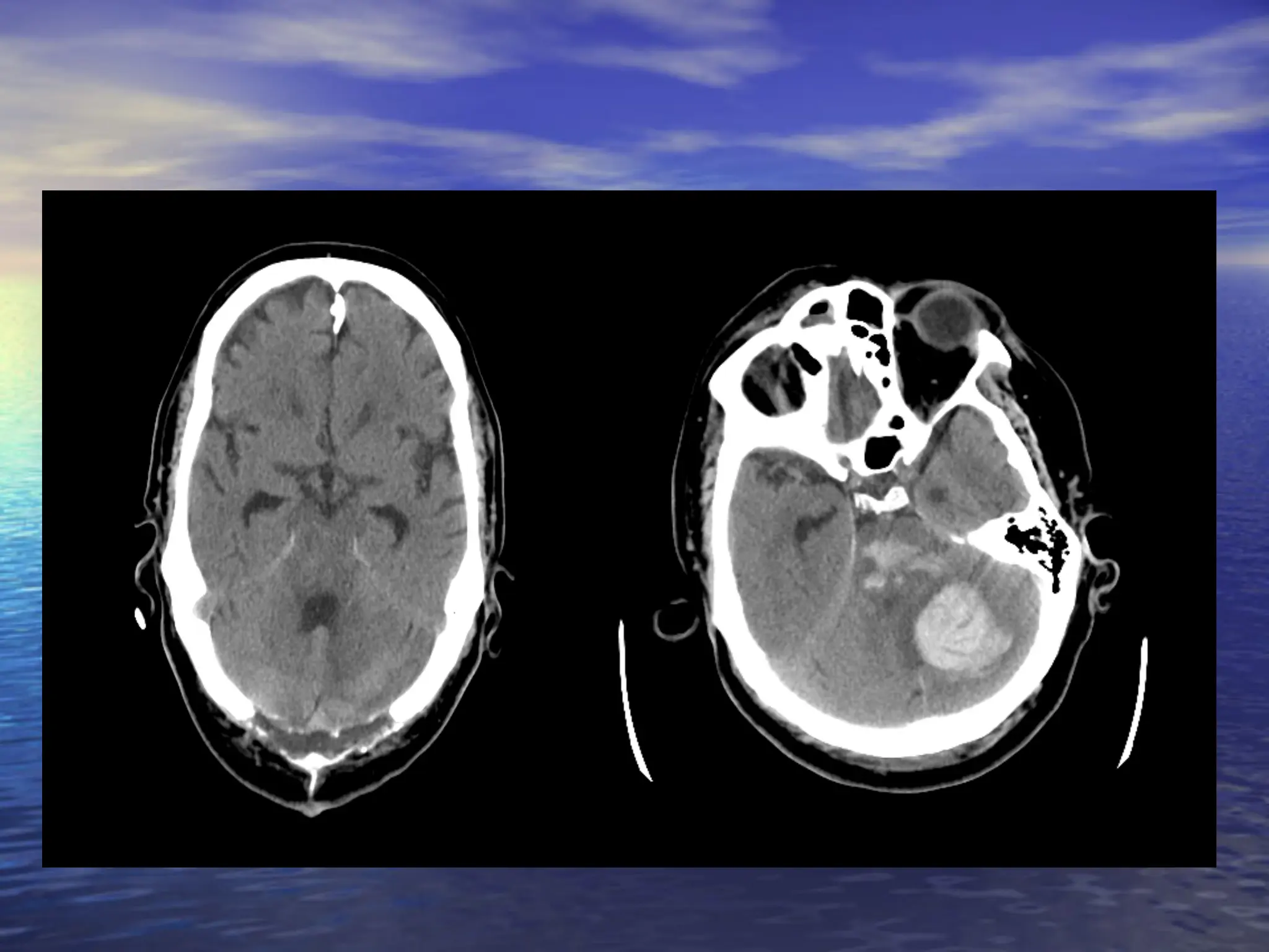
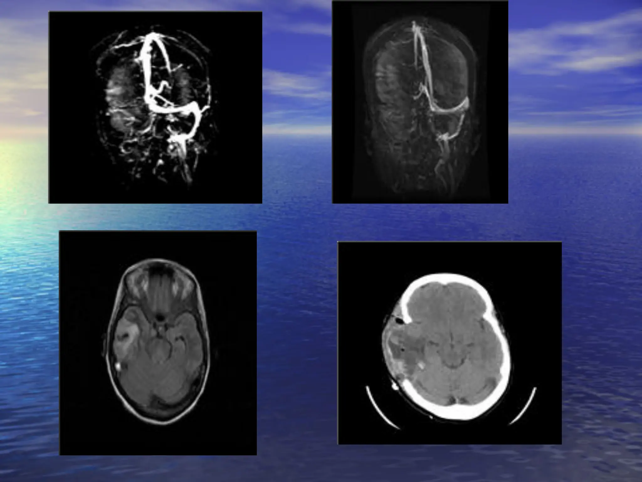
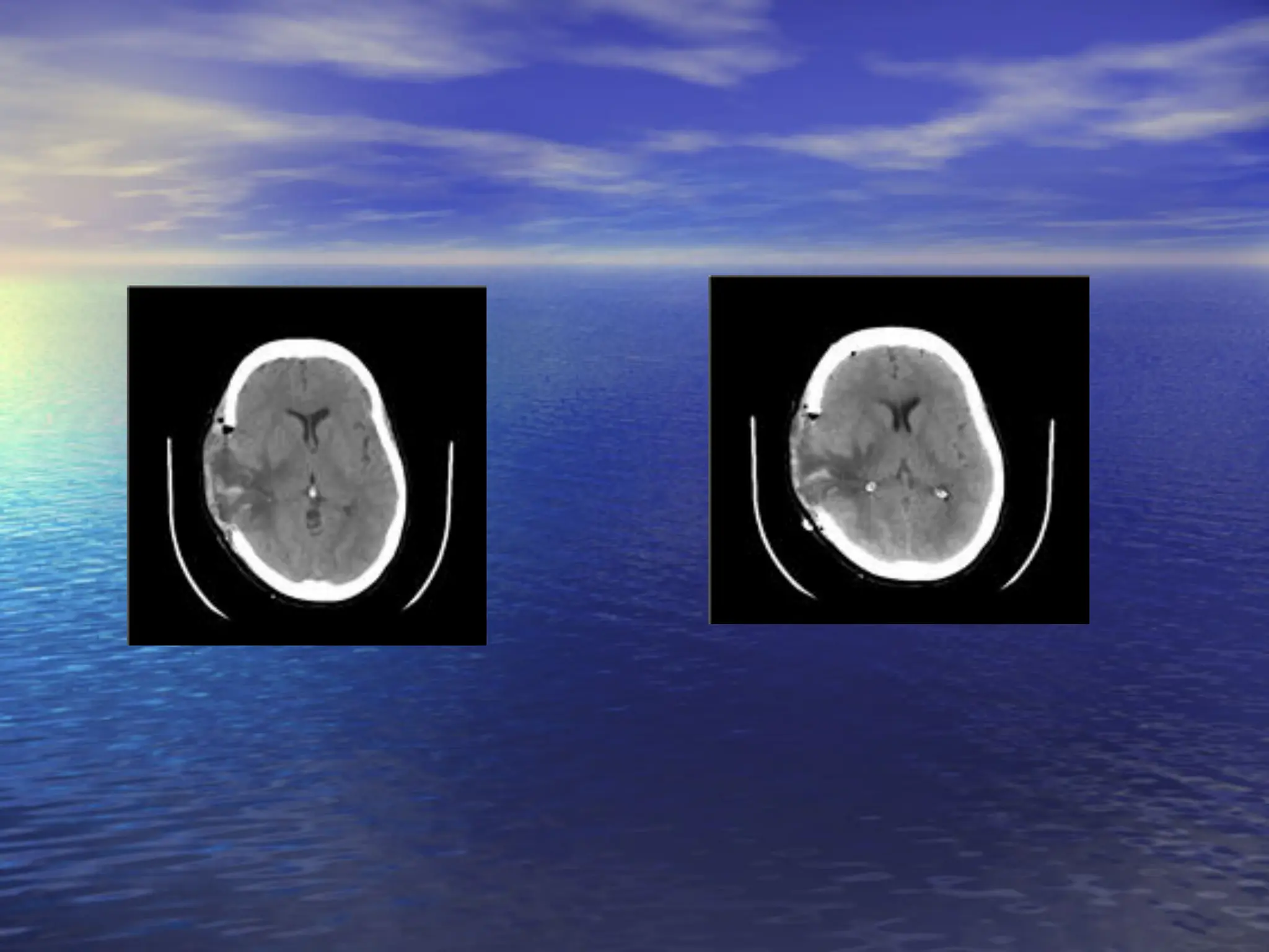
Decompressive craniectomy :
rationale, indications and outcome
KHALED ABDEEN M.D	

Ass. Professor of Neurosurgery	

Alexandria University
Decompressive craniectomy(DC) has been used	

as a final option in the management of refractory 	

intracranial hypertension caused by severe head 	

injury, cerebral infarction, subarachnoid 	

hemorrhage, intracerebral hematoma and so on. 	


Background
Decompressive craniectomy
(DC) is a method of “giving
room to the swelling brain”
and can be “ a life-saving 	

procedure.” 	


Mechanism by which DC
decreases compression of
brain stem structures and
minimizes herniation .	

(Kerr FWL; 1968)	

Pathophysiology : decompressive effects
Aims of Decompressive
Craniectomy
• Reduce ICP
• Improve blood flow
• Reduce damage to surrounding brain
tissue
• i.e. reduce secondary brain injury
Indication Criteria for Decompressive Craniectomy
	

INDICATION	

① coma or semicoma (GCS  9) 	

② pupillary abnormalities, but respond to mannitol	

③ supratentorial lesion with midline shift on CT	

④ refractory ICP despite best conventional therapy 	

⑤ age: initially  80 years , now ≦ 70 years	

(Of patients who were  70 years, 75% were dead)	

CONTRAINDICATION	

fatal brain stem damage :	

GCS  4 or fixed and dilated bilateral pupils
Increase in a focal CBF in the decompressed
brain related to the beneficial effect in patients	

(Yamakami  Yamaura, 1993) 	

Pre-DC	

 Post-DC	

8 days	


25 days	

99mTc-HMPAO 	

SPECT
Early DC prevents 	

secondary brain damage 	

Early DC reduces brain
edema formation by more
than 50% and prevents
secondary brain damage
when performed early
enough (i.e., during the
first 3 h after trauma).	


(Zweckberger K, et al.; 2006)
Cushing (1905) 	

 Miyazaki (1973)	

Kjellberg (1971)	

Ransohoff (1971)	

Makino (1975)	

Guerra (1999)	

skull	

Bilateral frontotemporo-	

parietal craniectomy	

A variety of operative techniques of DC	

Total 	

calvariectomy	

Subtemporal	

craniectomy	

Bifrontal	

craniectomy	

Hemicraniectomy	

History of decompressive craniectomy (DC)
Before and after…
Large (10 × 15 cm) frontotemporoparietal craniectomy 	

with the lower margin from the middle cranial fossa	

skin incision  craniectomy	

(Shima K, 2004)	

Surgical Technique for DC
Surgical Technique for DC (2)	

dural incision standard duraplasty	

Gore-tex 	

dura	

(Shima K, 2004)
Surgical Technique for DC (3)	

In the event of massive cerebral
swelling, extensive duraplasty with
internal decompression is performed.	

Dural closure 	

Dural incision
Malignant Middle Cerebral Artery
Infarction Syndrome
(MMCAIS)
How Common
Study	

 MMCAIS	

Ng L et al 1970	

 45 /353 supratentorial infarcts	

 12.74%	

Berrouschot J et al. 1998 	

 53/221 supratentorial infarcts	

 23.9%	

Kasner S et al, 2001	

 201/ 12000 stroke patients	

 1.675%	

Qureshi AI et al 2003	

 59 /1214 supratentorial infarcts	

 4.859%	

Wang KW et al 2006	

 40/418 MCA infarct	

 9.5%	

Reported between 5 to 10% of Acute Ischaemic Stroke.
What are the causes of
sudden deterioration in AIS?
Weimar C et al. Arch Neurol 2005
• 256/ 1964 patients (13%) had NIHSS ≥1 point after 48 to 72
hour
• 127 (6.5%) patients and 43 patients (2.2%) were intubated
• Attributable to
– Progressive stroke (33.6%)
– Increased ICP (27.3%)
– Recurrent cerebral ischaemia (11.3%)
– Secondary parenchymal haemorrhage (10.5%)
• Worsening of the NIH-SS ≥4: sensitivity 68.9%, specificity
68.4%
– Internal carotid occlusion [OR 3.323 (2.008 – 5.501),
p0.001]
– Middle cerebral artery (M1) occlusion [OR 3.019 (1.979 –
4.604), p0.001]
– territorial infarction [OR 1.917 (1.246 – 2.948), p = 0.003]
Malignant Middle Cerebral Artery
Infarction Syndrome
• Large hemispheric infarction involving
50% of MCA territory associated with a
massive cerebral oedema and brain-stem
herniation
• Caused by complete/ near complete
occlusion of either internal carotid artery
(ICA trunk) or proximal middle cerebral
artery
Can we predict brain oedema?
• Kasner S et al, 2001
– Hypertension, heart failure, ↑ WBC
– CT -  50% hypodensity and additional
vascular involvement
• Hofmeijer J et al 2008
– Infarct size  66%
– additional vascular involvement
• Thormalla G et al 2003
– Quantitative analysis of early DWI  PWI
can predict MMCAI
MMCAIS
• Dense pyramidal signs (initial)
• Neurological deterioration  24-72 hr1 due
to elevated ICP leading to brain stem
herniation
• Very high mortality despite maximal
medical treatment
– 70% (37/ 53 ) died in NICU (33/37 died
within first 5 days)2
– 78% (35/45) died within 1 week1
1. NG L et al. Stroke 1970
2. Berrouschot J et al. ICM 1998
Decompressive Hemicraniectomy
(DH)
• 1st described by Kocher in 1901 for the treatment of
TBI
• 1st reported by Rengachary S et al.1 for the
treatement of MMCAIS in 1981
• Removal of an ipsilateral bone flap ≥ 12 cm in
diameter and including parts of the frontal, parietal,
temporal and occipital squama plus Duraplasty
• To relieve ICP
• Inadequate craniectomy size is associated with
parencymal haemorrhage ± infarction and increased
mortality2 1. Rengachary S et al Neurosurgery 1981: vol 8/3, 321-328
2. Wagner S et al. Journal of Neurosurgery, May 2001, vol./is. 94/5(693-6)
Current Evidence on efficacy
of DH in management of
Malignant Middle Cerebral
Artery Infarction Syndr0me
Does decompressive hemicraniectomy improve
outcomes in management of malignant MCA
infarction syndrome?
• Survival (mortality)
• Functional outcomes: impairment,
disability, quality of life. Dependency =
GOS 2 or 3, mRS 4 to 5, BI  60.
• Can we predict malignant brain
oedema?
• Timing: when to operate?
Comparative Studies
• Schwab et al – 63 pts, Early (24 hr, b/4
MLS) vs. Late (24 h), early mortality was
16%vs. 34.4% and BI 68.8 vs. 62
• Cho et al – 52 pts, (6h vs.  6 h vs.
Medical), early mortality (7.8% vs. 36.7%
vs. 80%), better BI (70)and GOS (4)
• 6 studies compared DH with medical Rx.
Early mortality was 4.8% - 21% in DH
whereas 42-83% in Medical groups
Different outcomes in non-
randomised studies
• Age
• Timing of surgery – before or after signs
of brain herniation
• Additional vascular territory involvement
Systematic Reviews
• Cochrane (Morley N et al, 2002) – no RCT
evidence to support DH (reviewed non-
randomised studies from 1971-2001)
• Hofmeijer J et al (CCM 2003; 31/2:
617-25) - 2 large non-randomised studies
showed promising results in terms of
reduction in mortality and functional
outcome
Summary of Evidence
• Decompressive Hemicraniectomy if
performed early ( 48 hr) improve
survival and functional outcome in
patients ( 60 yr) with malignant MCA
infarction [RCT confirms the results of
observational study)
• Level of evidence 1+, Grade B
• Recommended by National Clinical
Guideline for Stroke, 4.6.1.k, 3rd edition
July 2008
Does decompressive hemicraniectomy improve
outcomes in management of malignant MCA
infarction syndrome?
• Survival (mortality)
• Functional outcomes: impairment,
disability, quality of life. Dependency =
GOS 2 or 3, mRS 4 to 5, BI  60.
• Can we predict malignant brain
oedema?
• Timing: when to operate?
The Cochrane Database of Systematic Reviews
The Cochrane Library, Copyright 2006,
The Cochrane Collaboration Volume (1), 2006
Decompressive craniectomy for the treatment of refractory high
intracranial pressure in traumatic brain injury
Sahuquillo, J; Arikan, F
•There is no evidence to support the routine use of
secondary DC to reduce unfavourable outcome in adults with
severe TBI and refractory high ICP. In the pediatric
population DC reduces the risk of death and unfavourable
outcome.
•However, the results of non-randomized trials and controlled
trials with historical controls involving adults, suggest that DC
may be a useful option when maximal medical treatment has
failed to control ICP
Brain Trauma Foundation
www.braintrauma.org
• Bifrontal decompressive craniectomy
within 48 hours of injury is a treatment
option for patients with diffuse,
medically refractory posttraumatic
cerebral edema and resultant
intracranial hypertension
Decompressive Hemicraniectomy
(DH)
• 1st described by Kocher in 1901 for the treatment of
TBI
• 1st reported by Rengachary S et al.1 for the
treatement of MMCAIS in 1981
• Removal of an ipsilateral bone flap ≥ 12 cm in
diameter and including parts of the frontal, parietal,
temporal and occipital squama plus Duraplasty
• To relieve ICP
• Inadequate craniectomy size is associated with
parencymal haemorrhage ± infarction and increased
mortality2 1. Rengachary S et al Neurosurgery 1981: vol 8/3, 321-328
2. Wagner S et al. Journal of Neurosurgery, May 2001, vol./is. 94/5(693-6)
“Management of 	

refractory intracranial hypertension 	

such as decompressive craniectomy 	

should be proactive 	

rather than reactive.”	

Thank you for your attention !

decompressive_craniectomy and it's managemnet

  • 1.
    Decompressive craniectomy : rationale,indications and outcome KHALED ABDEEN M.D Ass. Professor of Neurosurgery Alexandria University
  • 2.
    Decompressive craniectomy(DC) hasbeen used as a final option in the management of refractory intracranial hypertension caused by severe head injury, cerebral infarction, subarachnoid hemorrhage, intracerebral hematoma and so on. Background
  • 3.
    Decompressive craniectomy (DC) isa method of “giving room to the swelling brain” and can be “ a life-saving procedure.” Mechanism by which DC decreases compression of brain stem structures and minimizes herniation . (Kerr FWL; 1968) Pathophysiology : decompressive effects
  • 4.
    Aims of Decompressive Craniectomy •Reduce ICP • Improve blood flow • Reduce damage to surrounding brain tissue • i.e. reduce secondary brain injury
  • 5.
    Indication Criteria forDecompressive Craniectomy INDICATION ① coma or semicoma (GCS 9) ② pupillary abnormalities, but respond to mannitol ③ supratentorial lesion with midline shift on CT ④ refractory ICP despite best conventional therapy ⑤ age: initially 80 years , now ≦ 70 years (Of patients who were 70 years, 75% were dead) CONTRAINDICATION fatal brain stem damage : GCS 4 or fixed and dilated bilateral pupils
  • 6.
    Increase in afocal CBF in the decompressed brain related to the beneficial effect in patients (Yamakami Yamaura, 1993) Pre-DC Post-DC 8 days 25 days 99mTc-HMPAO SPECT
  • 7.
    Early DC prevents secondary brain damage Early DC reduces brain edema formation by more than 50% and prevents secondary brain damage when performed early enough (i.e., during the first 3 h after trauma). (Zweckberger K, et al.; 2006)
  • 8.
    Cushing (1905) Miyazaki (1973) Kjellberg (1971) Ransohoff (1971) Makino (1975) Guerra (1999) skull Bilateral frontotemporo- parietal craniectomy A variety of operative techniques of DC Total calvariectomy Subtemporal craniectomy Bifrontal craniectomy Hemicraniectomy History of decompressive craniectomy (DC)
  • 9.
  • 10.
    Large (10 ×15 cm) frontotemporoparietal craniectomy with the lower margin from the middle cranial fossa skin incision craniectomy (Shima K, 2004) Surgical Technique for DC
  • 11.
    Surgical Technique forDC (2) dural incision standard duraplasty Gore-tex dura (Shima K, 2004)
  • 12.
    Surgical Technique forDC (3) In the event of massive cerebral swelling, extensive duraplasty with internal decompression is performed. Dural closure Dural incision
  • 24.
    Malignant Middle CerebralArtery Infarction Syndrome (MMCAIS)
  • 25.
    How Common Study MMCAIS NgL et al 1970 45 /353 supratentorial infarcts 12.74% Berrouschot J et al. 1998 53/221 supratentorial infarcts 23.9% Kasner S et al, 2001 201/ 12000 stroke patients 1.675% Qureshi AI et al 2003 59 /1214 supratentorial infarcts 4.859% Wang KW et al 2006 40/418 MCA infarct 9.5% Reported between 5 to 10% of Acute Ischaemic Stroke.
  • 29.
    What are thecauses of sudden deterioration in AIS?
  • 30.
    Weimar C etal. Arch Neurol 2005 • 256/ 1964 patients (13%) had NIHSS ≥1 point after 48 to 72 hour • 127 (6.5%) patients and 43 patients (2.2%) were intubated • Attributable to – Progressive stroke (33.6%) – Increased ICP (27.3%) – Recurrent cerebral ischaemia (11.3%) – Secondary parenchymal haemorrhage (10.5%) • Worsening of the NIH-SS ≥4: sensitivity 68.9%, specificity 68.4% – Internal carotid occlusion [OR 3.323 (2.008 – 5.501), p0.001] – Middle cerebral artery (M1) occlusion [OR 3.019 (1.979 – 4.604), p0.001] – territorial infarction [OR 1.917 (1.246 – 2.948), p = 0.003]
  • 31.
    Malignant Middle CerebralArtery Infarction Syndrome • Large hemispheric infarction involving 50% of MCA territory associated with a massive cerebral oedema and brain-stem herniation • Caused by complete/ near complete occlusion of either internal carotid artery (ICA trunk) or proximal middle cerebral artery
  • 32.
    Can we predictbrain oedema? • Kasner S et al, 2001 – Hypertension, heart failure, ↑ WBC – CT - 50% hypodensity and additional vascular involvement • Hofmeijer J et al 2008 – Infarct size 66% – additional vascular involvement • Thormalla G et al 2003 – Quantitative analysis of early DWI PWI can predict MMCAI
  • 33.
    MMCAIS • Dense pyramidalsigns (initial) • Neurological deterioration 24-72 hr1 due to elevated ICP leading to brain stem herniation • Very high mortality despite maximal medical treatment – 70% (37/ 53 ) died in NICU (33/37 died within first 5 days)2 – 78% (35/45) died within 1 week1 1. NG L et al. Stroke 1970 2. Berrouschot J et al. ICM 1998
  • 34.
    Decompressive Hemicraniectomy (DH) • 1stdescribed by Kocher in 1901 for the treatment of TBI • 1st reported by Rengachary S et al.1 for the treatement of MMCAIS in 1981 • Removal of an ipsilateral bone flap ≥ 12 cm in diameter and including parts of the frontal, parietal, temporal and occipital squama plus Duraplasty • To relieve ICP • Inadequate craniectomy size is associated with parencymal haemorrhage ± infarction and increased mortality2 1. Rengachary S et al Neurosurgery 1981: vol 8/3, 321-328 2. Wagner S et al. Journal of Neurosurgery, May 2001, vol./is. 94/5(693-6)
  • 35.
    Current Evidence onefficacy of DH in management of Malignant Middle Cerebral Artery Infarction Syndr0me
  • 36.
    Does decompressive hemicraniectomyimprove outcomes in management of malignant MCA infarction syndrome? • Survival (mortality) • Functional outcomes: impairment, disability, quality of life. Dependency = GOS 2 or 3, mRS 4 to 5, BI 60. • Can we predict malignant brain oedema? • Timing: when to operate?
  • 37.
    Comparative Studies • Schwabet al – 63 pts, Early (24 hr, b/4 MLS) vs. Late (24 h), early mortality was 16%vs. 34.4% and BI 68.8 vs. 62 • Cho et al – 52 pts, (6h vs. 6 h vs. Medical), early mortality (7.8% vs. 36.7% vs. 80%), better BI (70)and GOS (4) • 6 studies compared DH with medical Rx. Early mortality was 4.8% - 21% in DH whereas 42-83% in Medical groups
  • 38.
    Different outcomes innon- randomised studies • Age • Timing of surgery – before or after signs of brain herniation • Additional vascular territory involvement
  • 39.
    Systematic Reviews • Cochrane(Morley N et al, 2002) – no RCT evidence to support DH (reviewed non- randomised studies from 1971-2001) • Hofmeijer J et al (CCM 2003; 31/2: 617-25) - 2 large non-randomised studies showed promising results in terms of reduction in mortality and functional outcome
  • 40.
    Summary of Evidence •Decompressive Hemicraniectomy if performed early ( 48 hr) improve survival and functional outcome in patients ( 60 yr) with malignant MCA infarction [RCT confirms the results of observational study) • Level of evidence 1+, Grade B • Recommended by National Clinical Guideline for Stroke, 4.6.1.k, 3rd edition July 2008
  • 41.
    Does decompressive hemicraniectomyimprove outcomes in management of malignant MCA infarction syndrome? • Survival (mortality) • Functional outcomes: impairment, disability, quality of life. Dependency = GOS 2 or 3, mRS 4 to 5, BI 60. • Can we predict malignant brain oedema? • Timing: when to operate?
  • 49.
    The Cochrane Databaseof Systematic Reviews The Cochrane Library, Copyright 2006, The Cochrane Collaboration Volume (1), 2006 Decompressive craniectomy for the treatment of refractory high intracranial pressure in traumatic brain injury Sahuquillo, J; Arikan, F •There is no evidence to support the routine use of secondary DC to reduce unfavourable outcome in adults with severe TBI and refractory high ICP. In the pediatric population DC reduces the risk of death and unfavourable outcome. •However, the results of non-randomized trials and controlled trials with historical controls involving adults, suggest that DC may be a useful option when maximal medical treatment has failed to control ICP
  • 50.
    Brain Trauma Foundation www.braintrauma.org •Bifrontal decompressive craniectomy within 48 hours of injury is a treatment option for patients with diffuse, medically refractory posttraumatic cerebral edema and resultant intracranial hypertension
  • 51.
    Decompressive Hemicraniectomy (DH) • 1stdescribed by Kocher in 1901 for the treatment of TBI • 1st reported by Rengachary S et al.1 for the treatement of MMCAIS in 1981 • Removal of an ipsilateral bone flap ≥ 12 cm in diameter and including parts of the frontal, parietal, temporal and occipital squama plus Duraplasty • To relieve ICP • Inadequate craniectomy size is associated with parencymal haemorrhage ± infarction and increased mortality2 1. Rengachary S et al Neurosurgery 1981: vol 8/3, 321-328 2. Wagner S et al. Journal of Neurosurgery, May 2001, vol./is. 94/5(693-6)
  • 52.
    “Management of refractoryintracranial hypertension such as decompressive craniectomy should be proactive rather than reactive.” Thank you for your attention !