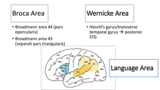
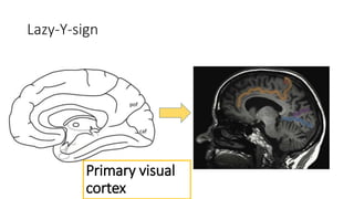
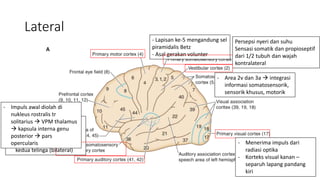
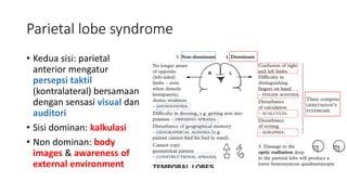
Cortical Anatomy & Function
-AIL-
Outline
• Cortical Anatomy
• Broadman Area
• Regional Brain Syndrome
Lateral
SFG
MFG
IFG
PRG
POG
SPG
SMG
AG
SOG
MOG
STG
MTG
ITG
IPG
LOG
M-around sign & T-sign
PTR
POP
POR hr
vr
PRG
Broca Area
• Broadmann area 44 (pars
opercularis)
• Broadmann area 45
(separuh pars triangularis)
• Heschl’s gyrus/transverse
temporal gyrus  posterior
STG
Wernicke Area
Language Area
Aphasia
Superior
SFG
MFG
IFG
PRG
POG
SPG
IPG
SC
pof
pcs
ips
Omega sign, L-sign, Thickness sign, Bracket sign
Primary motor &
sensory cortex
Inferior
MOG
LOG
AOG
POG
H-sign
LOTG
PHG
ITG
IOG
LIG
os
rs
acs
PHG
LOTG
FUG
Medial
CIG
SFG
GR
PCU
CUN
LIG
IOG
ITG
LOTG
PHG
PHG
pcs
cs
cis
rm-cis
pof
caf
Lazy-Y-sign
pof
caf
Primary visual
cortex
Brodmann Area
Lateral
- Lapisan ke-5 mengandung sel
piramidalis Betz
- Asal gerakan volunter
Persepsi nyeri dan suhu
Sensasi somatik dan propioseptif
dari 1/2 tubuh dan wajah
kontralateral
- Menerima impuls dari
radiasi optika
- Korteks visual kanan –
separuh lapang pandang
kiri
- Menerima impuls dari
korpus genikulatum medial
- Masing-masing sisi
mengolah impuls dari
kedua telinga (bilateral)
- Impuls awal diolah di
nukleus rostralis tr
solitarius  VPM thalamus
 kapsula interna genu
posterior  pars
opercularis
- Area 2v dan 3a  integrasi
informasi somatosensorik,
sensorik khusus, motorik
Medial
Regional Brain
Syndrome
Frontal lobe syndrome
• Prefrontal lobe:
• Prefontal korteks: penyimpanan cepat dan
analisis informasi objektif
• Prefrontal korteks dorsolateral (9,10,46):
perencanaan, motivasi dan berpikir
• Prefrontal korteks orbitalis (11,47): kontrol
pada perilaku, hubungan sosial
• Area olfaktori  Foster Kennedy Syndrome
1. Ipsilateral anosmia
2. Ipsilateral central scotoma (dengan optik
atropi e.c kompresi n.optikus)
3. Contralateral papiledema
Parietal lobe syndrome
• Kedua sisi: parietal
anterior mengatur
persepsi taktil
(kontralateral) bersamaan
dengan sensasi visual dan
auditori
• Sisi dominan: kalkulasi
• Non dominan: body
images & awareness of
external environment
Temporal lobe syndrome
1. Korteks auditori:
• Cortical deafness
• Amusia
• Halusinasi auditori
2. MTG & ITG: gangguan memori/proses belajar
3. Limbic lobe:
• Halusinasi olfaktori
• Kepribadian agresif atau anti sosial
• Tidak dpt membuat memori baru
4. Radiasi optika  kuadranopsia homonim
Occipital lobe syndrome
• Visual korteks: korteks striata dan
parastriata
• Anton syndrome:
opasien tidak sadar dan men-deny
visual loss
• Prosopagnosia:
otidak dapat naming keluarga
• Ilusi visual:
oMikropsia
oMakropsia
Kortex Striata
Kortex
parastriata
Cortical Anatomy & Function.pptx

Cortical Anatomy & Function.pptx

Editor's Notes

  • #5 Pars opercularis berhubungan dg gyrus precentral dan sulcus sentralis The inferior frontal sulcus and the precentral sulcus form a capital T tilted 90° clockwise (T-sign). The top of the T marks the precentral sulcus and the precentral sulcus identifies the precen- tral gyrus. The central sulcus is located immediately parallel and posterior to it
  • #14 Area 43  secondary gustatorik
  • #17 Mengapa terjadi kontralateral papiledema?
  • #20 Cortical deafness: Bilateral lesions are rare but may result in complete deafness of which the patient may be unaware. Lesions which involve surrounding association areas may result in difficulty in hearing spoken words (dominant) or difficulty in appreciating rhythm/music (non-dominant) – AMUSIA. Auditory hallucinations may occur in temporal lobe disease.
  • #21 Anton’s syndrome Involvement of both the striate and the parastriate cortices affects the interpretation of vision. The patient is unaware of his visual loss and denies its presence. This denial in the presence of obvious blindness characterizes Anton’s syndrome. Cortical blindness occurs mainly in vascular disease (posterior cerebral artery), but also following hypoxia and hypertensive encephalopathy or after surviving tentorial herniation. Balint’s syndrome Inability to direct voluntary gaze, associated with visual agnosia (loss of visual recognition) due to bilateral parieto-occipital lesions Prosopagnosia: the patient, though able to see a familiar face, e.g. a member of the family, cannot name it. This is usually associated with other disturbances of ‘interpretation’ and naming with intact vision such as colour agnosia (recognition of colours and matching of pairs of colours). Bilateral lesions at occipito-temporal junction are responsible.