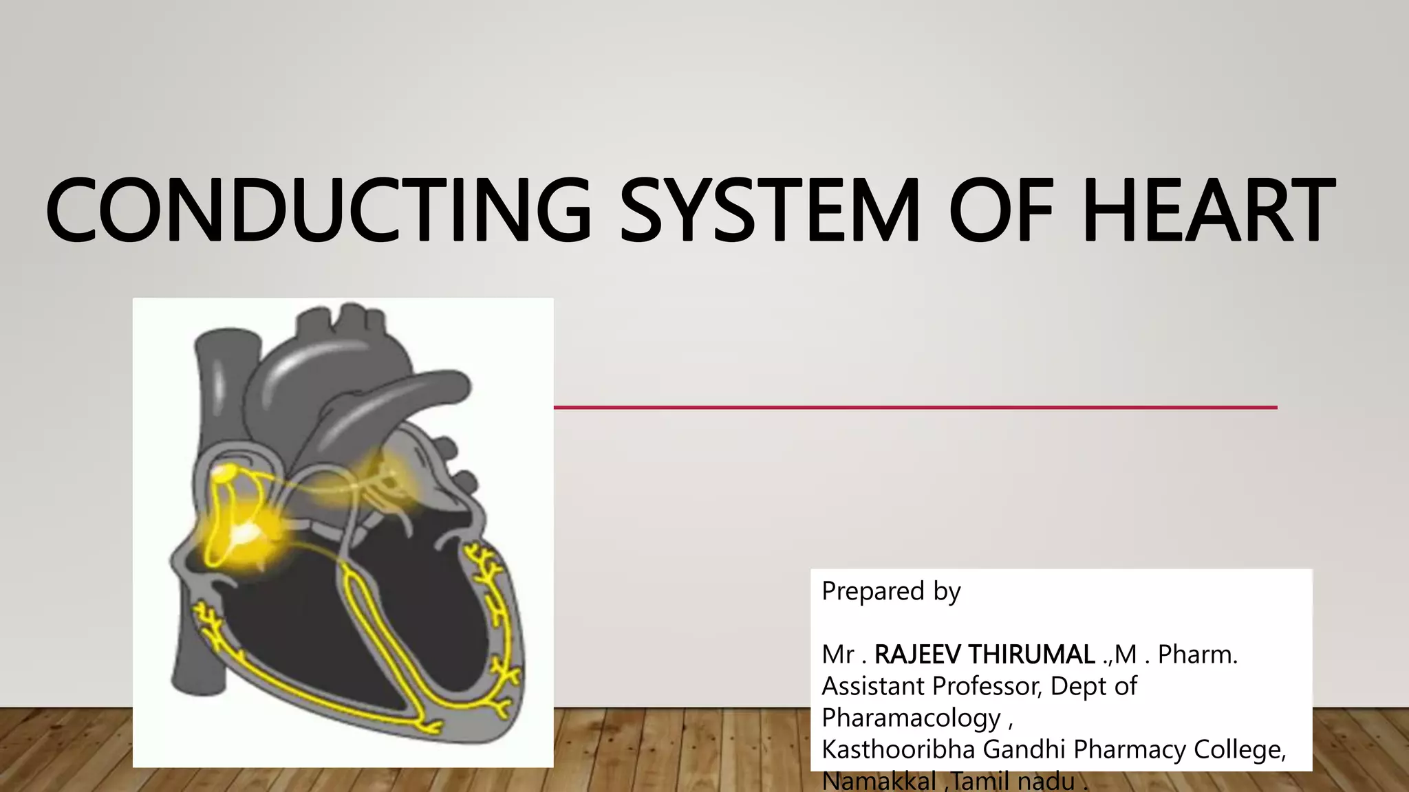
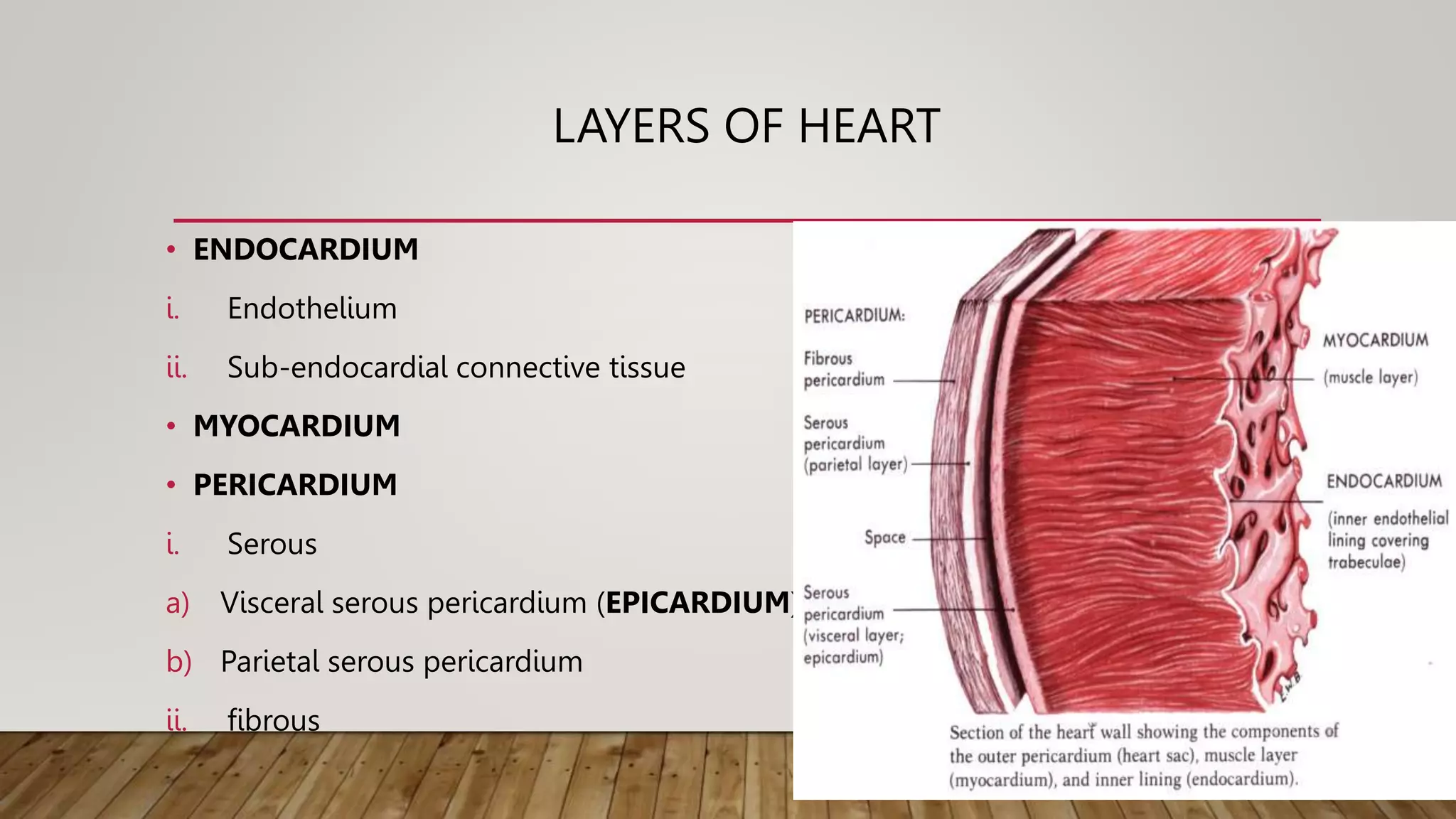
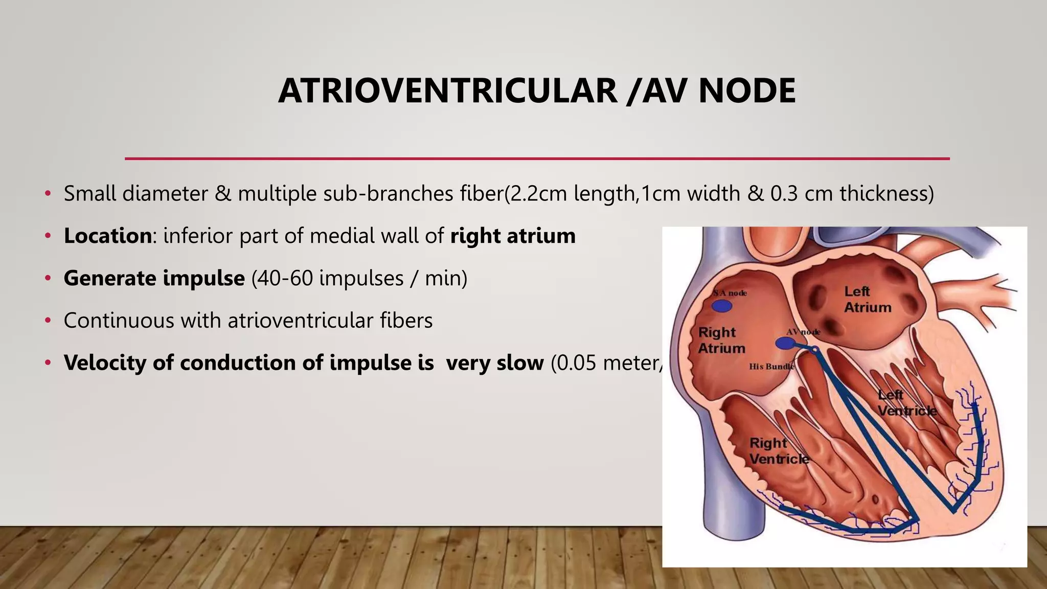
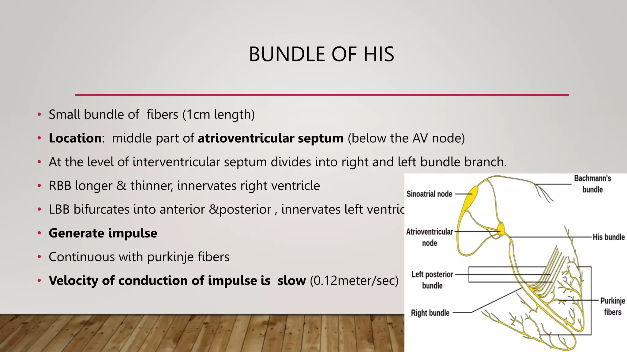
CONDUCTING SYSTEM OF HEART
Prepared by
Mr . RAJEEV THIRUMAL .,M . Pharm.
Assistant Professor, Dept of
Pharamacology ,
Kasthooribha Gandhi Pharmacy College,
Namakkal ,Tamil nadu .
LAYERS OF HEART
• ENDOCARDIUM
i. Endothelium
ii. Sub-endocardial connective tissue
• MYOCARDIUM
• PERICARDIUM
i. Serous
a) Visceral serous pericardium (EPICARDIUM)
b) Parietal serous pericardium
ii. fibrous
TYPES OF MYOCARDIAL FIBERS
1) Contractile fibers
a) Atrial muscle fibers
b) Ventricular muscle fibers
2) Non – contractile fibers (modified cardiac muscle fibers)
a) Specialized excitatory structure : SA node
b) Specialized conductive structure: AV node, Bundle of HIS and Purkinje fibers.
SINO ATRIAL / SA NODE
Pacemaker of mammalian heart
• Small strip (1.5 cm length & 0.5 cm wide) of modified cardiac muscle.
• Location: superior part of lateral wall of right atrium
• Contains Pacemaker or P cells –generate impulse (70-80 impulses / min)
• Continuous with atrial muscle fibers and intermodal fibers
• Velocity of conduction of impulse is very slow (0.05 meter/sec)
ATRIOVENTRICULAR /AV NODE
• Small diameter & multiple sub-branches fiber(2.2cm length,1cm width & 0.3 cm thickness)
• Location: inferior part of medial wall of right atrium
• Generate impulse (40-60 impulses / min)
• Continuous with atrioventricular fibers
• Velocity of conduction of impulse is very slow (0.05 meter/sec)
INTERNODAL FIBERS
• Connect SA node and AV node
1) Anterior internodal fibers / tract of Bachman
2) Middle internodal fibers / tract of Wenckebach
3) Posterior internodal fibers / tract of Thorel
• Location: wall of right atrium
• Velocity of conduction of impulse is fast (1 meter/sec)
BUNDLE OF HIS
• Small bundle of fibers (1cm length)
• Location: middle part of atrioventricular septum (below the AV node)
• At the level of interventricular septum divides into right and left bundle branch.
• RBB longer & thinner, innervates right ventricle
• LBB bifurcates into anterior &posterior , innervates left ventricle
• Generate impulse
• Continuous with purkinje fibers
• Velocity of conduction of impulse is slow (0.12meter/sec)
PURKINJE FIBERS
• Network of very small bundle of fibers
• Purkinje cell - larger diameter & numerous gap junction (low impedance electrical
synapse)
• Location: sub-endocardial regions of right and left ventricles
• Generate impulse (35-40 impulses / min)
• Velocity of conduction of impulse is very fast ( 4 meters/sec)
CONDUCTING SYSTEM OF HEART.pptx

CONDUCTING SYSTEM OF HEART.pptx

  • 1.
    CONDUCTING SYSTEM OFHEART Prepared by Mr . RAJEEV THIRUMAL .,M . Pharm. Assistant Professor, Dept of Pharamacology , Kasthooribha Gandhi Pharmacy College, Namakkal ,Tamil nadu .
  • 2.
    LAYERS OF HEART •ENDOCARDIUM i. Endothelium ii. Sub-endocardial connective tissue • MYOCARDIUM • PERICARDIUM i. Serous a) Visceral serous pericardium (EPICARDIUM) b) Parietal serous pericardium ii. fibrous
  • 3.
    TYPES OF MYOCARDIALFIBERS 1) Contractile fibers a) Atrial muscle fibers b) Ventricular muscle fibers 2) Non – contractile fibers (modified cardiac muscle fibers) a) Specialized excitatory structure : SA node b) Specialized conductive structure: AV node, Bundle of HIS and Purkinje fibers.
  • 4.
    SINO ATRIAL /SA NODE Pacemaker of mammalian heart • Small strip (1.5 cm length & 0.5 cm wide) of modified cardiac muscle. • Location: superior part of lateral wall of right atrium • Contains Pacemaker or P cells –generate impulse (70-80 impulses / min) • Continuous with atrial muscle fibers and intermodal fibers • Velocity of conduction of impulse is very slow (0.05 meter/sec)
  • 5.
    ATRIOVENTRICULAR /AV NODE •Small diameter & multiple sub-branches fiber(2.2cm length,1cm width & 0.3 cm thickness) • Location: inferior part of medial wall of right atrium • Generate impulse (40-60 impulses / min) • Continuous with atrioventricular fibers • Velocity of conduction of impulse is very slow (0.05 meter/sec)
  • 6.
    INTERNODAL FIBERS • ConnectSA node and AV node 1) Anterior internodal fibers / tract of Bachman 2) Middle internodal fibers / tract of Wenckebach 3) Posterior internodal fibers / tract of Thorel • Location: wall of right atrium • Velocity of conduction of impulse is fast (1 meter/sec)
  • 7.
    BUNDLE OF HIS •Small bundle of fibers (1cm length) • Location: middle part of atrioventricular septum (below the AV node) • At the level of interventricular septum divides into right and left bundle branch. • RBB longer & thinner, innervates right ventricle • LBB bifurcates into anterior &posterior , innervates left ventricle • Generate impulse • Continuous with purkinje fibers • Velocity of conduction of impulse is slow (0.12meter/sec)
  • 8.
    PURKINJE FIBERS • Networkof very small bundle of fibers • Purkinje cell - larger diameter & numerous gap junction (low impedance electrical synapse) • Location: sub-endocardial regions of right and left ventricles • Generate impulse (35-40 impulses / min) • Velocity of conduction of impulse is very fast ( 4 meters/sec)