How do we
knowthat
something is
alive?
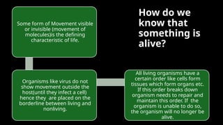
Some form of Movement visible
or invisible (movement of
molecules)is the defining
characteristic of life.
Organisms like virus do not
show movement outside the
host(until they infect a cell)
hence they are placed on the
borderline between living and
nonliving.
All living organisms have a
certain order like cells form
tissues which form organs etc.
If this order breaks down
organism needs to repair and
maintain this order. If the
organism is unable to do so,
the organism will no longer be
alive.
3.
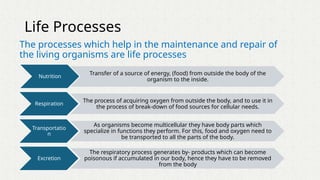
Life Processes
The processeswhich help in the maintenance and repair of
the living organisms are life processes
Nutrition
Transfer of a source of energy, (food) from outside the body of the
organism to the inside.
Respiration
The process of acquiring oxygen from outside the body, and to use it in
the process of break-down of food sources for cellular needs.
Transportatio
n
As organisms become multicellular they have body parts which
specialize in functions they perform. For this, food and oxygen need to
be transported to all the parts of the body.
Excretion
The respiratory process generates by- products which can become
poisonous if accumulated in our body, hence they have to be removed
from the body
4.
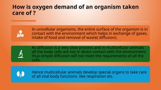
How is oxygendemand of an organism taken
care of ?
In unicellular organisms, the entire surface of the organism is in
contact with the environment which helps in exchange of gases,
intake of food and removal of waste( diffusion).
As diffusion is a very slow process and in multicellular animals
all the body cells are not in direct contact with the environment
thus simple diffusion will not meet the requirements of all the
cells.
Hence multicellular animals develop special organs to take care
of all vital body functions like respiration etc.
5.
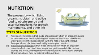
NUTRITION
The process bywhich living
organisms obtain and utilize
food to obtain energy and
essential nutrients for growth,
maintenance, and other life
processes
TYPES OF NUTRITION
(i) Autotrophic nutrition is that mode of nutrition in which an organism makes
its own food from the simple inorganic materials like carbon dioxide and
water present in the surroundings (with the help of sunlight energy).
Example: Green plants obtain food by autotrophic nutrition.
(ii) Heterotrophic nutrition is that mode of nutrition in which an organism
cannot make its own food from simple inorganic materials like carbon
dioxide and water, and depends on other organisms for its food. Example:
All animals obtain food by heterotrophic nutrition.
6.
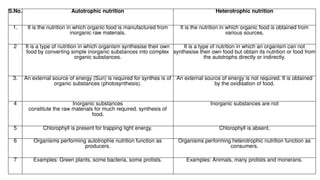
S.No. Autotrophic nutritionHeterotrophic nutrition
1. It is the nutrition in which organic food is manufactured from
inorganic raw materials.
It is the nutrition in which organic food is obtained from
various sources.
2 It is a type of nutrition in which organism synthesise their own
food by converting simple inorganic substances into complex
organic substances.
It is a type of nutrition in which an organism can not
synthesise their own food but obtain its nutrition or food from
the autotrophs directly or indirectly.
3. An external source of energy (Sun) is required for synthes is of
organic substances (photosynthesis).
An external source of energy is not required. It is obtained
by the oxidisation of food.
4 Inorganic substances
constitute the raw materials for much required. synthesis of
food.
Inorganic substances are not
5 Chlorophyll is present for trapping light energy. Chlorophyll is absent.
6 Organisms performing autotrophie nutrition function as
producers.
Organisms performing heterotrophic nutrition function as
consumers.
7 Examples: Green plants, some bacteria, some protists. Examples: Animals, many protists and monerans.
7.
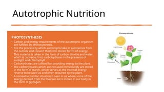
Autotrophic Nutrition
PHOTOSYNTHESIS
• Carbonand energy requirements of the autotrophic organism
are fulfilled by photosynthesis.
• It is the process by which autotrophs take in substances from
the outside and convert them into stored forms of energy.
• This material is taken in the form of carbon dioxide and water
which is converted into carbohydrates in the presence of
sunlight and chlorophyll.
• Carbohydrates are utilised for providing energy to the plant.
• The carbohydrates which are not used immediately are stored
in the form of starch, which serves as the internal energy
reserve to be used as and when required by the plant.
• A somewhat similar situation is seen in us where some of the
energy derived from the food we eat is stored in our body in
the form of glycogen.
8.
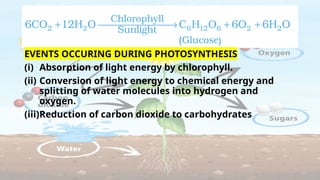
EVENTS OCCURING DURINGPHOTOSYNTHESIS
(i) Absorption of light energy by chlorophyll.
(ii) Conversion of light energy to chemical energy and
splitting of water molecules into hydrogen and
oxygen.
(iii)Reduction of carbon dioxide to carbohydrates
9.
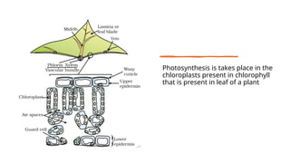
Photosynthesis is takesplace in the
chloroplasts present in chlorophyll
that is present in leaf of a plant
10.
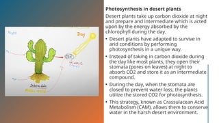
Photosynthesis in desertplants
Desert plants take up carbon dioxide at night
and prepare and intermediate which is acted
upon by the energy absorbed by the
chlorophyll during the day.
• Desert plants have adapted to survive in
arid conditions by performing
photosynthesis in a unique way.
• Instead of taking in carbon dioxide during
the day like most plants, they open their
stomata (pores on leaves) at night to
absorb CO2 and store it as an intermediate
compound.
• During the day, when the stomata are
closed to prevent water loss, the plants
utilize the stored CO2 for photosynthesis.
• This strategy, known as Crassulacean Acid
Metabolism (CAM), allows them to conserve
water in the harsh desert environment.
11.
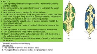
ACTIVITY 1
1. Takea potted plant with variegated leaves – for example, money
plant or crotons.
2. Keep the plant in a dark room for three days so that all the starch
gets used up.
3. Now keep the plant in sunlight for about six hours.
4. Pluck a leaf from the plant. Mark the green areas in it and trace them
on a sheet of paper. n Dip the leaf in boiling water for a few minutes.
5. After this, immerse it in a beaker containing alcohol.
6. Carefully place the above beaker in a water-bath and heat till the
alcohol begins to boil.
7. What happens to the colour of the leaf? What is the colour of the
solution?
8. Now dip the leaf in a dilute solution of iodine for a few minutes.
9. Take out the leaf and rinse off the iodine solution.
10.Observe the colour of the leaf and compare this with the tracing of
the leaf done in the beginning
11.What can you conclude about the presence of starch in various areas
of the leaf?
Questions asked from the activity.
Give reasons-
1. We boil leaf in alcohol over a water bath
2. Variegated leaves are used to test the presence of starch
12.
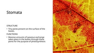
Stomata
STRUCTURE
• Tiny porespresent on the surface of the
leaves.
FUNCTIONS
• Massive amounts of gaseous exchange
takes place in the leaves through these
pores for the purpose of photosynthesis
13.
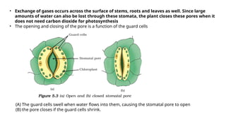
• Exchange ofgases occurs across the surface of stems, roots and leaves as well. Since large
amounts of water can also be lost through these stomata, the plant closes these pores when it
does not need carbon dioxide for photosynthesis
• The opening and closing of the pore is a function of the guard cells
(A) The guard cells swell when water flows into them, causing the stomatal pore to open
(B) the pore closes if the guard cells shrink.
14.
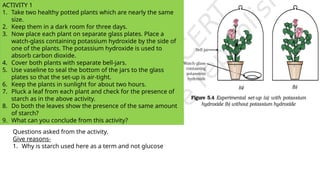
ACTIVITY 1
1. Taketwo healthy potted plants which are nearly the same
size.
2. Keep them in a dark room for three days.
3. Now place each plant on separate glass plates. Place a
watch-glass containing potassium hydroxide by the side of
one of the plants. The potassium hydroxide is used to
absorb carbon dioxide.
4. Cover both plants with separate bell-jars.
5. Use vaseline to seal the bottom of the jars to the glass
plates so that the set-up is air-tight.
6. Keep the plants in sunlight for about two hours.
7. Pluck a leaf from each plant and check for the presence of
starch as in the above activity.
8. Do both the leaves show the presence of the same amount
of starch?
9. What can you conclude from this activity?
Questions asked from the activity.
Give reasons-
1. Why is starch used here as a term and not glucose
15.
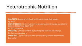
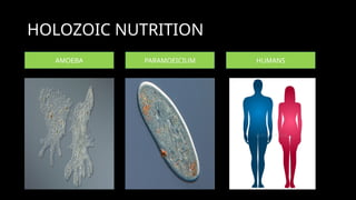
Heterotrophic Nutrition
HOLOZOIC
- Ingestwhole food, and break it inside their bodies
Eg. Man
SAPROTROPHIC
- Derive nutrition by breaking down the dead outside the
body and absorbing a part of it
Eg. Fungi, Bacteria
PARASITIC
- Derives nutrition by harming the host but not killing it
Eg. Cuscuta, tapeworm
SYMBIOTIC
- A partnership in which both the organisms are benefited.
E.g.. Lichen
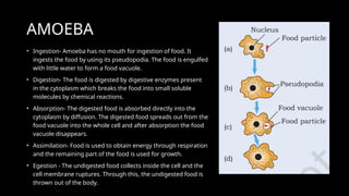
AMOEBA
• Ingestion- Amoebahas no mouth for ingestion of food. It
ingests the food by using its pseudopodia. The food is engulfed
with little water to form a food vacuole.
• Digestion- The food is digested by digestive enzymes present
in the cytoplasm which breaks the food into small soluble
molecules by chemical reactions.
• Absorption- The digested food is absorbed directly into the
cytoplasm by diffusion. The digested food spreads out from the
food vacuole into the whole cell and after absorption the food
vacuole disappears.
• Assimilation- Food is used to obtain energy through respiration
and the remaining part of the food is used for growth.
• Egestion - The undigested food collects inside the cell and the
cell membrane ruptures. Through this, the undigested food is
thrown out of the body.
18.
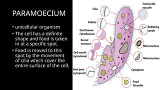
PARAMOECIUM
• unicellular organism
•The cell has a definite
shape and food is taken
in at a specific spot.
• Food is moved to this
spot by the movement
of cilia which cover the
entire surface of the cell.
19.
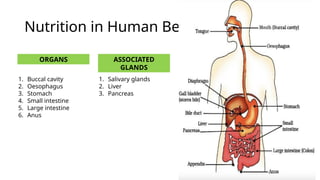
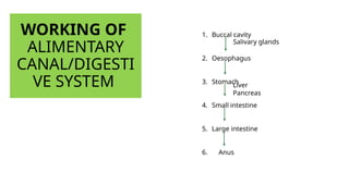
Nutrition in HumanBeings
ORGANS ASSOCIATED
GLANDS
1. Buccal cavity
2. Oesophagus
3. Stomach
4. Small intestine
5. Large intestine
6. Anus
1. Salivary glands
2. Liver
3. Pancreas
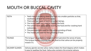
MOUTH OR BUCCALCAVITY
TEETH • Teeth help in breaking down the food into smaller particles so that,
swallowing of food becomes easier.
• There are four types of teeth in human beings.
The incisor teeth are used for cutting the food.
The canine teeth are used for tearing the food and for cracking hard
substances.
The premolars are used for the coarse grinding of food.
The molars are used for fine grinding of food
TOUNGE The tongue has gustatory receptors which perceive the sense of taste.
The tongue helps in turning over the food so that saliva can be properly
mixed in it.
SALIVARY GLANDS Salivary glands secrete saliva: Saliva makes the food slippery which makes
it easy to swallow the food. Saliva also contains the enzyme salivary
22.
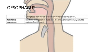
OESOPHAGUS
Taking food frommouth to stomach by Peristaltic movement.
Peristaltic
movement:
Rhythmic contraction of muscles of the lining of the alimentary canal to
push the food forward.
23.
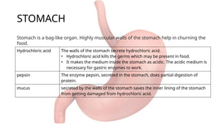
STOMACH
Hydrochloric acid Thewalls of the stomach secrete hydrochloric acid.
• Hydrochloric acid kills the germs which may be present in food.
• It makes the medium inside the stomach as acidic. The acidic medium is
necessary for gastric enzymes to work.
pepsin The enzyme pepsin, secreted in the stomach, does partial digestion of
protein.
mucus secreted by the walls of the stomach saves the inner lining of the stomach
from getting damaged from hydrochloric acid.
Stomach is a bag-like organ. Highly muscular walls of the stomach help in churning the
food.
24.
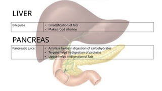
LIVER
Bile juice •Emulsification of fats
• Makes food alkaline
PANCREAS
Pancreatic juice • Amylase helps in digestion of carbohydrates
• Trypsin helps in digestion of proteins
• Lipase helps in digestion of fats
25.
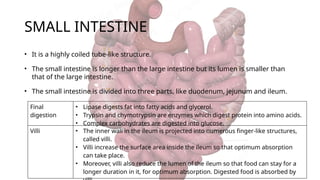
SMALL INTESTINE
• Itis a highly coiled tube-like structure.
• The small intestine is longer than the large intestine but its lumen is smaller than
that of the large intestine.
• The small intestine is divided into three parts, like duodenum, jejunum and ileum.
Final
digestion
• Lipase digests fat into fatty acids and glycerol.
• Trypsin and chymotrypsin are enzymes which digest protein into amino acids.
• Complex carbohydrates are digested into glucose.
Villi • The inner wall in the ileum is projected into numerous finger-like structures,
called villi.
• Villi increase the surface area inside the ileum so that optimum absorption
can take place.
• Moreover, villi also reduce the lumen of the ileum so that food can stay for a
longer duration in it, for optimum absorption. Digested food is absorbed by
26.
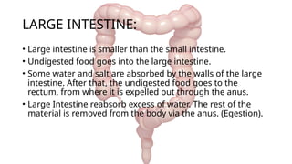
LARGE INTESTINE:
• Largeintestine is smaller than the small intestine.
• Undigested food goes into the large intestine.
• Some water and salt are absorbed by the walls of the large
intestine. After that, the undigested food goes to the
rectum, from where it is expelled out through the anus.
• Large Intestine reabsorb excess of water. The rest of the
material is removed from the body via the anus. (Egestion).
RESPIRATION
Respiration is thebiochemical process in which the
cells of an organism obtain energy by combining
oxygen and glucose, resulting in the release of carbon
dioxide, water, and ATP (the currency of energy in
cells).
29.
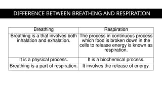
DIFFERENCE BETWEEN BREATHINGAND RESPIRATION
Breathing Respiration
Breathing is a that involves both
inhalation and exhalation.
The process in continuous process
which food is broken down in the
cells to release energy is known as
respiration.
It is a physical process. It is a biochemical process.
Breathing is a part of respiration. It involves the release of energy.
30.
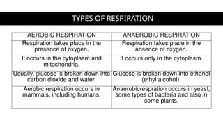
TYPES OF RESPIRATION
AEROBICRESPIRATION ANAEROBIC RESPIRATION
Respiration takes place in the
presence of oxygen.
Respiration takes place in the
absence of oxygen.
It occurs in the cytoplasm and
mitochondria.
It occurs only in the cytoplasm.
Usually, glucose is broken down into
carbon dioxide and water.
Glucose is broken down into ethanol
(ethyl alcohol).
Aerobic respiration occurs in
mammals, including humans.
Anaerobicrespration occurs in yeast,
some types of bacteria and also in
some plants.
31.
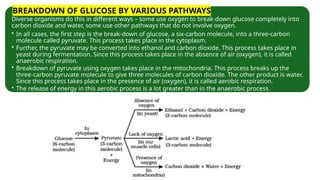
Diverse organisms dothis in different ways – some use oxygen to break-down glucose completely into
carbon dioxide and water, some use other pathways that do not involve oxygen.
• In all cases, the first step is the break-down of glucose, a six-carbon molecule, into a three-carbon
molecule called pyruvate. This process takes place in the cytoplasm.
• Further, the pyruvate may be converted into ethanol and carbon dioxide. This process takes place in
yeast during fermentation. Since this process takes place in the absence of air (oxygen), it is called
anaerobic respiration.
• Breakdown of pyruvate using oxygen takes place in the mitochondria. This process breaks up the
three-carbon pyruvate molecule to give three molecules of carbon dioxide. The other product is water.
Since this process takes place in the presence of air (oxygen), it is called aerobic respiration.
• The release of energy in this aerobic process is a lot greater than in the anaerobic process.
BREAKDOWN OF GLUCOSE BY VARIOUS PATHWAYS
32.
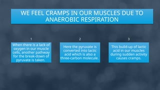
When there isa lack of
oxygen in our muscle
cells, another pathway
for the break-down of
pyruvate is taken.
Here the pyruvate is
converted into lactic
acid which is also a
three-carbon molecule.
This build-up of lactic
acid in our muscles
during sudden activity
causes cramps.
WE FEEL CRAMPS IN OUR MUSCLES DUE TO
ANAEROBIC RESPIRATION
1 2 3
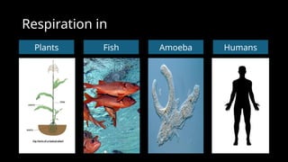
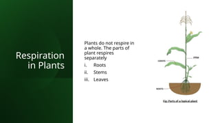
Respiration
in Plants
Plants donot respire in
a whole. The parts of
plant respires
separately
i. Roots
ii. Stems
iii. Leaves
35.
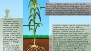
• The stemsof
herbaceous plants
takes place through
stomata. The oxygen
from the air diffuses
into the stem of a
herbaceous plant
through stomata and
reaches all the cells for
respiration. The carbon
dioxide produced
diffuses out through
stomata.
• In woody stems, the
bark has lenticels for
the exchange of gases
The leaves of a plant has tiny pores called stomata
through which the exchange of respiratory gases takes
place by diffusion. Oxygen from air diffuses into a leaf
through stomata and reaches all the cells, where it is
used for respiration and the carbon dioxide produced
diffuses out from the leaf into the air through stomata
The roots of a plant take the oxygen required
for respiration from the air present in-
between the soil particles by the process of
diffusion. The roots have extensions of
epidermal cells of a root called root hair which
are in contact with the air in the soil. Oxygen
diffuses from root hairs and reaches all the
other cells of the root for respiration. Carbon
dioxide gas produced in the cells of the root
during respiration moves out through the root
hairs by the process of diffusion. Thus, the
respiration in roots occurs by diffusion of
respiratory gases through the root hairs.
36.
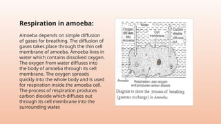
Respiration in amoeba:
Amoebadepends on simple diffusion
of gases for breathing. The diffusion of
gases takes place through the thin cell
membrane of amoeba. Amoeba lives in
water which contains dissolved oxygen.
The oxygen from water diffuses into
the body of amoeba through its cell
membrane. The oxygen spreads
quickly into the whole body and is used
for respiration inside the amoeba cell.
The process of respiration produces
carbon dioxide which diffuses out
through its cell membrane into the
surrounding water.
37.
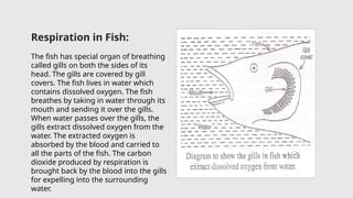
Respiration in Fish:
Thefish has special organ of breathing
called gills on both the sides of its
head. The gills are covered by gill
covers. The fish lives in water which
contains dissolved oxygen. The fish
breathes by taking in water through its
mouth and sending it over the gills.
When water passes over the gills, the
gills extract dissolved oxygen from the
water. The extracted oxygen is
absorbed by the blood and carried to
all the parts of the fish. The carbon
dioxide produced by respiration is
brought back by the blood into the gills
for expelling into the surrounding
water.
Respiratory
tract
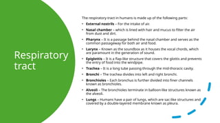
The respiratory tractin humans is made up of the following parts:
• External nostrils – For the intake of air.
• Nasal chamber – which is lined with hair and mucus to filter the air
from dust and dirt.
• Pharynx – It is a passage behind the nasal chamber and serves as the
common passageway for both air and food.
• Larynx – Known as the soundbox as it houses the vocal chords, which
are paramount in the generation of sound.
• Epiglottis – It is a flap-like structure that covers the glottis and prevents
the entry of food into the windpipe.
• Trachea – It is a long tube passing through the mid-thoracic cavity.
• Bronchi – The trachea divides into left and right bronchi.
• Bronchioles – Each bronchus is further divided into finer channels
known as bronchioles.
• Alveoli – The bronchioles terminate in balloon-like structures known as
the alveoli.
• Lungs – Humans have a pair of lungs, which are sac-like structures and
covered by a double-layered membrane known as pleura.
40.
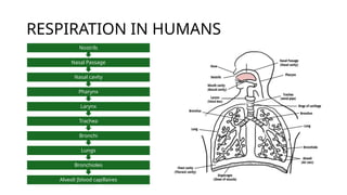
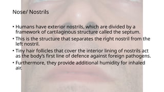
Nose/ Nostrils
• Humanshave exterior nostrils, which are divided by a
framework of cartilaginous structure called the septum.
• This is the structure that separates the right nostril from the
left nostril.
• Tiny hair follicles that cover the interior lining of nostrils act
as the body’s first line of defence against foreign pathogens.
• Furthermore, they provide additional humidity for inhaled
air.
41.
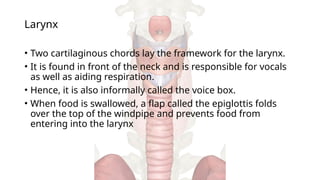
Larynx
• Two cartilaginouschords lay the framework for the larynx.
• It is found in front of the neck and is responsible for vocals
as well as aiding respiration.
• Hence, it is also informally called the voice box.
• When food is swallowed, a flap called the epiglottis folds
over the top of the windpipe and prevents food from
entering into the larynx
42.
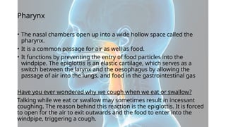
Pharynx
• The nasalchambers open up into a wide hollow space called the
pharynx.
• It is a common passage for air as well as food.
• It functions by preventing the entry of food particles into the
windpipe. The epiglottis is an elastic cartilage, which serves as a
switch between the larynx and the oesophagus by allowing the
passage of air into the lungs, and food in the gastrointestinal gas
Have you ever wondered why we cough when we eat or swallow?
Talking while we eat or swallow may sometimes result in incessant
coughing. The reason behind this reaction is the epiglottis. It is forced
to open for the air to exit outwards and the food to enter into the
windpipe, triggering a cough.
43.
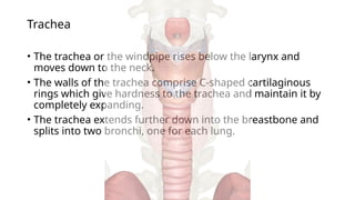
Trachea
• The tracheaor the windpipe rises below the larynx and
moves down to the neck.
• The walls of the trachea comprise C-shaped cartilaginous
rings which give hardness to the trachea and maintain it by
completely expanding.
• The trachea extends further down into the breastbone and
splits into two bronchi, one for each lung.
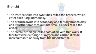
44.
Bronchi
• The tracheasplits into two tubes called the bronchi, which
enter each lung individually.
• The bronchi divide into secondary and tertiary bronchioles,
and it further branches out into small air-sacs called the
alveoli.
• The alveoli are single-celled sacs of air with thin walls. It
facilitates the exchange of oxygen and carbon dioxide
molecules into or away from the bloodstream.
45.
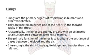
Lungs
• Lungs arethe primary organs of respiration in humans and
other vertebrates.
• They are located on either side of the heart, in the thoracic
cavity of the chest.
• Anatomically, the lungs are spongy organs with an estimates
total surface area between 50 to 75 sq meters.
• The primary function of the lungs is to facilitate the exchange of
gases between the blood and the air.
• Interestingly, the right lung is quite bigger and heavier than the
left lung.
46.
Breathing mechanism
• Airis inhaled with the help of nostrils, and in the nasal cavity, the
air is cleansed by the fine hair follicles present within them. The
cavity also has a group of blood vessels that warm the air. This air
then passes to the pharynx, then to the larynx and into the
trachea.
• The trachea and the bronchi are coated with ciliated epithelial cells
and goblet cells (secretory cells) which discharge mucus to
moisten the air as it passes through the respiratory tract. It also
traps the fine bits of dust or pathogen that escaped the hair in the
nasal openings. The motile cilia beat in an ascending motion, such
that the mucus and other foreign particles are carried back to the
buccal cavity where it may either be coughed out (or swallowed.)
• Once the air reaches the bronchus, it moves into the bronchioles,
and then into the alveoli.
47.
Functions
of
respiratory
system
1. Inhalation andExhalation- The respiratory system helps in breathing
(also known as pulmonary ventilation.) The air inhaled through the nose
moves through the pharynx, larynx, trachea and into the lungs. The air is
exhaled back through the same pathway. Changes in the volume and
pressure in the lungs aid in pulmonary ventilation.
2. Exchange of Gases between Lungs and Bloodstream- Inside the
lungs, the oxygen and carbon dioxide enter and exit respectively through
millions of microscopic sacs called alveoli. The inhaled oxygen diffuses into
the pulmonary capillaries, binds to haemoglobin and is pumped through
the bloodstream. The carbon dioxide from the blood diffuses into the
alveoli and is expelled through exhalation.
3. Exchange of Gases between Bloodstream and Body Tissues- The
blood carries the oxygen from the lungs around the body and releases the
oxygen when it reaches the capillaries. The oxygen is diffused through the
capillary walls into the body tissues. The carbon dioxide also diffuses into
the blood and is carried back to the lungs for release.
4. The Vibration of the Vocal Cords- While speaking, the muscles in the
larynx move the arytenoid cartilage. These cartilages push the vocal cords
together. During exhalation, when the air passes through the vocal cords,
it makes them vibrate and creates sound.
5. Olfaction or Smelling- During inhalation, when the air enters the nasal
cavities, some chemicals present in the air bind to it and activate the
receptors of the nervous system on the cilia. The signals are sent to the
olfactory bulbs via the brain.
48.
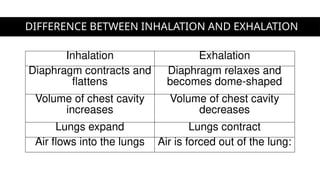
DIFFERENCE BETWEEN INHALATIONAND EXHALATION
Inhalation Exhalation
Diaphragm contracts and
flattens
Diaphragm relaxes and
becomes dome-shaped
Volume of chest cavity
increases
Volume of chest cavity
decreases
Lungs expand Lungs contract
Air flows into the lungs Air is forced out of the lung:
49.
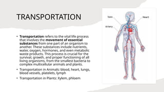
TRANSPORTATION
• Transportation refersto the vital life process
that involves the movement of essential
substances from one part of an organism to
another. These substances include nutrients,
water, oxygen, hormones, and even metabolic
waste products. This process is crucial for the
survival, growth, and proper functioning of all
living organisms, from the smallest bacteria to
complex multicellular animals and plants.
• Transportation in Animals: blood, heart, lungs,
blood vessels, platelets, lymph
• Transportation in Plants: Xylem, phloem
50.
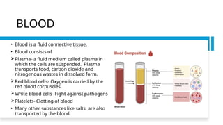
BLOOD
• Blood isa fluid connective tissue.
• Blood consists of
Plasma- a fluid medium called plasma in
which the cells are suspended. Plasma
transports food, carbon dioxide and
nitrogenous wastes in dissolved form.
Red blood cells- Oxygen is carried by the
red blood corpuscles.
White blood cells- Fight against pathogens
Platelets- Clotting of blood
• Many other substances like salts, are also
transported by the blood.
51.
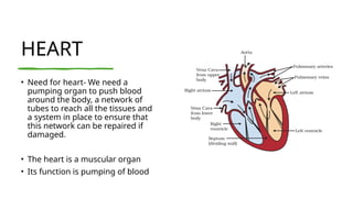
HEART
• Need forheart- We need a
pumping organ to push blood
around the body, a network of
tubes to reach all the tissues and
a system in place to ensure that
this network can be repaired if
damaged.
• The heart is a muscular organ
• Its function is pumping of blood
52.
Working of
heart
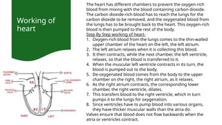
The hearthas different chambers to prevent the oxygen-rich
blood from mixing with the blood containing carbon dioxide.
The carbon dioxide-rich blood has to reach the lungs for the
carbon dioxide to be removed, and the oxygenated blood from
the lungs has to be brought back to the heart. This oxygen-rich
blood is then pumped to the rest of the body.
Step By Step working of heart-
1. Oxygen-rich blood from the lungs comes to the thin-walled
upper chamber of the heart on the left, the left atrium.
2. The left atrium relaxes when it is collecting this blood.
3. It then contracts, while the next chamber, the left ventricle,
relaxes, so that the blood is transferred to it.
4. When the muscular left ventricle contracts in its turn, the
blood is pumped out to the body.
5. De-oxygenated blood comes from the body to the upper
chamber on the right, the right atrium, as it relaxes.
6. As the right atrium contracts, the corresponding lower
chamber, the right ventricle, dilates.
7. This transfers blood to the right ventricle, which in turn
pumps it to the lungs for oxygenation.
8. Since ventricles have to pump blood into various organs,
they have thicker muscular walls than the atria do.
Valves ensure that blood does not flow backwards when the
atria or ventricles contract.
53.
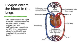
Oxygen enters
the bloodin the
lungs
• The separation of the right
side and the left side of the
heart is useful to keep
oxygenated and
deoxygenated blood from
mixing. Such separation
allows a highly efficient
supply of oxygen to the
body.
54.
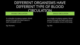
DIFFERENT ORGANISMS HAVE
DIFFERENTTYPE OF BLOOD
CIRCULATION
DOUBLE CIRCULATION SINGLE CIRCULATION
In a single circulatory system, blood
passes through the heart only once
in each complete circuit of the body
Eg: fish
In a double circulatory system, blood
passes through the heart twice in
each complete circuit
Eg: Humans
55.
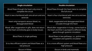
Single circulation Doublecirculation
Blood flows through the heart only once to
complete the circuit
Blood flows through the heart twice to complete
the full circuit
Heart is two-chambered with one atrium and one
ventricle
Heart is four-chambered with two atria and two
ventricles
Heart only transports venous blood, i.e.,
deoxygenated blood
Both, oxygenated and deoxygenated blood
circulate through the heart
Blood, after oxygenation in gills, does not return
to the heart and directly goes to body tissues
Blood after oxygenation in lungs, comes back to
the heart and then is pumped to different body
parts through systemic circulation
Blood flows in single pathway Blood flows in two pathways, i.e., pulmonary
circulation and systemic circulation
It is a less efficient system and blood flows at a
low pressure
It is a more efficient system and blood flows at a
high pressure
Occurs in fishes Occurs in birds and mammals
56.
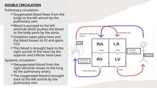
DOUBLE CIRCULATION
Pulmonary circulation-
Oxygenatedblood flows from the
lungs to the left atrium by the
pulmonary vein.
Blood is pumped to the left
ventricle which pushes the blood
to the body parts by the aorta.
Oxidation takes place here and
the blood looses its 02 and gains
CO2.
This blood is brought back to the
right auricle of the heart by the
superior and inferior vena cava.
Systemic circulation-
Deoxygenated blood from the
right ventricle moves to the lung
by the pulmonary artery.
The oxygenated blood is brought
back to the left auricle by the
pulmonary vein.
57.
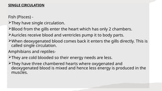
SINGLE CIRCULATION
Fish (Pisces)-
They have single circulation.
Blood from the gills enter the heart which has only 2 chambers.
Auricles receive blood and ventricles pump it to body parts.
When deoxygenated blood comes back it enters the gills directly. This is
called single circulation.
Amphibians and reptiles-
They are cold blooded so their energy needs are less.
They have three chambered hearts where oxygenated and
deoxygenated blood is mixed and hence less energy is produced in the
muscles.
58.
BLOOD PRESSURE
• Pressureexerted by blood in walls of blood vessel.
• Systolic Pressure : The pressure of blood inside the artery
during ventricular systole (contraction).
• Diastolic Pressure : The pressure in artery during ventricular
diastole (relaxation).
• Hypertension - high blood pressure (Construction of
arterioles)
59.
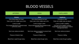
BLOOD VESSELS
ARTERIES VEINSCAPILLARIES
Arteries Veins Capillaries
Carry blood away from heart. Carry blood towards the heart. Link-artery to Vein.
Outer coat is thin middle coat is thick. Outer coat is thick middle coat is thin. Outer and middle coats are absent.
Lumen is small. Lumen is large in veins. Lumen is small in capillary.
Pure oxygenated blood is passed except
pulmonary artery.
Impure deoxygenated blood passed except
pulmonary vain.
First part of the capillary contains
oxygenated blood and last part contains
deoxygenated blood
Semi lunar valves are absent. Semi lunar valves are present to prevent back
flow of Blood.
Semi lunar valves are absent.
Pressure of blood is high. Pressure of blood is low. Pressure is falling.
Blood flow is rapid through artery. Blood flow is slow through veins. Blood flow is slow through capillary.
60.
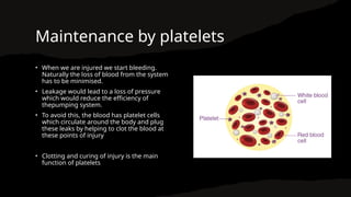
Maintenance by platelets
•When we are injured we start bleeding.
Naturally the loss of blood from the system
has to be minimised.
• Leakage would lead to a loss of pressure
which would reduce the efficiency of
thepumping system.
• To avoid this, the blood has platelet cells
which circulate around the body and plug
these leaks by helping to clot the blood at
these points of injury
• Clotting and curing of injury is the main
function of platelets
61.
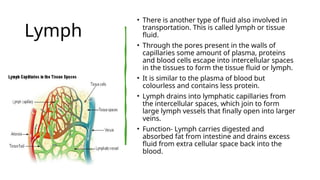
Lymph
• There isanother type of fluid also involved in
transportation. This is called lymph or tissue
fluid.
• Through the pores present in the walls of
capillaries some amount of plasma, proteins
and blood cells escape into intercellular spaces
in the tissues to form the tissue fluid or lymph.
• It is similar to the plasma of blood but
colourless and contains less protein.
• Lymph drains into lymphatic capillaries from
the intercellular spaces, which join to form
large lymph vessels that finally open into larger
veins.
• Function- Lymph carries digested and
absorbed fat from intestine and drains excess
fluid from extra cellular space back into the
blood.
62.
Transportation in
Plants
• Energyneeds differ between different body
designs.
• Plants do not move, and plant bodies have a
large proportion of dead cells in many tissues.
• As a result, plants have low energy needs, and
can use relatively slow transport systems.
• The distances over which transport systems
have to operate, however, can be very large in
plants such as very tall trees.
63.
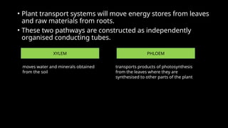
• Plant transportsystems will move energy stores from leaves
and raw materials from roots.
• These two pathways are constructed as independently
organised conducting tubes.
XYLEM PHLOEM
moves water and minerals obtained
from the soil
transports products of photosynthesis
from the leaves where they are
synthesised to other parts of the plant
64.
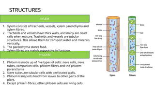
STRUCTURES
XYLEM
PHLOEM
1. Xylem consistsof tracheids, vessels, xylem parenchyma and
xylem fibres.
2. Tracheids and vessels have thick walls, and many are dead
cells when mature. Tracheids and vessels are tubular
structures. This allows them to transport water and minerals
vertically.
3. The parenchyma stores food.
4. Xylem fibres are mainly supportive in function.
1. Phloem is made up of five types of cells: sieve cells, sieve
tubes, companion cells, phloem fibres and the phloem
parenchyma
2. Sieve tubes are tubular cells with perforated walls.
3. Phloem transports food from leaves to other parts of the
plant.
4. Except phloem fibres, other phloem cells are living cells.
65.
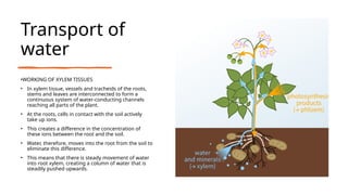
Transport of
water
•WORKING OFXYLEM TISSUES
• In xylem tissue, vessels and tracheids of the roots,
stems and leaves are interconnected to form a
continuous system of water-conducting channels
reaching all parts of the plant.
• At the roots, cells in contact with the soil actively
take up ions.
• This creates a difference in the concentration of
these ions between the root and the soil.
• Water, therefore, moves into the root from the soil to
eliminate this difference.
• This means that there is steady movement of water
into root xylem, creating a column of water that is
steadily pushed upwards.
66.
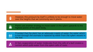
However, this pressureby itself is unlikely to be enough to move water
over the heights that we commonly see in plants.
Plants use another strategy to move water in the xylem upwards to the
highest points of the plant body.
Provided that the plant has an adequate supply of water, the water which
is lost through the stomata is replaced by water from the xylem vessels in
the leaf.
In fact, evaporation of water molecules from the cells of a leaf creates a
suction which pulls water from the xylem cells of roots.
67.
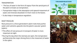
TRANSPIRATION
• The lossof water in the form of vapour from the aerial parts of
the plant is known as transpiration.
• Transpiration helps in the absorption and upward movement of
water and minerals dissolved in it from roots to the leaves.
• It also helps in temperature regulation.
ROOT PRESSURE
Root pressure is a force generated in plant roots that pushes
water and nutrients upwards into the plant's vascular system
(xylem)
The effect of root pressure in transport of water is more
important at night.
During the day when the stomata are open, the transpiration
pull becomes the major driving force in the movement of water
in the xylem
68.
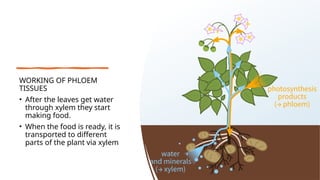
WORKING OF PHLOEM
TISSUES
•After the leaves get water
through xylem they start
making food.
• When the food is ready, it is
transported to different
parts of the plant via xylem
69.
TRANSLOCATIO
N
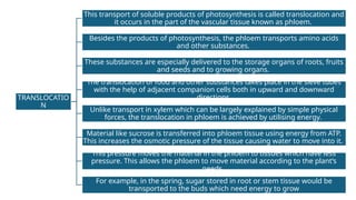
This transport ofsoluble products of photosynthesis is called translocation and
it occurs in the part of the vascular tissue known as phloem.
Besides the products of photosynthesis, the phloem transports amino acids
and other substances.
These substances are especially delivered to the storage organs of roots, fruits
and seeds and to growing organs.
The translocation of food and other substances takes place in the sieve tubes
with the help of adjacent companion cells both in upward and downward
directions.
Unlike transport in xylem which can be largely explained by simple physical
forces, the translocation in phloem is achieved by utilising energy.
Material like sucrose is transferred into phloem tissue using energy from ATP.
This increases the osmotic pressure of the tissue causing water to move into it.
This pressure moves the material in the phloem to tissues which have less
pressure. This allows the phloem to move material according to the plant’s
needs.
For example, in the spring, sugar stored in root or stem tissue would be
transported to the buds which need energy to grow
70.
EXCRETIO
N
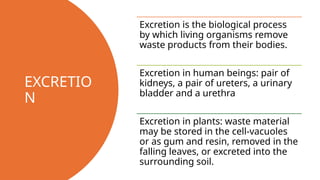
Excretion is thebiological process
by which living organisms remove
waste products from their bodies.
Excretion in human beings: pair of
kidneys, a pair of ureters, a urinary
bladder and a urethra
Excretion in plants: waste material
may be stored in the cell-vacuoles
or as gum and resin, removed in the
falling leaves, or excreted into the
surrounding soil.
71.
Excretion in
Humans
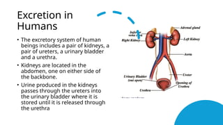
• Theexcretory system of human
beings includes a pair of kidneys, a
pair of ureters, a urinary bladder
and a urethra.
• Kidneys are located in the
abdomen, one on either side of
the backbone.
• Urine produced in the kidneys
passes through the ureters into
the urinary bladder where it is
stored until it is released through
the urethra
72.
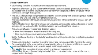
URINE FORMATION
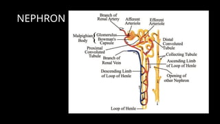
• Eachkidney contains many filtration units called as nephrons.
• Nephrons are made up of a cluster of thin walled capillaries called glomerulus which is
associated with a cup like structure called as Bowman's capsule and the long tube which
terminates through this capsule.
• The renal artery brings oxygenated blood to the kidneys along with the nitrogenous wastes
like urea and uric acid and many other substances.
The blood gets filtered through the glomerulus and this filtrate enters the tubular part of
nephron.
As this filtrate moves down the tubular part, glucose, amino acids, salts and excess of water
gets selectively reabsorbed by the blood vessels surrounding these tubules.
• The amount of water reabsorbed depends upon :
• How much excess of water is there in the body and,
• How much nitrogenous wastes need to be excreted out.
• So the fluid now flowing in the tubular part is urine which gets collected in collecting ducts of
nephrons.
• These collecting ducts together leave the kidney at a common point by forming the ureter.
Each ureter drains the urine in the urinary bladder where it is stored until the pressure of
expanded bladder leads to an urge to pass it out through urethra.
• This bladder is a muscular structure which is under nervous control.
180 litres of filtrate is formed daily but only 2 litres is excreted out as urine so the rest is
reabsorbed in the body.
73.
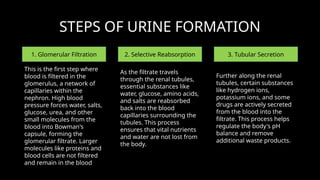
STEPS OF URINEFORMATION
1. Glomerular Filtration 2. Selective Reabsorption 3. Tubular Secretion
This is the first step where
blood is filtered in the
glomerulus, a network of
capillaries within the
nephron. High blood
pressure forces water, salts,
glucose, urea, and other
small molecules from the
blood into Bowman's
capsule, forming the
glomerular filtrate. Larger
molecules like proteins and
blood cells are not filtered
and remain in the blood
As the filtrate travels
through the renal tubules,
essential substances like
water, glucose, amino acids,
and salts are reabsorbed
back into the blood
capillaries surrounding the
tubules. This process
ensures that vital nutrients
and water are not lost from
the body.
Further along the renal
tubules, certain substances
like hydrogen ions,
potassium ions, and some
drugs are actively secreted
from the blood into the
filtrate. This process helps
regulate the body's pH
balance and remove
additional waste products.
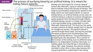
The process ofpurifying blood by an artificial kidney. It is meant for
kidney failure patients. Kidneys are vital organs for survival. Several
factors like infections, injury or restricted blood
flow to kidneys reduce the activity of kidneys. This
leads to accumulation of poisonous wastes in the
body, which can even lead to death. In case of
kidney failure, an artificial kidney can be used. An
artificial kidney is a device to remove nitrogenous
waste products from the blood through dialysis.
Artificial kidneys contain a number of tubes with
a semi-permeable lining, suspended in a tank
filled with dialysing fluid. This fluid has the same
osmotic pressure as blood, except that it is devoid
of nitrogenous wastes. The patient’s blood is
passed through these tubes. During this passage,
the waste products from the blood pass into
dialysing fluid by diffusion. The purified blood is
pumped back into the patient. This is similar to
the function of the kidney, but it is different since
there is no reabsorption involved. Normally, in a
healthy adult, the initial filtrate in the kidneys is
about 180 L daily. However, the volume actually
excreted is only a litre or two a day, because the
remaining filtrate is reabsorbed in the kidney
76.
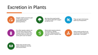
Excretion in Plants
Oxygenitself can be thought
of as a waste product
generated during
photosynthesis.
We have discussed earlier
how plants deal with oxygen
as well as CO2
They can get rid of excess
water by transpiration.
For other wastes, plants use
the fact that many of their
tissues consist of dead cells,
and that they can even lose
some parts such as leaves.
Many plant waste products
are stored in cellular
vacuoles. Waste products
may be stored in leaves that
fall off.
Other waste products are
stored as resins and gums,
especially in old xylem.
Plants also excrete some
waste substances into the
soil around them.

![chapter_1_life_processes-24_(3)[1]ff.pdf](https://cdn.slidesharecdn.com/ss_thumbnails/chapter1lifeprocesses-2431-250529135355-eab6d359-thumbnail.jpg?width=640&height=640&fit=bounds)