


![4 G.S. Pant
1.1.2.2 Electron Configuration Pauli in 1925 added a complementary rule for
arrangement of electrons around the nucleus. The pos-
Electrons around a nucleus can be described with tulation is now called Pauli’s exclusion principle and
wave functions [1]. Wave functions determine the states that no two electrons can have all quantum
location, energy, and momentum of the particle. numbers the same or exist in identical quantum states.
The square of a wave function gives the probability The filling of electrons in orbitals obeys the
distribution of the particle. At a given time, an elec- so-called Aufbau principle. The Aufbau principle
tron can be anywhere around the nucleus and have assumes that electrons are added to an atom starting
different probabilities at different locations. The with the lowest-energy orbital until all of the electrons
space around the nucleus in which the probability is are placed in an appropriate orbital. The sequence of
highest is called an orbital. In quantum mechanics, energy states and electron filling in orbitals of a multi-
the orbital is a mathematical concept that suggests electron atom can be represented as follows:
the average location of an electron around the
nucleus. If the energy of the electron changes, this 1s À 2s À 2p À 3s À 3p À 4s À 3d À 4p À 5s À 4d
average also changes. For the single electron of a À 5p À 6s À 4f À 5d À 6p À 7s À 5f À 6d À 7p
hydrogen atom, an infinite number of wave func-
tions, and therefore an infinite number of orbitals,
may exist.
An orbital can be completely described using the 1.1.2.3 Electron Binding Energies
corresponding wave function, but the process is
tedious and difficult. In simple terms, an orbital can The bound electrons need some external energy to
be described by four quantum numbers. make them free from the nucleus. It can be assumed
The principal quantum number n characterizes the that electrons around a nucleus have negative potential
energy and shell size in an atom. It is an integer and energy. The absolute value of the potential energy is
can have a value from 1 to 1, but practically n is called the binding energy, the minimum energy
always less than 8. The maximum number of elec- required to knock out an electron from the atom.
trons in orbital n is 2n2. The shells of electrons are
labeled alphabetically as Kðn ¼ 1Þ; Lðn ¼ 2Þ;
Mðn ¼ 3Þ; and so on based on the principal quan- 1.1.2.4 Atomic Emissions
tum number.
The orbital quantum number l relates to the angu- For stability, electrons are required to be in the mini-
lar momentum of the electron; l can take integer mum possible energy level or in the innermost orbi-
values from 0 to n À 1. In a stable atom, its value tals. However, there is no restriction for an electron to
does not go beyond 3. The orbital quantum num- transfer into outer orbitals if it gains sufficient energy.
ber characterizes the configuration of the electron If an electron absorbs external energy that is more
orbital. In the hydrogen atom, the value of l does than or equal to its binding energy, a pair of ions,
not appreciably affect the total energy, but in the electron and the atom with a positive charge, is
atoms with more than one electron, the energy created. This process is termed ionization. If the exter-
depends on both n and l. The subshells or orbitals nal energy is more than the binding energy of the
of electrons are labeled as sðl ¼0Þ, pðl = 1Þ, electron, the excess energy is divided between the
dðl = 2Þand fðl = 3Þ. two in such a way that conservation of momentum is
The azimuthal or magnetic quantum number ml preserved.
relates to the direction of the angular momentum If an electron absorbs energy and is elevated to the
of the electron and takes on integer values from À l outer orbitals, the original orbital does not remain
to þ l. vacant. Soon, the vacancy will be filled by electrons
The spin quantum number ms relates to the electron from the outer layers. This is a random process, and
angular momentum and can have only two values: the occupier may be any electron from the outer orbi-
À½ or +½. tals. However, the closer electron has a greater chance](https://image.slidesharecdn.com/basicsciencesofnuclearmedicine-121209103030-phpapp02/85/CIENCIAS-BASICAS-MN-17-320.jpg)
![6 G.S. Pant
1.1.3 Radioactivity the right of the line (area II) are neutron deficient,
and those above the line (area III) are too heavy
(excess of both neutrons and protons) to be stable.
For all practical purposes, the nucleus can be
An unstable nucleus sooner or later (nanoseconds
regarded as a combination of two fundamental parti-
to thousands of years) changes to a more stable
cles: neutrons and protons. These particles are
proton-neutron combination by emitting particles
together termed nucleons. The stability of a nucleus
such as alpha, beta, and gamma. The phenomenon of
depends on at least two different forces: the repulsive
spontaneous emission of such particles from the
coulomb force between any two or more protons and
nucleus is called radioactivity, and the nuclides are
the strong attractive force between any two nucleons
called radionuclides. The change from the unstable
(nuclear forces). The nuclear forces are strong but
nuclide (parent) to the more stable nuclide (daughter)
effective over short distances, whereas the weaker
is called radioactive decay or disintegration. During
coulomb forces are effective over longer distances.
disintegration, there is emission of nuclear particles
The stability of a nucleus depends on the arrange-
and release of energy. The process is spontaneous, and
ment of its nucleons, particularly the ratio of the
it is not possible to predict which radioactive atom will
number of neutrons to the number of protons. An
disintegrate first.
adequate number of neutrons is essential for stability.
Among the many possible combinations of protons
and neutrons, only around 260 nuclides are stable; 1.1.3.1 Modes of Decay
the rest are unstable.
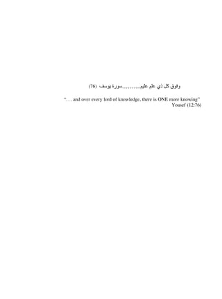
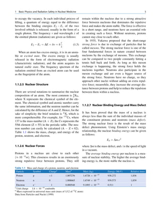
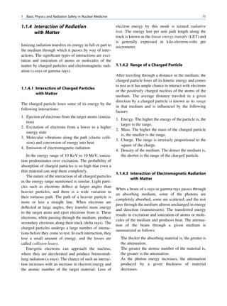
It seems that there are favored neutron-to-proton The radionuclide, which decays to attain stability, is
ratios among the stable nuclides. Figure 1.1 shows the called the parent nuclide, and the stable form so
function of number of neutron (N) against the number obtained is called the daughter. There are situations
of protons (Z) for all available nuclides. The stable when the daughter is also unstable. The unstable
nuclides gather around an imaginary line, which is nuclide may undergo transformation by any of the
called the line of stability. For light elements following modes.
(A 50), this line corresponds to N ¼ Z, but with
increasing atomic number the neutron-to-proton ratio
increases up to 1.5 (N ¼ 1.5Z). The line of stability Nuclides with Excess Neutrons
ends at A ¼ 209 (Bi), and all nuclides above that and
those that are not close to this line are unstable. Beta Emission
Nuclides that lie on the left of the line of stability Nuclides with an excess number of neutrons acquire a
(area I) have an excess of neutrons, those lying on stable form by converting a neutron to a proton. In this
process, an electron (negatron or beta minus) and an
antineutrino are emitted. The nuclear equation is given
100 III as follows:
n ! p þ e þ v þ Energy
Neutron number (N)
80
I
60
where n, p, e, and v represent the neutron, the proton,
the negatron (beta minus), and the antineutrino,
40 II respectively. The proton stays in the nucleus, but the
electron and the antineutrino are emitted and carry the
20 released energy as their kinetic energy. In this mode of
decay, the atomic number of the daughter nuclide is
one more than that of the parent with no change in
0 20 40 60 80 100
mass number. The mass of the neutron is more than the
Atomic number (Z)
sum of masses of the proton, electron, and the antineu-
Fig. 1.1 The line of stability and different regions around it. trino (the daughter is lighter than the parent). This
(Reproduced from [3]) defect in mass is converted into energy and randomly](https://image.slidesharecdn.com/basicsciencesofnuclearmedicine-121209103030-phpapp02/85/CIENCIAS-BASICAS-MN-19-320.jpg)
![8 G.S. Pant
be used for the production of carrier free radio- in knocking out an orbital electron from its own atom.
isotopes with high specific activity. This process is called internal conversion, and the
emitted electron is called a conversion electron. The
probability of K conversion electron is more than L or
Gamma Radiation and Internal Conversion M conversion electrons, and the phenomenon is more
common in heavy atoms. The internal conversion is
When all the energy associated with the decay process is followed by emission of characteristic x-rays or Auger
not carried away by the emitted particles, the daughter electrons as the outer shell electrons move to fill the
nuclei do not acquire their ground state. Such nuclei can inner shell vacancies.
be in either an excited state or a metastable (isomeric) It should be noted that there is no difference
state. In both situations, the excess energy is often between an x-ray and a gamma ray of equal energy
released in the form of one or more gamma photons. except that the gamma ray originates from the nucleus
The average lifetime of excited states is short, and energy and has a discrete spectrum of energy, whereas x-ray
is released within a fraction of a nanosecond. The aver- production is an atomic phenomenon and usually has a
age lifetime of metastable states is much longer, and continuous spectrum.
emission may vary from a few milliseconds to few days
or even longer. During this period, the nucleus behaves
as a pure gamma-emitting radionuclide. Some of the Laws of Radioactivity
metastable states have great clinical application. The
transition of a nucleus from a metastable state to a There is no information available by which one can
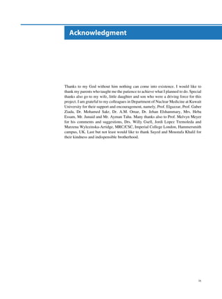
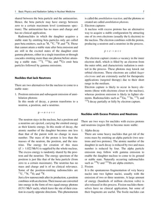
stable state is called an isomeric transition. The decay predict the time of disintegration of an atom; the
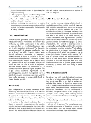
of 99mTc is the best example of isomeric transition. The interest really should not be in an individual atom
decay scheme of 99Mo-99mTc is shown in Fig. 1.2. because even an extremely small mass of any element
There are situations when the excited nuclei, consists of millions of identical atoms. Radioactive
instead of emitting a gamma photon, utilize the energy decay has been found to be a spontaneous process
67h
99
Mo μ
42
1.11
0.3% 0.922
17%
1.0%
0.513
82%
0.181
6h
0.142
0.140
2.12 x 10⁶y
Fig. 1.2 Decay scheme of 99
T
99
Mo. (Reproduced from [3]) 43 C](https://image.slidesharecdn.com/basicsciencesofnuclearmedicine-121209103030-phpapp02/85/CIENCIAS-BASICAS-MN-21-320.jpg)
![12 G.S. Pant
1.1.4.4 Linear Attenuation Coefficient The negative sign indicates that as dx increases, the
number of photons in the beam decreases. Equa-
The linear attenuation coefficient m is defined as the tion 1.13 can be rearranged as follows:
fractional reduction in the beam per unit thickness as
determined by a thin layer of the absorbing material. dN
m¼ (1.14)
N:dx
Fractional reduction in a thin layer
m¼ The formal definition of attenuation coefficient is
Thickness of the layers (cm)
derived from the integration of Eq. 1.14, which gives
The unit of the m is cm 1. the following relationship:
N ¼ No: e mx
(1.15)
1.1.4.5 Exponential Attenuation Equation 1.15 can also be expressed in terms of
beam intensity:
The exponential law can explain the attenuation of
radiation beam intensity. The mathematical derivation I ¼ Io: e mx
(1.16)
is given next.
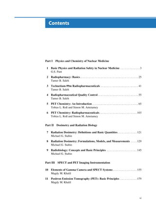
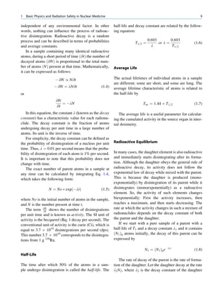
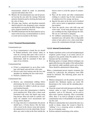
Let No be the initial number of photons in the beam where I and Io are the intensities of the beam as
and N be the number recorded by the detector placed recorded by the detector with and without absorbing
behind the absorber (Fig. 1.5). material, respectively. The attenuation coefficient may
The number dN, which gets attenuated, will be vary for a given material due to nonuniform thickness.
proportional to the thickness dx of the absorber This is particularly so if the absorbing material is
and to the number of photons N present in the malleable. It is therefore better to express the mass
beam. The number dN will depend on the number absorption coefficient, which is independent of thick-
of atoms present in the beam and the thickness of the ness of the absorbing material. The mass absorption
absorber. coefficient is obtained by dividing the linear attenua-
Mathematically, tion coefficient by the density of the material. The unit
of the mass attenuation coefficient is square centi-
dN / N: dx meters per gram. The electronic and atomic attenua-
(1.13)
or dN ¼ Àm:N:dx tion coefficients are also defined accordingly. The
electronic attenuation coefficient is the fractional
where m is a constant called the linear attenuation reduction in x-ray or gamma ray intensity produced
coefficient for the radiation used. by a layer of thickness 1 electron/cm2, whereas the
X
X-rays Attenuated
No N P
Primary
Fig. 1.5 Attenuation of a
radiation beam by an absorber.
The transmitted beam is
measured by detector P.
(Reproduced from [4])](https://image.slidesharecdn.com/basicsciencesofnuclearmedicine-121209103030-phpapp02/85/CIENCIAS-BASICAS-MN-25-320.jpg)
![1 Basic Physics and Radiation Safety in Nuclear Medicine 13
atomic attenuation coefficient is the fractional reduc- in medical applications of radiation. However, it has
tion by a layer of thickness 1 atom/cm2. Thus, the application in x-ray crystallography.
atomic attenuation coefficient will be Z times the elec-
tronic one.
Inelastic (Compton) Scattering
1.1.4.6 Half-Value Layer Compton elucidated the mechanism of inelastic (Comp-
ton) scattering. In this process, the photon interacts with
From Eq. 1.16, it can be seen that, for a certain thick- loosely bound (free) electrons. Part of the energy of the
ness (x ¼ d1/2) of the absorbing material, the intensity photon is used in ejecting the electron, and the rest is
becomes half of its original value, that is, I ¼ Io/2. scattered in different directions (Fig. 1.6).
Substituting these values, Eq. 1.16 can be rearranged In a so-called head-on collision, the photon turns
as follows: back along its original track (scattered through 180 ),
and maximum energy is transferred to the recoil elec-
d1=2 ðHVLÞ ¼ 0:693=m (1.17) tron. The change in wavelength dl of the photon is
given by
The half-value layer or thickness (HVL or HVT)
can be defined as the thickness of an absorbing mate- dl ¼ 0:024ð1 À cos ’ÞÅ (1.18)
rial that reduces the beam intensity to half of its origi-
nal value. Depending on the energy of radiation, where ’ is the angle of scattering of the gamma
various materials are used for the measurement of ˚
photon, and A is the angstrom unit for wavelength.
HVL, such as aluminum, copper, lead, brick, and The energy of the scattered photon is expressed as
concrete. The HVL for a broad beam is more than follows:
that for a narrow beam.
E1 ¼ E0 =½1 þ E0 =me c2 f1 À cos ’gŠ (1.19)
1.1.4.7 Mechanism of Attenuation
where E0 is the energy of the incident photon and E1 is
There are many modes of interaction between a photon that of the scattered photon, me is the mass of the
and matter, but only the types discussed next are of electron, and c is the velocity of light in a vacuum.
importance to us. Compton scattering involves interaction between
photons and electrons. The probability therefore
depends on the number of electrons present and inde-
Photon Scattering pendent of the atomic number. With the exception
of hydrogen, all elements contain nearly the same
Photon scattering may or may not result in transfer of number of electrons per gram (practically the same
energy during the interaction of the photon with an electron density). Compton scattering, therefore, is
atom of the medium.
γ-ray Electron (e−)
Elastic Scattering
In elastic scattering or unmodified scattering, the
photons are scattered in different directions without L M γ-ray
K
any loss of energy. The process thus attenuates the
beam without absorption. In this process, the photon
interacts with a tightly bound electron in an atom. The
electron later releases the photon in any direction
Fig. 1.6 Process of Compton scattering. The incoming photon
without absorbing energy from it. The contribution ejects the electron from outer orbit and is scattered with reduced
of this mode of interaction is relatively insignificant energy in a different direction. (Reproduced from [4])](https://image.slidesharecdn.com/basicsciencesofnuclearmedicine-121209103030-phpapp02/85/CIENCIAS-BASICAS-MN-26-320.jpg)
![14 G.S. Pant
independent of atomic number. This is the choice of x-ray photons. Such photons are characteristic of the
interaction required in radiation oncology, for which atom from which they are emitted. The K, L, M, and so
the delivered dose is homogeneous in spite of tissue on shells of a given atom have fixed energy, so the
inhomogeneity within the body. difference in their energies is also fixed. The radiation
The total probability s for the Compton process is emitted therefore is termed the characteristic x-rays.
given by Three types of possibilities exist during PEE:
1. Radiative transitions
s ¼ ss þ sa
As explained, during the electron transition from
the outer orbit to the inner orbit, a photon is emitted
where ss and sa are the probabilities for scattering and
with energy equal to the difference of the binding
absorption, respectively.
energies of the orbits involved. The vacancy moves
to a higher shell; consequently, a characteristic
photon of lower energy follows. The probability
1.1.4.8 Photoelectric Effect of emission of a photon is expressed as the fluores-
cent yield:
In the PEE process, the photon disappears when it
interacts with the bound electron. The photon energy Fluorescent yield
has to be higher than the binding energy of the electron Number of x À ray photons emitted
for this type of interaction to take place. ¼
Number of orbital vacancies created
hv ¼ BE þ kinetic energy Mostly, it is the K shell that is responsible for
fluorescent yield.
where hv is the energy of the photon and BE is the
binding energy of the electron in the shell (Fig. 1.7). If K shell fluorescent yield ( ok)
the photon energy is slightly higher than the binding
Number of K x À ray photons emitted
energy (BE), then the chance of PEE is high. For ¼
example, a photon of energy 100 keV has a high Number of K shell vacancies
probability of undergoing PEE when it interacts with
The yield increases with an increase in atomic
a Pb atom, for which the K shell binding energy is
number.
88 keV. The rest of the (100 to 88) 12-keV energy will
2. Auger electrons
be carried away by the ejected electron as its kinetic
The characteristic x-ray photon, instead of being
energy. The ejection of the electron creates a hole in
emitted, can eject another orbital electron from the
the inner shell, which is filled by an electron from any
atom. These electrons are called Auger electrons
of the outer shells. Since the electrons in the outer
(Fig. 1.8). The energy of the Auger electron is
shells possess higher energy than those in the inner
equal to the difference of the x-ray photon energy
shells, the difference in their energy is released as
and the binding energy of the shell involved in the
process. The process competes with radiative tran-
sition. The Auger yield is expressed as the ratio of
)
n (e electrons emitted due to vacancies in subshell i and
γ-ray Ele
ctro
the total number of atoms with a vacancy in sub-
shell i.
3. Coster Kronig electrons
The process for Coster Kronig electrons is exactly
like the Auger transition except that the electron
Fig. 1.7 Process of photoelectric absorption. The incoming filling the vacancy comes from the subshell of the
photon disappears (is absorbed), and the orbital electron is same principal shell in which the vacancy lies. The
knocked out. An electron from the outer shell falls (dotted
line) into the inner shell to fill the vacancy. (Reproduced kinetic energy of the emitted electrons can be cal-
from [4]) culated exactly as for Auger electrons. The energy](https://image.slidesharecdn.com/basicsciencesofnuclearmedicine-121209103030-phpapp02/85/CIENCIAS-BASICAS-MN-27-320.jpg)
![1 Basic Physics and Radiation Safety in Nuclear Medicine 15
Fig. 1.8 Mechanism of Characreristic Auger electron
Auger electron emission. x-ray e−
(Reproduced from [4]) hv
L L L
K K K
of Coster Kronig electrons is so small that they are -
(e )
tron
quickly absorbed in the medium. Elec
g-ray
511 keV
Pos
it
1.1.4.9 Pair Production
ion
(e +) e-
e+
511 keV
When a photon with energy in excess of 1.022 MeV
passes close to the nucleus of an atom, it may disap-
pear, and in its place two antiparticles (negatron and Fig. 1.9 Schematic representation of pair production. (Repro
duced from [4])
positron) may be produced as shown in Fig. 1.9. In this
process, energy converts into mass in accordance with
Einstein’s mass energy equivalence (E ¼ mc2). After While such enthusiasm is appreciable, adequate safe-
traversing some distance through the medium, the guards against radiation exposure are also necessary to
positron loses its energy, combines with an electron, minimize the radiation risk to occupational workers,
and annihilates. During combination, both the antipar- patients, and the public.
ticles disappear (annihilation) and two 0.511-MeV
photons are emitted in the opposite direction.
1.2.1 Types of Exposure
1.1.4.10 Photonuclear Reaction
The following three categories of people are likely to
be involved in radiation exposure in medical applica-
When photon energy is too high, either a neutron or a
tions of ionizing radiation:
proton may be knocked out (more likely the neutron)
from the nucleus. For the majority of atoms, the 1. Occupational staff
threshold energy for this effect is more than 10 MeV, 2. Patients
and the probability increases with increasing energy 3. Public
until a maximum is reached; above this maximum, the
Protection is aimed at achieving a dose as low as
probability falls rapidly.
reasonably achievable (ALARA) to these categories of
people. Spending a minimum of time near the radiation
sources, keeping a distance from them, and using
1.2 Radiation Safety shielding devices are the cardinal parameters for radia-
tion safety. The fourth parameter with unsealed sources
in nuclear medicine is to avoid or minimize the chance
The applications of radiopharmaceuticals for medical
of contamination.
diagnosis and therapy have rapidly increased due to
For safe use of radionuclides in nuclear medicine,
the favorable physical characteristics of artificially
the following basic requirements should be met:
produced radionuclides, progress in instrumentation,
and computer technology. Efforts are under way to 1. The nuclear medicine facility should be well
develop newer radiopharmaceuticals in nuclear medi- planned with a sufficient number of rooms for
cine for both diagnostic and therapeutic procedures. intended operations (including the storage and](https://image.slidesharecdn.com/basicsciencesofnuclearmedicine-121209103030-phpapp02/85/CIENCIAS-BASICAS-MN-28-320.jpg)

![20 G.S. Pant
1.2.3.5 Posttreatment et al. [12] published the relevant guidelines for radio-
iodine therapy for consultation.
The patient must be provided with an instruction card
detailing the type and duration of any radiation pro-
tection restrictions that must be followed at home. 1.2.3.8 Personal Hygiene and Laundering
This should also contain details of therapy and neces- Instructions for the First Week
sary radiation protection procedures. After Therapy
A normal toilet should be used in preference to a urinal
1.2.3.6 Contact with Spouse or Partner for voiding urine. The sitting posture is preferred to
and Others at Home standing. Spilled urine should be wiped with a tissue
and flushed. Hands should always be washed after
The patient should make arrangements to sleep apart using the toilet. Any linen or clothes that become
from his or her partner for some time as suggested by stained with urine should be immediately washed sep-
the RSO. The duration of such restriction actually arately from other clothes.
depends on the body burden of the patient at the time
of release from the hospital [5 8]. Contact with family
and friends at home should not be for prolonged per- 1.2.3.9 Records
iods for a few initial days after release from the hospi-
tal [5, 9]. Close contact with pregnant women and A proper logbook should be maintained with details of
young children on a regular basis should be avoided storage and disposal of radionuclides. The record of
for such time as suggested by the RSO. It would be dose administration to the patients, their routine mon-
ideal if an arrangement could be made for young itoring, transient storage of waste for physical decay,
children to stay with relatives or friends after the and the level of activity at the time of disposal should
treatment, at least for the initial few days or weeks. be properly recorded. Decontamination procedures
If such an arrangement is not possible, then prolonged and routine surveys should also be recorded in the
close contact with them should be avoided as per radiation safety logbook. The name of the authorized
advice of the RSO. Time duration to avoid close person who supervised the procedure should also be
contact can only be estimated on an individual basis recorded.
depending on the radioiodine burden and socioeco-
nomic status of the patient. Mathieu et al. [10] esti-
mated the radiation dose to the spouse and children at 1.2.4 Management of Radioactive Waste
home and observed that the dose to the spouse is
greater from patients treated for thyrotoxicosis than
for those treated for thyroid cancer. Pant et al. [11] Radioactive waste is generated as a result of handling
reported that the dose to family members of patients unsealed sources in the laboratory, leftover radioactive
treated with radioiodine (I-131) for thyrotoxicosis and material from routine preparations, dose dispensing to
cancer thyroid was within 1 mSv in the majority of the patients, contaminated items in routine use, and so on.
cases with proper counseling of the patient and the The waste arises in a large variety of forms depending
family members at the time of release from the hospital. on the physical, physiochemical, and biological prop-
erties of the material. In radionuclide therapy, the
waste may also consist of excreta.
1.2.3.7 Returning to Work
If work involves close contact with small children or 1.2.4.1 Storage of Radioactive Waste
pregnant women, then it should not be resumed by
treated patients for a few weeks; otherwise, routine The solid waste generated in the working area should
work can be assumed by avoiding close contact with be collected in polythene bags and transferred to suit-
fellow colleagues for a prolonged period. The Luster able containers in the storage room. The liquid waste](https://image.slidesharecdn.com/basicsciencesofnuclearmedicine-121209103030-phpapp02/85/CIENCIAS-BASICAS-MN-33-320.jpg)
![1 Basic Physics and Radiation Safety in Nuclear Medicine 21
has to be collected in either glass or preferably plastic conveniently dealt with by burial or incineration,
containers. The waste containing short-lived and long- depending on the national or international guidelines.
lived radionuclides should be collected in separate Incineration of refuse containing nonvolatile radio-
bags and stored in separate containers. If the labora- nuclides concentrates the activity in the ash. If the
tory is used for preparation of short-lived radiophar- ash contains undesirable high activity, special dis-
maceuticals, then it is advisable not to collect the posal methods should be adopted. The ash can be
waste until the next preparation. This will avoid diluted and disposed without exceeding the speci-
unnecessary exposure to the staff handling radioactive fied limits or buried. The design of the incinerator
waste. The storage room should have proper ventila- for handling the radioactive waste should be con-
tion and an exhaust system. The shielding around the sidered at the planning stage.
waste storage room should be adequate to prevent any
leakage of radiation. The waste must be stored for at
1.2.4.3 Management of Cadavers Containing
least ten half-lives for decay or until such a time
Radionuclides
disposal is conveniently possible.
An unfortunate situation arises if a patient dies after
administration of a high amount of radioactivity and
1.2.4.2 Disposal of Solid Waste
the radiation limits are more than the threshold level
for releasing the body from the hospital. If the activity
1. Low-activity waste
is concentrated in a few organs (as can be seen by
The solid waste comprised of paper tissues, swabs,
scanning the cadaver under the gamma camera), then
glassware, and similar materials that are of low
those organs should be removed, and the body released
activity (only a few becquerels) can be disposed
after ensuring that the limits recommended by the
with ordinary refuse provided no single item con-
competent authority are not exceeded. In case of wide-
tains concentrated activity and
spread disease for which organ removal is no solution,
(a) They do not contain alpha or beta emitters or the body may be put into an impermeable plastic bag
radionuclides with a long half-life. and stored in a mortuary (cold room) for physical
(b) The waste does not go through a recycling decay until the radiation level returns to an acceptable
procedure. limit. In any compelling social circumstances, the
(c) The radionuclide labels are intact (to guard advice of a regulatory body may be sought. Autopsy,
against misinterpretation). management of removed organs or a part of the body,
handing over the body, and burial or cremation should
2. High-activity wastes
be done under the direct supervision of the RSO.
Contaminated clothing and those items that need to
Removal of organs from the cadaver is socially not
be reused are segregated and stored for physical
permitted in some countries, the regulatory require-
decay of radioactivity or decontaminated sepa-
ment of that country shall be followed by the RSO.
rately. A derived working limit (DWL) of 3.7 Bq/
cm2 is indicated for personal clothing and hospital
bedding. Disposal methods for solid waste consist 1.2.4.4 Disposal of Liquid Waste
of decaying and disposal or ground burial. The
method chosen depends on the quantity of radioac- While disposal of liquid wastes through the sanitary
tive material present in the wastes. From each work sewage system, the limits of dilution and disposal
area, the wastes are collected in suitable disposable should not exceed the prescribed limits recommended
containers. Extra care for radiation protection is by the competent authority (normally 22.2 MBq/m3).
necessary during the accumulation, collection, and If the activity in the waste is too low, then it may be
disposal of radioactive wastes. Containers should disposed with proper dilution (dilute and dispose). If
be marked with the radiation symbol and suitable the activity level is moderate to high and the half-life
designation for segregation [13]. or lives of the radionuclides is relatively short, then the
Solid waste (e.g., animal carcasses, animal exc- waste should be stored for physical decay for a period
reta, specimens, biologically toxic material) can be of about ten half-lives (delay and decay).](https://image.slidesharecdn.com/basicsciencesofnuclearmedicine-121209103030-phpapp02/85/CIENCIAS-BASICAS-MN-34-320.jpg)
![22 G.S. Pant
The quantity of liquid radioactive waste generated out to prove the adequacy of the disposal system.
due to nuclear medicine investigations hardly poses any When large quantities of radionuclides are routinely
problem of storage or disposal. However, it is not the discharged to the environment, it is advisable to make
same for therapeutic nuclear medicine, for which a large environmental surveys in the vicinity since many
amount of radioactive waste is generated in the form of radionuclides will be concentrated on surfaces.
effluent from the isolation room or ward of thyroid In installations where large amounts of airborne
cancer patients. The large doses of radioiodine used for activity are involved, it may be necessary to use suitable
the treatment of thyroid cancer calls for planned storage air filtration (through charcoal filter) systems and to
and release of waste by sewage disposal. Amounts of discharge the filtered effluent through a tall stack. The
131
I as high as 7.4 11.1 GBq (200 300 mCi) are admi- height of the stack can be chosen to ensure that the radio-
nistered to patients with distant metastases. Approxi- activity is sufficiently diluted before it reaches ground
mately 80 90% of administered radioactivity is level. Combustible low-level radioactive waste may be
excreted through urine [5]. Therefore, management of incinerated with adequate precautions to reduce bulk.
radioactive urine poses a radiation safety problem. Security: The waste has to be protected from fire,
Various methods have been recommended for the insects, and extreme temperatures.
disposal of high-level radioactive liquid wastes. The
widely used technique is the storage delay tank sys-
tem. Storage of all effluent from the isolation room or
ward, or urine alone, in a storage delay tank system is References
the recommended method and is more feasible in
hospitals with tanks of appropriate volumes. The sys- 1. Pant GS, Rajabi H (2008) Basic atomic and nuclear physics.
tem allows collection of effluent from the isolation In: Basic physics and radiation safety in nuclear medicine.
Himalaya Publishing House, Mumbai
room or ward in the first tank. The tank is closed 2. Povh B, Rith K, Scholz C, Zetche F, Lavell M, Particles and
after it is completely filled, and collection takes place Nuclei: An introduction to the physical concept (2nd ed),
in the second tank. Until the second tank is completely Springer 1999
filled, the effluent in the first tank gets enough time to 3. Pant GS, Shukla AK (2008) Radioactivity. In: Basic physics
and radiation safety in nuclear medicine. Himalaya Publish
decay, which may make its release possible to the
ing House, Mumbai
sewage system. It will even be better if effluent in 4. Pant GS (2008) Basic interaction of radiation with matter.
each tank is allowed to decay for a given length of In: Basic physics and radiation safety in nuclear medicine.
time and then released into a big dilution tank before Himalaya Publishing House, Mumbai
5. Barrington SF, Kettle AG, O’Doherty MJ, Wells CP, Somer
its final release into the sewage line. A large number of
EJ, Coakley AJ (1996) Radiation dose rates from patients
small tanks is advisable for allowing decay of radio- receiving iodine 131 therapy for carcinoma of the thyroid.
iodine for at least ten half-lives. Provision of access to Eur J Nucl Med 23(2):123 130
the dilution tank is useful for monitoring the activity 6. Ibis E, Wilson CR, Collier BD, Akansel G, Isitman AT and
Yoss RG (1992) Iodine 131 contamination from thyroid
concentration at any time before its final release to the cancer patients. J Nucl Med 33(12):2110 2115
main sewer system. 7. Beierwalts WH, Widman J (1992) How harmful to others
are iodine 131 treated patients. J Nucl Med 33:2116 2117
8. de Klerk JMH (2000) 131I therapy: inpatient or outpatient?
J Nucl Med 41:1876 1878
1.2.4.5 Disposal of Gaseous Waste 9. O’Dogherty MJ, Kettle AG, Eustance CNP et al. (1993)
Radiation dose rates from adult patients receiving 131I ther
Gaseous wastes originate from exhausts of stores, apy for thyrotoxicosis, Nucl Med Commun 14:160 168
fume cupboards, and wards and emission from incin- 10. Mathieu I, Caussin J, Smeesters P et al. (1999) Recom
mended restrictions after 131I therapy: Measured doses in
erators. Points of release into the atmosphere should
family members. Health Phys 76(2):129 136
be carefully checked, and filters (including charcoal) 11. Pant GS, Sharma SK, Bal CS, Kumar R, Rath GK (2006)
may be used wherever possible. The concentration of Radiation dose to family members of hyperthyroidism and
radioactive materials in the air leaving the ventilation thyroid cancer patients treated with 131I. Radiat Prot Dosim
118(1):22 27
system should not exceed the maximum permissible
12. Luster M, Clarke SE, Dietlein M et al. (2008) Guidelines for
concentrations for breathing unless regular and ade- radioiodine therapy of differentiated thyroid cancer. Eur J
quate monitoring or environmental surveys are carried Nucl Med Mol Biol 35(10):1941 1959](https://image.slidesharecdn.com/basicsciencesofnuclearmedicine-121209103030-phpapp02/85/CIENCIAS-BASICAS-MN-35-320.jpg)


![Radiopharmacy: Basics
2
Tamer B. Saleh
Contents other drugs. The difference between a radiochemical
2.1 Introduction . . . . . . . . . . . . . . . . . . . . . . . . . . . . . . . . . . . . . . . . . . 25 and a radiopharmaceutical is that the former is not
2.2 An Ideal Radiopharmaceutical . . . . . . . . . . . . . . . . . . . . . 26 administered to humans due to the possible lack of
2.3 Production of Radionuclides . . . . . . . . . . . . . . . . . . . . . . . . 27 sterility and nonpyrogenicity; any material adminis-
2.3.1 Reactor Produced Radionuclides . . . . . . . . . . . . . 27
tered to humans must be sterile and nonpyrogenic. A
2.3.2 Cyclotron Produced Radionuclides . . . . . . . . . . . 28
2.3.3 Generator Produced Radionuclides . . . . . . . . . . . 29 radiopharmaceutical may be a radioactive element like
133
2.4 Common Radiopharmaceuticals . . . . . . . . . . . . . . . . . . . . 33 Xe or a labeled compound such as 99mTc-labeled
2.4.1 Thallium 201 . . . . . . . . . . . . . . . . . . . . . . . . . . . . . . . . 33 compounds [1].
2.4.2 Gallium 67 . . . . . . . . . . . . . . . . . . . . . . . . . . . . . . . . . . . 34
In nuclear medicine, about 95% of the radiophar-
2.4.3 Iodine Radiopharmaceuticals . . . . . . . . . . . . . . . . 34
2.4.4 Indium 111 Radiopharmaceuticals . . . . . . . . . . . 35 maceuticals are used for medical diagnosis; only about
2.4.5 Xenon 133 . . . . . . . . . . . . . . . . . . . . . . . . . . . . . . . . . . . 36 5% are used for therapeutic purposes. In designing a
2.4.6 Chromium 51 . . . . . . . . . . . . . . . . . . . . . . . . . . . . . . . . 36 radiopharmaceutical, a suitable pharmaceutical is cho-
2.4.7 Phosphorus 32 . . . . . . . . . . . . . . . . . . . . . . . . . . . . . . . 36
sen on the basis of its preferential localization in a
2.4.8 Strontium 89 . . . . . . . . . . . . . . . . . . . . . . . . . . . . . . . . . 36
2.4.9 Rhenium 186 . . . . . . . . . . . . . . . . . . . . . . . . . . . . . . . . 36 given organ or its participation in the physiological
2.4.10 Samarium 153 . . . . . . . . . . . . . . . . . . . . . . . . . . . . . . . 36 function of the organ. Then, a suitable radionuclide is
2.4.11 111In and 90Y Ibritumomab tagged onto the chosen pharmaceutical and adminis-
Tiuxetan (Zevalin) . . . . . . . . . . . . . . . . . . . . . . . . . . . 36
tered to the patient [2]. The radiation emitted from the
2.4.12 90Y Labeled Microspheres . . . . . . . . . . . . . . . . . . . 37
2.4.13 Lutetium 177 Compounds . . . . . . . . . . . . . . . . . . . 37 organ can be detected by an external radiation detector
References . . . . . . . . . . . . . . . . . . . . . . . . . . . . . . . . . . . . . . . . . . . . . . . . . . 37 for assessment of the morphological structure and the
physiological function of that organ. Radiopharma-
ceuticals in most cases have no pharmacological effect
as they are mainly administered in tracer amounts. So,
they mainly do not show any dose response relation-
2.1 Introduction ship. For the therapeutic radiopharmaceuticals, how-
ever, the observed biological effect is from the
radiation itself and not from the pharmaceutical [3].
A radiopharmaceutical is a radioactive compound that
Nuclear medicine procedures generally have two
has two components, a radionuclide and a pharmaceu-
classifications; the first is those that depend on single-
tical; it is used for the diagnosis and treatment of
photon emitters, for which planar and tomographic
human diseases. All radiopharmaceuticals are legend
imaging (single-photon emission computed tomogra-
drugs and are subject to all regulations that apply to
phy or SPECT) are the options of image acquisition.
The other type is positron emission tomography (PET),
for which the detection process relies on positron-
electron annihilation and the release of two opposing
photons (180 apart). The key component that distin-
T.B. Saleh guishes these techniques among other modalities is
King Fahed Specialist Hospital, Dammam, KSA the diversity and ability of their contrast agents to
e mail: tamirbayomy@yahoo.com
M.M. Khalil (ed.), Basic Sciences of Nuclear Medicine, DOI: 10.1007/978 3 540 85962 8 2, 25
# Springer Verlag Berlin Heidelberg 2011](https://image.slidesharecdn.com/basicsciencesofnuclearmedicine-121209103030-phpapp02/85/CIENCIAS-BASICAS-MN-38-320.jpg)
![26 T.B. Saleh
answer a clinical question. The contrast agents in radiotracers with short lifetimes mandate an injection
nuclear medicine are radiolabeled compounds or of a high-activity concentration using fast imaging
radiopharmaceuticals that, when localized in the systems and may also compromise image quality.
region of interest, emit important information about Thus, an optimal half-life satisfies imaging require-
the pathophysiologic status of the tissue involved. ments while maintaining the quality of the scan. Pro-
Both imaging techniques have high sensitivity in tein synthesis and peptide formation involve a slow
detecting molecular concentrations in the pico or kinetic process; thus, single-photon emitters provide
nano range, and their role in functional or molecular an opportunity to study the underlying functional dis-
imaging is well addressed. SPECT and PET radio- orders while the tracer still is able to emit a signal [1].
pharmaceuticals have a wide acceptance in molecular Suitable radionuclide emission: Radiopharmaceu-
imaging, biomedical research disciplines, and drug ticals emitting g-radiation by electron capture or iso-
development. However, many SPECT tracers are meric transition (energy between 30 and 300 keV) are
approved by the U.S. Food and Drug Administration commonly used in nuclear medicine diagnostic proce-
(FDA), widely available, well reviewed in the litera- dures. For therapeutic purposes, a-, b-, and Auger
ture, and relatively cheaper and perform for a signifi- electron emitters are used because of their high linear
cant patient population on a daily basis, whereas this energy transfer, which leads to maximum exposure
situation is not true for the use of PET compounds. and damage of the target cells. The a-particles and
SPECT radiotracers have a particular position in Auger electron emitters are mostly monoenergetic,
the matrix of molecular imaging due to their ability whereas the b-particles have a continuous energy
to image endogenous ligands such as peptides and spectrum up to their maximum energy Emax.
antibodies and their ability to measure relatively High target-to-nontarget ratio: In all diagnostic
slow kinetic processes due to the relatively long half- procedures, it is well known that the agent with better
life of the commonly used isotopes (in comparison to target uptake is a superior imaging agent since the
PET). In addition, the capability to measure two dif- activity from the nontarget areas can interfere with
ferent photon energies allows SPECT systems to the structural details of the organ imaged. Therefore,
depict two molecular pathways simultaneously by the target-to-nontarget activity ratio should be as large
measuring their corresponding photon emissions [4]. as possible.
In this chapter, we discuss some basic concepts about Target uptake rate: The rate at which an organ
properties of radiopharmaceuticals, production, and takes up the administrated radiopharmaceutical is
generator systems used in clinical practice. also considered a key characteristic of an ideal radio-
pharmaceutical because it influences the period after
which imaging acquisition is done. It is preferable to
get images as early as possible for patient conve-
2.2 An Ideal Radiopharmaceutical nience. For example, 99mTc-pertechnetate is prefera-
ble to 123I-NaI because the thyroid-imaging procedure
The definition of an ideal radiopharmaceutical in nuclear can be performed after 20 min of dose administration,
medicine procedures varies according to its use. The aim while with 123I-NaI it takes 4 6 h to launch the imag-
of a diagnostic radiopharmaceutical is to provide detect- ing session.
able photons with minimal biological effect to the cells Tracer excretion: The most common excretion
or organ, whereas it is desired to produce a cytotoxic route is renal clearance, which is rapid and can reduce
effect in a therapeutic procedure [5]. Generally, an ideal exposure to the blood, whole body, and marrow. In
radiopharmaceutical for diagnostic procedures should contrast, the gastrointestinal tract (GIT) and hepato-
meet the following characteristics: biliary excretion is slow and leads to higher GIT and
Short half-life: Radiopharmaceuticals should have whole-body exposures. With GIT excretion, reabsorp-
a relatively short effective half-life, which should not tion into the blood also occurs. Since organ visualiza-
exceed the time assigned to complete the study. It tion is better when the background tissues have less
provides a smaller radiation dose to the organ and uptake than the target organ, the radiopharmaceutical
ambient structures together with reduced exposure must be cleared from the blood and background tissue
to workers, family members, and others. However, to achieve better image contrast.](https://image.slidesharecdn.com/basicsciencesofnuclearmedicine-121209103030-phpapp02/85/CIENCIAS-BASICAS-MN-39-320.jpg)
![2 Radiopharmacy: Basics 27
Availability: The ideal radiopharmaceutical should that initiate the chain reaction. This chain reaction
be cost effective, inexpensive, and readily available in must be controlled to avoid the possible meltdown
any nuclear medicine facility. This feature also char- situation in the reactor using special neutron modera-
acterizes the spread and diffusion of gamma emitters tors (low molecular weight materials such as water,
compared to PET-based compounds. heavy water, and graphite, which are distributed in the
spaces between the fuel rods), and neutron absorbers
(e.g., cadmium rods placed in the fuel core) are used to
2.3 Production of Radionuclides thermalize and reduce the energy of the emitted neu-
trons to 0.025 eV to maintain equilibrium [1].
From the medical usefulness point of view, there
Naturally occurring radionuclides cannot be employed are two types of nuclear reactions used to produce
for medical diagnosis because of their long half-lives, radioisotopes of interest 2 types: thermal neutron reac-
which warrant the need for production of other radio- tions and fission (n, f) reactions.
nuclides that can be safely used for medical applica-
tions. Most of the radionuclides for medical use are
produced in nuclear reactors or cyclotrons. Some of 2.3.1.1 Thermal Neutron Reactions
the radionuclides are eluted from the generators in
which the parent radionuclide is produced from a The thermal neutrons around the core of the nuclear
reactor or a cyclotron [2]. reactor can induce the following types of nuclear
The process of all radionuclide production can be reactions:
described by the general equation
AY þn ! AYþ1 þg
Z Z
Xð‘‘BP’’; ‘‘EP’’ÞY
AZ þn ! AZ þp
Y Y
1
where
In the first type of reaction, the target atom A
X is the target element.
captures a neutron and emits gamma rays (g), also
Y is the product element.
written as an (n, g) reaction.
BP is the bombarding particle (projectile).
For example:
EP is the emitted product.
98
Pure metals are the best targets to use because of Mo (n,gÞ 99 Mo
their high ability to sustain the high temperature in
50
cyclotron and reactor systems. Cr (n,gÞ 51 Cr
In the second type of reactions, a proton is emitted
after absorption of the neutron, resulting in a new
2.3.1 Reactor-Produced Radionuclides element with different atomic number (Z).
For example:
The two major principles of a nuclear reactor are that
14
the neutrons induce fission in the fissile material con- Nðn; pÞ 14 C
structing the fuel rods (e.g., U235, P239) of the reactor
3
and the number of neutrons released in that fission Heðn; pÞ 3 H
reaction is about two or three neutrons with a mean
energy of 1.5 MeV. Although the (n, g) reaction produces a radioiso-
tope from a stable one with a low specific activity
U235 þ n ! fission products þ un product that are not carrier free, no sophisticated
chemical separation procedures are required.
These new neutrons are used to produce fission in The (n, p) reaction produces an isotope with a
other nuclei, resulting in the release of new neutrons different atomic number (element), enabling the](https://image.slidesharecdn.com/basicsciencesofnuclearmedicine-121209103030-phpapp02/85/CIENCIAS-BASICAS-MN-40-320.jpg)
![28 T.B. Saleh
production of high specific activity and carrier-free Molybdenum-99: For 99Mo separation, the irra-
radioisotopes. Specific activity can be defined as the diated uranium target is dissolved in nitric acid, and
amount of activity per unit mass of a radionuclide or a the solution is adsorbed on an alumina (Al2O3) col-
labeled compound. umn. The column is then washed with nitric acid to
Products of (n, g) reactions include parent isotopes, remove uranium and other fission products.
which are commonly used in nuclear generators to Iodine-131: For chemical separation of 131I from
235
produce daughter radionuclides, which are the iso- U, the latter is dissolved in 18% sodium hydrox-
topes of interest; these are usually separated from ide (NaOH) by heating, and hydroxides of many
their parents by column chromatography procedures. metal ions are then precipitated by cooling. The
For example: supernatant containing sodium iodide is acidified
with sulfuric acid. Iodide is oxidized to iodine by
t1=2¼67 h the effect of the acid, and iodine is collected in a
98
Moðn; gÞ 99 Mo ÀÀÀÀÀ! 99m Tc
ÀÀ
NaOH by distillation.
t1=2¼67 h
124
Xeðn; gÞ 125 Xe ÀÀÀÀÀ! 125 I
ÀÀÀ
2.3.2 Cyclotron-Produced Radionuclides
2.3.1.2 Fission or (n, f) Reactions Cyclotron systems, which were invented in 1930, have
an obvious role in the production of a wide range of
Fission is a process of breaking up a heavy nucleus nuclear medicine radiopharmaceuticals, especially
(e.g., 235U, 239Pu, 232Th) and any other material with those with short half-lives.
an atomic number greater than 90 into two fragments The basic principle of their operation is the accel-
(by-products). eration of charged particles such as protons, deuterons,
For example: and a-particles in a spiral path inside two semicircular,
flat, evacuated metallic cylinders called “dees.” The
U92 þn ! U92 ! Kr 89 þ Ba144 þ3n
235 236
36 56 dees are placed between the two poles of a magnet (see
Fig. 2.1) so that the ion beam is constrained within a
U92 þn ! U92 ! I53 þ Y39 þ3n
235 236 131 102
circular path inside the dees. At the gap between the
dees, the ions experience acceleration due to the impo-
U92 þn ! U92 ! Mo99 þ Sn135 þ2n
235 236
42 50 sition of the potential difference. The beam particles
originate at the ion source at the center of the cyclo-
The neutron interacts with the 235U nucleus to form tron, and as they spiral outward in the dees, they
unstable uranium atom 236U, which breaks into two acquire increasing energy for each passage across the
different smaller atoms and a number of neutrons. The gap of the dees. Eventually, the high-energy particles
isotopes produced may be employed in nuclear medi- reach the periphery of the dees, where they are
cine (99M, 131I, 133Xe); the greatest portion of these directed toward a target for bombardment [6].
radioisotopes is not useful in nuclear medicine as they Fixed-frequency cyclotrons can accelerate posi-
tend to decay by b emission. Unlike the radioisotopes tively charged ions up to only 50 MeV for protons
formed from (n, g) reactions, fission products can be due to the relativistic increase in the mass of the
chemically treated to produce carrier-free radionu- accelerated particle, while for linear accelerators, par-
clides. But, the major problem is how to separate ticle acceleration can occur up to several hundreds of
them from the other products to obtain the highest mega-electron-volts because of the ability of the accel-
level of radiochemical purity of the end product. The erator to compensate for the increase in mass of high-
radioisotopes are mainly separated by appropriate energy particles. Advanced techniques have been
chemical procedures that may involve precipitation, developed to use cyclotrons to accelerate particles to
solvent extraction, ion exchange, distillation, and much higher energies [7].
chromatography. Two of the most common isotopes The majority of cyclotrons built prior to 1980
are discussed as examples: accelerated positively charged ions (i.e., H+); medical](https://image.slidesharecdn.com/basicsciencesofnuclearmedicine-121209103030-phpapp02/85/CIENCIAS-BASICAS-MN-41-320.jpg)
![2 Radiopharmacy: Basics 29
Fig. 2.1 Layout of cyclotron Top view Side view
The accelerating electric
field reverses just at the
time the electrons finish
their half circle, so that
it accelerates them N S Uniform
across the gap. With magnetic
a higher speed, they field
move in a larger region
+
- Electric
accelerating
semicircle. After field between
repeating this process the magnetic
several times, they field regions
come out the exit port N S
at a high speed.
Injection of
electrons
Output beam of high
velocity electrons
cyclotrons accelerate negative ions (H ). This design In this case, a second nucleon may not be emitted
allows for a simple deflection system in which the because there is not enough energy left after the emis-
beam is intercepted by a thin carbon foil that extracts sion of the first neutron. The excitation energy insuffi-
the negative ions at the end of the trajectory, resulting cient to emit any more nucleons will be dissipated by
in the formation of a positively charged H+ beam. The g-ray emission.
beam then changes the direction without a deflector The target material must be pure and preferably
due to the magnetic field. The H+ bombards the target monoisotopic or at least enriched in the desired isotope
in a manner similar to that in a positively charged ion to avoid the production of extraneous radioisotopes. In
cyclotron. It is also possible to extract the beam at two addition, the energy and type of the irradiating particle
different points in the machine, allowing use of a must be chosen to avoid the presence of undesired
negative-ion cyclotron for production of two different radionuclides.
radioisotopes simultaneously [8]. Generally, when the Table 2.1 represents the most common commercial
target nuclei are irradiated by the accelerated particles, cyclotrons presented by the International Atomic
a nuclear reaction takes place. The incident particle, Energy Authority (IAEA) report for cyclotron distri-
after interaction, may leave some of its energy in the bution in member states in 2006 [9].
nucleus or be completely absorbed by it, depending on
its incident energy. In either case, the nucleus is
excited, resulting in the emission of nucleons (protons
2.3.3 Generator-Produced Radionuclides
and neutrons) followed by g-ray emission.
Depending on the energy deposited by the incident
particle, a number of nucleons may be emitted ran- The first commercial radionuclide generator was
domly from the irradiated nucleus, leading to the for- produced in the United States in the early 1960s
mation of different nuclides. As the energy of the (Brookhaven National Laboratories); since then, a
irradiating particle is increased, more nucleons are number of different types of generators have been
emitted and therefore a greater variety of nuclides developed for various purposes in nuclear medicine.
may be produced. Generators are “parent daughter systems involving a
An example of a simple cyclotron-produced radio- long-lived parent radionuclide that decays to short
nuclide is the production of 111In by irradiation of half-life daughter” and is called a generator because
111 of its ability to generate continuously a relatively
Cd with 12-MeV protons. The nuclear reaction
can be expressed as follows: short-lived daughter radionuclide. The parent and its
daughter nuclides are not isotopes; therefore, chemical
111
Cd (p, n)111 In separation is possible. Table 2.2 represents the most](https://image.slidesharecdn.com/basicsciencesofnuclearmedicine-121209103030-phpapp02/85/CIENCIAS-BASICAS-MN-42-320.jpg)
![30 T.B. Saleh
commonly used generators in nuclear medicine appli- Several methods can be adopted to obtain a steri-
cations. lized eluted radionuclide:
Radionuclide generators are formed by a glass or
The entire column of the generator is autoclaved.
plastic column fitted at the bottom with a filtered disk.
Column preparation occurs under general aseptic
The column is fitted with absorbent material such as
conditions.
alumina, on which the parent nuclide is absorbed.
Bacteriostatic agents are added to the generator
Daughter radionuclides are generated by the decay of
column.
the parent radionuclide until either a transient or secu-
A membrane filter unit is attached to the end of the
lar equilibrium is reached; after that, the daughter
column.
appears to decay with the half-life of the parent. The
Elution procedures are carried out under aseptic
daughter is eluted in a carrier-free state (because it is
conditions.
not an isotope of the parent radionuclide) with a sterile
and pyrogen-free appropriate solvent; then, the activ-
ity of the daughter starts to increase again up to equi-
librium, so the elution can be made multiple times. 2.3.3.1 Daughter Yield Equations
Figure 2.2 shows a typical generator system.
Assuming that there is initially no daughter activity in
the generator, the daughter activity at any given time
Table 2.1 Common commercial cyclotrons for cyclotron dis t is given by
tribution
Company Model Description l2 l1 t l2 t
11 MeV H , 40,60 mA A2 ¼ A0 ðe Àe Þ
CTI, Inc./ RDS 111 l2 À l1 1
Siemens RDS 112 11 MeV H , 40 mA
GE PETrace 16.5 MeV H , where
8.6 MeV D ,
80 mA A2 is the daughter activity at time t.
Ion Beam Cyclone 18/9 18 MeV H , 9 MeV A0 is the parent activity at time zero.
1
Applications Cyclone D , 80 mA l1 and l2 are decay constants for the parent and
(IBA) 30+ 30 MeV H , daughter, respectively.
15 MeV D , 60 mA
In case of transient equilibrium, as time t becomes
Sumitomo CYPRIS 370 16 MeV H+, 10 MeV
Heavy AVF D+, 60 mA sufficiently long, e l2 t is negligible compared with
Industries 930+ 90 MeV H+, 60 mA e l1 t , and the equation becomes
Scanditronix MC40+ 10 40 MeV H+,
Medical AB 5 20 MeV D+, l2 l1 t
60 mA A2 ¼ A0 ðe Þ
l 2 À l1 1
Table 2.2 Some generator systems used in nuclear medicine applications
Parent Parent Nuclear Daughter Daughter Mode of Principal keV Column Eluant
(T1/2) reaction (T1/2) daughter (% abundance)
decay
99 99m
Mo 66 h Fission Tc 6h IT 140(90) Al2O3 0.9% NaCl
87 88 87
Y 80 h Sr(p,2n) mSr 2.8 h IT 388(82) Dowex 0.15M
1Â8 NaHCO3
68 69 68
Ge 271 days Ga69 Ga 68 min B+ 511(178) Al2O3 0.005M
(p,2n) EDTA
62 63 62
Zn 9.3 h Cu(p,2n) Cu 9.7 min B+ 511(194) Dowex 2N HCL
1Â8
82 85 82
Sr 25.5 days Rb(p,4n) Rb 75 s B+ 511(190) SnO2 0.9% NaCl
Adapted from [2].](https://image.slidesharecdn.com/basicsciencesofnuclearmedicine-121209103030-phpapp02/85/CIENCIAS-BASICAS-MN-43-320.jpg)
![2 Radiopharmacy: Basics 31
Evacuated 99
Mo produced by the (n, f) fission reaction instead
vial Saline of the (n, g) reaction (to have a carrier-free 99Mo
radionuclide) has a half-life of 66 h and decays by
b-emission (87%) to metastable state technetium
Filter (99mTc) and in 13% to ground state (99Tc), while
99m
Tc has a half-life of 6 h and decays to 99Tc by an
isomeric transition with the emission of 140-keV
99Mo Column gamma photons [11].
Shielding
2.3.3.3 Liquid Column (Solvent Extraction)
Generator
The basic principle of the liquid column (solvent
extraction) generator involves placing a 20% NaOH
Fig. 2.2 Typical generator system solution of 99Mo in a glass column and then letting
methyl ethyl ketone (MEK) flow through that column
Since A0 ðe l1 t Þ is the parent activity at time t, we
1 to extract 99mTcO4, leaving 99Mo in an aqueous solu-
can express it by A1, and the equation can be rewritten tion. The advantage of this generator is that it is
as extremely cost effective, but it needs many manipula-
tions in the overall method and causes more radiation
l2
A2 ¼ A1 exposure to staff involved. Its use in nuclear medicine
l2 À l1
is diminishing.
In case of secular equilibrium, the parent activity
does not decrease dramatically even after many
2.3.3.4 Solid Column Generator
daughter half-lives. As such, the decay constant of
the parent l1 is much smaller than that of the daughter.
A solid column 99Mo-99mTc generator is made initially
So, we can make an approximation and assume that
with alumina (Al2O3) loaded in a plastic or glass
l2 À l1 % l2 and A1 ¼ A2; thus, the daughter activity
column where the99Mo radionuclide is adsorbed on
is equal to the parent activity.
alumina in the chemical form 99MoO4 (molybdate).
The column is washed with isotonic saline to remove
99 any undesirable activity. The amount of alumina used
2.3.3.2 Mo-99mTc Generator
is about 5 10 g, depending on the total 99Mo activity
used. 99mTc radionuclide is eluted as a product of
The 99Mo-99mTc generator has been the most com- 99
Mo decay in the form of sodium pertechnetate
monly used radionuclide generator in nuclear medi-
(Na99mTcO4) with a 0.9% NaCl solution. After elu-
cine practice worldwide since its first commercial
tion, the 99mTc activity starts to grow again up to
introduction in 1965. It has several characteristics
equilibrium [12]. Elution may be carried out even
and attractive properties, which are summarized as
before equilibrium if needed, and the amount of activ-
follows [10]:
ity obtained depends on the time elapsed between the
Cost effective and simple to use previous and the present elution.
Sterile and pyrogen free For radiation protection purposes, the generator
High radionuclide and radiochemical purity columns are shielded with lead or depleted uranium
Used to produce many 99mTc-labeled radiopharma- in generators with high 99Mo activity because 238U has
ceuticals frequently used in nuclear medicine a higher Z number and therefore attenuates g-rays
departments more efficiently.
Ideal half-life of the daughter nuclide (6 h) and There are two types of solid column 99Mo-99m
optimum energy (140 keV, ~90% abundance) Tc generators: wet and dry column. Dry column](https://image.slidesharecdn.com/basicsciencesofnuclearmedicine-121209103030-phpapp02/85/CIENCIAS-BASICAS-MN-44-320.jpg)
![32 T.B. Saleh
generators are preferable due to the repeated with- while the daughter 113In has a half-life of 100 min and
drawal of saline from the column after routine genera- energy of 393 keV [14]. The generator is made up of
tor usage by an evacuated tube, which prevents the hydrous zirconium oxide contained in a plastic or glass
formation of hydrogen peroxide (H2O2) and per- column. Sn-113 produced in a reactor by neutron
hydroxyl free radical (HO2), which if present in the irradiation and in the stannic form is adsorbed on the
99m
Tc eluate can interfere with the 99mTc labeling column, and the daughter 113In is eluted with 0.05N
procedures because they can act as oxidants. In addi- HCl [15].
tion, in wet column generators, saline in the tubing Due to the relatively long half-life of 113Sn
may possibly freeze in extremely cold weather, thus (117 days), the 113Sn-113In generator can be used for
preventing elution until thawed. 6 12 months, making it one of the most economical
generators. The disadvantage of this generator is the
improper energy of 393-keV photons from 113In with
99m routinely used gamma camera detectors. 113Sn-113In
2.3.3.5 Tc Yield in the 99Mo-99mTc Generator
generators generally have been replaced by moly gen-
erators; however, they are still useful in some devel-
Since 99Mo and 99mTc radionuclides decay to 99Tc, the
oping countries and isolated regions of the world [3].
generator eluate contains both 99mTc and 99Tc in vari-
ous concentrations. The fraction of 99mTc decreases
due to the rapid decay of 99mTc, especially when the 81
Rb-81Kr Generator
time between elutions increases. 99mTc and 99Tc have
the same chemical structure, so 99Tc can interfere with 81
Kr is a gamma-ray-emitting radionuclide with a
the preparation of 99mTc radiopharmaceuticals, espe-
photon energy of 190 keV (192% abundance). It is
cially with kits containing small amounts of stannous
commonly used as a lung and myocardial perfusion
ions. This situation becomes critical when the genera-
imaging agent [16]. The generator is formed of a
tors are left without elution for several days [13].
column containing a cation exchange resin (Bio-
The 99mTc content in the generator eluate can be
Rad AGMP-50), where the cyclotron-produced 81Rb
expressed by the following equation:
(t1/2 ¼ 47 h) is loaded. The noble gas 81Kr (t1/2 ¼ 13 s)
is eluted by passing humidified oxygen over the gen-
F ¼ NA =ðNA þ NB Þ
erator column [17]. The 81Kr and O2 are delivered to
the patient through a nonbreathing face mask. The
where F is the 99mTc mole fraction. NA and NB are the
major disadvantages of the 81Rb-81Kr generator are
number of 99mTc and 99Tc, respectively.
the high cost and the 12-h expiration time of the
The mole fraction of 99mTc (F) at any time t can be
nuclide of the generator [3].
calculated as
l1 t l2 t l1 t
F ¼ 0:87l1 ðe Àe Þ=ðl2 À l1 Þð1 À e Þ 82
Sr-82Rb Generator
where l1 and l2 are decay constants for the 99Mo and
99m Rubidium-82 is a positron-emitting radionuclide and
Tc, respectively. The factor 0.87 indicates that 87%
is used primarily as a myocardial perfusion agent for
of 99Mo decays to 99mTc.
PET imaging. It serves as an alternative to the
accepted oxygen-15 and nitrogen-13 with an increas-
ing trend for use in research and clinical practice [18].
2.3.3.6 Other Generator Systems Its importance also lies in the fact that the production
process does not require a cyclotron system and its
113
Sn-113In Generator associated complexities.
82
Sr (t1/2 ¼ 25 days) decays by electron capture to
Indium-113 can be used to prepare a number of radio- 82
Rb (t1/2 ¼ 75 s), which decays by b+ emission. To
pharmaceuticals used for imaging of lungs, liver, make the generator, the cyclotron-produced 82Sr is
brain, and kidneys. 113Sn has a half-life of 117 days, loaded on a SnO2 column, and 82Rb is eluted with](https://image.slidesharecdn.com/basicsciencesofnuclearmedicine-121209103030-phpapp02/85/CIENCIAS-BASICAS-MN-45-320.jpg)
![2 Radiopharmacy: Basics 33
0.9% NaCl solution to obtain it in the form of rubid- ator is the short half-life of the daughter radionuclide,
ium chloride. Because of its short half-life, 82Rb elu- limiting its use only to the day of delivery [25].
tion can be repeated every 10 15 min with maximum Most nuclear medicine procedures that use single-
yield [19]. The disadvantage of this generator is the photon emitters are based on Tc-99m or Tc-99m-labeled
short half-life of the 82Rb daughter radionuclide. In an compounds, and the recent shortage of this radionuclide
effort to overcome the short half-life, a calibrated (2010 international moly crisis) demonstrated the wide
continuous infusion system has been developed, and extensive importance of its clinical utility. However,
allowing elution of the generator directly into an intra- there are other radiopharmaceuticals that are of particu-
venous catheter [20]. lar interest in many diagnostic applications.
The activity of 82Rb produced from a 82Sr-82Rb
generator is dependent on elution conditions (volume
and eluent flow rate) and sampling conditions (time 2.4 Common Radiopharmaceuticals
and position of collection). There is a characteristic
curve for the elution of Rb-82 from the generator that
depends on the flow rate and the Sr-82 activity within
2.4.1 Thallium-201
the generator. This results in a variation of the infu-
sion profile, thus altering the amount of tracer Thallium-201 is a frequently used radiopharmaceuti-
injected [21]. cal in cardiac imaging in addition to its role in scan-
ning of tumors and parathyroid adenomas [26].
201
Tl, which is commercially available as thallium
68
Ge-68 Ga Generator chloride, is produced by exposing pure natural 203Tl to
a high-energy proton beam, resulting in production of
68 Pb-201:
Ga is primarily used for brain tumor imaging, but
with the availability of positron systems and its emis-
203
sion of 2.92-MeV positrons in 89% abundance, its use T1½p; 3nŠ Pb - 201
has increased in PET applications [22]. This generator
is made up of alumina loaded in a plastic or glass Pb-201 is then chemically separated from the target
column. Carrier-free 68Ge (t1/2 ¼ 271 days) in con- Tl-203 and allowed to decay to Tl-201.
centrated HCl is neutralized in EDTA (ethylenediami- Tl-201 decays to mercury (Hg-201) by Electron
netetraacetic acid) solution and adsorbed on the Capture with a half-life of 73 h and gives off a
column. Then, 68 Ga (t1/2 ¼ 68 min) is eluted with mercury-characteristic x-ray (69 80 keV) with 95%
0.005M EDTA solution. Alternatively, 68Ge is abundance and two gamma rays of 135 and 167 keV
adsorbed on a stannous dioxide column, and 68 Ga is with a combined abundance of 12%. The commer-
eluted with 1.0N HCl. This generator can be eluted cially produced thalous chloride should contain 95%
frequently with a maximum yield in a few hours [23]. of its content in the form of Tl-201 [1]. The maximum
concentration of thallium in the heart is obtained
approximately 10 30 min after injection in the resting
62
Zn-62Cu Generator state and 5 min after stress induced either physically
or pharmacologically. Uptake of Tl-201 into the myo-
Copper-62 is also a positron-emitting radionuclide cardium is dependent on tissue oxygenation, which
(98% abundance) and is used widely for PET imaging. governs the blood flow as oxygen is essential in
62
Zn (t1/2 ¼ 9.3 h) decays to 62Cu (t1/2 ¼ 9.7 min) by supporting Tl-201 uptake through the Na-K-ATPase
electron capture (92%) and b+ emission (8%). 62Cu (adenosine triphosphatase) concentration mechanism
decays by b+ emission (97%) and electron capture (Tl-201 and K+ are similarly involved in the Na-K-
(3%). 62Zn in 2N HCl is adsorbed on a Dowe 1 Â 8 ATPase pump) [27]. Tl-201 has been extensively
column, and 62Cu is converted to 62Cu-PTSM, copper- used as a myocardial perfusion imaging agent in
62 (II) pyruvaldehyde bis-(N-4-methyl)thiosemicarba- evaluating patients with coronary artery disease and
zone which is used for myocardial and brain perfusion in viability assessment. In addition, its role in tumor
imaging [24]. The biggest disadvantage of this gener- imaging has been recognized.](https://image.slidesharecdn.com/basicsciencesofnuclearmedicine-121209103030-phpapp02/85/CIENCIAS-BASICAS-MN-46-320.jpg)
![34 T.B. Saleh
2.4.2 Gallium-67 Iodine-131
Iodine-131 is produced as a by-product of uranium
fission. I-131 decays by b emission with a half-life
Gallium-67 is a cyclotron-produced radiopharmaceu-
of 8.05 days to X-133. As a result of that decay,
tical; it can be produced by one of the following
four g-rays are emitted with the following energies
nuclear reactions:
and abundances: 364 keV (82%), 637 keV (7%),
284 keV (6%), and 723 keV (2%). The 364-keV
Zn-67½p; nŠGa-67
energy photons are mainly used diagnostically. The
accepted radiochemical purity from the manufac-
Zn-68½p; 2nŠGa-67
turer for I-131 as NaI is 95%, as MIBG or Nor-
cholesterol is 95%, and as OIH is 97%. I-131 is
Ga-67 decays by EC with a half-life of 78 h with
used as NaI in thyroid therapy and diagnosis in
the following gamma-ray energy and abundances:
addition to imaging of the adrenal gland (as iodo-
93 keV (40%), 184 keV (24%), 296 keV (22%), and
methyl-norcholestrol or MIBG) and renal tubular
388 keV (7%).
system (as 131I-OIH). Due to the high thyroid radi-
Ga-67 is delivered from the manufacturer as gal-
ation uptake (1 rad/mCi), it has been replaced by
lium citrate with radiochemical purity greater than
I-123 for thyroid imaging and 99mTc-MAG3, Mer-
85%. Gallium is presented as Ga+3 in aqueous solu-
captoacetyltriglycine for renal tubular scan. Recently,
tions, making radiopharmaceutical production easier
I-131 was applied for radioimmunotherapy of non-
than that with 99mTc because reduction does not have
Hodgkin lymphoma (NHL) when labeled with anti-
to be performed.
CD20 monoclonal antibody (131I-tositumobab) in a
It is considered the master radiopharmaceutical for
therapeutic regimen called BEXXAR [29].
tumor imaging and detection of inflammatory sites. Its
Iodine-125
role is highly affected after the introduction of FDG-
Iodine-125 is produced from Xe-124 through the
PET, fluorodeoxyglucose applications. On injection of
following reaction:
gallium citrate, more than 90% of gallium becomes
bound to plasma proteins, mainly transferrin, resulting Xe-124½n; gŠ Xe-125
in slow clearance from plasma. The Ga-transferrin
binding procedure can be affected when transferrin is Then, Xe-125 decays by EC to I-125. I-125 decays
saturated with stable gallium or iron before gallium by EC with a half-life of 60 days and 35-keV
injection. Under these conditions, gallium distribution gamma rays. I-125, when labeled with albumin-
is shifted from soft tissue to bones with no change in producing radioiodinated serum albumin (RISA),
the tumor uptake, while increasing Ga-transferrin is frequently used for plasma volume and GFR,
binding causes an increase in soft tissue activity and Glomerular filtration rate determination. I-125-labeled
decreased tumor activity [28]. antibodies are used widely for radioimmunoassay.
Iodine-123
Iodine-123 is a cyclotron-produced radiopharma-
2.4.3 Iodine Radiopharmaceuticals ceutical. Different methods have been used for
I-123 production, although the current method of
production uses the following reactions with 31-
Radioisotopes of iodine are widely used in nuclear MeV protons from the cyclotrons:
medicine for diagnostic and therapeutic purposes.
Radioiodine can substitute into many iodine radio- 1. Xe-124[p, 2n]Cs-123. Cs-123 then decays by
pharmaceuticals (e.g., 131I-MIBG, 123I-MIBG, meta- EC and b+ emission to Xe-123, which decays
iodobenzylguanidine and orthoiodohippurate [OIH]), to the target radiopharmaceutical (I-123), also
and when oxidized for iodination (by chloramine T or by EC and b+ emission.
chloroglycoluril), it can attach itself to aromatic rings Xe-124½p; pnŠ Xe-123
to make different radiopharmaceuticals. The most
common iodine isotopes are I-131, I-125, I-123, and 2. Then, Xe-123 decays to I-123 by electron cap-
123
I-ioflupane. ture. I-125 is present as the only contaminant](https://image.slidesharecdn.com/basicsciencesofnuclearmedicine-121209103030-phpapp02/85/CIENCIAS-BASICAS-MN-47-320.jpg)
![2 Radiopharmacy: Basics 35
with the previous methods at a concentration of Indium-111 decays with a 67-h half-life by EC as a
less than 0.1%. I-123 decays by EC with a 13- pure gamma emitter with 173-keV (89%) abundance
h half-life and emits 159-keV photons with 83% and 247-keV (94% abundance) photons. The gamma
abundance (ideal for imaging). Radiochemical energies of In-111 are in the optimum range of detect-
purity of I-123 preparations from the manufac- ability for the commercially available gamma cam-
turer must exceed 95%. eras. Like Ga-67, In-111 in aqueous solutions exists
only as In3+ and behaves chemically like iron, forming
I-123 is the preferred thyroid imaging agent,
strong complexes with the plasma protein transferrin.
imparting 1% of the thyroid dose per microcurie
Because of the great stability of In-111 with trans-
when compared with I-131. 123I-labeled com-
ferrin, only strong chelates can be used in vivo to
pounds are commonly used as 123I-MIBG for an
direct the localization of the radiopharmaceuticals to
adrenal scan, 123I-OIH for a tubular renal scan, and
123 other sites. Strong chelators like DTPA, Diethlenetria-
I-iodoamphetamine (123I-IMP) for a cerebral
minepentaacetate or EDTA can be easily used with
perfusion scan [2].
indium using citrates or acetates as a transfer ligand.
123I-Ioflupane (DaTSCAN) 111
In-DTPA has been used for renal and brain imaging
DaTSCAN is a widely used 123I derivative for
and is currently used for cisternography. 111In-colloids
detection of the loss of nerve cells that release
can be used as liver/spleen imaging agents, and larger
dopamine in an area of the brain called the stria-
colloidal particles are commonly used for lung imaging.
tum; dopamine is a chemical messenger, and there-
The most common applications of In-111 are in
fore it will be useful in diagnosis of the following:
labeling blood cells (white blood cells and platelets)
1. Movement disorders: DaTSCAN is used to help for imaging inflammatory processes, thrombi, and
distinguish between Parkinson disease and proteins [33]. In protein labeling, by which proteins
essential tremor (tremors of unknown cause) are primarily labeled to DTPA, choosing proper the
with a sensitivity of 96.5% [30]. In-111-specific concentration is of greater impor-
2. Dementia: DaTSCAN is also used to help dis- tance. In blood cell labeling, the plasma transferrin
tinguish between “dementia with Lewy bodies” competes for the In-111 and reduces the labeling
and Alzheimer disease with 75.0 80.25% sensi- efficiency because In-111 binds with higher effi-
tivity [31]. ciency to transferrin than blood cells; therefore, iso-
lation of the desired blood component from plasma
Dopamine transporter (DAT) imaging with tropane
permits easy labeling of either platelets or white
derivatives such as FP-CIT Fluoropropyl-Carbo-
blood cells.
methoxy-Iodophenyl-Tropane (123I-Ioflupane) and 111
In has been conjugated to octreotide as an agent
b-CIT, Beta Carbomethoxy-Iodophenyl-Tropane
for the scintigraphic localization of primary and meta-
has been developed to directly measure degenera-
static somatostatin receptor-positive neuroendocrine
tion of dopamine presynaptic terminal and may be
tumors. A labeled form of octreotide is commercially
used to quantify changes in DAT density. Ioflupane
available as the DTPA chelated compound 111In-DTPA-
binds specifically to certain structures of the nerve
octriotide (111In-pentetreotide, Octreoscan) [34].
cells ending in the brain striatum that are responsi- 111
In contributes also in labeling of monoclonal
ble for the transport of dopamine. This binding can
antibodies (MAbs) using bifunctional chelates. The
be detected using tomographic imaging [32].
chelating agent (mainly DTPA) is first conjugated to
the antibody, and then 111In binds to the conjugated
MAb via the chelating agent [35]. The commercially
available kit is called Oncoscint. Indium In-111 satu-
2.4.4 Indium-111 Radiopharmaceuticals momab pendetide (Oncoscint) is indicated for use in
immunoscintigraphy in patients with known colorectal
In-111 is produced in a cyclotron through the follow- or ovarian cancer. It helps determine the extent and
ing reaction: location of extrahepatic foci of disease and can be
helpful in the preoperative determination of the resect-
Cd-111 ½p; nŠ In-111 ability of malignant lesions in these patients [36].](https://image.slidesharecdn.com/basicsciencesofnuclearmedicine-121209103030-phpapp02/85/CIENCIAS-BASICAS-MN-48-320.jpg)
![36 T.B. Saleh
89
A murine monoclonal antibody produced against Sr-chloride (Metastron) is used for relief of bone
prostate carcinoma and prostate hypertrophy is che- pain since the compound behaves biologically as cal-
lated to GYK-DTPA, glycyl-tyrosyl-(N,epsilon- cium does and localizes at the sites of active osteogen-
diethylenetriaminepentaacetic acid) and lypophilized esis [42].
to give capromab pendetide. After 111In labeling, a
111
In-capromab pendetide (ProstaScint) kit is produced
for detecting primary and metastatic prostate cancer [37].
2.4.9 Rhenium-186
2.4.5 Xenon-133 Rhenium-186 is a reactor-produced radiopharmaceuti-
cal that decays by b emission and has a half-life of
Xenon-133 is an inert gas used mainly for lung venti- 3.8 days. Re-186 is complexed with hydroxyethylene
lation scans and for the assessment of cerebral blood diphosphonate (HEDP) after reduction with stannous
flow. It is a by-product of uranium fission with a 5.3- ions like Tc-99m. 186Re-HEDP with radiochemical
day half-life and 35% abundance for 81-keV photons. purity over 97% is useful for bone pain palliative
This photon energy requires starting lung scans with therapy [43]. A whole-body scan can also be obtained
ventilation followed by the 99mTc-MAA (macroaggre- using its 137-keV energy photons [44].
gated albumin) perfusion scan [1].
2.4.10 Samarium-153
2.4.6 Chromium-51
Samarium-153 is a reactor-produced radionuclide that
Chromium-51 has a half-life of 27.7 days and has long decays by b emission and has a half-life of 46.3 h.
been used for labeling red blood cells as a method for Sm-153 is complexed with a bone-seeking agent, ethy-
determination of red cell mass and red cell survival [38]. lenediaminetetramethylene phosphonic acid (EDTMP),
which localizes in bone metastases by chemisorption.
153
Sm-EDTMP is approved by FDA for relief of meta-
static bone pain. Its duration of response is 1 12 months.
2.4.7 Phosphorus-32 In addition, its 103-keV photons allow scintigraphic
imaging of the whole body [45]46.
Phosphorus-32, which is delivered commercially as
32
P-sodium phosphate, is produced by irradiating sul-
fur with neutrons in a reactor. P-32 decays by
b emission with a half-life of 14.3 days. 32P-sodium 111
2.4.11 In- and 90Y-Ibritumomab
phosphate is indicated for therapeutic treatment of
polycythemia vera, chronic myelocytic leukemia, and Tiuxetan (Zevalin)
chronic lymphocytic leukemia and for palliation of
metastatic bone pain. Chromic 32P-phosphate is used Zevalin (111In- and 90Y-ibritumomab tiuxetan) con-
for treatment of peritoneal or pleural effusions caused sists of a murine monoclonal anti-CD20 antibody
by metastatic disease [39, 40]. covalently conjugated to the metal chelator DTPA,
which forms a stable complex with 111In for imaging
and with 90Y for therapy. 90Y-ibritumomab tiuxetan is
used for the treatment of some forms of B-cell NHL, a
2.4.8 Strontium-89 myeloproliferative disorder of the lymphatic system,
while its 111In derivative is used to scan the predicted
Strontium-89 (pure b emitter) is produced in the distribution of a therapeutic dosage of 90Y-ibritumo-
reactor and decays with a half-life of 50.6 days [41]. mab in the body [47].](https://image.slidesharecdn.com/basicsciencesofnuclearmedicine-121209103030-phpapp02/85/CIENCIAS-BASICAS-MN-49-320.jpg)
![2 Radiopharmacy: Basics 37
The antibody binds to the CD20 antigen found on the liver through a long and flexible plastic tube (cath-
the surface of normal and malignant B cells (but not B- eter) with guided fluoroscopy. This procedure allows a
cell precursors), allowing radiation from the attached large local dose of radiation to be delivered to the
isotope (yttrium-90) to kill it and some nearby cells. In tumor with less risk of toxicity to other parts of the
addition, the antibody itself may trigger cell death via body or the healthy liver tissues. The radiation from
antibody-dependent cell-mediated cytotoxicity, com- the induced 90Y activity is contained within the body
plement-dependent cytotoxicity, and apoptosis. and becomes minimally active within 7 days after
Together, these actions eliminate B cells from the treatment due to physical decay [52].
body, allowing a new population of healthy B cells
to develop from lymphoid stem cells [48].
An earlier version of anti-CD20 antibody, Rituxi-
mab, has also been approved under the brand name 2.4.13 Lutetium-177 Compounds
Rituxan for the treatment of NHL. Ibritumomab tiux-
etan was the first radioimmunotherapy drug approved Lutium-177 (T1/2 ¼ 6.71 days) is a radionuclide of
by the FDA in 2002 to treat cancer. It was approved for exciting potential. It is used in a manner similar to
the treatment of patients with relapsed or refractory, yttrium-90; however, it has slightly different advan-
low-grade or follicular B-cell NHL, including patients tages:
with rituximab-refractory follicular NHL. In September
1. It has both beta particle emissions (Emax ¼ 497,
2009, ibritumomab received approval from the FDA
384 and 176 keV) for therapeutic effect and
for an expanded label for the treatment of patients
gamma emissions (113 and 208 keV) for imaging
with previously untreated follicular NHL, who
purposes.
achieve a partial or complete response to first-line
2. It has a shorter radius of penetration than Y-90,
chemotherapy.
which makes it an ideal candidate for radioimmu-
notherapy for smaller and soft tumors.
90 Many Lu-177 derivatives have been developed for
2.4.12 Y-Labeled Microspheres many therapeutic purposes, such as
1. Lu-177 labeled EDTMP and 1,4,7,10-tetraazacy-
Treatment of hepatic carcinomas and metastasis have
clododecane-1,4,7,10-tetraaminomethylenepho-
experienced many trials [49] of the use of ceramic or
sphonate (DOTMP) for bone pain palliation [53]
resin microspheres with certain types of beta-emitting
2. Lu-177 labeled radioimmunoconjugates (Lu-177
radiation to enhance their response rate. However,
monoclonal antibody, 7E11) constructs for radio-
many side effects observed can contraindicate their
immunotherapy of prostate cancer [54]
usage for this purpose (e.g., secondary medullary tox-
3. Lu-177-DOTA, tetra-azacyclododecanetetra-acetic
icity with the release of 90Y from the microspheres).
acid octreotate for targeted radiotherapy of endo-
Accordingly, modification of these methods was
crine tumors [55].
applied to develop new microspheres containing
yttrium-90 in a stable form, preventing the release of
the radioactive material in the surrounding matrix.
Two products have been presented: SIR-Spheres with References
a 35-mm diameter (Medical Sirtec Ltd., Australia) and
TheraSphere with a 20- to 30-mm diameter (MDS
1. Wilson MA (1998) Textbook on nuclear medicine. Lippin
Nordion, Ottawa, Canada) [50]. This therapeutic mod-
cott Raven, Philadelphia
ule should be initiated with a diagnostic estimation of 2. Saha GB (2004) Fundamentals of radiopharmacy, 5th edn.
the possibility of locating and quantifying a possible Springer, Berlin
pulmonary shunt. This is achievable by injection of 3. Bernier D, Christian P, Langan LJ (1998) Nuclear medicine
99m technology and techniques, 3rd edn. Mosby, St. Louis
Tc-MAA into the hepatic artery [51]. When 4. Meikle S, Kench P, Kassiou M, Banati R (2005) Small
yttrium-90 is incorporated into the tiny glass beds, it animal SPECT and its place in the matrix of molecular
can be injected through the blood vessels supplying imaging technologies. Phys Med Biol 50(22):R45 R61](https://image.slidesharecdn.com/basicsciencesofnuclearmedicine-121209103030-phpapp02/85/CIENCIAS-BASICAS-MN-50-320.jpg)
![38 T.B. Saleh
5. Karesh S (1996) Radiopharmaceuticals a tutorial. Loyola 23. Asti M, De Pietri G, Fraternali A, Grassi E, Sghedoni R,
University Medical Education Network, Chicago Fioroni F, Roesch F, Versari A, Salvo D (2008) Validation
6. Saha GB, MacIntyre WJ, Go RT (1992) Cyclotrons and of (68)Ge/(68)Ga generator processing by chemical purifi
positron emission tomography for clinical imaging. Semin cation for routine clinical application of (68)Ga DOTA
Nucl Med 22:150 TOC. Nucl Med Biol 35(6):721 724
7. Lewis DM (1995) Isotope production and the future poten 24. Haynes NG, Lacy JL, Nayak N, Martin CS, Dai D, Mathias
tial of accelerators. In: Proceedings of the.14th international CJ, Green MA (2000) Performance of a 62Zn/62Cu gener
conference on cyclotrons and their applications, Cape Town ator in clinical trials of PET perfusion agent 62Cu PTSM.
8. Grey Morgan T, Hubbard RE (1992) The operation of J Nucl Med 41(2):309 314
cyclotrons used for radiopharmaceutical production. In: 25. Guillaume M, Brihaye C (1986) Generators for short lived
Proceedings of the 13th international conference on cyclo gamma and positron emitting radionuclides: current status
trons and their applications, Vancouver and prospects. Nucl Med Biol 13:89
9. IAEA (2006) Directory of cyclotrons used for radionuclide 26. Vasken D, Pasquale P F, Arrighi JA, Bacharach SL,
production in member states. IAEA DCRP, Vienna Quyyumi AA, Freedman NMT, Bonow RO (1993)
10. Eckelman WC, Coursey BM (eds) (1982) Technetium 99m: Coronary blood flow/perfusion and metabolic imaging: con
generators, chemistry and preparation of radiopharmaceu cordance and discordance between stress redistribution
ticals. Int J Appl Radiat Isot 33:793 reinjection and rest redistribution thallium imaging for
11. Marengo M, Apriele C, Bagnara C, Bolzati C, Bonada C, assessing viable myocardium: comparison with metabolic
Candini G, Casati R, Civollani S, Colompo FR, Compag activity by positron emission tomography. Circulation 88
none G, Del Dottore F, DI Gugliemo E, Ferretti PP, Lazzari (3):941 952
S, Minoia C, Pancaldi D, Ronchi A, Di Toppi GS, Saponaro 27. Fieno DS, Shea SM, Li Y, Harris KR, Finn JP, Li D (2004)
R, Torregionai T, Uccelli L, Vecchi F, Piffanelli A (1999) Myocardial perfusion imaging based on the blood oxygen
Quality control of Mo99/Tc99m generators: results of a level dependent effect using T2 prepared steady state free
survey of the radiopharmacy working group of the Italian precession magnetic resonance imaging. Circulation 110
Association of Nuclear Medicine (AIMN). Nucl Med Com (10):1284 1290
mun 20:1077 1084 28. Yutaka N, Akira F, Taiji T, Yukiya H, Susumu M (2004)
12. Zolle I (2007) Technetium 99m pharmaceuticals. Springer, Ga 67 citrate scintigraphy in the diagnosis of primary
Berlin hepatic lymphoma. Clin Nucl Med 29(1):53 54
13. Holland ME, Deutsch E, Heinemann HR (1986) Studies on 29. Capizzi R (2004) Targeted radio immunotherapy with Bex
commercially available 99Mo/99mTc generators. II. xar produces durable remissions in patients with late stage
Operating characteristics and behavior of 99M/99mTc low grade Non Hodgkin’s lymphomas. Trans Am Clin Cli
radionuclide generators. Appl Radiat Isotopes 37:173 matol Assoc 115:255 272
14. Sampson C (1995) Textbook of radiopharmacy, theory and 30. Booij J (2001) The clinical benefit of imaging striatal dopa
practice, 2nd edn. Taylor Francis, London mine transporters with [123I] FP CIT SPET in differentiat
15. Teranishi K, Yamaashi Y, Maruyama Y (2002) 113Sn ing patients with presynaptic parkinsonism from other
113mIn generator with a glass beads column. J Radioanal forms of parkinsonism. Eur J Nucl Med 28:266 272
Nucl Chem 254(2):369 371 31. Lorberboym M (2004) 123I FP CIT SPECT imaging of
¨
16. Kleynhans PH, Lotter MG, van Aswegen A, Herbst CP, dopamine transporters in patients with cerebrovascular dis
Marx JD, Minnaar PC (1982) The imaging of myocardial ease and clinical diagnosis of vascular parkinsonism. J Nucl
perfusion with 81mKr during coronary arteriography. Eur J Med 45:1688 1693
Nucl Med 7(9):405 409 32. Lavalaye J (2000) Effect of age and gender on dopamine
17. Johansson L, Stroak A (2006) Kr 81m calibration factor for transporter imaging with 123 I FP CIT SPECT in healthy
the npl ionisation chamber. Appl Radiat Isotopes 64(10 volunteers. Eur J Nucl Med 27:867 869
11):1360 1364 33. Thomas P, Mullan B (1995) Avid ln 111 labeled WBC
18. Klein R (2007) Precision controlled elution of a 82Sr/82Rb accumulation in a patient with active osteoarthritis of both
generator for cardiac perfusion imaging with positron emis knees. Clin Nucl Med 20(11):973 975
sion tomography. Phys Med Biol 52:659 673 34. Ha L, Mansberg R, Nguyen D (2008) Increased activity on
19. Aardaneh K, van der Walt TN, Davids C (2006) Radio In 111 octreotide imaging due to radiation fibrosis. Clin
chemical separation of 82Sr and the preparation of a sterile Nucl Med 33:46 48
82Sr/82Rb generator column. J Radioanal Nucl Chem 270 35. Paul BJ, George SC, Jack JE, Darlene F B, Freeman W,
(2):385 390 Conrad N, Howard DJ (1995) Indium 111 oncoscint CR/OV
20. Saha GB, Go RT, MacIntyre WJ et al (1990) Use of the and F 18 FDG in colorectal and ovarian carcinoma recur
82Sr/82Rb generator in clinical PET studies. Nucl Med Biol rences early observations. Clin Nucl Med 20(3):230 236
17:763 36. Bray D, Mills AP, Notghi A (1994) Imaging for recurrent/
21. Gennaro GP, Bergner BC, Haney PS, Kramer RH, Loberg residual colorectal carcinoma using 111In oncoscint. Nucl
MD (1987) Radioanalysis of 82Rb generator eluates. Int J Med Commun 15(4):248
Rad Appl Instrum A 38(3):219 225 37. Kimura M, Sivian T, Mouraviev V, Mayes J, Price M,
22. Breeman WAP, Verbruggen AM (2007) The 68Ge/68Ga Bannister M, Madden J, Wong T, Polascik T (2009) Utili
generator has high potential, but when can we use 68Ga zation of 111In capromab pendetide SPECT CT for detect
labelled tracers in clinical routine. Eur J Nucl Med Mol ing seminal vesicle invasion with recurrent prostate cancer
Imaging 34(7):978 981 after primary in situ therapy. Int J Urol 16(12):971 975](https://image.slidesharecdn.com/basicsciencesofnuclearmedicine-121209103030-phpapp02/85/CIENCIAS-BASICAS-MN-51-320.jpg)
![2 Radiopharmacy: Basics 39
38. Dworkin HJ, Premo M, Dees S (2007) Comparison of red 48. Han S, Iagaru AH, Zhu HJ, Goris ML (2006) Experience
cell and whole blood volume as performed using both with 90y ibritumomab (Zevalin) in the management of
chromium 51 tagged red cells and iodine 125 tagged albu refractory non Hodgkin’s lymphoma. Nucl Med Commun
min and using I 131 tagged albumin and extrapolated red 27(12):1022 1023
cell volume. Am J Med Sci 334(1):37 40 49. Gray BN et al (1989) Selective internal radiation (SIR)
39. Kaplan E, Fels IG, Kotlowski BR (1960) Therapy of carci therapy for treatment of liver metastases: measurement of
noma of the prostate metastatic to bone with P 32 labeled response rate. J Surg Oncol 42:192 196
condensed phosphate. J Nucl Med 1:1 13 50. Biersack HJ, Freeman LM (eds) (2007) Clinical nuclear
40. Lewington VJ (1993) Targeted radionuclide therapy for medicine. Springer, Berlin
bone metastases. Eur J Nucl Med 20:66 74 51. Ho S, Lau WY, Leung WT, Chan M, Chan KW, Johnson PJ,
41. Blake GM, Wood JF, Wood PJ, Zivanovic MA, Lewington Li AK (1997) Arteriovenous shunts in patients with hepatic
VJ (1989) 89Sr therapy: strontium plasma clearance in tumors. J Nucl Med 38:1201 1205
disseminated prostatic carcinoma. Eur J Nucl Med 15:49 54 52. Gray BN et al (2001) Randomized trial of SIR spheres plus
42. Poner A, Mertens W (1991) Strontium 89 in the treatment chemotherapy vs. chemotherapy alone for treating patients
of metastatic prostate cancer. Can J Oncol 1:11 18 with liver metastases from primary large bowel cancer. Ann
43. Ketring AR (1987) 153Sm EDTMP and 186Re HEDP as Oncol 12:1711 1720
bone therapeutic radiopharmaceuticals. Nucl Med Biol 53. Chang Y, Jeong J, Lee YS, Kim Y, Lee D, Key Chung J,
14:223 232 Lee M (2008) Comparison of potential bone pain palliation
44. de Klerk JMH, van Dijk A, van het Schip AD, Zonnenberg agents Lu 177 EDTMP and Lu 177 DOTMP. J Nucl Med
BA, van Rijk PP (1992) Pharmacokinetics of rhenium 186 49(Supplement 1):93
after administration of rhenium 186 HEDP to patients with 54. Tedesco J, Goeckeler W, Becker M, Frank K, Gulyas G,
bone metastases. J Nucl Med 33:646 651 Young S (2005) Development of optimal Lu 177 labeled
45. Holmes RA (1992) [153Sm] EDTMP: a potential therapy monoclonal antibody (7E11) constructs for radioimmu
for bone cancer pain. Semin Nucl Med 22:41 45 notherapy of prostate cancer. J Clin Oncol 23(June 1 Sup
46. Singh A, Holmes RA, Farhangi M (1989) Human pharma plement):4765
cokinetics of samarium 153 EDTMP in metastatic cancer. 55. Ezziddin S, Attassi M, Guhlke S, Ezziddin K, Palmedo H,
J Med 30:1814 1818 Reichmann K, Ahmadzadehfar H, Biermann K, Krenning E,
47. Otte A (2006) 90Y ibritumomab tiuxetan: new drug, inter Biersack HJ (2007) Targeted radiotherapy of neuroendo
esting concept, and encouraging in practice. Nucl Med crine tumors using Lu 177 DOTA octreotate with pro
Commun 27(7):595 596 longed intervals. J Nucl Med 48(Suppl 2):394](https://image.slidesharecdn.com/basicsciencesofnuclearmedicine-121209103030-phpapp02/85/CIENCIAS-BASICAS-MN-52-320.jpg)

![Technetium-99m Radiopharmaceuticals
3
Tamer B. Saleh
Contents
3.1 Technetium Chemistry . . . . . . . . . . . . . . . . . . . . . . . . . . . . . . 41
Technetium-99m is a transient metal, exists in many
3.1.1 Technetium 99m . . . . . . . . . . . . . . . . . . . . . . . . . . . . . . 41 oxidation states, and can combine with a variety of
3.1.2 Technetium and Technetium electron-rich compounds. 99mTc-labeled radiopharma-
Labeled Compounds . . . . . . . . . . . . . . . . . . . . . . . . . . 43 ceuticals are partners of 85% of all nuclear medicine
References . . . . . . . . . . . . . . . . . . . . . . . . . . . . . . . . . . . . . . . . . . . . . . . . . . 51
procedures because of the unique properties of 99mTc,
which is considered ideal for the following reasons:
1. It is a gamma emitter with an ideal energy of
140 keV for scintigraphy.
2. The half-life of 6.02 h is suitable for preparations
3.1 Technetium Chemistry and clinical applications.
3. Radiation burden to the patient is considerably
3.1.1 Technetium-99m reduced because of the absence of particulate radi-
ation and short half-life.
4. When labeled with a specific chemical substrate,
The name technetium was derived by the scientist
the labeled radiopharmaceutical provides a high
Mendeleyev from the Greek word technetos, meaning
ratio of target to nontarget.
“artificial.” Technetium-99m was discovered in 1937
5. It is readily available through a weekly delivery of
by Perrier and Segre in a sample of naturally occurring 99
98 Mo-99mTc generators.
Mo irradiated by neutrons and deuterons. The first
6. Quality control of its radiopharmaceuticals can be
generator as a source for Tc-99m was introduced in
achieved rapidly and by routinely available tools in
1957 at the Brookhaven National laboratory, and the
any nuclear medicine department [1].
first commercially available 99Mo-99mTc generator
was made available in 1965. Use of Tc-99m really Technetium can exist in eight oxidation states (i.e.,
revolutionized nuclear medicine procedures, particu- 1À to 7+); the stability of these oxidation states
larly with the modern gamma cameras coupled to depends on the type of the labeled ligand and the
advanced electronics and computing systems. This chemical environment. The 7+ and 4+ oxidation states
revolution was not completed until 1970, when the are the most stable. Tc-99 is presented at any
99
stannous ion reduction method of 99mTc-diethylene- Mo-99mTc generator elution due to the decay of
triaminepentaacetate (DTPA) production as an Mo99 to Tc99m. It represents about 70% of the total
“instant kit” was described, that simple and convenient technetium concentration in an eluate; however, this
“shake-and-bake” preparations for a large number of percentage may be increased to about 90% for the first
99m
Tc-labeled radiopharmaceuticals were possible. eluate and after weekend elutions of generator. Tc-
99m and Tc-99 are chemically identical, so they
compete for all chemical reactions. In preparations
T.B. Saleh containing only limited Sn2+ as a reducing agent
King Fahed Specialist Hospital, Dammam, KSA (e.g., 99mTc-HMPAO [hexamethylpropylene amine
e mail: tamirbayomy@yahoo.com
M.M. Khalil (ed.), Basic Sciences of Nuclear Medicine, DOI: 10.1007/978 3 540 85962 8 3, 41
# Springer Verlag Berlin Heidelberg 2011](https://image.slidesharecdn.com/basicsciencesofnuclearmedicine-121209103030-phpapp02/85/CIENCIAS-BASICAS-MN-54-320.jpg)
![42 T.B. Saleh
oxime] preparation), only freshly prepared Tc-99m Free pertechnetate (TcO4 ), which has not been
elution is used for obtaining maximum labeling yield reduced by tin or produced by the action of oxygen
[2, 3]. Hydrolyzed technetium that did not react with the
chelating agent or that was bound to hydrolyzed Sn2+
99m
3.1.1.1 Tc Reduction
99m
3.1.1.2 Tc Reduction Problems
Generator-produced Tc-99m is available in the form
of sodium pertechnetate (99mTc-NaTcO4 ), which has Presence of oxygen: The commercially available
99m
an oxidation state of 7+ for 99mTc. TcO4 has a con- Tc labeling kits are flushed with N2 gas to main-
figuration of a pyramidal tetrahedron with Tc7+ at the tain an inert gas atmosphere in it in addition to the
center and four oxygen atoms at the apex and corners implementation of antioxidants (e.g., ascorbic acid).
of the pyramid. TcO4 is chemically nonreactive and These arrangements are used to prevent the action
has no ability to label any compound by direct addi- of oxygen if sneaked into the preparation vial. Oxy-
tion; therefore, reduction of Tc7+ to a lower oxidation gen can cause oxidation of the stannous ions pres-
state is required. The reduction process is obtained ent, preventing the reduction to Tc7+ and thus
using a number of reducing agents (e.g., stannous increasing the percentage of free TcO4 in 99mTc-
chloride, stannous citrate, stannous tartarate, etc.), labeled radiopharmaceuticals. This problem can also
with stannous chloride preferred [4]. be avoided by using a relatively large quantity of
The TcO4 /stannous chloride (SnCl2.2H2O) reduc- stannous ions.
tion method is described as follows: Further, the high activity of 99mTc in the presence
of oxygen can cause radiolysis of water-producing
3 Sn2þ À 6e ! 3Sn4þ hydroxyl (OH), alkoxy (RO), and peroxy (RO2) free
2TcO4 þ 16Hþ þ 6e ! 2 TcO4 þ 8H2 O radicals, which can interact with 99mTc chelates, pro-
Overall; 3 Sn2þ þ 2 TcO4 þ 16Hþ ! 3Sn4þ ducing more TcO4 in the preparation [5].
þ2 TcO4 þ 8H2 O Hydrolysis of reduced technetium and tin: In aque-
ous solutions, 99mTc may undergo hydrolysis to form
Since the amount of 99mTc in the 99mTc eluant is many hydrolyzed species of 99mTcO2 complexes [6].
very small, only a little Sn2+ is required for reduction. Hydrolysis competes with the chelation process of the
However, a 106:1 ratio of Sn2+ ions to 99mTc atoms is desired compound, reducing the radiopharmaceutical
applied for all 99mTc-labeled radiopharmaceutical kits preparation yield. In addition, these compounds can
to ensure complete reduction of all 99mTc atoms. interfere with the diagnostic test when present in rela-
The Tc4+ is now in the appropriate chemical form tively high proportion. Hydrolysis of stannous com-
to react with an anion like PYP, Pyrophosphate meth- pounds can also occur in aqueous solutions at pH 6 7,
ylene diphosphonate (MDP), or DTPA. The complex producing insoluble colloids that have the ability to
formed is known as a chelate; the generic equation is bind to the reduced 99mTc, resulting in a low labeling
shown as follows: yield. An acid is added to the kit to change the pH
value of the solution and hence prevent hydrolysis of
Tc4þ þ chelating agentn ! Tc À chelate Sn2+ ions. Hydrolysis of reduced technetium and Sn2+
can be recovered by adding an excess amount of the
For example, chelating agent [7].
Tc4þ þ pyrophosphate4 ! Tc À pyrophosphate
3.1.1.3 Technitium-99m Labeling Kit
Generally, for any 99mTc-labeled preparation, there
Designation
are three species that may be present:
Bound 99mTc-labeled compound, which is the The kit designation is optimized to ensure that the
desired product, and its percentage reflects the desired 99mTc-labeled complex is obtained in its
yield of preparation higher yield [8]. Several factors influence the](https://image.slidesharecdn.com/basicsciencesofnuclearmedicine-121209103030-phpapp02/85/CIENCIAS-BASICAS-MN-55-320.jpg)
![3 Technetium-99m Radiopharmaceuticals 43
preparation process; they are primarily the nature and Fig. 3.1 Pertechnetate anion O
O
amounts of reducing agents and ligands, pH, and tem-
perature. Tc
An ideal Tc-99m labeling kit should contain the O O
following:
Ligand: For targeting 99mTc to its desired location
(e.g., DTPA, MDP, 2,6-dimethylphenylcarbamoyl-
Thyroid scintigraphy
methyl iminodiacetic acid [HIDA], and macroaggre-
Salivary gland scintigraphy
gated albumin [MAA])
Lachrymal duct scintigraphy
Reductant: For reduction of free pertechnetate
Meckle’s diverticulum scintigraphy
(TcO4 ) (e.g., stannous chloride)
Buffer: To provide a suitable pH environment for In blood, 70 80% of 99mTc-pertechnetate is bound
the formation of a specific 99mTc-labeled complex to proteins. The unbound fraction is preferentially
Antioxidant: To prevent reoxidation of the labeled concentrated in the thyroid gland and other related
compounds and hence increase the stability of the structures, such as salivary glands, gastric mucosa,
radiopharmaceutical (e.g., ascorbic acid, gentisic choroid plexus, and mammary tissue. 99mTc-pertech-
acid, and p-aminobenzoic acid) netate is excreted mainly by the kidneys, but other
Catalyst: Might be a ligand introduced to form an pathways may be relevant in certain circumstances,
intermediary coordination complex when the forma- such as saliva, sweat, gastric juice, milk, etc [10].
tion of the desired complex is slow relative to forma- Lactating women secrete 10% of pertechnetate
tion of hydrolyzed-reduced technetium (e.g., DTPA, in milk. Pertechnetate can also cross the placental
gluconate, and citrate) barrier [11].
Accelerators: To increase the radiochemical purity
and the rate of complex formation
Surfactants (optional): Required to solubilize lipo-
99m
philic 99mTc-labeled complexes (methoxyisobutyl iso- 3.1.2.2 Tc-Labeled Skeletal Imaging Agents
nitrile, MIBI) and particulate preparations (MAA and
microspheres) Since phosphonate and phosphate compounds are
Inert fillers: To achieve rapid solubilization of the localized with high concentrations to bony structures,
vial content through the control of particle size during phosphate derivatives were initially labeled with
99m
the lyophilization process Tc for skeletal imaging in 1972. However, phos-
phonate compounds are preferable because they are
more stable in vivo than phosphate compounds due to
3.1.2 Technetium and Technetium- the week P O P bond of phosphates, which can be
Labeled Compounds easily broken down by phosphatase enzymes, whereas
the P C P bond in diphosphonate is not affected [12]
99m (Fig. 3.2).
3.1.2.1 Tc-Sodium Pertechnetate
Three commonly used diphosphonate compounds
99m are MDP, hydroxymethylene diphosphonate (HDP),
Tc-sodium pertechnetate (TcO4 ) is eluted from
and 1-hydroxyethylidene diphosphonate (HEDP).
the 99mTc-99Mo generator with sterile isotonic saline.
The first two are shown in Fig. 3.3. 99mTc-diphospho-
Aseptic conditions should be maintained during the
nate agents are weak chelates and tend to degrade with
elution and dispensing processes, and the resultant
99m time in the presence of oxygen and free radicals.
Tc activity concentration depends on the elution
Excess tin in the labeling kit can prevent these oxida-
volume [9]. Figure 3.1 shows the pertechnetate anion.
99m tive reactions while maintaining the optimal value of
Tc-sodium pertechnetate is used clinically in the
the tin/chelating agent ratio to avoid the presence of
following applications:
undesired 99mTc-Sn-colloid.
Labeling with different chemical ligands to form 99m
Tc-diphosphonate complexes are used for multi-
99m
Tc-labeled complexes purpose bone imaging, whereas 99mTc-pyrophosphate](https://image.slidesharecdn.com/basicsciencesofnuclearmedicine-121209103030-phpapp02/85/CIENCIAS-BASICAS-MN-56-320.jpg)
![44 T.B. Saleh
99m
Fig. 3.2 Diphosphonic acid O O 3.1.2.3 Tc-Labeled Renal Imaging Agents
HO P O P OH
OH
Diethylenetriaminepentaacetate (DTPA)
HO
Diphosphoric acid
DTPA is produced commercially as a pentasodium or
calcium salts in the presence of an appropriate amount
of stannous chloride for reduction of the added free
technetium [16]. The structural formula is shown in
O H O
O H O Fig. 3.4. The exact oxidation state of the 99mTc-DTPA
HO P C P OH complex is not known, although several valency states
HO P C P OH
have been suggested (III V).
HO H OH OH OH
HO The 99mTc-DTPA complex is used successfully in
Methlene Hydroxymethylene
diphosphonic acid diphosphonic acid Renal studies and glomerular filtration rate (GFR)
determination
Fig. 3.3 MDP and HDP Cerebral scintigraphy when a blood-brain barrier
(BBB) leak is expected
Lung ventilation studies when used as an aerosol
compounds are used in red blood cell (RBC) labeling
Localization of inflammatory bowel disease
for gated blood pool and gastrointestinal tract (GIT)
bleeding procedures. The 99mTc-DTPA complex should undergo exten-
Many mechanisms of tracer uptake by the skeleton sive quality control testing when used for GFR deter-
have been proposed, and many factors play a role. The mination and pyrogenicity testing when used as a
most common and applicable principle states that the cerebral imaging agent since the brain is sensitive to
calcium and phosphate on mature bone are presented pyrogens. After intravenous injection, 99mTc-DTPA
as hydroxyapatite (HAB; sheets of calcium and penetrates the capillary walls to enter the extravascular
hydroxyl ions with phosphate bridges linking them), space within 4 min. Because of its hydrophilicity and
which constructs any bony structure. In immature negative charge, it is eliminated from cells to the extra-
HAB, in which the calcium-to-phosphorus molar cellular space. 99mTc-DTPA is removed from the cir-
ratio is low, phosphate groups of the tracer can be culation exclusively by the kidneys. 99mTc-DTPA
obtained in this reactive new bone formulation phase. cannot pass through the BBB unless there is a structural
This idea has been evidenced by the higher uptake of defect that permits diffusion of the tracer and hence is
these tracers in the joint and active areas of bone used for detection of vascular and neoplastic brain
formulation in children. It is not well known if the lesions. As an aerosol, diffusion of 0.5-mm diameter
technetium molecule can liberate itself from this bony particles is to the lung periphery and alveolar retention,
structure or if it remains [13]. whereas larger particles (!1 mm) tend to diffuse to the
About 40 50% of 99mTc-HDPs accumulate in the trachea and upper bronchial tree [17].
skeleton following intravenous injection, while the
rest is excreted mostly with urine.
Maximum bone accumulation occurs after 1 h and Dimercaptosuccinic Acid (DMSA)
remains constant for 72 h. Cumulative activity excre-
tion percentage varies according to the diphosphonate The kit contains an isomeric mixture of dimercapto-
agent; however, it has been observed that about 75 succinic acid (DMSA) as a mesoisomer (90%) in
80% of the activity is excreted through urine in the addition to D- and L-isomers (10%). The structural
first 24 h. formula of DMSA is shown in Fig. 3.5. Precaution
99m
Tc-MDP, 99mTc-HDP, and 99mTc-HEDP have should be taken with The 99mTc-DMSA labeling pro-
almost the same diagnostic efficiency, especially in cedure because of the high sensitivity of the formu-
detection of bone metastasis, trauma, infection, and lated complex to oxygen and light. In an acidic
vascular and metabolic diseases [14, 15]. medium, the 99mTc(III)-DMSA complex (renal agent)](https://image.slidesharecdn.com/basicsciencesofnuclearmedicine-121209103030-phpapp02/85/CIENCIAS-BASICAS-MN-57-320.jpg)
![3 Technetium-99m Radiopharmaceuticals 45
CH2 COOH Ac CO2
Ac
N - (CH2)2 -N- (CH2)2 - N O CH2
Ac Ac
Tc
Ac = CH2 COONa S N O
N N
Fig. 3.4 Pentetate O
O
O 99m
O Fig. 3.6 Tc(V) MAG3 (mercaptoacetyltriglycine) complex
C CH CH C
HO OH
SH SH HOOC HN O N COOH
(-)
Fig. 3.5 Dimercaptosuccinic acid Tc
S S
is formed with high yield (95%). On the other hand, Tc(V)oxo-L,L-EC
with elevated pH up to 7.5 8.0, the pentavalent 99mTc
Fig. 3.7 Tc 99m labeled ethylene dicysteine
(V)-DMSA complex is produced.
After intravenous injection, the 99mTc(III)-DMSA
complex is taken up in the renal parenchyma [18]. 2. Use 1 ml fresh eluate with maximum activity of
After 1 h, approximately 25% of the injected dose is 25 mCi and dilute to 4 ml to obtain a higher activity
found in the proximal tubules, 30% in the plasma, and concentration. A complex stable for only 1 h will
10% in the urine. The main uses of 99mTc-DMSA are be obtained.
to diagnose renal infection in children and in morpho- After intravenous injection, 99mTc-MAG3 is rapidly
logical studies of the renal cortex. Pentavalent 99mTc distributed in the extracellular fluid and excreted by
(V)-DMSA is used to detect medullary thyroid cancer the renal system. The maximum renal accumulation
cells and their soft tissue and bone metastases [19]. occurs after 3 4 min injection. 99mTc-MAG3 is used to
evaluate renal dynamic function in obstructive uropa-
thy, renovascular hypertension, and complications of
Mercaptoacetyltriglycine (DMSA)
transplant. It is also used to evaluate renal function as a
presurgical assessment for donors admitted for kidney
The 99mTc-labeled mercaptoacetyltriglycine (MAG3),
99m transplant [22].
Tc-MAG3 (99mTc-Mertiatide) (Fig. 3.6), is the
radiopharmaceutical of choice for renal tubular func-
tion assessment, particularly in renal transplants,
Ethylene Dicysteine (EC)
replacing 123I- and 131I-hippuran (orthoiodohippurate,
OIH) because of the unique characteristics of 99mTc
The ethylene dicysteine (EC) kit consists of three
for diagnostic purposes.
99m vials: A, B, and C. The labeling procedure is achieved
Tc-Mertiatide is obtained by adding 99mTcO4 to
by adding 99mTcO4 to vial A containing the EC active
the kit vial and heating in a water bath (100 C for
ingredient. Then, the dissolved contents of vial B
10 min) followed by cooling for 15 min [20, 21].
(stannous chloride as a reducing agent) are added to
Heating is required because at room temperature, for-
vial A to react for 15 min. Finally, the contents of vial
mation of the 99mTc-MAG3 is slow (52% in 2 h).
C (ascorbic acid in a buffer solution as a stabilizer) are
Generally, labeling should be performed with the
dissolved and injected in vial A [23].
highest possible specific concentration, although there
The 99mTc-EC complex (Fig. 3.7) has been used
are two recommended methods:
successfully since 1992 as a renal agent for the deter-
1. Use 3 ml of fresh eluate with an activity up to mination of the tubular extraction rate and examina-
30 mCi and dilute to 10 ml, then add 99mTcO4 to tion of renal function in patients with transplanted
the vial and heat for 10 min in a water bath. The 10 kidneys, but it is not widely used because of its long
ml of complex volume is stable for 4 h. and complicated preparation procedure in comparison](https://image.slidesharecdn.com/basicsciencesofnuclearmedicine-121209103030-phpapp02/85/CIENCIAS-BASICAS-MN-58-320.jpg)
![46 T.B. Saleh
99m
to MAG3. It has similar clinical applications as MAG3 Tc-sestamibi is used for
[24].
1. Myocardial perfusion studies; diagnosis of ische-
Many other radiopharmaceuticals have been intro-
mic heart disease, diagnosis and localization of
duced for renal scan purposes, as 99mTc-p-aminohippuric
myocardial infarctions, and assessment of global
acid (99mTc-PAH) and 99mTc-DACH, diaminocyclohex-
ventricular function
ane but they have possessed inferior diagnostic and
2. Diagnosis of parathyroid hyperfunctioning ade-
biokinetic features [25].
noma
3. Tumor (breast, bone and soft tissue, lymphoma,
and brain) imaging
99m
3.1.2.4 Tc-Labeled Myocardial Perfusion 99m
Tc-sestamibi accumulates in the viable myocar-
Agents
dial tissue according to blood flow like thallous chlo-
ride but is not dependent on the functional capability
Methoxyisobutyl Isonitrile
of the Na/K pump. Elimination of the complex from
99m blood is fast, and the hepatobiliary tract is the main
Tc-sestamibi was introduced initially under the
clearance pathway of the complex [28].
brand name Cardiolite as a technetium-based radio-
pharmaceutical to replace Tl-201 in myocardial perfu-
sion imaging.
The Cardiolite kit contains a lyophilized mixture of Tc-99m-Labeled Tetrofosmin
the MIBI chelating agent in the form of copper salt to
facilitate labeling with ligand exchange at elevated With the brand name Myoview, tetrofosmin was intro-
temperature [26]. duced as a myocardial imaging agent with the advantage
Labeling is achieved by addition of 1 3 ml of that heating is not required. The 99mTc-tetrofosmin
99m
Tc-pertechnetate (25 150 mCi) to the reaction complex (Fig. 3.9) is formulated by adding up to
vial and shaking vigorously. Figure 3.8 shows the 240 mCi of free technetium in a 4- to 8-ml volume
Tc-99m-labeled sestamibi complex. As with all other with a specific concentration not greater than 30 mCi/
types of preparations that need heat, vial pressure ml to the reaction vial and shaking gently. Although
normalization is essential in 99mTc-sestamibi prepara- the complex formation could be enhanced by heating,
tion procedures. Heating in a water bath (100 C for it is formed rapidly at room temperature in high yield.
10 min) or in a microwave oven for 10 s is required to Tetrofosmin is sensitive to the preparation variables,
complete the formulation procedure [27]. especially the age of the eluate (less than 6 h) and the
time interval from the last elution [29]. 99mTc-tetro-
fosmin is used in the diagnosis of myocardial perfu-
sion abnormalities in patients with coronary artery
OCH3
H3C CH3 +
+ OEt EtO
H3C N CH3
CH3O N C N OCH3
CH3 C C CH3 O
EtO P P OEt
Tc - v
BF4 Tc
CH3 C C CH3 EtO P P OEt
C
CH3O N N O
OCH3
H3C N
CH3
H3C CH3
OCH3 OEt EtO
99m 99m
Fig. 3.8 Tc(l) sestamibi complex Fig. 3.9 Tc(V) tetrofosmin complex](https://image.slidesharecdn.com/basicsciencesofnuclearmedicine-121209103030-phpapp02/85/CIENCIAS-BASICAS-MN-59-320.jpg)
![3 Technetium-99m Radiopharmaceuticals 47
disease [30 32]. 99mTc-tetrofosmin is diffused to the Neutral lipophilic molecules may cross the BBB by
viable myocardial tissue proportional to blood flow. diffusion or active transport depending on the molec-
After 5 min of intravenous injection, myocardial ular size and structural configuration. The lipophilic
99m
uptake is 1.2% at rest and 1.3% during stress and Tc-HMPAO complex can cross the BBB and be
remains constant for at least 4 h. Elimination from extracted from blood with high efficiency. Cerebral
blood is fast, and the major pathway of tetrofosmin extraction is about 5% of the total injected radioactiv-
clearance is the hepatobiliary tract. ity. In vivo, the primary complex decomposes rapidly
to a charged, less-lipophilic complex and then is
trapped in the brain [35, 36].
99m After injection, 50% of the radioactivity is elimi-
3.1.2.5 Tc-Labeled Brain Perfusion Agents
nated from blood within 2 3 min. After 5 min when
the lipophilic complex has disappeared from blood
Hexamethylpropylene Amine Oxime (HMPAO)
and brain, the persisting image of the brain due to the
trapped activity will be stable for 24 h.
Use of 99mTc-HMPAO (exametazime) (Fig. 3.10) as a
brain perfusion agent is based on its ability to cross the
BBB as a lipophilic complex. The complex is obtained
by adding up to 30 mCi of 99mTcO4 to the reaction vial Ethyl Cysteinate Dimer (ECD)
and inverting gently for 10 s. The 99mTc-HMPAO
complex (primary complex) is initially unstable and Ethyl cysteinate dimer (ECD) is also called Neurolite
tends with time to convert to a less-lipophilic form and contains two vials, A and B. The 99mTc-ECD
(secondary complex) that has less ability to cross the (bicisate) complex (Fig. 3.11) is obtained as follows:
BBB [33]. Hence, it should be used within 30 min
1. Add 3 ml saline to vial A, which contains the active
postpreparation after labeling to obtain radiochemical
ingredients, and then invert the vial to dissolve the
purity more than 85%. Instability of the complex has
kit contents.
been attributed to the following three main factors:
2. Add 2 ml of 99mTcO4 (25 100 mCi) aseptically to
1. High (9 9.8) pH vial B, which contains 1 ml phosphate buffer.
2. Radiolysis by hydroxy free radicals 3. Withdraw 1 ml of vial A contents, add to vial B,
3. Excess stannous ions and allow the reaction to take place for 30 min at
room temperature. The produced 99mTc-bicisate
Stabilization of the 99mTc-HMPAO primary com-
complex shows high in vitro stability.
plex for up to 6 h could be achieved by adding stabi-
99m
lizers like methylene blue in phosphate buffer or Tc-ECD is indicated for brain scintigraphy for
cobalt (II)-Chloride to the reaction vials [34]. diagnosis of acute cerebral ischemia, epilepsy, head
As mentioned, 99mTc-HMPAO is primarily used in trauma, and dementia [37]. After intravenous injec-
brain perfusion imaging, although it is used for leuko- tion, 99mTc-ECD is distributed in the normal brain
cyte labeling substituting 111In-oxine. Methylene blue proportional to blood flow. The lipophilic complex
and phosphate buffer should not be used when the crosses the BBB with a percentage of 6.5% of the
complex formulation is designed for labeling of leu- total injected activity after 5 min. After 1 h, less than
kocytes. 5% of the radioactivity is present, mainly as the non-
lipophilic complex. Excretion of 99mTc-ECD from the
H3C CH3 body is primarily by the kidneys, approximately 50%
during the first 2 h [38].
H3C N O N CH3
Tc H
Fig. 3.10 99mTc(V) D,L H3C N N CH3 EtO2C N O N CO2Et
HMPAO
O O Tc
(hexamethylpropylene amine Fig. 3.11 99mTc(V)O L,L
oxime) complex H ECD (ethyl cysteinate dimer) S S](https://image.slidesharecdn.com/basicsciencesofnuclearmedicine-121209103030-phpapp02/85/CIENCIAS-BASICAS-MN-60-320.jpg)
![48 T.B. Saleh
99m
3.1.2.6 Tc-Labeled Hepatobiliary Agents measured at 10 min. Whole activity is seen in biliary
trees and the gall bladder in 15 25 min and in the
Iminodiacetic Acid Derivatives (IDA) intestines in 45 60 min. Hepatobiliary excretion of
99m
Tc-IDA complexes is governed by molecular size
Since the first iminodiacetic acid (IDA) derivative, and affected by the type of the substituents attached to
HIDA, was developed, several N-substituted IDA the phenyl ring.
derivatives have been prepared: 2,6-diethyl (DIDA or
etilfenin), paraisopropyl (PIPIDA or iprofenin), para-
99m
butyl (BIDA or butilfenin), diisopropyl (DISIDA 3.1.2.7 Tc-Labeled Human Serum Albumin
or disofenin), and bromotrimethyl (mebrofenin)
(Fig. 3.12) [39]. Human Serum Albumin (HSA)
99m
Tc-IDA complexes are formed by mixing 1
5 ml of 99mTcO4 (8 40 mCi) with the vial contents 99m
Tc-labeled albumin is a product derived from
and allowing the reaction to be completed in 15 min. human serum albumin (HSA), which is a natural con-
99m
Tc-IDA complexes are formed easily with reduced stituent of human blood. The labeling of a kit contain-
technetium at room temperature. Yield, radiochemical ing HSA and Sn+2 is carried out by adding up to
purity, and stability are not affected by the size of the 60 mCi of 99mTcO4 in a volume of 1 8 ml to the kit
substituents attached to the phenyl ring [40]. vial; labeling efficiency should be greater than 90%.
99m
Tc-IDA derivatives are used to The contents of the vial should be thoroughly mixed
before drawing the patient dosage. The oxidation state
Evaluate hepatocyte function
of technetium in 99mTc-HSA is not known but has
Rule out or prove acute cholecystitis
been postulated to be 5+ [41]. 99mTc-HSA is used for
Demonstrate common bile duct partial or complete
cardiac blood pool imaging and first-pass and gated
obstruction
equilibrium ventriculography [42].
Verify hepatic bile duct atresia in infants
After intravenous injection, the 99mTc-IDA com-
plex is bound to the plasma protein (mainly albumin) Macroaggregated Human Serum Albumin
and carried to the liver; maximum liver uptake is
MAA is obtained by aggregation of HSA at acidic pH.
The number of particles varies from 1 to 12 million
CH2CH3 O particles per milligram of aggregated albumin. The
CH2COOH
shape of the particles is irregular, and the size ranges
NH C CH2 N
between 10 and 90 mm, with no particles larger than
CH2COOH 150 mm.
CH2CH3
Etifenin Preparation of 99mTc-MAA is carried out by adding
2 10 ml of 99mTc-pertechnetate with activity up to
CH3 O 100 mCi aseptically to the reaction vial; the complete
CH2COOH
CH2
reaction time is from 5 to 20 min. Prior to injection and
H3C NH C N
dosage administration, the contents of the vial should
CH2COOH
Br CH3 be agitated gently to make a homogeneous suspension.
Mebrofenin Similarly, the contents of the syringe also should be
thoroughly mixed before administration [43].
CH(CH3)2
O Following intravenous injection, more than 90%
CH2COOH
CH2
of the 99mTc-MAA is localized in lung capillaries
NH C N
and arterioles. Organ selectivity is directly related to
CH2COOH
CH(CH3)2
particle size, small particles (8 mm) pass the capil-
Disofenin
laries and retained in the reticuloendothelial system
while relatively larger particles (15 mm) accumulate
Fig. 3.12 Iminodiacetic derivatives in the lung capillaries [44]. 99mTc-MAA is the](https://image.slidesharecdn.com/basicsciencesofnuclearmedicine-121209103030-phpapp02/85/CIENCIAS-BASICAS-MN-61-320.jpg)
![3 Technetium-99m Radiopharmaceuticals 49
in addition to the basic ingredients of thiosulfate and
an acid, may contain gelatin as a protective colloid and
ethylenediaminetetraacetic acid (EDTA) to remove by
chelation any aluminum ion present in the 99mTc-elu-
ate [47]. The particle size ranges from 0.1 to 1 mm,
with a mean size of 0.3 mm, and the size distribution
can vary from preparation to preparation and from kit
to kit.
The 99mTc-SC complex formation procedure is a
two-step procedure:
Step 1: The acid reacts with sodium thiosulfate in
the presence of 99mTcO4 and forms colloidal
99m
Tc2S7:
Fig. 3.13 Albumin microspheres (40 mm) (small square
50 Â 50 mm, 150 fold), From [7] with permission from
SpringerþBusiness Media. 2Na99m TcO4 þ 7Na2 S2 O3 þ 2HCl!99m Tc2 S7
þ 7Na2 SO4 þ H2 O þ 2NaCl
radiopharmaceutical of choice for lung perfusion scan
with ventilation scan to exclude pulmonary embolism. Step 2: Colloidal sulfur is precipitated as
In addition it is also used for radionuclide venography
for detection of deep vein thrombosis (DVT). Na2 S2 O3 þ 2HCl ! H2 SO3 þ S þ 2NaCl
Certain precautions should be taken to eliminate
Human Serum Albumin Microspheres (HSAM) large colloidal particles and to avoid usage of 99mTc-
pertechnetate with eluate containing an alumina con-
Human serum albumin microspheres (HSAM; centration above 10 mg aluminum/ml [48].
99m
Fig. 3.13) are produced by heat denaturation of HSA Tc-SC may be useful for the following diagnos-
in vegetable oil [45]. A particle size between 12 and tic applications:
45 mm is regularly obtained from the commercial kit. Determination of GIT bleeding sites
The commercial kit may contain 10 mg of micro- Gastric-emptying scan
spheres, corresponding to 800,000 1,600,000 micro- Bone marrow imaging
spheres per vial. Complex preparation is obtained Liver-spleen scintigraphy
when 5 150 mCi of 99mTc-pertechnetate in 2 10 ml
is added to the reaction vial followed by labeling time
at room temperature for 15 min. Clinically, 99mTc-
Tin Colloid (TC)
HSAM is used for pulmonary perfusion scintigraphy
and determination of right-to-left shunts [46]. 99m
Tc-tin colloid (TC) is now considered the agent of
choice for liver-spleen scintigraphy since it does not
need special labeling conditions (heating or pH adjust-
99m
3.1.2.8 Tc-Labeled Colloids ment). The complex is formed simply by adding up to
100 mCi of 99mTc-pertechnetate to the reaction vial.
Sulfur Colloid (SC) Labeling occurs with high efficiency after 20 min at
room temperature. 99mTc-tin colloid shows a particle
99m
Tc-sulfur colloid (SC) complex is obtained by mix- size distribution between 0.2 and 0.8 mm [49]. Biodis-
ing 99mTcO4 and the kit contents of sodium thiosulfate tribution of colloids is typically dependent on the
in an acidic medium and then heating at 95-100 C in a particle size. With a particle size of 0.3 0.6 mm, 80
water bath for 5 10 min. The pH of the mixture is 90% of the radioactivity accumulates in the liver, with
adjusted from 6 to 7 with a suitable buffer. Kits of 5 10% in the spleen and 5 9% in the bone marrow
99m
Tc-SC available from commercial manufacturers, [50]. Larger particles tend to localize in the spleen and](https://image.slidesharecdn.com/basicsciencesofnuclearmedicine-121209103030-phpapp02/85/CIENCIAS-BASICAS-MN-62-320.jpg)
![50 T.B. Saleh
smaller ones in the bone marrow. Colloids are rapidly cleared rapidly by the urinary tract and plasma clear-
removed from blood by phagocytosis, mainly in the ance due to its small particle size [53].
liver. It has similar application as those of sulfur The IMMU-4 antibody is targeted against the CEAs
colloid and is more commonly used because of its of colorectal tumors; therefore, 99mTc-CEA Scan is
easier preparation, which avoids the need for boiling. used for the detection of recurrence or metastatic car-
cinomas of the colon or rectum, particularly when high
levels of CEA are detected [54]. However, it is an
Albumin Nanocolloid uncommon procedure following positron emission
tomographic/computed tomographic (PET/CT) scan.
The commercial nanocolloid kit contains the HSA
colloid and stannous dihydrate and is characterized
by small size particles (almost 95% of the particles Sulesomab (LeukoScan)
are less than 0.08 mm with a mean size of 0.03 mm).
Complex labeling is obtained when proper 99mTcO4 The kit vial for LeukoScan contains the active ingre-
activity (up to 150 mCi) in a small volume is added dient Fab fragment, called sulesomab, obtained from
aseptically to the vial and the reaction is continued for the murine monoclonal antigranulocyte antibody
10 min at room temperature [51]. Because of the IMMU-MN3. It is a single-dose kit introduced by
smaller size of the particles, more nanocolloid loca- Immunomedics Europe in 1997. Labeling is carried
lizes in the bone marrow (%15%) relative to 99mTc-SC out by adding 0.5 ml of isotonic saline and swirling the
(2 5%). content for 30 s; immediately after dissolution, 1 ml of
99m 99m
Tc-albumin nanocolloid is used in TcO4, corresponding to an activity of 30 mCi, is
added to the vial. The reaction is allowed for 10 min,
Sentinel lymph node (SLN) scintigraphy [52]
and the labeling yield should be more than 90%. After
Lymphoscintigraphy
intravenous injection, elimination of 99mTc-LeukoS-
Bone marrow scintigraphy
can from the blood is indicated by 34% of the baseline
Inflammation scintigraphy
activity after 1 h. The route of excretion is essentially
Biodistribution of 99mTc-albumin nanocolloid was renal, with 41% of the activity excreted in urine over
discussed in regard to a small size colloidal particle. the first 24 h [55]. 99mTc-sulesomab targets the gran-
ulocytes and therefore is primarily used to detect
infection and inflammation, particularly in patients
99m
3.1.2.9 Tc-Labeled Monoclonal Antibodies with osteomyelitis, joint infection involving implants,
inflammatory bowel disease, and foot ulcers of dia-
Arcitumomab (CEA Scan) betics [56].
Carcinoembryonic antigen (CEA) (CEA Scan kit
99m
introduced by Mallinckrodt Medical) as a single-dos- 3.1.2.10 Tc-Labeled Peptides and proteins
age kit contains the active ingredient Fab fragment of
arcitumomab, a murine monoclonal antibody IMMU- Depreotide (NeoSpect)
4. 99mTc-CEA labeling is carried out by adding 1 ml of
99m
TcO4 (20 30 mCi) to the reaction vial and incubat- The depreotide (NeoSpect) kit vial contains a lyophi-
ing the mixture for 5 min at room temperature. The lized mixture of depreotide, sodium glucoheptonate,
labeling yield should be more than 90%. The complex stannous chloride, and sodium EDTA. Labeling of the
is stable for 4 h after labeling. CEA is expressed in a depreotide (cyclic decapeptide) with 99mTc is per-
variety of carcinomas, particularly of the GIT, and can formed by ligand exchange of intermediary 99mTc-
be detected in the serum. IMMU-4 is specific for the glucoheptonate. The 99mTc-NeoSpect complex is
classical 200,000-Da CEA, which is found predomi- obtained by aseptically adding up to 50 mCi 99mTc-
nantly on the cell membrane. 99mTc-CEA Scan com- pertechnetate in a volume of 1 ml to the vial. After
plexes the circulating CEA and binds to CEA on the normalizing the pressure, the vial should be agitated
cell surface. The Fab fragment of arcitumomab is carefully for 10 s and then placed in a water bath for](https://image.slidesharecdn.com/basicsciencesofnuclearmedicine-121209103030-phpapp02/85/CIENCIAS-BASICAS-MN-63-320.jpg)
![3 Technetium-99m Radiopharmaceuticals 51
10 min; when the labeling procedure is completed, the mercaptobutyl)-annexin V (Mallinckrodt, Petten, The
vial should be left for cooling at room temperature for Netherlands). This mixture is incubated for 2 h at room
15 min (running water should not be used for cooling) temperature [63].
99m
[57]. Depreotide is a synthetic peptide that binds with Tc-Annexin strongly accumulates, with a bio-
high affinity to somatostatin receptors (SSTRs) in logical half-life of 62 h, in the kidneys (21%) and the
normal as well as abnormal tissues. This agent is liver (12.8%) after 4 h of injection. Accumulation in
used to detect SSTR-bearing pulmonary masses in myocardium and colon is limited, and excretion
patients proven or suspected to have pulmonary is obtained exclusively by urine (75%) and feces
lesions by CT or chest x-ray. Negative results with (25%) [63].
99m
Tc-depreotide can exclude regional lymph node
metastasis with a high degree of probability [58].
Some reports showed that 99mTc-depreotide was References
accumulated in pulmonary nodules 1.5 2 h following
the intravenous injection [58, 59]. 99mTc-depreotide
1. Murray IPC, Ell PJ (1998) Nuclear medicine in clinical
can be also seen in the spine, sternum, and rib ends
diagnosis and treatment. Churchill Livingstone, Edinburgh
[60]. The main route of elimination of the compound is 2. Dewanjee MK (1990) The chemistry of 99mTc labeled
the renal system. This is an uncommon procedure radiopharmaceuticals. Semin Nucl Med 20:5
when PET/CT is available that can show lesions as 3. Fleming WK, Jay M, Ryo UY (1990) Reconstitution and
fractionation of radiopharmaceutical kits. J Nucl Med
small as 5 mm.
31:127 128
4. Kowalsky RJ, Falen SW (2004) Radiopharmaceuticals in
nuclear pharmacy and nuclear medicine, 2nd edn. American
Apcitide (AcuTect) Pharmacists Association, Washington
5. Hung JC, Iverson BC, Toulouse KA, Mahoney DW (2002)
99m
Tc-apcitide was introduced under the brand name Effect of methylene blue stabilizer on in vitro viability and
of AcuTect as a single-dosage kit. The kit vial contains chemotaxis of Tc 99m exametazime labeled leukocytes.
J Nucl Med 43(7):928 932
bibapcitide, which consists of two apcitide monomers, 6. Bogsrud TV, Herold TJ, Mahoney DW, Hung JC (1999)
stannous chloride and sodium glucoheptonate. Label- Comparison of three cold kit reconstitution techniques for
ing is performed by adding up to 50 mCi of 99mTcO4 to the reduction of hand radiation dose. Nucl Med Commun 20
the kit vial and heating for 15 min in a boiling water (8):761 767
7. Zolle I (2007) Technetium 99m pharmaceuticals. Springer,
bath. The labeling yield should be greater than 90%. Berlin
99m
Tc-apticide binds to the GP glycoprotein IIb/IIIa 8. Eckelman WC, Steigman J, Paik CH (1996) Radiopharma
receptors on activated platelets that are responsible for ceutical chemistry. In: Harpert J, Eckelman WC, Neumann
aggregation in forming the thrombi and therefore is used RD (eds) Nuclear medicine: diagnosis and therapy. Thieme
Medical, New York, p 213
for the detection of acute DVT in lower extremities [40]. 9. European Pharmacopoeia (Ph. Eur.) ver. (5.0). Council of
Europe, Maisonneuve, Sainte Ruffine, 2005
10. Oldendorf WH, Sisson WB, Lisaka Y (1970) Compartmen
Annexin V (Apomate) tal redistribution of 99mTc pertechnetate in the presence of
perchlorate ion and its relation to plasma protein binding.
Annexin V (Apomate) is a human protein with a J Nucl Med 11:85 88
11. Ahlgren L, Ivarsson S, Johansson L, Mattsson S, Nosslin B
molecular weight of 36 kDa, has a high affinity for (1985) Excretion of radionuclides in human breast milk
cell membranes with bound phosphatidyl serine (PS) after administration of radiopharmaceuticals. J Nucl Med
[61]. In vitro assays have been developed that use 26:1085 1090
annexin V to detect apoptosis in hematopoietic cells, 12. Tofe AJ, Bevan JA, Fawzi MB, Francis MD, Silberstein EB,
Alexander GA, Gunderson DE, Blair K (1980) Gentisic
neurons, fibroblasts, endothelial cells, smooth muscle acid: a new stabilizer for low tin skeletal imaging agents:
cells, carcinomas, and lymphomas. 99mTc-annexin V concise communication. J Nucl Med 21:366 370
has also been suggested as an imaging agent to detect 13. Wilson MA (1998) Textbook on nuclear medicine. Lippin
thrombi in vivo because activated platelets express cott Raven, Philadelphia
14. Francis MD, Ferguson DL, Tofe AJ, Bevan JA, Michaels
large amounts of PS on their surface [62]. The radio- SE (1980) Comparative evaluation of three diphosphonates:
pharmaceutical is prepared by adding about 1,000 MBq in vitro adsorption (C14 labeled) and in vivo osteogenic
99m
Tc-pertechnetate to 1 mg freeze-dried (n-1-imino-4- uptake (Tc 99m complexed). J Nucl Med 21:1185 1189](https://image.slidesharecdn.com/basicsciencesofnuclearmedicine-121209103030-phpapp02/85/CIENCIAS-BASICAS-MN-64-320.jpg)
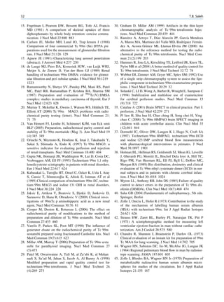
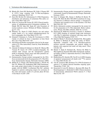

![Radiopharmaceutical Quality Control
4
Tamer B. Saleh
Contents 4.2 Physiochemical Tests
4.1 Introduction . . . . . . . . . . . . . . . . . . . . . . . . . . . . . . . . . . . . . . . . . . 55
4.2 Physiochemical Tests . . . . . . . . . . . . . . . . . . . . . . . . . . . . . . . . 55
4.2.1 Physical Tests . . . . . . . . . . . . . . . . . . . . . . . . . . . . . . . . 55 4.2.1 Physical Tests
4.2.2 pH and Ionic Strength . . . . . . . . . . . . . . . . . . . . . . . . 55
4.2.3 Radionuclidic Purity . . . . . . . . . . . . . . . . . . . . . . . . . 55
4.2.4 Radiochemical Purity . . . . . . . . . . . . . . . . . . . . . . . . 56
Physical characteristics should be observed for any
4.2.5 Precipitation . . . . . . . . . . . . . . . . . . . . . . . . . . . . . . . . . 59 radiopharmaceutical for the first and frequent use.
4.2.6 Distillation . . . . . . . . . . . . . . . . . . . . . . . . . . . . . . . . . . . 59 Color alterations should be identified for both true
4.2.7 Ion Exchange . . . . . . . . . . . . . . . . . . . . . . . . . . . . . . . . 59 solution and colloidal preparations. True solutions
4.2.8 Solvent Extraction . . . . . . . . . . . . . . . . . . . . . . . . . . . 61
4.2.9 Electrophoresis . . . . . . . . . . . . . . . . . . . . . . . . . . . . . . . 62
should also be checked for turbidity and presence of
4.2.10 Gel Filtration . . . . . . . . . . . . . . . . . . . . . . . . . . . . . . . . . 62 any particulate matter; in colloidal preparations, deter-
4.3 High-Performance Liquid Chromatography . . . . . . 62 mination of particle size is of most interest.
4.3.1 Sep Pak Analysis . . . . . . . . . . . . . . . . . . . . . . . . . . . . . 63
4.4 Biological Tests . . . . . . . . . . . . . . . . . . . . . . . . . . . . . . . . . . . . . . 63
4.4.1 Sterility . . . . . . . . . . . . . . . . . . . . . . . . . . . . . . . . . . . . . . . 63
4.4.2 Pyrogenicity . . . . . . . . . . . . . . . . . . . . . . . . . . . . . . . . . . 63
4.4.3 Toxicity . . . . . . . . . . . . . . . . . . . . . . . . . . . . . . . . . . . . . . . 63
4.2.2 pH and Ionic Strength
References . . . . . . . . . . . . . . . . . . . . . . . . . . . . . . . . . . . . . . . . . . . . . . . . . . 64
The administered radiopharmaceutical should have a
proper pH (hydrogen ion concentration) with an ideal
value of 7.4 (pH of blood), but it can vary between
2 and 9 because of the high buffer capacity of the
blood.The pH of any radiopharmaceutical preparation
can be measured by a pH meter [4].
4.1 Introduction Ionic strength, isotonicity, and osmolality should
be observed properly in any radiopharmaceutical prep-
Like all other drugs intended for human administra- aration so it is suitable for human administration.
tion, radiopharmaceuticals should undergo strict and Note: Since pH and ionic strength are important
routine quality testing procedures in addition to their factors for the stability of a radiopharmaceutical, the
own specific tests for radionuclidic and radiochemical preparation diluent is preferred to be the same solvent
purity. Radiopharmaceutical quality control tests can used in the original preparation.
be simply classified as Physiochemical and Biological
tests [1 3].
4.2.3 Radionuclidic Purity
T.B. Saleh
The term radionuclidic purity refers to the presence
Senior Medical Physicist, RSO, King Fahed Specialist Hospital,
Dammam, Kingdom of Saudi Arabia of radionuclides other than the one of interest and is
e mail: tamirbayomy@yahoo.com defined as the proportion of the total radioactivity
M.M. Khalil (ed.), Basic Sciences of Nuclear Medicine, DOI: 10.1007/978 3 540 85962 8 4, 55
# Springer Verlag Berlin Heidelberg 2011](https://image.slidesharecdn.com/basicsciencesofnuclearmedicine-121209103030-phpapp02/85/CIENCIAS-BASICAS-MN-68-320.jpg)
![56 T.B. Saleh
present as the stated radionuclide. Measurement of determine the radiochemical purity of a given radio-
radionuclide purity requires determination of the iden- pharmaceutical, and these methods are discussed next.
tity and amounts of all radionuclides that are present.
The radionuclide impurities which, vary according to
the method of radionuclide production, can affect dra-
matically the obtained image quality in addition to the 4.2.4.1 Thin-Layer Chromatography
overall patient radiation dose.
Radionuclidic impurities may belong to the same Thin-layer chromatography (TLC), which was devel-
element of the desired radionuclide or to a different oped by Hoye in 1967 is considered the most com-
element. Examples of radionuclidic impurities are monly used method for determination of radiochemical
99
Mo in 99mTc-labeled preparations and iodide isotopes purity in nuclear medicine. The main principle of a
in 131I-labeled preparations [5]. Radionuclidic purity TLC chromatography system is that a mobile phase
is determined by measuring the half-lives and charac- (solvent) migrates along a stationary phase (adsorbent)
teristic radiations emitted by each radionuclide. The by the action of the capillary forces [7]. Depending on
g-emitters can be differentiated by identification of the distribution of components between the stationary
their energies on the spectra obtained by an NaI(Tl) and the mobile phases, a radiopharmaceutical sample
crystal or a lithium-drifted germanium [Ge(Li)] detec- spotted onto an adsorbent will migrate with different
tor coupled to a multichannel analyzer (MCA). The velocities, and thus impurities are separated.
b-emitters can be tested by a b-spectrometer or a liquid In TLC, each component in a given sample is
scintillation counter. Since a given radiation energy identified by an Rf value, which is defined as “the
may belong to a number of radionuclides, half-life ratio of the distance traveled by the sample component
must be established to identify each radionuclide. to the distance the solvent front has advanced from the
original point of starting the chromatography test in
the stationary phase.”
4.2.4 Radiochemical Purity Rf ¼ Distance traveled by the component/distance
of the solvent front
Radiochemical purity is defined as the proportion of
the stated radionuclide that is present in the stated The Rf values range from 0 to1. If a component
chemical form. Image quality (as a function of migrates with the solvent front, the Rf is [1], while the
the radiopharmaceutical biological distribution) and Rf for the component remaining at the origin is [0]. Rf
the radiation absorbed dose are directly related to the values are established with known components and
radiochemical purity. Radiochemical impurities are may vary under different experimental conditions.
produced from decomposition due to the action of The main principles of separation are adsorption (elec-
solvent, change in temperature or pH, light, presence trostatic forces), partition (solubility), and ion
of oxidation or reducing agents, and radiolysis. exchange (charge), and the movement of the mobile
Free and hydrolyzed 99mTc forms in many phase may take either ascending or descending modes.
99m
Tc-labeled preparations; secondary hexamethyl- When the solvent front moves to the desired distance,
propylene amine oxime (HMPAO) complex in the strip is removed from the testing container, dried,
99m
Tc-HMPAO preparations and free 131I-iodide in and measured in an appropriate radiation detection
131
I-labeled proteins are good examples of the radio- system; histograms are obtained for the activity of all
chemical impurities [6]. sample components [8].
The stability of a compound is time dependent on
exposure to light, change in temperature, and radioly-
sis, and the longer the time of exposure is, the higher
the probability of decomposition will be. Sodium Stationary Phases
ascorbate, ascorbic acid, and sodium sulfite are nor-
mally used for maintaining the stability of a radiophar- Standard TLC materials: Standard TLC plates are
maceutical. Several analytical methods are used to available as glass plates, as plastic or aluminum foils](https://image.slidesharecdn.com/basicsciencesofnuclearmedicine-121209103030-phpapp02/85/CIENCIAS-BASICAS-MN-69-320.jpg)
![4 Radiopharmaceutical Quality Control 57
covered with the stationary phase. A wide range of Mobile Phases
stationary phases are commercially available, includ-
ing silica gel, reversed-phase silica, aluminum oxide, Saline, Water, Acetone, Methyl Ethyl Ketone (MEK),
synthetic resins, and cellulose. The main advantage of Ethanol, Acetic Acid, Chloroform, and Acetonitrile
standard TLC materials is that they have the ability to represent the most common group of mobile phases
provide relatively high- resolution tests, while the used as the mobile partner in most TLC chro-
relatively long developing time of the mobile phase matographic procedures.
(mainly 30 min) through the high-particle-size
adsorbent material (20 mm) is considered its main
disadvantage [9].
4.2.4.2 TLC Chromatography Procedure
High-performance TLC (HPTLC): HPTLC pro-
vides a good solution to the long chromatographic
To determine the radiochemical purity by ITLC or
developing time by use of materials with a smaller
paper chromatography for a radiopharmaceutical sam-
particle size.
ple, the following steps should be applied [11]:
Instant TLC (ITLC): ITLC plates are made of
fiberglass sheets integrated with an adsorbent, usually Fill a small beaker with about 10 ml (3 5 mm high)
silica gel, and can be cut to any size, developing an with the proper solvent and then cover the beaker
economic chromatographic solution. Due to the fine with a glass plate or with a foil sheet.
mesh material, the migration properties are increased Prepare the chromatography strip (5 10 cm long)
manyfold compared to the standard TLC materials, and mark the solvent front (Rf = 0) with a colored
reducing the chromatographic time to less than 5 min pen and the start point (Rf = 1) with a pencil.
without affecting the separation of radiochemical Using a 1-ml syringe with a fine needle (25 gauge),
impurities. Because of the advantages mentioned, put one small drop of the sample onto the starting
ITLC materials are the most frequently used for point on the strip.
the stationary phase in nuclear medicine since they Immediately, insert the strip into the beaker so the
fulfill the need for a rapid and accurate method spot sample does not dry,observing that initially the
for testing the radiochemical purity of a radiophar- solvent level is below the starting point.
maceutical sample. Silica stationary phases have When the solvent has reached the front, the strip
been produced for ITLC as silica gel (ITLC-SG) should be removed and dried.
and silicic acid (ITLC-SA). ITLC-SG is the most Quantify the regional distribution of radioactivity
widely used adsorbent for routine radiochemical on the strip using any of the radiation detection
purity determination [10]. methods discussed next.
Paper: Papers (e.g., Whatman no. 1 and Whatman
Figure 4.1 shows a sample procedure for determi-
3MM) were commonly used in the early days of chro-
nation of radiochemical purity of a radiopharmaceuti-
matography, although they are still used and recom-
cal by ITLC using two different solvents.
mended for many chromatographic procedures.
The main disadvantage of paper chromatography is
the poor resolution it provides for radiochemical
purity tests; however, Whatman 3MM is the material 4.2.4.3 Methods for Regional Radioactivity
of choice for partition chromatography procedures. Measurement in TLC Chromatography
Aluminum oxide: Aluminum-coated plates are
commonly used for separation of some radiopharma- Many analytical methods were developed for determi-
ceuticals (e.g., Sesta-MIBI[methoxyisobutyl isoni- nation of the radioactivity percentage of the different
trile]) depending on the aluminum oxide (Al2O3) radiochemical species along the TLC chromatography
polar properties. strip, starting with the autoradiography method by
Cellulose: Cellulose can interact with water and which a chromatogram is placed on an x-ray film and
serve as a stationary phase for separation of polar exposed in the dark for about 1 h. However, the most
substances by paper chromatography; also, it can be popular methods in nuclear medicine were achieved
used in the powder form as an adsorbent for TLC. using one of the following systems [9]:](https://image.slidesharecdn.com/basicsciencesofnuclearmedicine-121209103030-phpapp02/85/CIENCIAS-BASICAS-MN-70-320.jpg)
![58 T.B. Saleh
99m
TcO−4
TcO−4
99m 99m
Tc-exametazime
99m
Tc-2° amine complex
A
E
99m
Tc-exametazime
B 99m
Tc-2° amine complex
99m
TcO2 F 99m
TcO2
0.9% NaCl
ITLC-SG Acetonitrile:water
99m
TcO−4 (1:1)
99m
Tc-exametazime Whatman 1
C
1. % 99mTcO−4 = [A/(A+B)].100
2. % 99mTc-exametazime= [A/(A+B)]-[D/(C+D)].100
99m
D Tc-2° amine complex
3. % 99mTcO2 = [F/(E+F)].100 99m
TcO2
MEK
ITLC-SG
Fig. 4.1 Chromatographic system for determination of radiochemical purity of Tc 99m labeled HMPAO. (Adapted from [7])
Gamma camera: A dried strip is placed close to the Linear analyzer: This device operates as a position-
detector head of the camera, and an image is sensing proportional counter, measuring a fixed
obtained. The region of interest (ROI) is drawn for number of channels along the length of the chro-
each radioactive area. The radiochemical purity is matographic plate. It gives us the most sensitive
expressed as a fraction of the total recovered activity. results, but resolution is less than that obtained with
Ionization chamber: The dried strip is cut into two NaI(Tl) scintillation counters [14].
segments and measured in the chamber. This
A comparison of four different methods of quantifi-
method is frequently used for simple separation
cation is shown in Fig. 4.2, in which a phantom chro-
techniques (compounds of Rf = 0 or 1) but is not
matographic plate with increasing amount of
applicable in multisegmental strips or samples with
radioactivity (0.25, 1.0, 4.0, 16.0, and 64.0 kBq) spotted
low radioactive concentration [12].
at exact intervals was measured bythe following:
NaI(Tl) scintillation counter: Chromatographic
plates are cut in segments (up to ten) and counted A: Linear analyzer
in the scintillation counter. This method is prefer-
B: Conventional scanner
rede to the ionization chamber because it provides
more sensitive results, giving the chance to deter- C: Cut and measure scintillation counter
mine the radiochemical purity of a sample at dif- D: Ionization chamber
ferent and close Rf values.
Chromatography scanner: A slit-collimated radia- The highest sensitivity was achieved in A and C,
tion detector [mainly NaI(Tl) scintillation counter] while the best resolution occured with B and C.
is moved along the thin-layer plate, and the radio- The ionization chamber indicated a low detection
activity distribution between the start and the sol- efficiency of radioactivity below 20 kBq (peaks 1, 2, 3,
vent front points is recorded and plotted as and 4).
radioactive peaks [13]. This method is considered For 99mTc, the main impurities are the free pertech-
the most accurate one because it provides values netate (TcO4) and the reduced, hydrolyzed technetium
with high sensitivity and resolution . (TcO2) ( in another words, colloidal 99mTc).](https://image.slidesharecdn.com/basicsciencesofnuclearmedicine-121209103030-phpapp02/85/CIENCIAS-BASICAS-MN-71-320.jpg)
![4 Radiopharmaceutical Quality Control 59
Fig. 4.2 Phantom a b
0,25 1,0 4,0 16 64 [kBq] 0,25 1,0 4,0 16 64 [kBq]
chromatography plate
1 2 3 4 5 1 2 3 4 5
analyzed by four different
methods of measurement. The
highest sensitivity was
achieved with a and c, the best
resolution with b and c. Using
the ionization chamber (d),
activities less than 20 kBq
(peaks 1, 2, 3, 4) were not
resolved accurately. (From [9]
with permission from
Springer) Linear analyser with standard detector TLC-scanner with scintillation detector
c 1 2 3 4 5 d 1 2 3 4 5
Cut and count in a scintillation counter Cut and count in an ionisation chamber
Free pertechnetate migration properties and hence 4.2.6 Distillation
its Rf value are governed by the type of mobile and
stationary phases used for the TLC test, while reduced,
Distillation is a method applicable to compounds with
hydrolyzed technetium is present mainly at the origin
different vapor pressures. The two compounds can be
(Rf = 0) due to its insolubility properties (like all other
separated by simple distillation at a specific tempera-
colloidal components), explaining why reduced,
ture. The compound with higher vapor pressure is
hydrolyzed technetium species cannot be recognized
distilled off first, leaving the other compound in the
in colloidal and particulate preparations (e.g., macro-
distilling container. Iodide impurities can be separated
aggregated albumin [MAA] preparations).
from any iodine-labeled compound by distillation.
Tables 4.1 and 4.2 summarize the TLC chro-
matographic data for Tc-99m and non-Tc-99m radio-
pharmaceuticals, respectively.
4.2.7 Ion Exchange
Ion exchange is performed simply by passing a radio-
4.2.5 Precipitation pharmaceutical sample through a column of ionic
resin and eluting the column with a suitable solvent.
Precipitation is the method of separating a radio- Separation of different chemical forms is dependent
chemical form from another with an appropriate on the ability and affinity of exchange of ions from a
chemical reagent. The precipitate is separated by solution onto the resin. Polymerized and high molecu-
centrifugation. 51Cr3+ in 51Cr-sodium chromate can lar weight resins are of two kinds: cation exchange and
be estimated by precipitating chromates as lead anion exchange resins.
chromate and determining the radioactivity of the An example of that method is the determination of
supernatant. the presence of 99mTcO4– in 99mTc-labeled albumins,](https://image.slidesharecdn.com/basicsciencesofnuclearmedicine-121209103030-phpapp02/85/CIENCIAS-BASICAS-MN-72-320.jpg)
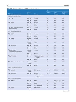
![4 Radiopharmaceutical Quality Control 61
Table 4.1 (continued)
Rf
99m 99m
Stationary TcO4 Tc Hydrolyzed
99m 99m
Tc Radiopharmaceutical Phase Solvent Complex Tc
Miscellaneous
99m
Tc MAA ITLC SG Acetone 1.0 0.0 0.0
99m
Tc HSA (human serum albumin) ITLC SG Ethanol:NH4OH:H2O 1.0 1.0 0.0
(2:1:5)
99m
Tc SC (sulfur colloid) ITLC SG Acetone 1.0 0.0 0.0
99m
Tc nanocolloid Whatman Saline 0.8 0.0 0.0
ET 31
99m
Tc architumomab ITLC SG Acetone 1.0 0.0 0.0
99m
Tc nofetumomab ITLC SG 12% TCA in H2O 1.0 0.0 0.0
Data adapted from [9, 21] and UKRG Handbook (Bev Ellis, United Kingdom Radiopharmaceutical Group, London, 2002)
Adapted from [2].
ITLC SA instant thin layer chromatography silicic acid, ITLC SG instant thin layer chromatography silica gel
Table 4.2 Chromatographic data of radiopharmaceuticals other than 99mTc complexes
Rf values
Labeled
Radiopharmaceutical Stationary phase Solvent product Impurities
125I RISA (radioiodinated serum ITLC SG 85% methanol 0.0 1.0(IÀ)
albumin)
131I hippuran ITLC SG CHCl3:acetic acid (9:1) 1.0 1.0(IÀ)
131I NP 59 ITLC SG Chloroform 1.0 1.0(IÀ)
131I MIBG Silica gel plated Ethyl acetate:ethanol (1:1) 0.0 0.6(IÀ)
plastic
131I NaI ITLC SG 85% methanol 1.0 0.2(IOÀ3)
51Cr sodium chromate ITLC SG n Butanol saturated with 1N HCl 0.9 0.2(Cr3+)
67Ga citrate ITLC SG CHCl3:acetic acid(9:1) 0.1 1.0
111In DTPA ITLC SG 10% ammonium acetate: 1.0 0.1(In3+)
(diethylenetriaminepentaacetate) methanol (1:1)
111In capromab pendetide ITLC SG Saline 0.0 1.0(In3+)
18F FDGh ITLC SG CH3CN/H2O (95:5) 0.37 0.0
90Y ,111In ibritumomab tiuxetan 0.9%NaC1 solution 0.0 1.0
Adapted from [21]
ITLC SG instant thin layer chromatography silica gel
by which the 99mTcO4– is adsorbed to the Dowex-1 4.2.8 Solvent Extraction
resin, leaving the 99mTc-labeled albumins and the
hydrolyzed 99mTc to go with the elute when using
When two immiscible solvents are shaken together,
0.9% NaCl as a solvent to wash the resin column.
any solute present will distribute between the two](https://image.slidesharecdn.com/basicsciencesofnuclearmedicine-121209103030-phpapp02/85/CIENCIAS-BASICAS-MN-74-320.jpg)
![62 T.B. Saleh
phases according to its solubility in each phase. Equi- Sequential fractions of the eluate are collected by an
librium is reached and governed by the partition coef- automated fraction collector, and the radioactivity is
ficient D: measured for each fraction.
Gel chromatography is the method of choice for
D ¼ Concentration in organic phase/Concentration separating proteins of different molecular weights. In
in aqueous phase addition, this method is equally important in detecting
impurities in 99mTc-labeled radiopharmaceuticals. To
Lipid-soluble molecules may have D ! 100, so it obtain the percentages of free, bound, and unbound
becomes highly concentrated in the organic phase. If hydrolyzed 99mTc species, a Sephadex gel-saline
the solvent is volatile (e.g., ether or chloroform), the filtration system is widely used. In this case, 99mTc-
solute can be recovered by distillation or evaporation. chelate is eluted first, followed by the free 99mTcO4–,
An example of this method is the separation of 99mTc- while the hydrolyzed 99mTc is retained by the column.
pertechnetate by dissolving 99Mo in a strongly alkaline It has been observed that some 99mTc chelates may
solution (e.g., potassium hydroxide) and then extract- bind to or dissociate on the column, producing inaccu-
ing with MEK. 99mTc-pertechnetate dissolved in the rate results.
organic phase can be recovered by evaporation to
dryness; this method has been used initially to prepare
99m
Tc-pertechnetate for 99mTc-labeled radiopharma-
ceuticals [15].
4.3 High-Performance Liquid
Chromatography
4.2.9 Electrophoresis
High-performance liquid chromatography (HPLC) is
The basic idea of electrophoresis is the property of a recent modification of the gel chromatography
charged molecules (atoms) to migrate in an electric method; the liquid phase is forced at a high pressure
field. The migration rate is dependent on the charge through an adsorbent, densely packed column. When
and size of the molecule. The apparatus used in elec- the different species of the sample are eluted from
trophoresis consists simply of a direct current power the column, they pass through a radiation detector or
supply to provide a potential difference of 400 V or any other detector, which records its presence, nor-
greater, connected through an electrolyte buffer solu- mally in a graph form. The stationary phase most
tion (barbitone/barbituric acid) to either end of a strip frequently consists of beads of silica bounding an
of support medium, which may be cellulose acetate or organic pad containing long-chain (C18) carbon
filter paper. Since 99mTc-pertechnetate is considered a groups. A wide range of stationary and mobile
small ion, it migrates rapidly and is readily separated phases has been used; by suitable choice, this tech-
from larger negatively charged complexes, such as nique is extremely valuable in the development and
99m
Tc-DTPA (diethylenetriaminepentaacetate) or evaluation of new radiopharmaceuticals. In addition,
insoluble 99mTcO2, which remain at the origin [16]. HPLC is considered a suitable technique for estima-
tion of short-lived radiopharmaceuticals because of
its rapidity [17].
If the components are irreversibly adsorbed on the
4.2.10 Gel Filtration stationary phase of the HPLC system (e.g., hydrolyzed
99m
Tc species), the system required is expensive;
Gel filtration chromatography is a process obtained therefore, the use of HPLC is not routine in hospital
when a mixture of solutes is passed down a column practice. As an example, HPLC has been used for
of suitable medium, such as cross-linked dextran separation of 99mTc-MDP (methylene diphosphonate)
Sephadex, and then eluted with a proper solvent, prepared by borohydride reduction into several differ-
allowing the large molecules to be released first ent components. A schematic diagram of the general
while the smaller ones are selectively retarded due to components of the radio-HPLC system can be seen in
penetrating the pores of the gel polymeric structure. Fig. 4.3.](https://image.slidesharecdn.com/basicsciencesofnuclearmedicine-121209103030-phpapp02/85/CIENCIAS-BASICAS-MN-75-320.jpg)
![4 Radiopharmaceutical Quality Control 63
Fig. 4.3 Components of a
radio high performance liquid
chromatographic (HPLC) Mobile
system phase Pump Injector Column Detector Recorder
reservoir
4.3.1 Sep-Pak Analysis 4.4.2 Pyrogenicity
Sep-Pak cartridges (Millipore Water) have been devel- Pyrogens are either polysaccharides or proteins pro-
oped for isolation and cleanup of sample components duced by the metabolism of microorganisms. They are
as a modification of the HPLC technology. This mainly soluble and heat stable, represented primarily
method is applicable for radiochemical analysis as bacterial endotoxins. Pyrogens produce symptoms
of 99mTc-MAG3 (mercaptoacetyltriglycine), 99mTc- of fever, chills, malaise, joint pain, sweating, head-
sestaMIBI, and radiolabeled MIBG. ache, and dilation of the pupils within 30 min to 2 h
after administration.
Pyrogenicity testing was developed from the rabbit
test (monitoring of the temperature of three healthy
4.4 Biological Tests
rabbits for 3 h after injection of the test sample) to a
more sophisticated and rapid method called the limu-
The presence of microorganisms (bacteria, fungi, etc.) lus amebocyte lysate (LAL) method. LAL, which is
should be examined for all pharmaceuticals intended isolated from the horseshoe crab (limulus), reacts with
for human administration and is defined by the sterility gram-negative bacterial endotoxins in nanogram or
test. greater concentrations, forming an opaque gel. The
Living organisms also can produce metabolic by- thicker the gel, the greater the concentration of pyro-
products (endotoxins) that can undesirably affect the gens in the sample. Gram-negative endotoxins are
radiopharmaceutical preparation, so special testing known as the most important source of pyrogen con-
procedures should be applied (pyrogenicity and toxic- tamination, so the LAL test is a rapid and sensitive
ity tests). pyrogenicity test [20].
Generally, manufacturers are required to perform
the sterility and pyrogenicity tests prior to release of
4.4.1 Sterility their products to the end users. However, the short
half-lives of 99mTc radiopharmaceuticals prohibit
their testing for sterility and pyrogenicity, emphasiz-
The objective of the sterility test is to ensure that the
ing aseptic labeling techniques by using laminar flow
sterilization processes, mainly by autoclaving for
enclosures containing high-efficiency particle air
long-lived radiopharmaceuticals and membrane filtra-
(HEPA) filters improves the environment for radio-
tion for short-lived ones, are conducted properly [18].
pharmaceutical formulation.
A proper sterility test involves the incubation of the
radiopharmaceutical sample for 14 days in
Fluid thioglycollate medium for growth of aerobic 4.4.3 Toxicity
and anaerobic bacteria
Soybean-casein digest medium for growth of fungi
Toxicity tests should be applied for all radiopharma-
and molds
ceuticals approved for human use. A quantity called
The sterility test requires 14 days, so 99mTc-labeled LD50/60 describes the toxic effect of a radiopharma-
compounds and other short-lived radiopharmaceuti- ceutical by determination of the dose required to pro-
cals could be released prior to the completion of the duce mortality of 50% of a species in 60 days after
test [19]. administration of a radiopharmaceutical dose [21].](https://image.slidesharecdn.com/basicsciencesofnuclearmedicine-121209103030-phpapp02/85/CIENCIAS-BASICAS-MN-76-320.jpg)
![PET Chemistry: An Introduction
5
Tobias L. Roß and Simon M. Ametamey
Contents even those which are toxic at higher concentrations,
5.1 Introduction . . . . . . . . . . . . . . . . . . . . . . . . . . . . . . . . . . . . . . . . . . 65 become considerable for the development of radiophar-
5.2 Choice of the Radionuclide . . . . . . . . . . . . . . . . . . . . . . . . . . 66 maceuticals and use in nuclear medicine. In contrast to
5.2.1 Labelling Methods Introduction the wide range of employable bioactive molecules, the
of the Radionuclide . . . . . . . . . . . . . . . . . . . . . . . . . . . 67
range of suitable radioactive nuclides is much more
5.2.2 Labelling Methods for Fluorine 18 . . . . . . . . . . . 70
5.2.3 Labelling Methods for Carbon 11 . . . . . . . . . . . . 83 restricted by their nuclear physical and chemical prop-
5.2.4 Fast Reactions for Oxygen 15 erties. In particular, radionuclides for diagnostic appli-
and Nitrogen 13 . . . . . . . . . . . . . . . . . . . . . . . . . . . . . . . 87 cations should provide appropriate (short) half-lives
5.2.5 Non standard Positron Emitters . . . . . . . . . . . . . . . 87
and radiation properties for detection and imaging,
5.3 Conclusions . . . . . . . . . . . . . . . . . . . . . . . . . . . . . . . . . . . . . . . . . . 90
References . . . . . . . . . . . . . . . . . . . . . . . . . . . . . . . . . . . . . . . . . . . . . . . . . . 91 but at the same time the radiation dose of patients and
personnel have to be kept to minimum. Nonetheless, to
date, a couple of radionuclides have proven suitable for
both nuclear medical diagnostic applications, single
photon emission computed tomography (SPECT), and
5.1 Introduction positron emission tomography (PET).
As indicated by their names, SPECT is based on
photon or g-ray emitting nuclides while PET is derived
One major advantage of radioactivity is its extremely from those nuclides which belong to the group of
high sensitivity of detection. Regarding the medical neutron-deficient nuclides and emit positrons (b+-
applicability of radioactivity, it permits non-invasive decay). Large scale production of positron emitting
in vivo detection of radiolabelled compounds at nano- radionuclides became possible for the first time by
to picomolar levels. The use of substances at such the invention of the cyclotron by Ernest Orlando
low concentrations usually precludes a physiological, Lawrence in 1929 [1]. Since then, many (medical)
toxic or immunologic response of the investigated cyclotrons have been built and have been in use at
biological system. Consequently, the considered physi- various nuclear medicine PET facilities. As a result,
ological process or system is examined in an unswayed short-lived positron emitters such as most commonly
situation. Furthermore, a wide range of substances, employed fluorine-18 and carbon-11 are routinely pro-
duced at most nuclear medicine centres on a daily basis.
In the b+-decay of a neutron-deficient nucleus, a
T.L. Roß (*)
positron (b+) and a neutrino (n) are synchronously
Radiopharmaceutical Chemistry, Institute of Nuclear Chemistry,
Johannes Gutenberg University Mainz, Fritz Strassmann Weg emitted, while in the nucleus, a proton is converted
2, D 55128 Mainz, Germany into a neutron. Neutrinos show practically no interac-
e mail: ross@uni mainz.de tion with matter and thus they are not detectable by
S.M. Ametamey
PET cameras. In contrast, the emitted positron is able
Animal Imaging Center PET, Center for Radiopharmaceutical
¨
Sciences of ETH, PSI and USZ, ETH Honggerberg, D CHAB to interact with an electron, its anti-particle. As a
IPW HCI H427, Wolfgang Pauli Str. 10, CH 8093, Zurich, result, both particles annihilate and give two g-rays
Switzerland with a total energy of 1.022 MeV, the sum of the
e mail: amsimon@ethz.ch
M.M. Khalil (ed.), Basic Sciences of Nuclear Medicine, DOI: 10.1007/978 3 540 85962 8 5, 65
# Springer Verlag Berlin Heidelberg 2011](https://image.slidesharecdn.com/basicsciencesofnuclearmedicine-121209103030-phpapp02/85/CIENCIAS-BASICAS-MN-78-320.jpg)
![66 T.L. Roß and S.M. Ametamey
masses of positron and electron, 511 keV each. Both g- control of PET radiopharmaceuticals as well as their
rays show a nearly 180 distribution and each carries use in drug development are introduced and briefly
the characteristic energy of 511 keV. Accordingly, the summarised. Both chapters principally cover literature
decay of positron emitters which are used as label for until the beginning of 2009.
PET radiopharmaceuticals results in two g-rays and
as these are body-penetrating photons, they can be
detected by an appropriate PET camera. This physical
phenomenon provides the base of PET imaging. 5.2 Choice of the Radionuclide
In PET scanners, a circular ring of detector pairs,
which record only coincidence events, registers the There is a variety of basic functions and effects which
in vivo generated pairs of g-rays. An appropriate com- can generally be followed and visualised by PET such as
puter-aided data acquisition provides PET images with metabolism, pharmacokinetics, (patho)physiological
information about in vivo distribution and levels of and general biochemical functions; receptor-ligand bio-
accumulation of the radionuclide and the radiophar- chemistry; enzyme functions and inhibition; immune
maceutical, respectively. Consequently, biochemical reactions and response; pharmaceutical and toxicologi-
processes can be visualised and a dynamic data acqui- cal effects. However, a close look into the designated
sition further allows for registration of a temporal processes and the related biochemistry is necessary to
component such as pharmacokinetics of a certain find a positron emitter with appropriate characteristics.
drug. In combination with bio-mathematical models Although fluorine-18 is the most commonly pre-
and individual corrections of attenuation, transmission ferred positron emitter for PET radiopharmaceuticals,
and scatter effects, physiological and pharmacological monoclonal antibodies labelled with fluorine-18 for
processes can be precisely acquired and quantified [2]. immuno-PET imaging are normally not useful because
The most important radionuclides for PET imaging the physical half-life of 110 min does not fit to the
are fluorine-18 and carbon-11. Particularly, the 18F- slow accumulation (normally 2 4 days) of most mono-
labelled glucose derivative 2-deoxy-2-[18F]fluoro-D- clonal antibodies in solid tumours [12]. In such cases,
glucose ([18F]FDG) represents the most widely used longer-lived PET nuclides as iodine-124 (T½ ¼ 4.18
PET radiopharmaceutical which has contributed most days) and zirconium-89 (T½ ¼ 3.27 days) are more
to the worldwide success of clinical PET imaging. The suitable for this particular application. On the other
combination of a highly efficient radiochemistry and hand, longer half-lives increase radiation doses to the
a high yielding 18O(p,n)18F nuclear reaction makes patients and thoughtful considerations towards a
[18F]FDG available in large amounts and also enables health/risk benefit analysis are mandatory.
shipment and distribution by commercial producers. As a basic principle, short-lived radionuclides should
Since its development in the 1970s [3], [18F]FDG has preferably be used if their suitability is similarly good
been employed in many PET studies in oncology, with respect to a certain application. Blood flow tracers
neuroscience and cardiology [4 7]. However, further are a perfect example for the use of extremely short-
substances have followed and to date, several PET lived radionuclides such as oxygen-15 (T½ ¼ 2 min),
radiopharmaceuticals for specific targets have been nitrogen-13 (T½ ¼ 10 min) and rubidium-82 (T½ ¼
developed and evaluated for a wide range of applica- 1.3 min). The scanning times of blood flow studies
tions in clinical nuclear medicine as well as in preclin- using PET are normally very short and not longer than
ical research [8 11]. 2 5 min. Hence, radiolabelled substances such as [15O]
The following chapter deals with the develop- water, [15O]butanol, [13N]ammonia and [82Rb]RBCl
ment and the use of PET radiopharmaceuticals. Here are particularly suitable. However, the relatively short
a comprehensive overview of basic considerations half-lives of the these radionuclides place some con-
and possibilities in development of PET radio- straints on imaging procedure and execution.
pharmaceuticals is given. An outline of commonly Besides half-lives, there are further physical aspects
employed clinically established PET radiopharma- to be considered. One is the b+-energy (Eb+) of the
ceuticals, their most important production routes and emitted positrons. The Eb+ also clearly affects the radia-
clinical applications follows in the next chapter, in tion dose to the patients and thus the lower the Eb+ the
which also aspects of routine production and quality better it is for the patients. Since the Eb+ is also](https://image.slidesharecdn.com/basicsciencesofnuclearmedicine-121209103030-phpapp02/85/CIENCIAS-BASICAS-MN-79-320.jpg)
![5 PET Chemistry: An Introduction 67
responsible for the positron range (travelling distance of Table 5.1 Important positron emitters used for PET and their
the positron) and a short positron range enhances the nuclear data from [16, 17]
spatial resolution in PET, a low Eb+ is also very favor- Eb+,max
Nuclide Half life Decay mode (%) [keV]
able for high resolution PET imaging. However, in
Organic
human PET scanners, the distance of the detectors to
the object is long and the positron range is no longer 11
C 20.4 min b+ (99.8) EC (0.2) 960
significant for the absolute spatial resolution as demon- 13
N 9.96 min b (100)
+
1,190
strated in comparable studies using different positron 15
O 2.03 min b+ (99.9) EC (0.1) 1,720
emitters in imaging phantoms [13, 14]. In contrast, high-
resolution small animal PET scanners show dramati-
30
P 2.5 min b+ (99.8) EC (0.2) 3,250
cally degraded image quality by the use of positron Analogue
emitters with high Eb+ or complex decay schemes [15]. 18
F 109.6 min b+ (97) EC (3) 635
In comparison with most of the available positron
emitters for PET, it is already quite evident from the
73
Se 7.1 h b (65) EC (35)
+
1,320
nuclear properties that fluorine-18 is the most pre- 75
Br 98 min b (75.5) EC (24.5)
+
1,740
ferred radionuclide for PET. The optimal half-life of 76
Br 16.2 h b (57) EC (43)
+
3,900
fluorine-18 offers multi-step radiochemistry, extended 77
Br 2.38 days b (0.7) EC (99.3)
+
343
PET studies of slower biochemistry as well as the
shipment of the 18F-labelled radiopharmaceuticals to 120
I 81.1 min b (64) EC (36)
+
4,100
clinics without an on-site cyclotron or a radiochemis- 124
I 4.18 days b (25) EC (75)
+
2,140
try facility. Furthermore, it has one of the lowest Eb+
Metallic
among the PET nuclides and provides high-resolution
PET images. An overview of the nuclear data of
38
K 7.6 min b+ (100) 2,680
important positron emitters for PET is given in 45
Ti 3.09 h b (85) EC (15)
+
1,040
Table 5.1. 60
Cu 23.7 min b (93) EC (7)
+
3,772
In the same way as the radionuclide must fulfil the
physical requirements of the PET imaging, it needs to
61
Cu 3.33 h b (61) EC (39)
+
1,215
exhibit suitable chemical properties with respect to 62
Cu 9.7 min b (98) EC (2)
+
2,930
available labelling techniques. Thereby, the labelling 64
Cu 12.7 h + À
b (18) b (37) EC (45) 655
strategy depends on the initial situation and attendant 68
Ga 68.3 min b (90) EC (10)
+
1,900
restrictions. If a certain radionuclide is given by rea-
sons such as availability or imaging characteristics, the 72
As 26 h b (88) EC (12)
+
2,515
target structure often needs to be modified towards its 82
Rb 1.3 min b (96) EC (4)
+
3,350
suitability for corresponding labelling methods. In 86
Y 14.7 h b (34) EC (66)
+
1,300
contrast, if the structure of a biomolecule is stipulated,
a combination of a radionuclide with an appropriate
89
Zr 3.27 days b+ (33) EC (77) 902
and efficient labelling procedure needs to be found. 94m
Tc 52 min b (72) EC (28)
+
2,470
However, a restricted number of PET radionuclides
and a limited selection of reactions for their intro- acceptable dosimetry and favourable pharmacological
duction into biomolecules generally necessitate the and PET imaging properties.
approach of tailored structures. Noteworthy, those
structural modifications of the parent biomolecule are
mostly accompanied by changes in the pharmacologi-
5.2.1 Labelling Methods – Introduction
cal behaviour and usually a compromise covering
pharmacological performance, radiochemistry, dosim- of the Radionuclide
etry and PET imaging requirements must be found.
In general, the choice for the right positron emitter Organic positron emitters: The introduction of the
for a new PET radiopharmaceutical can be described radionuclide into a biomolecule or a structure of
as the best match between efficient radiochemistry, (patho)physiological interest obviously is one of the](https://image.slidesharecdn.com/basicsciencesofnuclearmedicine-121209103030-phpapp02/85/CIENCIAS-BASICAS-MN-80-320.jpg)
![68 T.L. Roß and S.M. Ametamey
essential steps in the development of radiopharmaceu- pharmacologically relevant substances which are orig-
ticals. Biomolecules and pharmaceuticals mainly con- inally carrying fluorine, bromine or iodine [20, 21].
sist of carbon, hydrogen, oxygen, nitrogen, sulphur Consequently, the advantages of authentic labelling
and phosphorous due to that fact the so-called organic and longer half-lives accrue and simplify the develop-
radionuclides (see Table 5.1), carbon-11, oxygen-15, ment of a corresponding radiopharmaceutical.
ammonia-13 and phosphorous-30 allow the so-called Metallic positron emitters: In a third group, there are
authentic labelling without any changes in (bio)chem- also some metallic positron emitters which are suitable
ical and physiological behaviour of the radiolabelled for PET imaging (see Table 5.1). The half-lives vary
molecule. However, these organic radionuclides are from minutes to days and offer a broad range of
extremely short-lived isotopes with half-lives only applicability. In contrast to organic or analogue PET
from 2 to 20 min and that strongly limits their applica- nuclides, some of the metallic radionuclides are achiev-
bility. Only the half-life of 20 min of carbon-11 offers able from generator systems (e.g. 62Zn/62Cu, 68Ge/68Ga
the possibility of radiosyntheses with more than one and 82Sr/82Rb) which make them available in places
step and the detection of physiological processes with without an on-site cyclotron. Metallic PET nuclides can
slower pharmacokinetics. Besides an unchanged phar- be used either directly in their free cationic forms or as
macology, the major advantage of such short half-lives complexes. Rubidium-82 has been evaluated as a myo-
is a low radiation dose to the patients and possible cardial perfusion PET tracer [22, 23]. In form of [82Rb]
repeat studies within a short period. RbCl, it is used as radiopharmaceutical for perfusion
Analogue positron emitters: Biomolecules and phar- PET imaging on the market for almost 20 years
maceuticals are generally relatively complex organic (CardioGen-82#, approved by the FDA in 1989). The
compounds and claim for multi-step radiosyntheses for similarities of rubidium to the potassium cation lead to
their radiolabelled counterparts. In addition, many a rapid uptake of rubidium-82 into the myocardium and
(patho)physiological processes are slower and thus not allow the identification of regions of insufficient
detectable with the extremely short-lived radionuclides. perfusion by PET imaging [24, 25]. In complexes,
Alternatively, the so-called analogue radionuclides with the metallic radionuclides are usually incorporated
longer half-lives from 80 min to 4 days are commonly into biomolecules which carry suitable chelators (i.e.
introduced into biomolecules. The labelling with ana- Fig. 5.27 for the somatostatin receptor ligand [68Ga-
logue radionuclides makes use of similarities in steric DOTA, Tyr3]octreotide [26]).
demand and/or in electronic character of the substituted In addition to differences in chemical, physical and
atom or functional group. The steric demand of an atom nuclear properties of the radionuclides, the production
or a functional group refers to the amount of space routes or processes can also influence the labelling
occupied by an atom or a functional group. Accord- approach. The production route as well as the work-
ingly, selenium-73 can be used in the manner of sulphur. up provides the radionuclide in a certain chemical
Selenium as the next homologue to sulphur has very form which requires suitable (radio)chemistry in the
similar steric and chemical properties. The analogue following synthetic steps. From the production pro-
radiopharmaceuticals L-[73Se]selenomethionine [18] cess, PET radionuclides are obtained only in a nano- to
and L-homocysteine[73,75Se]selenolactone [19] are picomolar range while they are still very well detect-
examples for such a selenium-sulphur-analogy. Simi- able by their radioactive decay. As a result, the final
larly, 75,76,77Br and 120,124I can be regarded as structural PET radiopharmaceuticals are so attractive to medici-
analogues for methyl groups. nal purposes. In the body, they can be detected with
In the majority of cases, the analogue radionu- non-invasive methods while the quantity of material is
clides evoke only small insignificant structural dif- extremely small and generally toxic and pharmacolog-
ferences, but the arising electronic changes and those ical effects are negligible.
of chemical reactivity can be important. In each indi- Specific activity: Owing to the desired insignificant
vidual case, the pharmacological behaviour and prop- quantities, a fundamental criterion of the quality of a
erties of such analogue radiotracers have to be tested radionuclide and the final radiopharmaceutical is its
for changes in characteristics. In the last decades, the specific (radio)activity (SA) which depends on the
number of new pharmaceuticals has increased rapidly amount of stable isotopes (carrier) present. Carrier
and more and more compounds have been identified as can be divided into:](https://image.slidesharecdn.com/basicsciencesofnuclearmedicine-121209103030-phpapp02/85/CIENCIAS-BASICAS-MN-81-320.jpg)
![5 PET Chemistry: An Introduction 69
Isotopic carrier: isotopes of the same element as Carrier free (c.f): Ideally carrier-free systems are
the radionuclide and not achievable with PET radionuclides as they all
Non-isotopic carrier: isotopes of other elements have naturally occurring stable isotopes. For example,
mostly with very similar chemical and physical carbon is the fourth most abundant element on earth
properties to the radionuclide and it is present in almost every kind of material. Thus,
especially for carbon-11 high specific activities are an
On this account, SA is defined as the mass-related
exceptional challenge. However, in radiochemistry of
radioactivity:
PET radionuclides, traces of stable isotopes are omni-
SA ¼ A=m½Bq=gŠ present and act as isotopic carrier. Sources of isotopic
carrier are the air, target and vessel materials, transport
where A is the radioactivity in Becquerel and m is the lines and tubes, chemicals and solvents.
mass of the radioactive material including all impuri- No carrier added (n.c.a): Contaminations in che-
ties and carrier, respectively. In (radio)chemistry such micals and solvents are below normal chemical purifi-
a specification related to the mass is inconvenient and cation limits, but they are still in the quantity of the
thus SA is generally expressed on the molar basis as radionuclide. Those conditions are referred to as no-
radioactivity related to the amount of substance: carrier-added (n.c.a.) conditions and correspond to a
state of practically highest SA attainable.
SA ¼ A=n½Bq=molŠ Carrier added (c.a): On the contrary, some circum-
stances require the addition of stable isotopes what is
where m is replaced by n for the amount of substance
termed as carrier-added (c.a.). Predominantly, c.a. con-
in moles. In the absence of impurities or isotopic
ditions are employed to achieve weighable quantities
carrier, the theoretically attainable maximum molar
of a product for characterisation by non-radioactive
SA equals to:
analytical methods or to increase radiochemical yields.
SA ¼ NA (ln2/T1=2 )[Bq/mol] or As a widely used c.a. procedure the production of
electrophilic fluorine-18 is well known. The addition
SA ¼ 1:16 Â 10 =T1=2 ½Bq/molŠ
20
of the isotopic carrier fluorine-19 is necessary to mobi-
lise n.c.a. [18F]F2 which is too reactive and adheres to
where NA is Avogadro’s number (6.023 Â 1023) in
the walls of targets and tubes.
atoms/mol and T½ is the half-life of the radionuclide
Labelling reactions and radiosyntheses on the n.c.a.
in hours. The general abundance of stable isotopes of
scale mean to work at a subnanomolar level regarding
the PET radionuclides smaller the theoretically attain-
the amount of radioactive substance while all other
able SA and the quantity of material become higher
reactants and solvents are still present at a macro-
by natural isotopic carrier, but it is normally still at a
scopic scale. Hence, the course of reaction may differ
nano- to picomolar level (6.3 Â 104 versus 300
strongly from that of classical chemical reactions at
600 GBq/mmol for theoretical and practical SA, respec-
balanced stoichiometric ratios, where all substrates
tively, for fluoride-18 produced from 18O(p,n)18F).
and reagents are present in amounts in a similar
Most applications in molecular imaging call for high
or equal range. Such labelling reactions under non-
(molar) specific activities and a lot of effort is put into
equilibrium conditions generally proceed according
this issue. Especially for brain receptor PET imaging,
to pseudo-first-order kinetics where the precursor
high specific activities are essential when receptor
amounts are in extreme excess to the radionuclide
systems of low density can be saturated by radioli-
and can approximately be set as constant. On the
gands with low SA. Besides poor PET imaging results,
other hand, the radionuclide and the labelled product
because of an unfavourable signal-to-noise ratio, phar-
exist on a n.c.a. scale and thus a consecutive labelling
macological or toxic effects have also to be consid-
reaction or an interaction of two radioactive species
ered. In general, for radiochemical practice, the
can be statistically excluded.
radionuclide situations can be classified as:
In labelling procedures and radiosyntheses, obvi-
Carrier-free (c.f.) ously, the decay has to be taken into account and thus
No-carrier-added (n.c.a.) the half-life of the employed radionuclide. With res-
Carrier-added (c.a.) pect to the PET imaging, the final radiopharmaceutical](https://image.slidesharecdn.com/basicsciencesofnuclearmedicine-121209103030-phpapp02/85/CIENCIAS-BASICAS-MN-82-320.jpg)
![70 T.L. Roß and S.M. Ametamey
must be obtained in reasonable amounts sufficient for (isotopic carrier) for an isotopic exchange and removal
the following PET procedures. As a rule of thumb, the of the n.c.a. fluorine-18 out of the target. Due to this
radiosynthesis including purification, formulation and fact, the procedure dramatically lowers the obtainable
quality control of a PET radiopharmaceutical should not specific activity which is one of the major disadvan-
exceed three half-lives of the radionuclide. Conse- tages of these production routes.
quently, the extremely short-lived PET radionuclides Nonetheless, many compounds of (radio)pharma-
call for very fast chemistry and preclude multi-step cological interest call for electrophilic labelling meth-
procedures. ods and thus necessitate c.a. [18F]F2 or its derived
The efficacy of radiolabelling reactions is generally secondary labelling agents. The most popular PET
quantified by the radiochemical yield (RCY) which radiopharmaceutical which is routinely produced via
corresponds to the decay-corrected yield related to an electrophilic c.a. 18F-labelling (18F-fluorodestanny-
the starting activity. In contrast, the real yield reflects lation) is 6-[18F]fluoro-L-DOPA ([18F]F-DOPA) (see
the amount of isolated radioactive material, but is Fig. 5.3) [34, 35]. So far, an efficient nucleophilic
not functional as an appraisal factor of the labelling approach for a n.c.a. 18F-fluorination of [18F]F-DOPA
procedure. is still lacking.
However, the nucleophilic production route using
18
O-enriched water as target material is the most effi-
cient procedure and also provides the n.c.a. [18F]fluoride
5.2.2 Labelling Methods for Fluorine-18 in high specific activities. As a result, the 18O(p,n)18F
reaction is the most widely used method to produce
The indisputable importance of fluorine-18 in PET fluorine-18. The required proton energy of
makes 18F-labelled radiopharmaceuticals the most 16 ! 3 MeV for the nuclear reaction is achievable
favoured ones; thus, especially procedures for the without problems from small cyclotron, so-called
introduction of fluorine-18 are of great interest and medical cyclotrons. Normal batches of 50 100 GBq
several methods and strategies have been developed for the production of 18F-labelled clinically utilised
[27 31]. There are many established nuclear produc- PET radiopharmaceuticals can be obtained within
tion pathways for fluorine-18; the most commonly 30 60 min depending on the target construction and
used are listed in Table 5.2 [32, 33]. the corresponding beam current.
The main difference between various nuclear reac- Regarding the chemical concepts for the introduc-
tions is the target material which is either gas or liquid tion of fluorine-18 into organic molecules, the meth-
(water) and determines the final chemical form of ods of the macroscopic organic chemistry could be
fluorine-18. From gas targets, fluorine-18 is achieved principally transferred. In general chemistry, the com-
as electrophilic c.a. [18F]fluorine gas ([18F]F2) and monly used fluorination procedures are based on the
from the water targets, nucleophilic n.c.a. [18F]fluo- Wallach reaction [36] and the Balz Schiemann reac-
ride in aqueous solution is obtained. As mentioned tion [37]. However, in n.c.a. 18F-radiosyntheses, these
before, in case of the electrophilic [18F]F2, adsorption procedures led only to very low radiochemical yields
of the produced n.c.a. fluorine-18 on the walls of the [38, 39]. Effects of the unusual stoichiometric ratios
target requires the addition of non-radioactive F2 under n.c.a. conditions as well as principle aspects of
Table 5.2 Most common nuclear reactions for production of fluorine 18
18
Reaction O(p,n)18F 16
O(3He,p)18F 20
Ne(d,a)18F 18
O(p,n)18F
Target filling H218O H2O Ne (200 mmol F2) 18
O2, Kr (50 mmol F2)
Particle energy [MeV] 16 ! 3 36 ! 0 14 ! 0 16 ! 3
18 18 18
Chemical product form [ F]fluoride (aq) [ F]fluoride (aq) [ F]F2 [18F]F2
Yield [GBq/mAh] 2.22 0.26 0.37 0.44 ~0.35
Specific activity [GBq/mmol] 40 Â 10 3
40 Â 10 3
~0.04 0.40 ~0.35 2.00](https://image.slidesharecdn.com/basicsciencesofnuclearmedicine-121209103030-phpapp02/85/CIENCIAS-BASICAS-MN-83-320.jpg)
![5 PET Chemistry: An Introduction 71
the reactions’ mechanisms and reactants led to these process for electrophilic fluorine-18, although its pro-
results. Both reaction types revealed inappropriate for duction rates are lower [32, 33]. As all production
fluorine-18 chemistry under n.c.a. conditions. processes for electrophilic fluorine-18 require carrier
Generally, radiofluorination methods can be divided addition, c.a. [18F]F2 or milder reagents derived from
into electrophilic and nucleophilic reactions (substitu- it cannot be used in preparations of PET radiopharma-
tions) according to the chemical form of fluorine-18 ceuticals where high specific activities are mandatory
and thus the production route. Both methods represent [41, 42].
direct 18F-fluorinations and can be completed by two Generally, the methods of electrophilic fluorina-
additionally indirect methods, the 18F-fluorinations via tions from organic chemistry can be directly trans-
prosthetic groups and the 18F-fluorinations via built-up ferred into c.a. fluorine-18 chemistry. Due to the fact
syntheses. In general, the indirect methods are based that carrier is added here, the stoichiometric ratios are
on direct methods for the 18F-labelling of the required more balanced than under n.c.a. conditions and thus
prosthetic group or synthon. Frequently, the nucleo- closer to macroscopic chemistry. In organic chemistry,
philic 18F-methods are employed here due to higher elemental fluorine is known for its high reactivity and
specific activities, higher radiochemical yields and a its poor selectivity. Therefore c.a. [18F]F2 is often
better availability of n.c.a. [18F]fluoride. transferred into less reactive and more selective elec-
trophilic fluorination agents such as [18F]acetyl hypo-
fluoride ([18F]CH3COOF) [43], [18F]xenon difluoride
([18F]XeF2) [44, 45] or [18F]fluorosulfonamides [46].
5.2.2.1 Electrophilic Substitutions The maximum radiochemical yield in electrophilic
radiofluorinations is limited to 50% as only one fluo-
Fluorine-18 for electrophilic substitution reactions is rine in [18F]F2 is substituted by a 18F atom. Conse-
available as c.a. [18F]F2 directly from targets. 20Ne and quently, that is also the situation for all secondary
enriched [18O]O2 can be used as target materials (cf. electrophilic radiofluorination agents derived from
Table 5.2). Both alternatives come along with an c.a. [18F]F2.
adsorption of the fluorine-18 on the target walls and The most popular example of electrophilic radio-
entail an addition of [19F]F2 to mobilise the produced fluorinations using c.a. [18F]F2 is the first method to
fluorine-18 by isotopic exchange. In the 18O(p,n)18F produce 2-deoxy-2-[18F]fluoro-D-glucose ([18F]FDG)
reaction, the enriched [18O]O2 target filling is removed by Ido et al. in 1978 (see Fig. 5.1) [3]. [18F]F2 was used
after bombardment and the target is filled with 0.1% in an electrophilic addition to the double bond of
[19F]F2 in Kr and repeatedly irradiated for the [18F]F2 triacetoxyglucal and gave [18F]FDG in a radiochemi-
formation [40]. In comparison, the 20Ne(d,a)18F reac- cal yield of 8%. As a radioactive side product 3% of
tion is more practical as 0.1% [19F]F2 is directly added the 18F-labelled mannose derivative (2-deoxy-2-[18F]
with the neon and an additional step for recovery of fluoro-D-mannose, [18F]FDM) was obtained. In 1982,
the enriched material and the consecutive irradiation is a higher RCY of 20% and an improved product-to-
saved. Furthermore, the process does not require byproduct-ratio of 7:1 were achieved in the approach
enriched material and is less expensive. Therefore, of Shiue et al. using the milder radiofluorination agent
the 20Ne(d,a)18F reaction is the commonly employed [18F]acetyl hypofluoride [47]. Many other approaches
OAc OH OH
18
O 1. c.a. [18F ]F2 O
F
O
AcO HO + HO
AcO 2. Hydrolysis HO HO
18
F OH OH
[18F]FDG [18F]FDM
Fig. 5.1 Original radiosynthesis of [18F]FDG (RCY 8%) by Ido et al. using c.a. [18F]F2. As a side product, the 18
F labelled
mannose derivative ([18F]FDM) was obtained in a RCY of 3%](https://image.slidesharecdn.com/basicsciencesofnuclearmedicine-121209103030-phpapp02/85/CIENCIAS-BASICAS-MN-84-320.jpg)
![72 T.L. Roß and S.M. Ametamey
were made to increase radiochemical yields of [18F] Several attempts have been made to improve radio-
FDG in electrophilic procedures [48 50], including chemical yields and regioselectivity in the direct
also attempts with [18F]XeF2 [51 53]. radiofluorination of L-DOPA [58, 59]. So far, the
Another example for a direct electrophilic 18F- most efficient procedures for 6-[18F]F-DOPA which
fluorination is 5-[18F]fluorouracil which is the 18F- provide adequate RCY of up to 33% for clinical
labelled analogue of 5-fluorouracil. 5-Fluorouracil is PET imaging are based on 18F-fluorodemetallation
a chemotherapeutic and thus its 18F-labelled analogue reactions [60 62]. Among the 18F-demetallation reac-
can be used for therapy control, for visualisation of tions, to date, the 18F-fluorodestannylation is the
various tumours and for prediction of therapy response most commonly used reaction for routinely produced
in liver metastases [54, 55]. 5-[18F]fluorouracil can be 6-[18F]F-DOPA (see Fig. 5.3) [34, 63]. An automation
prepared by direct 18F-fluorination of uracil using c.a. of this radiosynthesis and recently improved precursor
[18F]F2 [56]. synthesis and quality control allows reliable routine
The most important PET radiopharmaceutical which productions for clinical PET imaging using 6-[18F]
is routinely produced via electrophilic 18F-fluorination fluoro-L-DOPA [35, 64].
methods is 6-[18F]fluoro-L-DOPA ([18F]F-DOPA). To date, the 18F-fluorodestannylations are generally
After [18F]FDG, [18F]F-DOPA ranks second in its the preferred methods for electrophilic 18F-labelling of
frequency of clinical use. The direct radiofluorination complex molecules as they provided satisfactory
of 3,4-dihydroxyphenyl-L-alanine using [18F]F2 leads radiochemical yields and high regioselectivity.
to three possible 18F-labelled regioisomers namely For higher specific activities in electrophilic 18F-
2-[18F]F-DOPA (12%), 5-[18F]F-DOPA (1.7%) and fluorinations, [18F]F2 can be obtained from n.c.a.
6-[18F]F-DOPA (21%) (see Fig. 5.2) and requires a [18F]CH3F via an electric gaseous discharge reaction
complex HPLC purification to obtain the desired in the presence of [19F]F2 (150 nmol) (see Fig. 5.4).
6-[18F]F-DOPA in only 3% RCY [57]. This provides specific activities of up to 55 GBq/mmol
COOH COOH COOH COOH
NH2 NH2 NH2 NH2
c.a. [18F]F2 18F 18F
+ +
-65°C, HF
18F
OH OH OH OH
OH OH OH OH
L- DOPA 2- [18F]F-DOPA 5-[18F] F-DOPA 6-[18F]F-DOPA
12% 1.7% 21%
Fig. 5.2 Direct electrophilic radiofluorination of [18F]F DOPA using c.a. [18F]F2. The product mixture contains 21% of the desired
18
F labelled regioisomer 6 [18F]F DOPA
Fig. 5.3 Electrophilic COOEt COOH
radiofluorination of 6 [18F]F
DOPA by regioselective 18F NHBoc NH2
fluorodestannylation. After Me3Sn 1. c.a. [18F]F 2 18F
45 50 min 6 [18F]F DOPA is
obtained in RCY of 26 33% 2. HBr, 130°C
OBoc OH
OBoc OH
6-[18F]Fluoro-L-DOPA
RCY = 26–30 %](https://image.slidesharecdn.com/basicsciencesofnuclearmedicine-121209103030-phpapp02/85/CIENCIAS-BASICAS-MN-85-320.jpg)
![5 PET Chemistry: An Introduction 73
in case of the [18F]F2 which leads to SA of ~15 GBq/m For saving the costly, enriched material, the
mol of final 18F-labelled products [65]. first step after the irradiation is the separation of
However, electrophilic substitution reactions using the [18F]fluoride from the [18O]water. Commonly,
[18F]F2 and secondary milder fluorination agents [18F]fluoride is trapped on an anionic exchange resin
derived from it can be used in clinically routine pro- (solid phase extraction cartridge systems) while the
duction of PET radiopharmaceuticals where low spe- [18O]water is recovered. [18F]Fluoride in aqueous
cific activities and moderate radiochemical yields are solution is strongly hydrated and inactivated for nucle-
not essential. PET imaging of receptor systems and ophilic reactions. For an activation of the [18F]fluo-
other PET imaging investigations which require high ride, generally, the water is removed by azeotropic
specific activities, still necessitate 18F-radiopharma- distillation with acetonitrile and the remaining dry
ceuticals produced under no-carrier-added conditions [18F]fluoride is available for nucleophilic substitution
and thus derive from nucleophilic substitution using reaction as an activated nucleophile.
n.c.a. [18F]fluoride. Due to the strong tendency of fluoride ions to form
hydrogen fluoride, the 18F-labelling reactions must be
carried out under dry and aprotic conditions. Hence,
nucleophilic 18F-labelling is usually performed in
5.2.2.2 Nucleophilic Substitutions dipolar aprotic organic solvents. For further activation
and increased nucleophilicity of the [18F]fluoride, it is
As mentioned earlier, the 18O(p,n)18F reaction using used in combination with weak and soft cations, those
enriched [18O]water as target material is the most of caesium or rubidium. As a result, a so-called
efficient and most widely used production route for ‘naked’ [18F]fluoride of high nucleophilicity is pro-
(nucleophilic) fluorine-18. The required proton energy duced. Similarly, phase transfer catalyst such as tetra-
of 16 MeV can be easily generated by medial cyclo- alkylammonium salts, mainly as their carbonates,
trons and so 50 100 GBq of n.c.a. fluorine-18 can be hydroxides or hydrogen carbonates, can be used. One
produced within 30 60 min. The fluorine-18 is of the most efficient and commonly applied system in
obtained directly from the target as nucleophilic n.c.a. radiofluorinations is the combination of a cryptand, the
[18F]fluoride in aqueous solution without any carrier aminopolyether Kryptofix#2.2.2, and potassium car-
addition. bonate (see Figs. 5.5 and 5.6) [66, 67]. In case of base-
n.c.a. [18F]Fluoride Electric discharge
CH3I 18
[ F]CH3F [18F]F2
F2/Ne
Fig. 5.4 Production of c.a. [18F]F2 of higher specific activities derived from electric gaseous discharge of n.c.a. [18F]fluoromethane
under carrier added conditions. [18F]F2 is obtained with specific activities of 55 GBq/mmol
18 –
F
18
H2 O H218O Anionic Elution Azeotropic Precursor
Labelling
Protons 16 Mev Target exchanger distillation
~4min 85°C
separation Kryptofix (acetonitrile) (TATM)
K2CO3
18
H2 O
Recovery
Sterile Neutralisation
[18F]FDG Hydrolysis
Formulation and
RCY ~60% (base or acid)
filter purification
Fig. 5.5 Steps of the [18F]FDG routine production. TATM 1,3,4,6 tetra O acetyl 2 O trifluoro methanesulfonyl beta D
mannopyranose](https://image.slidesharecdn.com/basicsciencesofnuclearmedicine-121209103030-phpapp02/85/CIENCIAS-BASICAS-MN-86-320.jpg)
![74 T.L. Roß and S.M. Ametamey
Fig. 5.6 Principle of
[18F]fluoride activation by O N
removal of water in H H
O O O
combination with the H H
Azeotropic distillation 18F
Kryptofix#2.2.2/potassium O 18
F O K
carbonate system H H K2.2.2/K2CO3
O O O
H H
O N
[18F]Fluoride [18F]Fluoride
hydrated “naked”
OAc OH
Fig. 5.7 Most commonly 1. n.c.a. [18F]Fluoride,
used radiosynthesis of n.c.a. TfO
O K2.2.2/K2CO3 O
[18F]FDG (RCY = 50 70%) AcO HO
by Hamacher et al. AcO OAc 2. Hydrolysis HO OH
18
F
[18F]FDG
sensitive compounds the carbonate can be exchanged mechanism and suitable leaving groups are required.
by oxalate which provides less basic conditions. In The most efficient leaving groups are sulphonic
another method, the [18F]fluoride is separated from acid esters such as the methane sulphonic acid ester
[18O]water by an electrochemical anodic adsorption (mesylate), the trifluoromethane sulphonic acid ester
[68]. For drying the cell is flushed two times with (triflate), the para-toluene sulphonic acid ester (tosy-
acetonitrile or dimethylamide. A polarity change of late) and the para-nitrobenzene sulphonic acid ester
the electrical field provides a subsequent desorption (nosylate). Further suitable leaving groups are halo-
and release of the [18F]fluoride into a dipolar aprotic gens. The most important and prominent example
solvent containing a phase transfer catalyst system of such an aliphatic nucleophilic substitution using
[69]. In recent studies, the use of ionic liquids showed n.c.a. [18F]fluoride is the synthesis of [18F]FDG using
very high 18F-labelling efficiency of up to 90% RCY an acetyl-protected mannose precursor (1,3,4,6-tetra-
without previous drying procedures [70]. Small O-acetyl-2-O-trifluoro-methanesulfonyl-beta-D-man-
volumes of aqueous 18F-solution are directly added nopyranose, TATM) carrying a triflate leaving group
to the reaction mixture containing a base, precursor which was developed by Hamacher et al. in 1986 (see
and ionic liquid. The best results were obtained from Fig. 5.7) [67]. This procedure provides [18F]FDG after
the combination of caesium carbonate and the ionic deprotection and purification in very high radiochemi-
liquid 1-butyl-3-methylimidazolium triflate ([bmim] cal yields of 50 70% with high specific activities of
[OTf]). This method was also applied for [18F]FDG ~300 500 GBq/mmol. To date, this is the most widely
productions and showed good RCY of 50 60%, but used method for the production of [18F]FDG towards
so far, it has been tested just with small amounts of preclinical and clinical applications.
[18F]fluoride of less than 1 GBq [71]. Regarding the reaction conditions for aliphatic
Generally, the most important procedures to get nucleophilic substitutions using n.c.a. [18F]fluoride,
18
F-labelled radiopharmaceuticals are based on the best results are typically obtained from acetonitrile as
nucleophilic substitution using n.c.a. [18F]fluoride solvent and the Kryptofix#2.2.2/potassium carbonate
which is so far also the only way to get 18F-radio- system. Applied reaction temperatures vary from 80 C
pharmaceuticals of high specific activities. Nucleo- to 110 C and depend on the individual precursor mol-
philic substitution reactions can be divided into ecule. Due to the low boiling point of acetonitrile of
aliphatic and aromatic substitutions. 82 84 C, temperatures higher than 110 C are not
Aliphatic substitution: In case of aliphatic nucleo- practical. Further suitable solvents are dimethylfor-
philic substitutions, the reactions follow the SN2 mamide, dimethylsulfoxide and dimethylacetamide](https://image.slidesharecdn.com/basicsciencesofnuclearmedicine-121209103030-phpapp02/85/CIENCIAS-BASICAS-MN-87-320.jpg)
![5 PET Chemistry: An Introduction 75
which also allow higher temperatures up to 160 and halogens in ortho- or para-position to the substi-
190 C. In recent studies, Kim et al. found increased tution (see Fig. 5.8).
radiochemical yields in aliphatic nucleophilic 18F- Suitable leaving groups (LG) are nitro, halogens
labelling by the use of tert-alcohols (frequently tert- and especially trimethylammonium salts as their
butanol) as co-solvents to acetonitrile. A beneficial triflate, tosylate, perchlorate or iodide [30, 31, 78].
effect was shown for a number of clinically important Generally, dimethylsulfoxide is the solvent of choice
18
F-labelled PET radiopharmaceuticals [72]. for the nucleophilic aromatic substitution, but also
Generally, the aliphatic nucleophilic substitution is dimethylamide and dimethylacetamide or solvent
high yielding and does not take much longer than 10 mixtures have been found beneficial. The nucleophilic
15 min for completion. Often, a subsequent deprotec- aromatic substitution usually requires higher energy
tion step is necessary, but can also be accomplished than its aliphatic variant, especially in case of the
within short reaction times of 5 10 min. As a result, fluoro-for-nitro exchange. Therefore, the dipolar apro-
aliphatic nucleophilic substitution is widely applied in tic solvents with higher boiling points are preferred
18
F-labelling chemistry and several routinely pro- and the use of acetonitrile is rare.
duced 18F-labelled PET radiopharmaceuticals are An example of a nucleophilic aromatic substitution
obtained from this reaction type. Besides [18F]FDG, is the direct 18F-fluorination of the butyrophenone
the most popular examples are 3-deoxy-30 -[18F]fluoro- neuroleptic N-methyl-[18F]fluorospiperone using the
L-thymidine ([18F]FLT) [73], [18F]fluoromisonidazole corresponding nitro-precursor which gave a RCY of
([18F]FMISO) [74], O-(2-[18F]fluoroethyl-L-tyrosine) ~20% (isolated product) after 70 min synthesis time
([18F]FET) [75, 76] and [18F]fluorocholine ([18F]FCH) (see Fig. 5.9) [79]. The aromatic system is activated by
[77]. the electron-withdrawing effect of the para-ketone
Aromatic substitution: The nucleophilic aromatic functionality. However, butyrophenones are base sen-
n.c.a. 18F-fluorinations require an activated aromatic sitive and the direct 18F-labelling of N-methyl-[18F]
system, an electron deficient system. Otherwise, the fluorospiperone could be realised only with the less
desired target ring is not attractive for a nucleophilic basic Kryptofix#2.2.2/potassium carbonate/oxalate
attack by n.c.a. [18F]fluoride. Such activation can be buffer system. In the same manner, [18F]haloperidol
reached by strong electron-withdrawing groups [79, 80], [18F]altanserin [81] and p-[18F]MPPF (4-
(EWG) such as nitro, cyano, carbonyl functionalities [18F]fluoro-N-[2-[4-(2-methoxyphenyl)-1-piperazinyl]
18F
LG
n.c.a. [18F]Fluoride
o-/p-EWG o-/p-EWG
Fig. 5.8 Nucleophilic
aromatic substitution using EWG = NO2, CN, COR, CHO, COOR, Br, Cl
n.c.a. [18F]fluoride LG = NO2, Alkyl3N+ (OTs-, OTf-, OCl4- or I-), Br, Cl, I
O O
N N
O O
n.c.a. [18F]Fluoride
N N N N
K2.2.2/K2CO3/K2C2O4
18F
O2N
N-methyl-[18F]fluorospiperone
Fig. 5.9 Nucleophilic aromatic 18F fluorination of n.c.a. N methyl [18F]fluorospiperone](https://image.slidesharecdn.com/basicsciencesofnuclearmedicine-121209103030-phpapp02/85/CIENCIAS-BASICAS-MN-88-320.jpg)
![76 T.L. Roß and S.M. Ametamey
ethyl]-N-2-pyridinyl-benzamide) [82, 83] have been butyrophenone neuroleptics [79, 80], [18F]altanserin
successfully labelled with n.c.a. [18F]fluoride by the [81], [18F]methylbenperidol [94], p-[18F]MPPF [82,
fluoro-for-nitro exchange. 83], [18F]flumazenil [95], 18F-labelled MAO-B inhibi-
Another possibility for nucleophilic aromatic sub- tor [93], 18F-labelled epibatidine analogues [87 89]
stitutions is given by electron-deficient heteroaromatic and further ligands for the nicotine acetylcholine
systems such as pyridines which do not need further receptor system (nAChR) [91, 92].
activating electron-withdrawing groups [84 86]. 18F- In general, radiolabelling chemistry benefits
fluoroanalogues of epibatidine have been labelled via from microwave heating which usually dramatically
a nucleophilic (hetero)aromatic substitution in the enhances reaction (labelling) kinetics and provides
ortho-position of the pyridinyl group (see Fig. 5.10) products within minutes and often with higher (radio-
and gave radiochemical yields of 55 65% using the chemical)yields [96]. However, the aromatic fluoro-
trimethylammonium triflate leaving group [87 89]. for-nitro exchange, particularly, benefits usually from
However, the 18F-labelled epibatidines revealed very microwave heating and increased radiochemical
toxic [88, 90] and further less toxic 18F-labelled yields within markedly reduced reaction times can be
ligands for the nicotine acetylcholine receptor system obtained [81, 97, 98].
have been developed, again via the nucleophilic If an aromatic system is somehow non-activated
(hetero)aromatic substitution on the ortho-position of or even deactivated (electron-rich) for nucleophilic
18
a pyridinyl group [91, 92]. In case of meta-substitu- F-fluorination, a possible strategy is the introduction
tions, the activation of the pyridine is normally not of auxiliary activating groups transferring the deacti-
efficient enough and additional activating groups are vated arene into an activated system. Such supple-
necessary to obtain sufficient 18F-incorporation [86] as mentary groups or functions need to be removed or
shown by the 18F-labelling of a MAO-B inhibitor in modified after the 18F-labelling which implies a multi-
the meta-position of the pyridinyl moiety using the step radiosynthesis. Aldehydes and ketone functions
fluoro-for-nitro exchange (see Fig. 5.11); 10% RCY are particularly suitable as activating groups as they
after 120 min total synthesis time [93]. can be removed by reductive decarbonylation [99
Using the direct nucleophilic aromatic substitution, 101]. This method has been applied for nucleo-
several 18F-labelled PET radiopharmaceuticals have philic 18F-labelling approaches towards n.c.a.
been successfully synthesized including 18F-labelled 6-[18F]FDOPA which resulted in only 3 5% RCY
18F
Boc N N(CH3)3OTf 1. n.c.a. [18F]Fluoride, H N
K2.2.2/K2CO3, DMSO
N N
2. TFA
Norchloro-[18F]fluoroepibatidine
18
Fig. 5.10 F Fluoroanalogue of epibatidine. 18F labelling via nucleophilic (hetero)aromatic substitution
NO2 18F
1. n.c.a. [18F]Fluoride,
H K2.2.2/K2CO3, DMSO H
N N
BocHN N 2. HCl (20%) H2N N
O O
N-(2-aminoethyl)-5-[18F]fluoro-
pyridine-2-carboxamide
Fig. 5.11 18F labelling of N (2 aminoethyl) 5 [18F]fluoropyridine 2 carboxamide, a MAO B inhibitor, using nucleophilic (hetero)
aromatic substitution in pyridine’s meta position](https://image.slidesharecdn.com/basicsciencesofnuclearmedicine-121209103030-phpapp02/85/CIENCIAS-BASICAS-MN-89-320.jpg)
![5 PET Chemistry: An Introduction 77
18F
X-
I+ S n.c.a. [18F]Fluoride S 18F
+
K2.2.2 /K2CO3
R R
X = Br , I , OTs, OTf [18F]fluoroarene
R = 2-OCH3, 3-OCH3, 4-OCH3 , 4-CH3, 4-OBn, H, 4-I, 4-Br, 4-Cl
18
Fig. 5.12 Nucleophilic aromatic F labelling of various arenes including electron rich systems using aryl(2 thienyl)iodonium
salts as precursors
after a three-step radiosynthesis [102] and towards n. conjugated to the target structure forming the final 18F-
c.a. 2-[18F]fluoroestradiol which could be achieved labelled PET radiopharmaceutical. Principally, both
in 10 24% RCY [103]. electrophilic and nucleophilic 18F-labelling are suitable
Another method which allows a direct nucleophilic for the 18F-introduction into prosthetic groups, but due
aromatic 18F-labelling of deactivated systems is the use to high specific activities, higher RCY and better avail-
of diaryliodonium or aryl(heteroaryl)iodonium salts ability of n.c.a. [18F]fluoride, the nucleophilic methods
(see Fig. 5.12) [104, 105]. The resulting product distri- clearly outperform the electrophilic procedures.
bution after the nucleophilic attack of the n.c.a. [18F] The prosthetic group: A variety of prosthetic groups
fluoride strongly depends on the electronic and steric have been developed so far, whereas only limited
character of each aryl ring and its substituents, respec- methods for their introduction into biomolecules are
tively. Generally, the more electron-deficient ring of available: acylation [111 122], alkylation [123 125],
the iodonium salt is preferred for the 18F-introduction. amidation [126 130], imidation [125], thiol-coupling
Thus, the use of electron-rich heteroaryl systems as one [131, 132], oxime-formation [133, 134] and photo-
iodonium moiety such as the 2-thienyl group leads to a chemical conjugation [122, 135] (see Fig. 5.13).
regioselective 18F-labelling on the counter ring [105]. Most of the procedures for preparation of prosthetic
So far, some attempts of using diaryliodonium salts as groups are multi-step radiosyntheses and with the final
precursors for complex structures towards 18F-labelled coupling step to bioactive molecules they end as 4 5
radiopharmaceuticals have been made, but the step radiosynthesis. Furthermore, the methods for
18
F-labelling of complex structures via diaryliodonium introduction of certain prosthetic groups require cer-
salts still remains a challenge [103, 106]. One success- tain functionalities in the target structure and some
ful example is the PBR ligand [18F]DAA1106 which suffer from low RCY or poor in vivo stability, but
was recently 18F-labeled in radiochemical yields of prosthetic groups are still indispensable, because of
46% from a diaryliodonium precursor [107]. the limitations of direct nucleophilic 18F-labelling.
[18F]SFB: The most commonly applied 18F-labelled
prosthetic group is N-succinimidyl-4-[18F]fluorobenzo-
18
5.2.2.3 F-Fluorinations Via Prosthetic Groups ate ([18F]SFB) which cannot be obtained in a single step
[116, 117]. Generally, [18F]SFB derives from n.c.a.
18 18
F-labelling via prosthetic groups is based on small F-labelling of the triflate salt of 4-trimethylammo-
molecules which are first 18F-labelled and then intro- nium-ethylbenzoate yielding 4-[18F]fluorobenzoic acid
duced into appropriate biomolecules [31, 108 110]. As ([18F]FBA) after basic hydrolysis; in the next step,
mentioned before, the direct nucleophilic 18F-labelling [18F]FBA is converted into activated succinimidyl esters
methods which usually provide the 18F-labelled PET using activating agents like N-hydroxysuccinimidine/
radiopharmaceutical fast and in high RCY are generally 1,3-dicyclohexalcarbodiimide (NHS/DCC) [118],
inappropriate for multifunctionalised structures such as N,N0 -disuccinimidyl carbonate (DSC) [119] or O-
peptides, oligonucleotides or antibodies. For that rea- (N-succinimidyl)-N-N,N0 ,N0 -tetramethyluronium tetra-
son, small organic molecules are labelled with fluorine- fluoroborate (TSTU) [121] to give [18F]SFB. To date,
18 using a direct method and subsequently, they are the TSTU-mediated procedure is the fastest and most](https://image.slidesharecdn.com/basicsciencesofnuclearmedicine-121209103030-phpapp02/85/CIENCIAS-BASICAS-MN-90-320.jpg)
![78 T.L. Roß and S.M. Ametamey
Prosthetic groups for acylation: O
O
O O
O
N N
O O O O OH
18 18
F F 18 O
F O 18 [18F]SFB 18
O O F F
[111] [112] [113] [117-122] [120]
Prosthetic groups for amidation: Photochemical conjugation
O NO2
18F
NH2
N NH2
n
18 H 18
F 18
F
n = 1,3 F H2N N3
[126] [128,130] [129] [135]
Prosthetic groups for...
Alkylation Oxime-formation Thiol-coupling Imidation
O
O O O NO2 NH
Br 18
F N
N O
H
18 18 18
O 18
F HS F F F
[123,132] [125] [133] [131] [123]
18
Fig. 5.13 Examples of prosthetic groups and their application in n.c.a. F labelling of biomolecules. References are given in
brackets
O O Peptide
N
O O 1. [18F]fluoride O OH O O O NH
K2.2.2/K2CO3 NH2
DMSO TSTU Peptide
2. Hydrolysis
(OH-)
18 18
N+(CH3)3 18
F F F
-OTf
18 F-labelled
[18F]FBA [18F]SFB peptide
Fig. 5.14 Principle of prosthetic group 18F labelling of biomolecules using n.c.a. [18F]SFB (TSTU mediated). TSTU: O (N
succinimidyl) N N,N0 ,N0 tetramethyluronium tetrafluoroborate
convenient method to produce [18F]SFB (see Fig. 5.14) chemistry’ [136] has been applied to fluorine-18
[121]. [18F]SFB can then be coupled to an amino func- chemistry [137 139]. Very mild reaction conditions
tion of the target structure. accompanied by high efficiency, high selectivity and
Recently, the Cu(I)-catalysed 1,3-dipolar cycload- excellent yields make this click reaction particularly
dition between alkynes and azides which is the suitable for biological applications as well as for the
most prominent representative of the so-called ‘click synthesis of PET radiopharmaceuticals.](https://image.slidesharecdn.com/basicsciencesofnuclearmedicine-121209103030-phpapp02/85/CIENCIAS-BASICAS-MN-91-320.jpg)
![5 PET Chemistry: An Introduction 79
OH
O O RRPYIL
OH
O NH RRPYIL CuSO4 NH
N Na ascorbate O
H
+
borax buffer sol.
40°C, 20min N N
H
N N 18F
N3
18F-labelled
18
F neurotensin(8-13)
Fig. 5.15 N.c.a. 18F labelling of neurotensin(8 13) using click chemistry
18
As an example, the hexapeptide neurotensin(8 13) F-labelling is based on organoboron and organosili-
was successfully n.c.a. 18F-labelled using the click reac- con bioconjugates which can be labelled with n.c.a.
tion of the 18F-alkyne n.c.a. 4-[18F]fluoro-N-(prop-2- [18F]fluoride in one step under aqueous conditions
ynyl)benzamide and the azide-functionalised with high RCY [142 144]. In a similar approach,
N3(CH2)4CO-neurotensin(8 13) (see Fig. 5.15) [140]. organosilicon building blocks were introduced into a
Under very mild conditions of only 40 C reaction tem- peptide structure and facilitated direct nucleophilic
perature and in borax buffer solution, radiochemical n.c.a. 18F-labelling of peptides in one step under very
yields of 66% were achieved within 20 min. mild aqueous and even slightly acidic conditions with-
In each individual case, the choice of the prosthetic out the need for protection group chemistry (see
group, and therewith the method of conjugation, Fig. 5.16) [145]. Depending on the type of precursor,
depends on the chemical and pharmacological proper- either 45% RCY or 53% RCY is achieved after 15 min
18
ties of the target structure. Furthermore, the in vivo F-labelling of the silane precursor or the silanol
stability of the prosthetic group and the influence precursor, respectively.
on the pharmacological behaviour of the 18F-labelled
compound has to be considered. In terms of the most
important requirements for prosthetic group 18F-label-
ling, to date, the [18F]SFB group seems to be the most 5.2.2.5 18
F-Labelled Synthons for Built-Up
suitable prosthetic group. However, the wide scope Radiosyntheses
and the very mild conditions of the 18F-click cycload-
dition have added a new and wide flexibility to the The growing number of complex and multifunctional
18
F-labelling prosthetic groups. pharmaceuticals poses a particular challenge to radi-
olabelling methods. Frequently, the target structure is
not suitable for direct 18F-labelling and only an indi-
5.2.2.4 Direct 18F-Labelling of Multifunctional rect 18F-labelling method can be applied. Besides the
Molecules prosthetic group 18F-labelling, the 18F-labelling via
built-up radiosynthesis offers another indirect alterna-
As mentioned above, the method of choice to intro- tive [27 31, 86, 146]. Both methods are very similar as
duce the 18F-label into structures like peptides is the they are based on 18F-labelled small organic molecules
use of small 18F-labelled prosthetic groups which and indeed the lines between them are often blurred.
are coupled to the biomolecule (see previous para- Generally, the 18F-labelling via built-up radiosynth-
graph). Recently, the first successful approaches of eses using synthons are used in the direction of small
direct nucleophilic 18F-labelling were reported. Pep- monomeric radiotracers while the 18F-labelled pros-
tides can be selectively functionalised with a highly thetic groups are mostly applied towards 18F-labelling
activated aromatic system bearing a trimethylammo- of macromolecular structures such as peptides or anti-
nium leaving group which enables a direct one-step body fragments. Obviously, the indirect 18F-labelling
nucleophilic aromatic n.c.a. 18F-labelling under very methods imply multi-step radiosyntheses of minimum
mild conditions [141]. Another new strategy of direct two steps.](https://image.slidesharecdn.com/basicsciencesofnuclearmedicine-121209103030-phpapp02/85/CIENCIAS-BASICAS-MN-92-320.jpg)
![80 T.L. Roß and S.M. Ametamey
18F
X
R R
Si Si
R R
O n.c.a. [18F]Fluoride, O
K2.2.2/K2CO3
HN DMSO (1% AcOH) HN
NH NH
O O
NH O NH O
O O
X = OH, R = iso-propyl HN HN
X = H, R = tert-butyl NH2 NH2
O O
[18F ]fluorosilanotetrapeptide
Fig. 5.16 Direct nucleophilic n.c.a. 18F labelling of a silicon tetrapeptide
O N
-OTf NH
18F HN
N+(CH3)3 O N
O
CHO CHO N
n-Bu4N18F NaBH3CN
18F
DMSO CH3OH, CH3COOH
130 °C, 15 min HN
O
2-[18F]fluoro-CP-118,954
Fig. 5.17 Reductive amination with 2 [18F]fluorobenzaldehyde forming the AChE inhibitor 2 [18F]fluoro CP 118,954
The Synthons: The built-up radiosynthesis approach transferred into other functionalities. Thus, [18F]fluoro-
is based on small activated organic molecules which benzaldehydes can be reduced to their [18F]fluoro-
are subsequent to the 18F-fluorination used for a benzamines or amides and subsequently used in
built-up synthesis of the final target compound. Such amination reactions towards N-[18F]fluorobenzylamines
18
F-labelled synthons are generally derivatives of [147 152]. Recently, the AChE inhibitor 5,7-Dihydro-
[18F]fluorobenzene or similar 18F-labelled aryls. 3-[2-[1-(2-[18F]fluorobenzyl)-4-piperidinyl]ethyl]-6H-
Regarding the 18F-introduction they usually bear a pyrrolo[3,2,f]-1,2-benzisoxazol-6-one (2-[18F]fluoro-CP-
leaving group and an activating group. In addition, 118,954) has been labelled with fluorine-18 via reduc-
they need to be functionalised towards further coupling tive amination using 2-[18F]fluorobenzaldehyde (see
or built-up reactions. Either the activation group is Fig. 5.17) [152].
modified or the synthons bear additional substituents Additional useful derivatives from [18F]fluoroben-
which provide further derivatisation and allow coupling zaldehydes are the [18F]fluorobenzyl halides which
reactions. Frequently, the activation group is modified can be used as alkylation agents for amino [153
for following coupling or built-up reaction steps. 155], hydroxyl [156] or thiol [156] functions. 2-[18F]
[18F]Fluorobenzaldehydes give several possibilities fluoro-4,5-dimethoxybenzaldehyde was prepared from
for built-up syntheses and represent the most versatile its trimethylammonium triflate precursor and used as
class of synthons. The aldehyde moiety can be easily synthon in a five-step enantioselective radiosynthesis](https://image.slidesharecdn.com/basicsciencesofnuclearmedicine-121209103030-phpapp02/85/CIENCIAS-BASICAS-MN-93-320.jpg)
![5 PET Chemistry: An Introduction 81
Br- O
N+
N
-
OTf
18F 18F
N+(CH3)3
CHO CH2X COOH
i, ii, iii 1. (Ph)2CNCH2COOtBu
2. HI , HPLC NH2
MeO MeO HO
OMe OMe OH
6-[18F]fluoro-L-DOPA
i = [18F]fluoride, K2.2.2/K2CO3,DMSO
ii = NaBH4 (aq)
iii = HX (g) (X = Br, I)
Fig. 5.18 2 [18F]fluoro 4,5 dimethoxybenzyl halides as synthons for n.c.a. radiosynthesis of 6 [18F]fluoro L DOPA
18F 18F 18F
OH OH
CHO NO2 NH2
C2H5NO2 HCOONH4
NaOH Pd/C
OBn OBn OH
6-[18F]fluorometaraminol
Fig. 5.19 N.c.a. radiosynthesis of 6 [18F]fluorometaraminol via nucleophilic addition of nitroethane to 3 benzyloxy 6 [18F]
fluorobenzaldehyde
of n.c.a. 6-[18F]fluoro-L-DOPA (see Fig. 5.18) [157, [18F]Fluorobenzaldehydes have also been applied in
158]. After reduction of the aldehyde group with nucleophilic additions [162, 163]. In this way, the nucle-
sodium borhydride to the benzylalkohol function, the ophilic addition of nitroethane to n.c.a 3-benzyloxy-6-
treatment with the corresponding hydrogen halide [18F]fluorobenzaldehyde and following reductive
leads to the 2-[18F]fluoro-4,5-dimethoxybenzyl halide. deprotection led to n.c.a. 6-[18F]fluorometaraminol in
N.c.a. 6-[18F]fluoro-L-DOPA was achieved from an a diastereomeric mixture from which the stereoisomers
enantioselective coupling with N-(diphenylmethylene) could be separately isolated by two subsequent semi-
glycine tert-butyl ester, deprotection and semi-preparative preparative HPLC purifications (see Fig. 5.19) [164].
HPLC in RCY of 25 30% with an enantiomeric In the same manner, also the n.c.a 4-[18F]fluorometar-
excess of 95%. aminol was synthesised.
Besides the conversion reactions of the aldehyde Similar to the carbonyl chemistry of [18F]fluoroben-
group, [18F]fluorobenzaldehydes can also function as zaldehydes, [18F]fluoroacetophenones offer a broad
direct reaction partner according to organic carbonyl range of synthetic possibilities [164, 165]. Moreover,
chemistry. Prominent representatives of such chemistry secondary derived synthon/prosthetic group 4-[18F]
which have also been applied to 18F-radiochemistry are fluorophenacylbromide can be conjugated to peptides
the Wittig reaction [159], the Horner Wadsworth and proteins via alkylation reaction or thiol-coupling
Emmons reaction [160] and the Knoevenagel conden- reactions [125, 134].
sation [161]. Another group of versatile synthons derive from
In addition, the electrophilic character of aldehydes the [18F]fluoro-4-haloarenes which can be used in
also offers the possibility of nucleophilic additions. palladium(0)-catalysed C C-bond formation reactions](https://image.slidesharecdn.com/basicsciencesofnuclearmedicine-121209103030-phpapp02/85/CIENCIAS-BASICAS-MN-94-320.jpg)
![82 T.L. Roß and S.M. Ametamey
18F
[18F]fluvastatin
[167]
ONa
18F
N
[18F]RP62203 [173] OH OH O
(5-HT2Aligand) HO
O N
N
O S N 18F
Stille
reaction 18F-mestranol
MeO [167]
Hartwig–Buchwald (N-arylation) Sonogashira reaction
18F
X
Suzuki cross coupling Li or Mg [157]
[167] X = Br, I
18F 18F 18F
or
18F-biphenyls
R BrMg Li
For organometallic 18F-fluoroarylation
Fig. 5.20 N.c.a. 4 [18F]fluorohalobenzenes as versatile synthons for palladium(0) catalysed coupling reactions and their transfor
mation into metalorganic reagents for 18F fluoroarylation reaction
such as the Stille reaction [166 170], the Sonogashira In addition to the most widely used 18F-labelling
reaction [171] and Suzuki cross-coupling reactions synthons [18F]fluorobenzaldehydes, [18F]fluorobenzyl
[172] (see Fig. 5.20). Furthermore, 4-bromo and 4- halides and [18F]fluorohalobenzenes, further primary
iodo-[18F]fluorobenzenes have been used in palla- and secondary 18F-aryls have been developed and
dium-mediated N-arylation reactions, also referred to proven to be useful for 18F-labelling via built-up radio-
as Hartwig-Buchwald reactions [173, 174]. In addi- synthesis. Accordingly, n.c.a. 4-cyano-1-[18F]fluoro-
tion, n.c.a. [18F]fluoro-4-haloarenes can also be easily benzene or 4-[18F]fluorobenzonitrile was employed
transferred into reactive species such as Grignard for built-up radiosyntheses of several 18F-butyrophe-
reagents or into 4-[18F]fluorophenyl lithium which none neuroleptics [180]. On the other hand, it can
can be employed in various metalorganic coupling also be transferred into the secondary 18F-labelling
reactions [175]. synthon n.c.a. 4-[18F]fluorobenzyl amine which can
Due to their broad applicability, [18F]fluorohalo- be used as prosthetic group [130, 132] or further con-
benzenes and their secondary derived 18F-labelling verted into N-4-[18F]fluorobenzyl-a-bromoacetamide
synthons have become more and more attractive. In as prosthetic group for the 18F-labelling of oligonu-
the past decade, several methods for an efficient prep- cleotides [129]. More in a sense of a prosthetic group
aration of [18F]fluorohaloarenes have been developed n.c.a. 4-[18F]fluorobenzyl amine was recently used
and make this class of 18F-labelled synthons readily for the 18F-labelling of the first 18F-labelled folic
available [105, 166, 176 179]. acid derivatives [132].](https://image.slidesharecdn.com/basicsciencesofnuclearmedicine-121209103030-phpapp02/85/CIENCIAS-BASICAS-MN-95-320.jpg)
![5 PET Chemistry: An Introduction 83
N.c.a. [18F]fluoronitrobenzenes, which is available of them still do not provide the desirable broad appli-
from high-yielding 18F-labelling of the appropriate cability and call for very special conditions. Thus,
dinitrobenzene precursors, can be easily reduced to there is still room for improvement and new develop-
the corresponding [18F]fluoroanilines by the use of ment of 18F-labelling methods. However, many 18F-
common reducing agents such as NaBH4, SnCl2, labelled PET radiopharmaceuticals from various clas-
N2H2/Pd, H2/Pd-C, BH3 or LiAlH4 [181]. N.c.a. ses of compounds have been prepared and some are
[18F]fluoroanilines have been employed for the 18F- routinely produced and employed in nuclear medicine
labelling of several anilinoquinazolines as epidermal practice.
growth factor receptor (EGFR) ligands [183 185] as
well as for fluorophenylureas [183]. A subsequent
treatment of the 4-[18F]fluoroaniline with nitrites
leads to the 4-[18F]fluorophenyldiazonium derivative 5.2.3 Labelling Methods for Carbon-11
which was used for the preparation of 18F-labelled 5-
HT2 receptor ligands [182]. Besides fluorine-18, carbon-11 is the most commonly
Since various biologically active compounds bear a used positron emitter for PET radiopharmaceuticals.
4-fluorophenoxy moiety [186], the secondary synthon Although the short half-life of only 20.4 min of car-
n.c.a. 4-[18F]fluorophenol is of great interest. The first bon-11 does not allow time-consuming radiosyntheses
radiosynthesis of this versatile synthon was based on a or the shipment of produced 11C-labelled radiophar-
hydrolysis of the 4-[18F]fluorophenyldiazonium salt maceuticals, several important 11C-radiopharmaceuti-
[187]. In recent years, new synthetic strategies towards cals are routinely employed in the clinics.
4-[18F]fluorophenol and several improvements of the Similar to the requirements for fluorine-18 produc-
radiosyntheses have made 4-[18F]fluorophenol readily tions, the production of carbon-11 can be facilitated
available for built-up radiosyntheses [188, 189]. Thus, with small medical cyclotrons using protons in an
it was applied for the radiosynthesis of a highly selec- energy range of 15 ! 7 MeV. The 14N(p,a)11C
tive dopamine D4 receptor ligand [190] as well as in a nuclear reaction is applied as the general production
catalysed variant of the Ullmann ether coupling to method [191]. The reaction is carried out with 14N-gas
provide 2-(4-[18F]fluorophenoxy)-benzylamines (see targets. Small portions of oxygen ( 2%) added to the
Fig. 5.21) [190]. target gas cause [11C]CO2 formation and in case of
Finding the right 18F-labelling strategies for new hydrogen (5 10%) addition, [11C]CH4 is the final
radiopharmaceuticals is generally limited by the target product form [192, 193].
structures themselves. Although a variety of Several further production routes are known for
18
F-fluorination methods have been developed, many carbon-11, but generally, they are of much less
18F 18F
O
NH2
Br Br
18F
H
O Br O N
O
Ullmann
coupling R R
OH O N O N
O N BH3-THF
R
Br O O
18F 18F
Fig. 5.21 N.c.a. 4 [18F]fluorophenol as versatile synthon in built up radiosyntheses](https://image.slidesharecdn.com/basicsciencesofnuclearmedicine-121209103030-phpapp02/85/CIENCIAS-BASICAS-MN-96-320.jpg)
![84 T.L. Roß and S.M. Ametamey
Fig. 5.22 Secondary and [11C]CH2N2
further 11C labelling synthons [11C]CH3I
derived from the primary
[11C]CO2
[11C]CHCl3
[11C]CH3Li [11C]CH4 [11C]CCl4 [11C]COCl2
[11 C]HCN
[11C]CH3NO2 [11C]CH3I [11C]CH3OH [11C]CO
[11C]CO2
11
[ C]CH2O
[11C]CH3IOTf
R[11C]CH2O R[11C]COCl
R[11C]CO2H
Fig. 5.23 Radiosynthesis of LiAlH4 Iodination
[11C]CH3I according to the [11C]CO2 [11C]CH3OH [11C]CH3I
solvent
‘wet’ method starting from
primary [11C]CO2
importance than the 14N(p,a)11C reaction [16, 32, 194, ‘Wet method’: The first efficient radiosynthesis for
195]. Furthermore, using the 14N(p,a)11C nuclear [11C]CH3I was developed by Comar et al. in 1973
reaction, carbon-11 can be obtained in high radio- [201, 202]. This so-called ‘wet’ method is based on
chemical yields with high specific activities. reduction of [11C]CO2 to [11C]CH3OH by means
11
C precursors: Regarding the two product forms of lithium aluminium hydride (LiAlH4) in solvents
and thus the two primary 11C-labelling synthons such as ethyleneglycol dimethylether, tetrahydrofuran
[11C]CH4 and [11C]CO2, the latter is the most pre- or diethylether. [11C]CH3OH is then iodinated using
ferred labelling precursor. [11C]carbon dioxide offers hydroiodic acid or triphenylphosphite ethyliodide (see
the possibility of direct 11C-introductions into organic Fig. 5.23). Diphosphorous tetraiodide [203] and tri-
molecules. Accordingly, [11C]CO2 reacts with pri- phenylphosphine diiodide [204] can also be employed
mary amino functions to form [11C]ureas and as iodination agents. Although the ‘wet’ method pro-
[11C]isocyanates [196]. Another direct 11C-labelling vides reliable and high radiochemical yields, it has one
possibility is given by the reaction with organometal- major drawback: the use of LiAlH4. LiAlH4 is a source
lic systems. Thus, the treatment of the Grignard of non-radioactive carbon dioxide which in turn brings
reagents CH3MgBr or CH3MGCl with [11C]CO2 in isotopic carrier carbon-12 and thus dramatically
gives [1-11C]acetate which is the most important reduces the specific radioactivity of the [11C]CH3I
11
C-labelled radiopharmaceutical derived from direct and the following products.
11
C-carboxylation [197, 198]. Dry method: More recently, a new approach to
Even though the half-life of 20.4 min of carbon- [11C]CH3I, the so-called ‘gas phase’ or ‘dry’ method
11 allows only reactions and conversions with fast was developed [205, 206]. Starting from [11C]CO2,
kinetics, most 11C-labelling methods are based on hydrogen reduction in presence of a nickel catalyst
secondary 11C-labelling synthons derived from provides [11C]CH4 which is passed through a heated
[11C]CO2 (see Fig. 5.22) [199]. Along with all the glass tube (~720 C) with iodine vapour for iodination
possible pathways, the ones using [11C]CH3I are the (see Fig. 5.24). The product [11C]CH3I is trapped on a
preferred routes for 11C-labelling. However, [11C] Porapak column and after completion of the iodin-
HCN and [11C]CO are also important 11C-labelling ation, [11C]CH3I is released by heating and a stream
synthons. Especially, [11C]CO has been proven for its of helium. The iodination process can be performed in
applicability in palladium- or selenium-catalysed a single pass reaction where the [11C]CH4 slowly
reactions [200]. passes the heated glass tube for iodination only once](https://image.slidesharecdn.com/basicsciencesofnuclearmedicine-121209103030-phpapp02/85/CIENCIAS-BASICAS-MN-97-320.jpg)
![5 PET Chemistry: An Introduction 85
Fig. 5.24 Radiosynthesis of H2 Iodination
[11C]CH3I according to the [11C]CO2 [11C]CH4 [11C]CH3I
Ni-catalyst I2,~720°C
‘dry’ method starting from
primary [11C]CO2
O O
N N
O O
N N
[N-methyl-11C]flumazenil
NH N
F F 11CH
O [11C]CH3I or O 3
[11C]CH3OTf
11CH
Cl Cl 3
Base
OH O
H H [O-methyl-11C]raclopride
N N
Cl N Cl N
OH O OH O
COOH COOH
H311C [S-methyl-11C]methionine
HS NH2 S NH2
Fig. 5.25 N , O and S heteroatom 11C methylation reactions based on [11C]CH3I and/or [11C]CH3OTf
[207] or in a circulation process where the [11C]CH4 is can be converted to the more reactive [11C]CH3OTf
circularly pumped through the iodination system until by means of silver triflate at elevated temperatures.
complete iodination [208]. The 11C-methylation with [11C]CH3OTf generally
Alternatively, the [11C]CH4 can be produced in situ offers higher RCY in reduced reaction times and at
in the target and used directly for the iodination pro- lower temperatures in comparison to the [11C]CH3I
cess. This variant saves one reaction step and methylation as it has already been demonstrated for
thus time. Furthermore, the in situ production of several important 11C-labelled PET radiopharmaceu-
[11C]CH4 in the target generally provides higher spe- ticals [212 215].
cific radioactivity. To date, the highest reported spe- Generally, 11C-labelling via methylation is per-
cific radioactivity of [11C]CH3I was 4,700 GBq/mmol formed as N-, O- or S-heteroatom 11C-methylation
and was obtained from iodination of in situ produced using the desmethyl precursors. Accordingly, the
[11C]CH4 in a single pass reaction [209, 210]. Due to routinely used 11C-labelled PET radiopharmaceuticals
that fact, an easier automation of the process and more [N-methyl-11C]flumazenil [210, 216, 217], [O-
convenient ongoing maintenance of the synthesis sys- methyl-11C]raclopride [218, 219] or L-[S-methyl-11C]
tem, the ‘dry’ method almost superseded the ‘wet’ methionine [203, 220] are prepared via N-, O- or
alternative for [11C]CH3I productions. Particularly, S-11C-methylation, respectively (see Fig. 5.25).
when high specific radioactivity is required as for Heteroatom 11C-methylation reactions are usually
PET studies of receptor systems in the CNS, carried out in solvents such as dimethylformamide,
the ‘dry’ process is the method of choice for the dimethylsulfoxide or acetonitrile. The 11C-methylation
[11C]CH3I production. agents is directly transferred into the solution which
In some cases, [11C]CH3I is not reactive enough for contains the desmethyl precursor and mostly a base
sufficient 11C-methylation and a more reactive 11C- such as sodium hydroxide, sodium hydride, potassium
methylation agent is needed [211]. Hence, [11C]CH3I carbonate or tetrabutylammonium hydroxide. 11C-](https://image.slidesharecdn.com/basicsciencesofnuclearmedicine-121209103030-phpapp02/85/CIENCIAS-BASICAS-MN-98-320.jpg)
![86 T.L. Roß and S.M. Ametamey
Methylations are normally completed within 10 min Another technical advancement which has recently
under elevated temperatures. entered the PET radiochemistry field is the microflui-
Solid phase: Over the years, the basic reaction dic radiosyntheses systems. The systems are based on
conditions have not been changed so much, but several continuous-flow microreactors and use only micro- or
interesting and innovative technical improvements nanolitre volumes. Some systems have been devel-
have been developed. As a consequence, most of the oped so far (see Sect. 5.2) and have already been
radiosyntheses of the routinely employed 11C-labelled successfully applied for 11C-labelling of several car-
PET radiopharmaceuticals can be performed on solid- boxylic acid esters [230].
11
phase. As resin or solid phase material, commercially C C bond reactions: [11C]CH3I can also be
available C-18 solid-phase-extraction (SPE) car- applied in 11C C bond formation reactions. Due to the
tridges can be applied. The cartridges are loaded short half-life, the most limiting factor is the reaction/
with precursor, base and small amounts of solvent synthesis time of such 11C C bond formations. None-
and the 11C-methylation agent is passed through the theless, there are several examples of C C bond forma-
cartridge by a gentle stream of nitrogen or helium. tions applied in 11C-labelling using [11C]CH3I. Some
The reactions are normally efficient at ambient tem- examples can be found for the use of [11C]CH3I in
perature and completed after short reaction times. Wittig reactions as its corresponding triphenylpho-
The 11C-labelled product is eluted from the cartridge sphorane [11C]CH2PPh3 [231] or triphenylarsonium
with an appropriate solvent and often it is directly [11C]CH2ArPh3 [232]. Most examples of various 11C
eluted into a loop of the HPLC system for the C bond formation reactions can be found for 11C-
subsequent purification. For example, the 5-HT1A labelled amino acids using methods like enzymatic
antagonist [11C]WAY 100635 have been prepared 11
C C bond formations [233, 234] or enantioselective
11
and isolated within 25 min synthesis time in good C C bond formations based on Schiff-base Ni-
yields of ~40% (related to [11C]CH3I, not decay cor- complexes as chiral auxiliaries [235, 236]. Further-
rected) [221]. Several important 11C-labelled PET more, such multi-step radiosyntheses of 11C C bond
radiopharmaceuticals have also shown applicability formations towards 11C-labelled amino acids have
for solid-phase-supported radiosynthesis [222 224]. been shown to be transferable into automated synthesis
Loop method: A further development of the solid- systems [237]. However, besides amino acids, also sev-
phase supported radiosyntheses is the so-called loop eral other pharmacologically relevant substances have
method. A conventional HPLC loop is coated with been 11C-labelled by C C bond formations [238 243].
a film of the precursor solution and the 11C-methyla- Other approaches for 11C C bond formations are
tion agent is passed through by a gentle stream of palladium-supported cross-coupling reactions which
nitrogen or helium. Subsequently, the loop content have been developed for various 11C-labelled radio-
is washed out and simultaneously injected into pharmaceuticals. The most prominent representatives
the HPLC system. The method saves reaction time of these reaction type are the Stille reaction [244 247],
and reduces the technical assembly to a bare mini- the Suzuki cross coupling reaction [245, 247, 248] and
mum. A variety of 11C-labelled radiopharmaceuticals the Sonogashira reaction [248, 249]. The Stille reac-
can be prepared by this convenient and fast method tion is the most intensively employed variant of palla-
[225 229]. dium-catalysed 11C C bond formations (see Fig. 5.26)
11CH
SnBu3 3
1. [11C]CH3I,
Pd2dba3,
O2N O2N
P(o-CH3C6H5)3, DMF
N N 2. TFA N N
NBoc NH
[11C]MNQP
Fig. 5.26 Radiosynthesis of the serotonin transporter ligand 5 [11C]methyl 6 nitroquipazine ([11C]MNQP) using [11C]CH3I in a
palladium catalysed Stille reaction](https://image.slidesharecdn.com/basicsciencesofnuclearmedicine-121209103030-phpapp02/85/CIENCIAS-BASICAS-MN-99-320.jpg)
![5 PET Chemistry: An Introduction 87
and has proven its applicability for many 11C-labelled obtained in the form of 13N-labelled nitrites
PET radiopharmaceuticals [245 248, 250 254]. and nitrates, which are subsequently reduced to [13N]
Most 11C-labelling procedures are clearly based on ammonia by titanium(III)chloride or Devarda’s alloy
[ C]CH3I as the most versatile 11C-labelling synthon
11
in alkaline medium [260]. Another method uses addi-
or precursor. The most convenient methods are the tional ethanol in the target gas as radical scavenger to
very fast N-, O- and S-heteroatom 11C-methylation avoid nitrite and nitrate formation [261]. This method
reactions which can be accomplished even with simple leads directly to [13N]ammonia.
technical equipment such as a conventional HPLC [13N]NH3 is a perfusion tracer and the most com-
loop in case of a [11C]CH3I loop reaction. Further- monly used 13N-labelled PET radiopharmaceutical in
more, also multi-step radiosyntheses like 11C C bond PET imaging. In addition to the direct clinical applica-
formations have been proven as useful 11C-labelling tions of [13N]NH3, there are a few examples of
strategy. Particularly, the palladium-promoted 11C C 13
N-labelled compounds which derive from [13N]NH3,
bond formations have broadened the applicability of but generally without clinical relevance. The L-[13N]
11
C-labelling towards PET radiopharmaceuticals. amino acids L-[13N]LEU, L-[13N]VAL and L-[13N]
GLU were 13N-labelled via an enzyme-supported
amino acid synthesis and used for investigations of
their pharmacokinetics in the myocardium [262]. The
5.2.4 Fast Reactions for Oxygen-15
half-life of 10 min of nitrogen-13 offers a little bit more
and Nitrogen-13 flexibility than does oxygen-15, but its half-life is still
unsuitable for extensive radiosyntheses.
The half-lives of 2 and 10 min of the extremely short-
lived positron emitter oxygen-15 and nitrogen-13, respec-
tively, allow only very fast conversions without time- 5.2.5 Non-standard Positron Emitters
consuming labelling procedures. Moreover, in PET
imaging using 15O- or 13N-labelled radiopharmaceuti-
5.2.5.1 Labelling Using Radioiodine
cals, only simple physiological processes with very fast
kinetics such as perfusion or blood flow can be studied.
From more than 30 radioactive isotopes of iodine, only
The extremely short half-life of oxygen-15 allows
iodine-120 and iodine-124 have suitable properties for
only very fast online reactions in terms of radiochem-
use as PET radionuclides. However, the low abun-
istry. A number of nuclear reactions exist for the
dance of positron emission (56% for 120I and 22%
production of oxygen-15, but the most commonly
for 124I), their high positron energies (4.1 MeV for
used method is the 14N(d,n)15O nuclear reaction 120
I and 2.1 MeV for 124I) and an extensive production
[255]. The target material is aluminium and the target
route make them less attractive for routine PET imag-
content is a mixture of nitrogen and 0.2 1.0% of
ing. Significantly more importance in nuclear medi-
oxygen. An example for such an online reaction is
cine and general life science have iodine-123 (100%
the preparation of the perfusion tracer [15O]water.
EC, 159 keV g-line [main]) as SPECT nuclide, iodine-
The target release is mixed with hydrogen and passed
125 (100% EC, 35 keV g-line [main]) for long-
over a palladium/activated charcoal catalyst at 200 C
term in vitro studies and radioimmunoassays and the
to give [15O]water [256, 257]. Another 15O-labelled
b -emitter iodine-131 as nuclide in radiotherapy of
perfusion and blood flow tracer is n-[15O]butanol
thyroid gland and tumours. Because of the conve-
[258]. In this case, a solid phase-supported (cartridge
nient longer half-life (T½ ¼ 8.02 days), the well-
extraction) reaction of tri-n-butylborane and the target
detectable g-line of 364 keV (85.5%) and the good
released c.a. [15O]O2 furnishes the n-[15O]butanol.
availability, 131I lends itself as model isotope for
The radiosyntheses of 15O-labelled PET radiopharma-
radiotracer development.
ceuticals are restricted to such fast online processes
The main pathways for radioiodine labelling can be
and the application of 15O-tracers in PET imaging is
classified into four general procedures [10, 263 264]:
limited to perfusion or blood flow studies.
The general production route to nitrogen-13 is the Direct electrophilic radioiodination
16
O(p,a)13N nuclear reaction [259]. The nitrogen-13 is Electrophilic demetallation](https://image.slidesharecdn.com/basicsciencesofnuclearmedicine-121209103030-phpapp02/85/CIENCIAS-BASICAS-MN-100-320.jpg)
![88 T.L. Roß and S.M. Ametamey
Non-isotopic exchange (nucleophilic labelling) 5.2.5.3 Electrophilic Demetallation
Prosthetic group labelling (indirect method)
Contrary to the direct electrophilic procedure, the elec-
trophilic demetallation provides an almost regiospecific
radioiodination. Especially for automated syntheses, it
5.2.5.2 Direct Electrophilic Radioiodination offers simple purification and isolation of the radio-
tracer and is therefore the first choice. Nonetheless,
The direct electrophilic substitution is the most com- the syntheses of the organometallic precursors may
monly used radioiodination method. A lot of various become complex and extensive [273]. Suitable precur-
techniques are available, which lead to high RCY in sors for demetallation radioiodine-labelling are organo-
uncomplicated labelling reactions and which can be metallic compounds of thallium [274], boron [275],
often carried out at room temperature. Due to its high mercury [276] and particularly, the organometallics of
volatility, low reactivity and the need for carrier addi- the elements of the group IVb. Of these, an exceptional
tion, molecular iodine (I2) is excluded for the n.c.a. position is taken by the organotins, which show, many
scale. These problems to achieve reactive electrophilic times, excellent RCY in very short reaction time (few
species are easily circumvented by an in situ oxidation minutes); generally the RCY increases with Si Ge
of iodide, which is obtained straight from the target. Sn [277]. Currently, the radioiodo-destannylation is the
The generally used oxidants are Chloramine-T (CAT; most suitable radioiodination procedure and thus is the
para-tosylchloramide sodium), IodogenTM (1,3,4,6- most commonly employed method.
tetrachloro-3a,6a-diphenylglycouril) and N-halogen-
succinimides.
The exact chemical nature and oxidation state of 5.2.5.4 Non-isotopic Exchange (Nucleophilic
the iodinating species are not fully clarified so far. In Labelling)
case of aqueous solutions with strong acidic condi-
tions, a hypoiodite, and for neutral and alkaline con- Another labelling procedure for regiospecific radio-
ditions, an iodine-analogue of, for example, CAT are iodine introduction is the non-isotopic exchange.
postulated [265]. Due to the insignificant differences Non-isotopic exchange is generally Cu(I)-catalysed
in their redox potentials, the choice of the proper oxi- and is suitable for electron-rich as well as for electron-
dant is depended on the reaction conditions and the deficient aromatic molecules [278]. In case of iodine-
character of the iodine substrate. CAT allows oxida- for-bromine exchange, high specific activities are
tions in homogeneous aqueous solutions, whereas Iodo- available. In Cu(I)-promoted reactions, the readiness
genTM is insoluble in water and thus it is the proper of the displacement follows the nucleofugality of the
substance for a heterogenic reaction route, which is halogens (I Br Cl ). In the Cu(I)-mediated
advantageous for oxidation-sensitive precursors. In the substitution mechanism, a quadratic-planar complex
group of N-halogensuccinimides N-chlorotetrafluoro- was suggested, including Cu(I) as coordinated central
succinimide (NCTFS), N-chlorosuccinimide (NCS) atom, whereby the activation energy for the substitu-
and rarely N-bromosuccinimide (NBS) are applied tion process is reduced and the iodine can be intro-
for in situ oxidation [266, 267]. When using NCS in duced [279]. In variations, the Cu(I)-salts are in
trifluoromethane sulphonic acid, even deactivated aro- situ synthesised by a mild reduction of Cu(II)-salts
matic compounds can be labelled with radioiodine in (reducing agent: ascorbic acid, bisulphite or Sn(II)-
acceptable RCY [268]. Besides these oxidants, con- compounds). Hereby, Cu2SO4 is more applicable
ventional oxidising reagents are in use, such as hydro- than the use of copper halides, because the formation
gen peroxide, respectively, peracids [269] and metal of halogenated side products is excluded. One of the
cations (Ag+, Tl3+, Pb4+ and Ce4+) [270]. Rather important advantages is the much easier precursor
unconventional, but also useful are enzymatic [271] preparation and their high stability. Moreover, it
or electrochemical [272] methods for oxidation. As a is again a highly regiospecific labelling route for
disadvantage, the electrophilic radioiodination may radioiodine. In comparison to the electrophilic radio-
raise the problem of a regio-unselective attack, as a iodination, disadvantages are relatively high reaction
result of which isomeric derivatives may occur. temperatures of up to 180 C and vastly longer reaction](https://image.slidesharecdn.com/basicsciencesofnuclearmedicine-121209103030-phpapp02/85/CIENCIAS-BASICAS-MN-101-320.jpg)
![5 PET Chemistry: An Introduction 89
times up to hours. In given cases, the separation and (T½ ¼ 98 min, 75% b+), 76Br (T½ ¼ 16.2 h, 57% b+)
isolation of the radiotracer provokes difficulties due to and 77Br (T½ ¼ 57 h, 0.7% b+). Among these nucli-
its chemical and physical similarities to the bromine des, the most preferred one is bromine-76. It has a
precursor. longer and more convenient half-life than bromine-75
and a much higher b+-abundance than bromine-77.
Bromine-77 is more attractive for radiotherapy than
5.2.5.5 Prosthetic Group Labelling for PET imaging as it decays also by Auger electron-
emission [288 291]. It has been demonstrated that
If molecules are sensitive to oxidative reagents or bromine-77 is highly lethal when it is incorporated
functional groups for iodination are lacking, the into DNA of mammalian cells [289].
above-mentioned direct radioiodination methods fail. In small medical cyclotrons, bromine-76 can be
As an alternative, small molecules can be radioiodi- produced via the 76Se(p,n)76Br nuclear reaction using
nated as labelling synthons and subsequently coupled a Cu2Se target. The bromine-76 is isolated from the
with the desired compound. This is principally the target by a dry distillation process and usually trapped
same procedure as for the 18F-labelling via prosthetic in alkaline solution [292]. In the same way as radio-
groups (cf. Sect. 5.4.3.1). iodine, for electrophilic demetallation reactions
The first approach on prosthetic groups for radio- (mostly destannylations), radiobromine can be easily
iodination was the so-called Bolton Hunter reagent, oxidised in situ using oxidants such as CAT, NCS
N-succinimidyl-3-(4-hydroxyphenyl)propionate (SHPP), or simply hydrogen peroxide in combination with
an activated ester as labelling synthon for proteins via acetic acid. As an example, the proliferation marker
coupling with a free amino function, normally of the [76Br]bromofluorodeoxyuridine has been radiobromi-
amino acid lysine [280, 281]. It is still widely used nated via in situ oxidation by CAT and electrophilic
for radioiodination of proteins and macromolecules; destannylation of the corresponding trimethyltin pre-
thus a 124I-labelled VEGF antibody (VEGF ¼ vascu- cursor [293 295]. An alternative radiobromination
lar endothelial growth factor) for measuring angiogen- method is the nucleophilic non-isotopic exchange.
esis was recently radioiodinated via a derivative of the Again the conditions of nucleophilic radioiodination
Bolton Hunter reagent [282]. The Bolton Hunter reactions are transferable, thus Cu(II)-mediated
principle for radioiodination of proteins led to further exchange reactions are particularly suitable. Accord-
developments of prosthetic groups such as methyl- ing to this, a 76Br-labelled derivative of epibatidine
p-hydroxybenzimidate (Wood reagent) which is an was synthesised for PET imaging studies of the nico-
activated imidate ester and also a versatile and conve- tinic acetylcholine receptor system [296].
nient radioiodination synthon [283]. In addition, alde- In general, radiobromine is less available than radio-
hydes, isothiocyanates [284] and activated a-carbonyl iodine, due to more complicated target work-up and
halides [285] are further prosthetic groups for labelling isolation procedures. In the radiochemistry of radiobro-
via free amino functions. mine, methods from radioiodine labelling can often be
In case of aldehydes, the radioiodo-tyramine-cello- directly adopted and the radiochemistry is more conve-
biose is an important compound which, for example, nient to accomplish than fluorine-18 labelling. Predom-
was used for labelling monoclonal antibodies [286]. inantly, the electrophilic destannylation reactions are
Several other coupling methods of prosthetic groups employed for radiobromination chemistry. However, a
with functional groups of proteins or complex mole- few 76Br-labelled radiopharmaceuticals have been
cules are known. Another common example for suitable developed to date [294 301], but they have only little
functions is the thiol group of cysteine, where appropri- relevance in clinical PET imaging.
ate prosthetic groups are malimide derivatives [287].
5.2.5.7 Complexes for Labelling with Metallic
5.2.5.6 Labelling Using Radiobromine PET Radionuclides
In case of positron emitting radioisotopes of bromine, Among the metallic positron emitters which are suit-
three nuclides are suitable for PET imaging, 75Br able for PET imaging, the production routes can be](https://image.slidesharecdn.com/basicsciencesofnuclearmedicine-121209103030-phpapp02/85/CIENCIAS-BASICAS-MN-102-320.jpg)
![90 T.L. Roß and S.M. Ametamey
O O OH
O O
HO O HO
N NH
OH
HN N N N
HO N O
O
O
N N N N
HO
NH2 N OH
HO O HO O OH
O O
Desferrioxamine-B NOTA DOTA
Fig. 5.27 Chelator systems for labelling with metallic nuclides such as gallium 68
N N N N
N N N N
Cu Cu
N S N N S N
S S H
H H H
Cu-PTSM Cu-ATSM
Fig. 5.28 Chelator systems for (radio)copper
divided into cyclotron-produced nuclides such as using the DOTA and the NOTA system; however, of
copper-64, titanium-45, yttrium-86 or zirconium-89 these, the most promising candidate is the DOTA con-
and generator-produced nuclides such as gallium-68, jugate [68Ga-DOTA, Tyr3]octreotide ([68Ga]DOTA-
rubidium-82, or copper-62. The main advantage of the TOC) [26, 303 305].
latter is clearly their availability which is not limited In a similar manner, radiocopper forms complexes
to facilities with an on-site cyclotron. Gallium-68 (T½ such as Cu-PTSM (pyruvaldehyde-bis(N4-methy-
¼ 68 min) is available from the 68Ge-68Ga generator. thiosemicarbazone)) or Cu-ATSM (diacetyl-bis(N4-
In a similar manner, rubidium-82 (T½ ¼ 1.3 min) can methylthiosemicarbazone)) (see Fig. 5.28) [306 309].
be obtained from the 82Sr-82Rb generator and copper- These Cu-complexes are both employed in the clinics.
62 (T½ ¼ 10 min) from the 62Zn-62Cu generator. 62
Cu-labelled PTSM is used as perfusion and blood
Especially, gallium-68 has more and more drawn the flow agent for heart and brain, whereas 62Cu-labelled
attention of radiopharmaceutical research, due to its ATSM has been shown to accumulate in hypoxic
favourable nuclear characteristics. tumour cells.
In terms of radiochemistry, labelling with metallic
nuclides is based on chelatoring systems which are
coupled to biomolecules or which have interesting
biological properties themselves. Prominent examples 5.3 Conclusions
of chelating systems for gallium-68 are DOTA
(1,4,7,10-tetraazacyclododecane-N,N0 ,N00 ,N000 -tetraace- A variety of labelling methods has already been devel-
tic acid), NOTA (1,4,7-triazacyclononane-N,N0 ,N00 - oped, but some methods are only suitable for certain
triacetic acid) and DFO (desferrioxamine-B) (see radionuclides and they are often limited in their app-
Fig. 5.27). The latter was used as an octreotide conju- licability. On the other hand, more and more molecules
gate forming a 68Ga-labelled octreotide derivative for of biological or pharmacological interest are dis-
tumour imaging of somatostatin receptor-positive covered and pose new challenges to radiolabelling and
tumours [302]. Octreotide was also 68Ga-labelled radiochemistry. Consequently, the development and](https://image.slidesharecdn.com/basicsciencesofnuclearmedicine-121209103030-phpapp02/85/CIENCIAS-BASICAS-MN-103-320.jpg)
![5 PET Chemistry: An Introduction 91
improvement of new labelling strategies and methods 14. Herzog H, Qaim SM, Tellmann L, Spellerberg S, Kruecker D,
for PET radiopharmaceuticals are of paramount interest. Coenen HH (2006) Assessment of the short lived non pure
positron emitting nuclide 120I for PET imaging. Eur J Nucl
In particular, the expansion of the labelling methods for Med Mol Imaging 33:1249 1257
fluorine-18 as the most commonly used and preferred 15. Laforest R, Rowland DJ, Welch MJ (2002) MicroPET
radionuclide in PET imaging are of great importance. imaging with nonconventional isotopes. IEEE Trans
However, many PET radiopharmaceuticals have Nucl Sci 49:2119 2126
16. Qaim SM (2001) Nuclear data relevant to the production
been developed and a few of them found the way and application of diagnostic radionuclides. Radiochim
into clinical routine. PET chemistry forms the basis Acta 89:223 232
of PET radiopharmaceuticals and PET imaging and 17. Magill J, Pfennig G, Galy J (2006) The Karlsruhe chart of
will always be a major contributor to the success of the the nuclides, 7th edn. ISBN 92 79 02175 3
18. Plenevaux A, Guillaume M, Brihaye C, Lemaire C, Canti
growing field of this molecular imaging modality. neau R (1990) Chemical processing for production of no
carrier added Selenium 73 from germanium and arsenic
targets and synthesis of L 2 amino 4 ([73Se]methylseleno)
butyric acid (L [73Se] selenomethionine). Appl Radiat Isot
41:829 835
References 19. Emert J, Blum T, Hamacher K, Coenen HH (2001) Alter
native syntheses of [73, 75Se]selenoethers exemplified for
homocysteine[73, 75Se]selenolactone. Radiochim Acta
1. Lawrence EO, Livingston MS (1932) The production of 89:863 866
high speed light ions without the use of high voltages. Phys ¨
20. Muller K, Faeh C, Diederich F (2007) Fluorine in pharma
Rev 40:19 35 ceuticals: looking beyond intuition. Science 317:1881
2. Herzog H (2001) In vivo functional imaging with SPECT 1886
and PET. Radiochim Acta 89:203 214 21. Hagmann WK (2008) The many roles for fluorine in
3. Ido T, Wan C N, Casella V, Fowler JS, Wolf AP, Reivich medicinal chemistry. J Med Chem 51:4359 4369
M, Kuhl DE (1978) Labeled 2 deoxy D glucose analogs. 22. Love WD, Romney RB, Burgh GE (1954) A comparison
18
F labeled 2 deoxy 2 fluoro D glucose, 2 deoxy 2 of the distribution of potassium and exchangeable rubid
fluoro D mannose and 14C 2 deoxy 2 fluoro D glucose. ium in the organs of dog using rubidium 86. Cir Res
J Labeled Compd Radiopharm 14:175 182 2:112 122
4. Coleman RE (2000) FDG imaging. Nucl Med Biol 23. Selwyn AP, Allan RM, L’Abbate A, Horlock P, Camici P,
27:689 690 ´
Clark J, oBrien HA, Grant PM (1982) Relation between
5. Reske SN, Kotzerke J (2001) FDG PET for clinical use. regional myocardial uptake of rubidium 82 and perfusion:
Eur J Nucl Med 28:1707 1723 absolute reduction of cation uptake in ischemia. Am J
6. Gambhir SS, Czerni J, Schwimmer J, Silverman DHS, Cardiol 50:112 121
Coleman RE, Phelps ME (2001) A tabulated summary of 24. Gould KL (1991) PET perfusion imaging and nuclear
FDG PET literature. J Nucl Med 42:1S 93S cardiology. J Nucl Med 32:579 606
7. Adam MJ (2002) Radiohalogenated carbohydrates for use 25. Machac J, Bacharach SL, Bateman TM, Bax JJ, Beanlands
in PET and SPECT. J Labelled Compd Radiopharm R, Bengel F, Bergmann SR, Brunken RC, Case J, Delbeke
45:167 180 D, DiCarli MF, Garcia EV, Goldstein RA, Gropler RJ,
8. Shiue C Y, Welch MJ (2004) Update on PET radiophar Travin M, Patterson R, Schelbert HR (2006) Positron
maceuticals: life beyond fluorodeoxyglucose. Radiol Clin emission tomography myocardial perfusion and glucose
N Am 42:1033 1053 metabolism imaging. J Nucl Cardiol 13:e121 e151
9. Couturier O, Luxen A, Chatal J F, Vuillez J P, Rigo P, 26. Hofmann M, Oei M, Boerner AR, Maecke H, Geworski L,
Hustinx R (2004) Fluorinated tracers for imaging cancer Knapp WH, Krause T (2005) Comparison of Ga 68
with positron emission tomography. Eur J Nucl Med Mol DOTATOC and Ga 68 DOTANOC for radiopeptide
Imaging 31:1182 1206 PET. Nuklearmedizin 44:A58
10. Adam MJ, Wilbur DS (2005) Radiohalogens for imaging ´ ´
27. Lasne MC, Perrio C, Rouden J, Barre L, Roeda D, Dolle F,
and therapy. Chem Soc Rev 34:153 163 Crouzel C (2002) Chemistry of b+ emitting compounds
11. Schubiger PA, Lehmann L, Friebe M (eds) (2007) PET based on Fluorine 18. Topics Curr Chem 222:201 258
chemistry the driving force in molecular imaging. 28. Wester HJ (2003) 18F labeling chemistry and labeled com
Springer, Berlin ¨
pounds. In: Rosch F (ed) Handbook of nuclear chemistry.
12. Van Dongen GAMS, Visser GWM, Lub De Hooge MN, Kluwer, Dordrecht, pp 167 209
Vries D, Perk LR (2007) Immuno PET: a navigator in 29. Coenen HH (2007) Fluorine 18 labelling methods: fea
monoclonal antibody development and applications. tures and possibilities of basic reactions. In: Schubiger
Oncologist 12:1279 1390 PA, Lehmann L, Friebe M (eds) PET chemistry the
13. Dehdashti F, Mintun MA, Lewis JS, Bradley J, Govindan R, driving force in molecular imaging. Springer, Berlin, pp
Laforest R, Welch MJ, Siegel BA (2003) In vivo assess 15 50
ment of tumor hypoxia in lung cancer with 60Cu ATSM. 30. Ametamey SM, Honer M, Schubiber PA (2008) Molecular
Eur J Nucl Med Mol Imaging 30:844 850 imaging with PET. Chem Rev 108:1501 1516](https://image.slidesharecdn.com/basicsciencesofnuclearmedicine-121209103030-phpapp02/85/CIENCIAS-BASICAS-MN-104-320.jpg)
![92 T.L. Roß and S.M. Ametamey
31. Miller PW, Long NJ, Vilar R, Gee AD (2008) Synthesis of 47. Shiue CY, Salvadori AP, Wolf AP, Fowler JS, MacGregor
11C, 18F, 15O, and 13N radiolabels for positron emission RR (1982) A new improved synthesis of 2 deoxy 2 [18F]
tomography. Angew Chem Int Ed 47:8998 9033 fluoro D glucose from 18F labeled acetyl hypofluorite.
32. Qaim SM, Clark JC, Crouzel C, Guillaume M, Helmeke HJ, J Nucl Med 23:899 903
¨
Nebeling B, Pike VW, Stocklin G (1993) PET radionuclide 48. Ehrenkaufer RE, Potocki JF, Jewett DM (1984) Simple
¨
production. In: Stocklin G, Pike VW (eds) Radiopharma synthesis of F 18 labeled 2 fluoro 2 deoxy D glucose.
ceuticals for positron emission tomography methodologi J Nucl Med 25:333 337
cal aspects. Kluwer, Dordrecht, pp 1 43 49. Levy S, David RE, Livni E (1982) A new method using
33. Guillaume M, Luxen A, Nebeling B, Argentini M, Clark anhydrous [18F]fluoride to radiolabel 2 [18F]fluoro 2
JC, Pike VW (1991) Recommendations for Fluorine 18 deoxy D glucose. J Nucl Med 23:918 922
production. Appl Radiat Isot 42:749 762 50. Bida TG, Satyamurthy N, Barrio JR (1984) The synthesis
34. Namavari M, Bishop A, Satyamurthy N, Bida G, Barrio JR of 2 [F I8]fluoro 2 deoxy D glucose using glycals: a reex
(1992) Regioselective radiofluorodestannylation with amination. J Nucl Med 25:1327 1334
[18F]F2, and [18F]CH3COOF: a high yield synthesis of 51. Korytnyk W, Valentekovic Horvat S (1980) Reactions of
6 [18F]fluoro L dopa. Appl Radiat Isot 43:989 996 glycals with xenon fluoride: an improved synthesis of
¨
35. De Vries EFJ, Luurtsema G, Brussermann M, Elsinga PH, 2 deoxy 2 fluoro saccharides. Tetrahedron Lett 21:
Vaalburg W (1999) Fully automated synthesis module for 1493 1496
the high yield one pot preparation of 6 [18F]fuoro L 52. Shiue C Y, To K C, Wolf AP (1983) A rapid synthesis of
DOPA. Appl Radiat Isot 51:389 394 2 deoxy 2 fluoro D glucose from xenon difluoride suit
¨
36. Wallach O (1886) Uber das Verhalten einiger Diazo und able for labelling with 18F. J Label Comp Radiopharm
Diazoamidoverbindungen. Justus Liebig Ann Chem 20:157 162
235:242 255 53. Sood S, Firnau G, Garnett ES (1983) Radiofluorination
¨
37. Balz G, Schiemann G (1927) Uber aromatische Fluorver with xenon difluoride: a new high yield synthesis of [18F]
bindungen, I.: Ein neues Verfahren zu ihrer Darstellung. 2 fluoro 2 deoxy D glucose. J Nucl Med 24:718 721
Chem Ber 60:1186 1190 54. Strauss LG, Conti PS (1991) The application of PET in
38. Atkins HL, Christmann DR, Fowler JS, Hauser W, Hoyte clinical oncology. J Nucl Med 32:623 648
RM, Kloper JF, Lin SS, Wolfe AP (1972) Organic radio 55. Dimitrakopoulou Strauss A, Strauss LG, Schlag P, Hohen
pharmaceuticals labelled with isotopes of short half life. ¨
berger P, Muhler M, Oberdorfer F, van Kaick G (1998)
V. 18F labeled 5 and 6 fluorotryptophan. J Nucl Med Fluorine 18 fluorouracil to predict therapy response in
13:713 719 liver metastases from colorectal carcinoma. J Nucl Med
39. Tewson TJ, Welch MJ (1979) Preparation of fluorine 18 39:1197 1202
aryl fluorides: piperidyl triazenes as a source of diazonium 56. Oberdorfer F, Hofmann E, Maier Borst W (1989) Prepa
salts. J Chem Soc Chem Commun 1149 1150 ration of 18F labelled 5 fluorouracil of very high purity.
40. Hess E, Blessing G, Coenen HH, Qaim SM (2000) J Labelled Compd Radiopharm 27:137 145
Improved target system for production of high purity 57. Firnau G, Chirakal R, Garnett ES (1984) Aromatic radio
[18F]fluorine via the 18O(p, n)18F reaction. Appl Radiat fluorination with [18F]fluorine gas: 6 [18F]fluoro L Dopa.
Isot 52:1431 1440 J Nucl Med 25:1228 1233
41. Bauer A, Zilles K, Matusch A, Holzmann C, Riess O, von ¨
58. Coenen HH, Franken F, Kling P, Stocklin G (1988) Direct
¨
Horsten S (2005) Regional and subtype selective changes electrophilic radiofluorination of phenylalanine, tyrosine
of neurotransmitter receptor density in a rat transgenic and dopa. Appl Radiat Isot 39:1243 1250
for the Huntington’s disease mutation. J Neurochem 94: 59. Chirakal R, Vasdev N, Schrobilgen GJ, Nahmias C (1999)
639 650 Radiochemical and NMR spectroscopic investigation of
42. Ametamey SM, Honer M, Schubiger PA (2008) Molecular the solvent effect on the electrophilic elemental fluorina
imaging with PET. Chem Rev 108:1501 1516 tion of L DOPA: synthesis of [18F]5 Fluoro L DOPA.
43. Fowler JS, Shiue CY, Wolf AP, Salvador AP, MacGregor RR J Fluorine Chem 99:87
(1982) Synthesis of 18F labeled acetyl hypofluoride for 60. Adam MJ, Jivan S (1988) Synthesis and purification of
radiotracer synthesis. J Labelled Compd Radiopharm L 6 [18F]Fluorodopa. Appl Radiat Isot 39:1203 1206
19:1634 1635 61. Luxen A, Perlmutter M, Bida GT, Van Moffaert G,
44. Chirakal R, Firnau G, Schrobigen GJ, MacKay J, Garnett ES Cook JS, Satyamurthy N, Phelps ME, Barrio JR (1990)
(1984) The synthesis of [18F]xenon difluoride from [18F] Remote, semiautomated production of 6 [18F]fluoro L
fluorine gas. Appl Radiat Isot 35:401 404 dopa for human studies with PET. Appl Radiat Isot
45. Constantinou M, Aigbirhio FI, Smith RG, Ramsden CA, 41:275 281
Pike VW (2001) Xenon difluoride exchanges fluoride 62. Szajek LP, Channing MA, Eckelman WC (1998) Auto
under mild conditions: a simple preparation of [18F] mated synthesis 6 [18F]fluoro L DOPA using polystyrene
xenon difluoride for PET and mechanistic studies. J Am supports with 6 mercuric of modified bound DOPA pre
Chem Soc 123:1780 1781 cursors. Appl Radiat Isot 49:795 804
46. Satyamurthy N, Bida GT, Phelps ME, Barrio J (1990) ´
63. Dolle F, Demphel S, Hinnen F, Fournier D, Vaufrey F,
N [18F]Fluoro N alkylsulfonamides: novel reagents for Crouzel C (1998) 6 [18F]Fluoro L DOPA by radiofluoro
mild and regioselective radiofluorination. Appl Radiat destannylation: a short and simple synthesis of a new label
Isot 41:733 738 ling precursor. J Labelled Compd Radiopharm 41:105 114](https://image.slidesharecdn.com/basicsciencesofnuclearmedicine-121209103030-phpapp02/85/CIENCIAS-BASICAS-MN-105-320.jpg)
![5 PET Chemistry: An Introduction 93
¨
64. Fuchtner F, Angelberger P, Kvaternik H, Hammerschmidt nucleophilic aromatic substitution reaction. J Labelled
F, Simovc P, Steinbach J (2002) Aspects of 6 [18F]fluoro Compd Radiopharm 21:1223 1225
L DOPA preparation: precursor synthesis, preparative 79. Hamacher K, Hamkens W (1995) Remote controlled one
HPLC purification and determination of radiochemical step production of 18F labeled butyrophenone neuroleptics
purity. Nucl Med Biol 29:477 481 exemplified by the synthesis of n.c.a. [18F]N methylspi
65. Bergman J, Solin O (1997) Fluorine 18 labeled fluorine perone. Appl Radiat Isot 46:911 916
gas for synthesis of tracer molecules. Nucl Med Biol 80. ¨
Katsifis A, Hamacher K, Schnittler J, Stocklin G (1993)
24:677 683 Optimization studies concerning the direct nucleophilic
¨ ¨ ¨
66. Coenen HH, Klatte B, Knochel A, Schuller M, Stocklin G fluorination of butyrophenone neuroleptics. Appl Radiat
(1986) Preparation of n.c.a. 17 [18F]fluoroheptadecanoic Isot 44:1015 1020
acid in high yields via aminopolyether supported, nucleo 81. Lemaire C, Cantineau R, Guillaume M, Plenevaux A,
philic fluorination. J Labelled Compd Radiopharm Christiaens L (1991) Fluorine 18 altanserin: a radioligand
23:455 467 for the study of serotonin receptors with PET: radiolabel
¨
67. Hamacher K, Coenen HH, Stocklin G (1986) Efficient ing and in vivo biologic behavior in rats. J Nucl Med
stereospecific synthesis of no carrier added 2 [18F] 32:2266 2272
fluoro 2 deoxy D glucose using aminopolyether sup 82. Shiue C Y, Shiue GG, Mozley D, Kung M P, Zhuang Z P,
ported nucleophilic substitution. J Nucl Med 27:235 238 Kim H J, Kung HF (1997) p [18F] MPPF: a potential
68. Alexoff D, Schlyer DJ, Wolf AP (1989) Recovery of [18F] radioligand for PET studies of 5 HT1A receptors in
Fluoride from [18O]water in an electrochemical cell. Appl humans. Synapse 25:147 154
Radiat Isot 40:1 6 83. Le Bars D, Lemaire C, Ginovart N, Plenevaux A, Aerts J,
69. Hamacher K, Hirschfelder T, Coenen HH (2002) Electro Brihaye C, Hassoun W, Leviel V, Mekhsian P, Weissmann D,
chemical cell for separation of [18F]Fluoride from irra Pujol JF, Luxen A, Comar D (1998) High yield radio
diated O 18 water and subsequent no carrier added synthesis and preliminary in vivo evaluation of p [18F]
nucleophilic Fluorination. Appl Radiat Isot 56:519 523 MPPF, a fluoro analog of WAY 100635. Nucl Med Biol
70. Kim DW, Choe YS, Chi DY (2003) A new nucleophilic 25:343 350
fluorine 18 labeling method for aliphatic mesylates: reac 84. Irie T, Fukushi K, Ido T (1982) Synthesis of 18F 6 flour
tion in ionic liquids shows tolerance for water. Nucl Med opurine and 18F 6 flouro 9 b D ribofuranosylpurine. Int J
Biol 30:345 350 Appl Radiat Isot 33:445 448
71. Kim HW, Jeong JM, Lee YS, Chi DY, Chung KH, Lee DS, 85. ¨ ¨
Knust EJ, Muller Platz C, Schuller M (1982) Synthesis,
Chung JK, Lee MC (2004) Rapid synthesis of [18F]FDG quality control and tissue distribution of 2 [18F] nicotinic
without an evaporation step using an ionic liquid. Appl acid diethylamide, a potential agent for regional cerebral
Radiat Isot 61:1241 1246 function studies. J Radioanal Chem 74:283 291
72. Kim DW, Ahn D S, Oh Y H, Lee S, Kil HS, Oh SJ, Lee SJ, 86. ´
Dolle F (2005) Fluorine 18 labelled fluoropyridines:
Kim JS, Ryu JS, Moon DH, Chi SY (2006) A new class of advances in radiopharmaceutical design. Curr Pharm Des
SN2 reactions catalyzed by protic solvents: facile fluorina 11:3221 3235
tion for isotopic labeling of diagnostic molecules. J Am 87. Horti A, Ravert HT, London ED, Dannals RF (1996)
Chem Soc 128:16394 16397 Synthesis of a radiotracer for studying nicotinic acetylcho
73. Martin SJ, Eisenbarth JA, Wagner Utermann U, Mier W, line receptors: (+/ ) exo 2 (2 [18F]fluoro 5 pyridyl) 7
Henze M, Pritzkow H, Haberkorn U, Eisenhut M (2002) A azabicyclo[2.2.1]heptane. J Labelled Compd Radiopharm
new precursor for the radiosynthesis of [18F]FLT. Nucl 38:355 365
Med Biol 29:263 273 88. Ding Y S, Liang F, Fowler JS, Kuhar MJ, Carroll FI
¨ ¨ ¨ ¨ ¨ ¨
74. Kamarainen E L, Kyllonen T, Nihtila O, Bjork H, Solin O (1997) Synthesis of [18F]norchlorofluoroepibatidine and
(2004) Preparation of fluorine 18 labelled fluoromisonida its N methyl derivative: new PET ligands for mapping
zole using two different synthesis methods. J Labelled nicotinic acetylcholine receptors. J Labelled Compd
Compd Radiopharm 47:37 45 Radiopharm 39:827 832
75. Hamacher K, Coenen HH (2002) Effcient routine produc 89. ´
Dolci L, Dolle F, Valette H, Vaufrey F, Fuseau C,
tion of the 18F labelled amino acid O (2 [18F]fluoroethyl) Bottlaender M, Crouzel C (1999) Synthesis of a fluorine
L tyrosine. Appl Radiat Isot 57:205 212 18 labeled derivative of epibatidine for in vivo nicotinic
76. Krasikova RN, Kuznetsova OF, Fedorova OS, Maleev VI, acetylcholine receptor PET imaging. Bioorg Med Chem
Saveleva TF, Belokon YN (2008) No carrier added syn 7:467 479
thesis of O (20 [18F]fluoroethyl) l tyrosine via a novel type 90. Horti A, Scheffel U, Stathis M, Finley P, Ravert HT,
of chiral enantiomerically pure precursor, NiII complex of London ED, Dannals RF (1997) Fluorine 18 FPH for
a (S) tyrosine Schiff base. Bioorg Med Chem 16:4994 PET imaging of nicotinic acetylcholine receptors. J Nucl
5003 Med 38:1260 1265
77. DeGrado TR, Baldwin SW, Wang S, Orr MD, Liao RP, 91. Dolle F, Valette H, Bottlaender M, Hinnen F, Vaufrey F,
Friedman HS, Reiman R, Price DT, Coleman RE (2001) Guenther I, Crouzel C (1998) Synthesis of 2 [18F]fluoro 3
Synthesis and evaluation of 18F labeled choline analogs as [2(S) 2 azetidinylmethoxy]pyridine, a highly potent radi
oncologic PET tracers. J Nucl Med 42:1805 1814 oligand for in vivo imaging central nicotinic acetylcholine
78. Angeli G, Speranza M, Wolf AP, Shiue CY, Fowler JS, receptors. J Labelled Compd Radiopharm 41:451 463
Watanabe M (1984) New developments in the synthesis of 92. Ding Y S, Liu N, Wang T, Marecek J, Garza V, Ojima I,
no carrier added (nca) 18F labeled aryl fluorides using the Fowler JS (2000) Synthesis and evaluation of 6 [18F]](https://image.slidesharecdn.com/basicsciencesofnuclearmedicine-121209103030-phpapp02/85/CIENCIAS-BASICAS-MN-106-320.jpg)
![94 T.L. Roß and S.M. Ametamey
fluoro 3 (2(S) azetidinylmethoxy)pyridine as a PET tracer 107. Zhang MR, Kumata K, Suzuki K (2007) A practical route
for nicotinic acetylcholine receptors. Nucl Med Biol for synthesizing a PET ligand containing [18F]fluoroben
27:381 389 zene using reaction of diphenyliodonium salt with [18F]FÀ.
93. Beer H F, Haeberli M, Ametamey S, Schubiger PA (1995) Tetrahedron Lett 48:8632 8635
Comparison of two synthetic methods to obtain N (2 ami 108. Okarvi SM (2001) Recent progress in fluorine 18 labelled
noethyl) 5 [18F]fluoropyridine 2 carboxamide, a potential peptide radiopharmaceuticals. Eur J Nuc Med 28:929 938
MAO B imaging tracer for PET. J Labelled Compd Radio 109. Wester H J, Schottelius M (2007) Fluorine 18 labeling of
pharm 36:933 945 peptides and proteins. In: Schubiger PA, Lehmann L,
94. Moerlein SM, Perlmutter JS, Markham J, Welch MJ Friebe M (eds) PET chemistry the driving force in
(1997) In vivo kinetics of [18F](N Methyl)benperidol: a molecular imaging. Springer, Berlin, pp 79 111
novel PET tracer for assessment of dopaminergic D2 like 110. Dolle F (2007) [18F]Fluoropyridines: from conventional
´
receptor binding. J Cereb Blood Flow Metab 17:833 845 radiotracers to labeling of macromolecules such as pro
95. Ryzhikov NN, Seneca N, Krasikova RN, Gomzina NA, teins and oligonuclides. In: Schubiger PA, Lehmann L,
´
Shchukin E, Fedorova OS, Vassiliev DA, Gulyas B, Hall Friebe M (eds) PET chemistry the driving force in
H, Savic I, Halldin C (2005) Preparation of highly specific molecular imaging. Springer, Berlin, pp 113 157
radioactivity [18F]flumazenil and its evaluation in cyno ¨ ¨
111. Muller Platz CM, Kloster G, Legler G, Stocklin G (1982)
molgus monkey by positron emission tomography. Nucl [18F]fluoroacetate: an agent for introduction no carrier
Med Biol 32:109 116 added Fluorine 18 into Urokinase without loss of biological
96. Stone Elander S, Elander N (2002) Microwave applica activity. J Labelled Compd Radiopharm 19:1645 1646
tions in radiolabelling with short lived positron emitting 112. Block D, Coenen HH, Stocklin G (1988) N.c.a. 18F fluor
¨
radionuclides. J Labelled Compd Radiopharm 45:715 746 oacylation via fluorocarboxylic acid esters. J Labelled
97. Hwang D R, Moerlein SM, Lang L, Welch MJ (1987) Compd Radiopharm 25:185 200
Application of microwave technology to the synthesis of 113. Jacobson KA, Furlano DC, Kirk KL (1988) A prosthetic
short lived radiopharmaceuticals. J Chem Soc Chem Com group for the rapid introduction of fluorine into peptides
mun 1799 1801 and functionalized drugs. J Fluor Chem 39:339 347
98. Stone Elander S, Elander N (1993) Fast chemistry in ¨
114. Guhlke S, Coenen HH, Stocklin G (1994) Fluoroacylation
microwave fields: nucleophilic 18F radiofluorinations of agents based on small n.c.a. [18F]fluorocarboxylic acids.
aromatic molecules. Appl Radiat Isot 44:889 893 Appl Radiat Isot 45:715 727
99. Ding Y S, Shiue C Y, Fowler JS, Wolf AP, Plenevaux A 115. Guhlke S, Wester H J, Burns C, Stocklin G (1994) (2 [18F]
¨
(1990) No carrier added (NCA) aryl [18F]fluorides via the fluoropropionyl (D)phe1) octreotide, a potential radio
nucleophilic aromatic substitution of electron rich aro pharmaceutical for quantitative somatostatin receptor
matic rings. J Fluorine Chem 48:189 206 imaging with PET: synthesis, radiolabeling, in vitro vali
100. Chakraborty PK, Kilbourn MR (1991) [18F]Fluorination/ dation and biodistribution in mice. Nucl Med Biol 21:
decarbonylation: new route to aryl [18F]fluorides. Appl 819 825
Radiat Isot 42:1209 1213 116. Garg PK, Garg S, Zalutsky MR (1991) Fluorine 18 label
101. Plenevaux A, Lemaire L, Palmer AJ, Damhaut P, Comar D ing of monoclonal antibodies and fragments with preser
(1992) Synthesis of non activated 18F fluorinated aromatic vation of immunoreactivity. Bioconjugate Chem 2:44 49
compounds through nucleophilic substitution and decar 117. Vaidyanathan G, Bigner DD, Zalutsky MR (1992)
boxylation reactions. Appl Radiat Isot 42:1035 1040 Fluorine 18 labeled monoclonal antibody fragments: a
102. Reddy GN, Haeberli M, Beer H F, Schubiger PA (1993) potential approach for combining radioimmunoscintigra
An improved synthesis of no carrier added (NCA) 6 [18F] phy and positron emission tomography. J Nucl Med
Fluoro l DOPA and its remote routine production for PET 33:1535 1541
investigations of dopaminergic systems. Appl Radiat Isot 118. Vaidyanathan G, Zalutsky MR (1992) Labeling proteins
44:645 649 with fluorine 18 using N succinimidyl 4 [18F]fluorobenzo
103. Hostetler ED, Jonson SD, Welch MJ, Katzenellenbogen JA ate. Nucl Med Biol 19:275 281
(1999) Synthesis of 2 [18F]fluoroestradiol, a potential 119. Vaidyanathan G, Zalutsky MR (1994) Improved synthesis
diagnostic imaging agent for breast cancer: strategies to of N succinimidyl 4 [18F]fluorobenzoate and its applica
achieve nucleophilic substitution of an electron rich aro tion to the labeling of a monoclonal antibody fragment.
matic ring with [18F]FÀ. J Org Chem 64:178 185 Bioconjugate Chem 5:352 356
104. Pike VW, Aigbirhio FI (1995) Reactions of cyclotron 120. Lang L, Eckelman WC (1994) One step synthesis of 18F
produced [18F]fluoride with diaryliodonium salts a labeled [18F] N succinimidyl 4 (fluoromethyl)benzoate
novel single step route to no carrier added [18F]fluoroar for protein labelling. Appl Radiat Isot 45:1155 1163
enes. J Chem Soc Chem Commun 2215 2216 ¨
121. Wester H J, Hamacher K, Stocklin G (1996) A compara
105. Ross TL, Ermert J, Hocke C, Coenen HH (2007) Nucleo tive study of n.c.a. Fluorine 18 labeling of proteins via
philic 18F fluorination of heteroaromatic iodonium salts acylation and photochemical conjugation. Nucl Med Biol
with no carrier added [18F]fluoride. J Am Chem Soc 23:365 372
129:8018 8025 122. Lang L, Eckelman WC (1997) Labeling proteins at high
106. ¨
Wust FR, Carlson KE, Katzenellenbogen JA (2003) Syn specific activity using N succinidyl 4 [18F](fluoromethyl)
thesis of novel arylpyrazolo corticosteroids as potential benzoate. Appl Radiat Isot 48:169 173
ligands for imaging brain glucocorticoid receptors. Ster 123. Kilbourn MR, Dence CS, Welch MJ, Mathias CJ (1987)
oids 68:177 191 Fluorine 18 labeling of proteins. J Nucl Med 28:462 470](https://image.slidesharecdn.com/basicsciencesofnuclearmedicine-121209103030-phpapp02/85/CIENCIAS-BASICAS-MN-107-320.jpg)
![5 PET Chemistry: An Introduction 95
124. Block D, Coenen HH, Stocklin G (1988) N.c.a. 18F fluor
¨ 139. Ross TL (2010) Recent advances in Fluorine 18 radio
oalkylation of H Acidic compounds. J Labelled Compd pharmaceuticals: the click chemistry approach applied to
Radiopharm 25:201 216 Fluorine 18. Curr Radiopharm 3:200 221
125. Glaser M, Karlsen H, Solbakken M, Arukwe J, Brady F, ¨
140. Ramenda T, Bergmann R, Wust FR (2007) Synthesis of
Luthra SK, Cuthbertson A (2004) 18F fluorothiols: a new 18F labelled neurotensin(8 13) via copper mediated 1, 3
approach to label peptides chemoselectively as potential dipolar [3+2]cycloaddition reaction. Lett Drug Des Disc
tracers for positron emission tomography. Bioconjugate 4:279 285
Chem 15:1447 1453 141. Becaud J, Karramkam M, Mu L, Schubiger PA,
126. Shai Y, Kirk KL, Channing MA, Dunn BB, Lesniak MA, Ametamey SM, Smits R, Koksch B, Graham K, Cyr JE,
Eastman RC, Finn RD, Roth J, Jacobson KA (1989) Fluo Dinkelborg L, Suelzle D, Stellfeld T, Brumby T, Lehmann
rine 18 labeled insulin: a prosthetic group methodology L, Srinivasan A (2008) Development of new direct meth
for incorporation of a positron emitter into peptides and ods for 18F labeling of peptides. J Labelled Compd Radio
proteins. Biochem 28:4801 4806 pharm 50:S215
´
127. Dolle F, Hinnen F, Vaufrey F, Tavitian B, Crouzel C 142. Ting R, Adam MJ, Ruth TJ, Perrin DM (2005) Arylfluor
(1997) A general method for labeling oligodeoxynucleo oborates and alkylfluorosilicates as potential PET imaging
tides with 18F for in vivo PET imaging. J Labelled Compd agents: high yielding aqueous biomolecular 18F labeling.
Radiopharm 39:319 330 J Am Chem Soc 127:13094 13095
128. Haradahira T, Hasegawa Y, Furuta K, Suzuki M, Wata ¨
143. Schirrmacher R, Bradtmoller G, Schirrmacher E,
nabe Y, Suzuki K (1998) Synthesis of a F 18 labeled Thews O, Tillmanns J, Siessmeier T, Buchholz HG,
analog of antitumor prostaglandin delta 7 PGA1 methyl ¨
Bartenstein P, Wangler B, Niemeyer CM, Jurkschat K
ester using p [18F]fluorobenzylamine. Appl Radiat Isot (2006) 18F labeling of peptides by means of an organosi
49:1551 1556 licon based fluoride acceptor. Angew Chem Int Ed
129. Jelinski M, Hamacher K, Coenen HH (2002) C Terminal 45:6047 6050
18
F fluoroethylamidation exemplified on [Gly OH9] oxy ¨
144. Schirrmacher E, Wangler B, Cypryk M, Bradtmoller G,¨
tocin. J Labelled Compd Radiopharm 45:217 229 ¨
Schafer M, Eisenhut M, Jurkschat K, Schirrmacher R
¨ ¨ ¨
130. Bettio A, Honer M, Muller C, Bruhlmeier M, Muller U, (2007) Synthesis of p (Di tert butyl[18F]fluorosilyl)benz
Schibli R, Groehn V, Schubiger PA, Ametamey SM aldehyde ([18F]SiFA A) with high specific activity by
(2006) Synthesis and Preclinical evaluation of a folic isotopic exchange: a convenient labeling synthon for the
acid derivative labeled with 18F for PET Imaging of folate 18
F labeling of N amino oxy derivatized peptides. Bio
receptor positive tumors. J Nucl Med 47:1153 1160 conjugate Chem 18:2085 2089
131. Shiue CY, Watanabe M, Wolf AP, Fowler JS, Salvadori P ¨
145. Mu L, Hohne A, Schubiger PA, Ametamey SM, Graham
(1984) Application of the nucleophilic substitution reac K, Cyr JE, Dinkelborg L, Stellfeld T, Srinivasan A,
tion to the synthesis of No carrier added [18F]fluoroben Voigtmann U, Klar U (2008) Silicon based building
zene and other 18F labeled aryl fluorides. J Labelled blocks for one step 18F radiolabeling of peptides for PET
Compd Radiopharm 21:533 547 imaging. Angew Chem Int Ed 47:4922 4925
132. Downer JB, McCarthy TJ, Edwards WB, Anderson CJ, ¨
146. Wust FR (2007) Fluorine 18 labelling of small molecules:
Welch MJ (1997) Reactivity of p [18F]fluorophenacyl bro the use of 18F labeled aryl fluorides derived from no
mide for radiolabeling of proteins and peptides. Appl carrier added [18F]fluoride as labeling precursors. In:
Radiat Isot 48:907 916 Schubiger PA, Lehmann L, Friebe M (eds) PET chemistry
133. Poethko T, Schottelius M, Thumshirn G, Hersel U, Herz the driving force in molecular imaging. Springer, Berlin,
M, Henriksen G, Kessler H, Schwaiger M, Wester H J pp 51 78
(2004) Two step methodology for high yield routine 147. Wilson AA, Dannals RF, Ravert HT, Wagner HN (1990)
radiohalogenation of peptides: 18F labeled RGD and Reductive amination of [18F]fluorobenzaldehydes: radio
octreotide analogs. J Nucl Med 45:892 902 synthesis of 2 [18F] and 4 [18F]fluorodexetimides. J
134. Poethko T, Schottelius M, Thumshirn G, Herz M, Haubner R, Labelled Compd Radiopharm 28:1189 1199
Henriksen G, Kessler H, Schwaiger M, Wester H J 148. Negash K, Morton TE, VanBrocklin HF (1997) [18F]
(2004) Chemoselective pre conjugate radiohalogenation Fluorobenzyltrozamicol: an efficient synthetic approach.
of unprotected mono and multimeric peptides via oxime J Labe Compd Radiopharm 40:40 42
formation. Radiochim Acta 92:317 327 149. Mishani E, McCarthy TJ, Brodbeck R, Dence DS, Krause
135. Lange CW, VanBrocklin HF, Taylor SE (2002) Photocon JE, Welch MJ (1997) Synthesis and evaluation of a fluo
jugation of 3 azido 5 nitrobenzyl [18F]fluoride to an oli rine 18 labeled NK 1 antagonist. J Labelled Compd
gonucleotide aptamer. J Labelled Compd Radiopharm Radiopharm 40:653 655
45:257 268 150. Lee SY, Choe YS, Kim YR, Paik JY, Choi BW, Kim SE
136. Kolb HC, Finn MG, Sharpless KB (2001) Click chemistry: (2004) Synthesis and evaluation of 5, 7 dihydro 3[2 [1 (4
diverse chemical function from a few good reactions. [18F]fluorobenzyl) 4 piperidinyl]ethyl] 6H pyrrolo[3, 2 f]
Angew Chem Int Ed 40:2004 2021 1, 2 benzisoxazol 6 ome for in vivo mapping of acetyl
137. Marik J, Sutcliffe JL (2006) Click for PET: rapid prepara cholinesterase. Nucl Med Commun 25:591 596
tion of [18F]fluoropeptides using CuI catalyzed 1, 3 dipo ¨ ¨
151. Mading P, Fuchtner F, Hilger CS, Halks Miller M, Horuk
lar cycloaddition. Tetrahedron Lett 47:6681 6684 R (2004) 18F labelling of a potent nonpeptide CCR1 antag
138. Glaser M, Robins EG (2009) ‘Click labelling’ in PET radio onist for the diagnosis of the Alzheimer’s disease.
chemistry. J Labelled Compd Radiopharm 52:407 414 J Labelled Compd Radiopharm 47:1053 1054](https://image.slidesharecdn.com/basicsciencesofnuclearmedicine-121209103030-phpapp02/85/CIENCIAS-BASICAS-MN-108-320.jpg)
![96 T.L. Roß and S.M. Ametamey
152. Ryu EK, Choe YS, Park EY, Pail EY, Kim YR, Lee KH, (1993) Fluorine 18 labeled progestin ketals: synthesis
Choi Y, Kim SE, Kim BT (2005) Synthesis and evaluation and target tissue uptake selectivity of potential imaging
of 2 [18F]fluoro CP 118, 954 for the in vivo mapping of agents for receptor positive breast tumors. J Med Chem
acetylcholinesterase. Nucl Med Biol 32:185 191 36:1120 1127
153. Hatano K, Ido T, Iwata R (1991) The Synthesis of o and 166. Allain Barbier L, Lasne MC, Perrio Huard C, Moreau B,
p [18F]fluorobenzyl bromides asn their application to the Barre L (1998) Synthesis of 4 [18F]Fluorophenyl alkenes
`
preparation of labelled neuroleptics. J Labelled Compd and arenes via palladium catalyzed coupling of 4 [18F]
Radiopharm 29:373 380 Fluoroiodobenzene with vinyl and aryl tin reagents. Acta
154. Dence CS, John CS, Bowen WD, Welch MJ (1997) Syn Chem Scand 52:480 489
thesis and evaluation of [18F] labelled benzamide: high 167. ˚ ¨
Forngren T, Andersson Y, Lamm B, Langstrom B (1998)
affinity sigma receptor ligands for PET imaging. Nucl Synthesis of [4 18F] 1 bromo 4 fluorobenzene and its use
Med Biol 24:333 340 in palladium promoted cross coupling reactions with
155. Mach RH, Elder ST, Morton TE, Nowak PA, Evora PH, organostannanes. Acta Chem Scand 52:475 479
Scripko JG, Luedtke RR, Unsworth CD, Filtz T, Rao AV, 168. `
Marriere E, Rouden J, Tadino V, Lasne MC (2000) Syn
Molinoff PB, Ehrenkaufer RLE (1993) The use of 4[18F] thesis of analogues of ( ) cytisine for in vivo studies of
fluorobenzyl iodide (FBI) in PET radiotracer synthesis: nicotinic receptors using positron emission tomography.
model alkylation studies and its application in the design Org Lett 2:1121 1124
of dopamine D1 and D2 receptor based imaging agents. 169. Wust FR, Kniess T (2004) Synthesis of 18F labelled
¨
Nucl Med Biol 20:777 794 nucleosides using Stille cross coupling reactions with
156. Iwata R, Pascali C, Bogni A, Horvath G, Kovacs Z, Yanai [4 18F]fluoroiodobenzene. J Labelled Compd Radiopharm
K, Ido T (2000) A new, convenient method for the prepa 47:457 468
ration of 4 [18F]fluorobenzyl halides. Appl Radiat Isot 170. Wust FR, Hohne A, Metz P (2005) Synthesis of 18F
¨ ¨
52:87 92 labelled COX 2 inhibitors via Stille reaction with 4 [18F]
157. Lemaire C, Damhaut P, Plenevaux A, Comar D (1994) fluoroiodobenzene. Org Biomol Chem 3:503 507
Enantioselective synthesis of 6 [Fluorine 18] fluoro L 171. Wust FR, Kniess T (2003) Synthesis of 4 [18F]fluoroiodo
¨
Dopa from no carrier added Fluorine 18 fluoride. J Nucl benzene and its applicationin the Sonogashira cross
Med 35:1996 2002 coupling reaction with terminal alkynes. J Labelled
158. Lemaire C, Gillet S, Guillouet S, Plenevaux A, Aerts J, Compd Radiopharm 46:699 713
Luxen A (2004) Highly enantioselective synthesis of 172. Steiniger B, Wust FR (2006) Synthesis of 18F labelled
¨
no carrier added 6 [18F]fluoro L dopa by chiral phase biphenyls via Suzuki cross coupling with 4 [18F]fluoroio
transfer alkylation. Eur J Org Chem 2899 2904 dobenzene. J Labelled Compd Radiopharm 49:817 827
`
159. Piarraud A, Lasne MC, Barre L, Vaugois JM, Lancelot JC 173. `
Marriere E, Chazalviel L, Dhilly M, Toutain J,
(1993) Synthesis of no carrier added [18F]GBR 12936 via Perrio C, Dauphin F, Lasne MC (1999) Synthesis of
Wittig reaction for use in adopamine reuptake site study. [18F]RP 62203, a potent and selective serotonine 5 HT2A
J Labelled Compd Radiopharm 32:253 254 receptor antagonist and biological evaluation with ex
¨
160. Gerster S, Wust FR, Pawelke B, Bergmann R, Pietzsch J vivo autoradiography. J Labelled Compd Radiopharm
(2005) Synthesis and biodistribution of a 18F labelled res 42:S69 S71
veratrol derivative for small animal positron emission 174. Wust FR, Kniess T (2005) Synthesis of 18F labelled
¨
tomography (PET). Amino Acids 29:415 428 sigma 2 receptor ligands for positron emission tomograpy
161. Lemaire C, Guillaume M, Christiaens L, Palmer AJ, (PET) via latN arylation with 4 [18F]fluoroiodobenzene.
Cantineau R (1987) A new route for the synthesis of J Labelled Compd Radiopharm 48:31 43
[18F]fluoroaromatic substituted amino acids: no carrier 175. Ludwig T, Gail R, Coenen HH (2001) New ways to n.c.a.
added L p [18F]fluorophenylalanine. Appl Radiat Isot radiofluorinated aromatic compounds. Isotop Lab Compds
38:1033 1038 7:358 361
162. Ding YS, Fowler JS, Gatley SJ, Dewey SL, Wolf AP 176. Gail R, Coenen HH (1994) A one step preparation of the n.
(1991) Synthesis of high specific activity (+) and ( ) 6 c.a. fluorine 18 labelled synthons: 4 fluorobromo benzene
[18F]fluoronorepinephrine via the nucleophilic aromatic and 4 fluoroiodobenzene. Appl Radiat Isot 45:105 111
substitution reaction. J Med Chem 34:767 771 177. Gail R, Hocke C, Coenen HH (1997) Direct n.c.a. 18F
163. Langer O, Dolle F, Valette H, Halldin C, Vaufrey F, fluorination of halo and alkylarenes via corresponding
˚
Fuseau C, Coulon C, Ottaviani M, Nagren K, Bottlaender M, diphenyliodonium salts. J Labelled Compd Radiopharm
Maziere B, Crouzel C (2001) Synthesis of high 40:50 52
specific radioactivity 4 and 6 [18F]fluorometaraminol 178. Shah A, Pike VW, Widdowson DA (1998) The synthesis
PET tracers for the adrenergic nervous system of the of [18F]Fluoroarenes from the reaction of cyclotron
heart. Bioorg Med Chem 9:677 694 produced [18F]fluoride ion with diaryliodonium salts.
164. Banks WR, Hwang DR, Borcher RD, Mantil JC (1993) J Chem Soc Perkin Trans 1:2043 2046
Production optimization of a bifunctional fluorine 18 179. Ermert J, Hocke C, Ludwig T, Gail R, Coenen HH (2004)
labelled radiopharmaceutical intermediate: fluorine 18 Comparison of pathways to the versatile synthon of no
fluoroacetophenone. J Labelled Compd Radiopharm carrier added 1 bromo 4 [18F]fluorobenzene. J Labelled
32:101 103 Compd Radiopharm 47:429 441
165. Kochanny MJ, VanBrocklin HF, Kym PR, Carlson KE, 180. Shiue C Y, Fowler JS, Wolf AP, Watanabe M, Arnett CD
O’Neil JP, Bonasera TA, Welch MJ, Katzenellenbogen JA (1985) syntheses and specific activity determinations of](https://image.slidesharecdn.com/basicsciencesofnuclearmedicine-121209103030-phpapp02/85/CIENCIAS-BASICAS-MN-109-320.jpg)
![5 PET Chemistry: An Introduction 97
no carrier added fluorine 18 labeled neuroleptic drugs. ylation of primary amines. J Labelled Compd Radiopharm
J Nucl Med 26:181 186 42:537 551
181. Collins M, Lasne MC, Barre L (1992) Rapid synthesis of 197. Pike VW, Horlock PL, Brown C, Clarck JC (1984) The
N, N0 disubstituted piperazines. Application to the prepa remotely controlled preparation of a 11C labelled radiophar
ration of no carrier added 1 (4 [18F]fluorophenyl)pipera maceutical [1 11C]acetate. Appl Radiat Isot 35:623 627
zine and of an [18F] selective ligand of serotoninergic 198. Kruijer PS, Ter Linden T, Mooij R, Visser FC, Herscheid
receptors (5HT2 antagonist). J Chem Soc Perkin Trans JDM (1995) A practical method for the preparation of
1:3185 3188 [11C]acetate. Appl Radiat Isot 46:317 321
182. VanBrocklin HF, O’Neil JP, Hom DL, Gibbs AR (2001) 199. ˚ ¨
Langstrom B, Kihlberg T, Bergstrom M, Antoni G, Bjorkman
Synthesis of [18F]fluoroanilines: precursors to [18F]fluor M, Forngren BH, Forngren T, Hartvig P, Markides K,
oanilinoquilazolines. J Labelled Compd Radiopharm 44: Yngve U, Ogren M (1999) Compounds labelled with
S880 S882 short lived beta + emitting radionuclides and some appli
183. Olma S, Ermert J, Coenen HH (2005) Preparation of n.c.a. cations in life sciences. The importance of time as a
[18F]fluorophenylureas. J Labelled Compd Radiopharm parameter. Acta Chem Scand 53:651 669
48:S175 200. Antoni G, Kihlberg T, Langstrom B (2003) 11C: labelling
˚ ¨
184. Vasdev N, Dorff PN, Gibbs AR, Nandanan E, Reid LM, chemistry and labelled compounds. In: Vertes A, Nagy S,
O’Neil JP, VanBrocklin HF (2005) Synthesis of 6 acryla Klencsar Z (eds) Handbook of nuclear chemistry, vol 4,
mido 4 (2 [18F]fluoroanilino)quinazoline: a prospective Radiochemistry and Radiopharmaceutical chemistry in
irreversible EGFR binding probe. J Labelled Compd life science. Kluwer, Dordrecht, pp 119 165
Radiopharm 48:109 115 201. Comar D, Maziere M, Crouzel M (1973) Synthesis and
185. Seimbille Y, Phelps ME, Czernin J, Silverman DHS metabolism of 11C chlorpromazine methiodide. Radio
(2005) Fluorine 18 labeling of 6, 7 disubstituted anilino pharm Labeled Compd 7:461 469
quinazoline derivatives for positron emission tomography 202. ˚ ¨
Langstrom B, Lunquvist H (1976) The preparation of
(PET) imaging of tyrosine kinase receptors: synthesis of [11C]methyl iodide and its use in the synthesis of [11C]
18F Iressa and related molecular probes. J Labelled methyl L methionine. Appl Radiat Isot 27:357 363
Compd Radiopharm 48:829 843 203. Oberdorfer F, Hanisch M, Helus F, Maier Borst W (1985)
186. Kirk KL, Creveling CR (1984) The chemistry and biology A new procedure for the preparation of 11C labelled
of ring fluorinated biogenic amines. Med Res Rev 4: methyl iodide. Appl Radiat Isot 36:435 438
189 220 204. ¨
Holschbach MH, Schuller M (1993) A new simple on line
187. `
Barre L, Barbier L, Lasne MC (1993) Investigation of method for the preparation of n.c.a. [11C]methyl iodide.
possible routes to no carrier added 4 [18F]fluorophenol. Appl Radiat Isot 44:779 780
Labelled Compd Radiopharm 35:167 168 205. Larsen P, Ulin J, Dahlstrom K (1995) A new method for
188. Ludwig T, Ermert J, Coenen HH (2002) 4 [18F]fluoroar production of 11C labelled methyl iodide from [11C]meth
ylalkylethers via an improved synthesis of n.c.a. 4 [18F] ane. J Labelled Compd Radiopharm 37:73 75
fluorophenol. Nucl Med Biol 29:255 262 206. Link JM, Clark JC, Larsen P, Krohn KA (1995) Production
189. Stoll T, Ermert J, Oya S, Kung HF, Coenen HH (2004) of [11C]methyl iodide by reaction of [11C]CH4 with I2.
Application of n.c.a. 4 [18F]fluorophenol in diaryl ether J Labelled Compd Radiopharm 37:76 78
syntheses of 2 (4 [18F]fluorophenoxy) benzylamines. J 207. Link JM, Krohn KA, Clark JC (1997) Production of [11C]
Labelled Compd Radiopharm 47:443 455 CH3I by single pass reaction of [11C]CH4 with I2. Nucl
190. Ludwig T, Ermert J, Coenen HH (2001) Synthesis of the Med Biol 24:93 97
dopamine D4 receptor ligand 3 (4 [18F]fluorophenoxy) 208. Larsen P, Ulin J, Dahlstrom K, Jensen M (1997) Synthesis
propyl (2 (4 tolyloxy)ethyl)amine via optimised n.c.a. of [11C]Iodomethane by iodination of [11C]methane. Appl
4 [18F]fluorophenol. J Labelled Compd Radiopharm 44: Radiat Isot 48:153 157
S1 S3 209. Noguchi J, Suzuki K (2003) Automated synthesis of the
191. Casella V, Christman DR, Ido T, Wolf AP (1978) Excita ultra high specific activity of [11C]Ro15 4513 and its
tion function for N 14 (p, alpha) C 11 reaction up to 15 application in an extremely low concentration region to
MeV. Radiochim Acta 25:17 20 an ARG study. Nucl Med Biol 30:335 343
192. Buckley KR, Huser J, Jivan S, Chun KS, Ruth TJ (2000) 210. Zhang MR, Suzuki K (2005) Sources of carbon which
11
C methane production in small volume, high pressure decrease the specific activity of [11C]CH3I synthesized
gas targets. Radiochim Acta 88:201 205 by the single pass I2 method. Appl Radiat Isot 62:447 450
193. Buckley KR, Jivan S, Ruth TJ (2004) Improved yields for 211. Jewett DM (1992) A simple synthesis of [11C]methyl tri
the in situ production of [11C]CH4 using a niobium target flate. Appl Radiat Isot 43:1383 1385
chamber. Nucl Med Biol 31:825 827 212. ˚ ¨
Nagren K, Muller L, Halldin C, Swahn CG, Lehikoinen P
194. Ferrieri RA, Wolf AP (1983) The chemistry of positron (1995) Improved synthesis of some commonly used PET
emitting nucleogenic (hot) atoms with regard to prepara radioligands by the use of [11C]methyl triflate. Nucl Med
tion of labelled compounds of practical utility. Radiochim Biol 22:235 239
Acta 34:69 83 213. ˚ ¨
Nagren K, Halldin C, Muller L, Swahn CG, Lehikoinen P
195. Wolf AP, Redvanly CS (1977) Carbon 11 and radiophar (1995) Comparison of [11C]methyl triflate and [11C]
maceuticals. Appl Radiat Isot 28:29 48 methyl iodide in the synthesis of PET radioligands such
196. Schirbel A, Holschbach MH, Coenen HH (1999) N.c.a. as [11C]beta CIT and [11C]beta CFT. Nucl Med Biol
[11C]CO2 as a safe substitute for phosgene in the carbon 22:965 979](https://image.slidesharecdn.com/basicsciencesofnuclearmedicine-121209103030-phpapp02/85/CIENCIAS-BASICAS-MN-110-320.jpg)
![98 T.L. Roß and S.M. Ametamey
˚
214. Lundkvist C, Sandell J, Nagren K, Pike VW, Halldin C 228. Iwata R, Pascali C, Bogni A, Yanai K, Kato M, Ido T,
(1998) Improved synthesis of the PET radioligands [11C] Ishiwata K (2002) A combined loop SPE method for the
FLB 457, [11C]MDL 100907 and [11C]b CIT FE, by the automated preparation of [11C]doxepin. J Labelled Compd
use of [11C]methyl triflate. J Labelled Compd Radiopharm Radiopharm 45:271 280
41:545 556 229. Studenov AR, Jivan S, Adam MJ, Ruth TJ, Buckley KR
˚
215. Nagren K, Halldin C (1998) Methylation of amide and (2004) Studies of the mechanism of the in loop synthesis of
thiol functions with [11C]methyl triflate, as exemplied by radiopharmaceuticals. Appl Radiat Isot 61:1195 1201
[11C]NMSP, [11C]Flumazenil and [11C]Methionine. J 230. Lu S Y, Watts P, Chin FT, Hong J, Musachio JL, Briard E,
Labelled Compd Radiopharm 41:831 841 Pike VW (2004) Syntheses of 11C and 18F labeled car
216. Maziere M, Hantraye P, Prenant C, Sastre J, Comar D boxylic esters within a hydrodynamically driven micro
(1984) Synthesis of ethyl 8 fluoro 5, 6 dihydro5 [11C] reactor. Lab Chip 4:523 525
methyl 6 oxo 4H imidazo[1, 5 a] [1, 4]benzodiazepine 231. Kihlberg T, Gullberg P, Langstrom B (1990) [11C]Methy
¨
3 carboxylate (RO 15.1788 11C): a specific radioligand lenetriphenylphosphorane, a new 11C precursor, used in a
for the in vivo study of central benzodiazepine receptors one pot Wittig synthesis of [beta 11C]styrene. J Labelled
by positron emission tomography. Appl Radiat Isot Compd Radiopharm 28:1115 1120
35:973 976 232. Zessin J, Steinbach J, Johannsen B (1999) Synthesis of
217. Suzuki K, Inoue O, Hashimoto K, Yamasaki T, Kuchiki M, triphenylarsonium [11C]methylide, a new 11C precursor.
Tamate K (1985) Computer controlled large scale produc Application in the preparation of [2 11C]indole. J Labeled
tion of high specific activity [11C]RO 15 1788 for PET Compd Radiopharm 42:725 736
studies of benzodiazepine receptors. Appl Radiat Isot 233. Bjurling P, Watanabe Y, Tokushige M, Oda T, Langstrom ¨
36:971 976 B (1989) Syntheses of beta 11C labeled L tryptophan and
218. Farde L, Ehrin E, Eriksson L, Greitz T, Hall H, Hedstrom 5 hydroxy L tryptophan using a multi enzymatic reaction
C G, Litton J E, Sedvall G (1985) Substituted benzamides route. J Chem Soc Perkin Trans 1:1331 1334
as ligands for visualization of dopamine receptor binding 234. Ikemoto M, Sasaki M, Haradahira T, Yada T, Omura H,
in the human brain by positron emission tomography. Proc Furuya Y, Watanabe Y, Suzuki K (1999) Synthesis of
Natl Acad Sci USA 82:3863 3867 L [beta 11C]amino acids using immobilized enzymes.
219. Farde L, Hall H, Ehrin E, Sedvall G (1986) Quantitative Appl Radiat Isot 50:715 721
analysis of D2 dopamine receptor binding in the living ¨
235. Fasth KJ, Langstrom B (1990) Asymmetric synthesis
human brain by PET. Science 231:258 261 of L [beta 11C]amino acids using a chiral nickel complex
˚ ¨
220. Langstrom B, Antoni G, Gullberg P, Halldin C, Malmborg of the Schiff base of (S) O [(N benzylprolyl) amino]ben
˚ ¨
P, Nagren K, Rimland A, Svard H (1987) Synthesis of zophenone and glycine. Acta Chem Scand 44:720 725
L and D [methyl 11C]methionine. J Nucl Med 28:1037 236. Mosevich IK, Kuznetsova OF, Vasil’ev DA, Anichkov
1040 AA, Korsakov MV (1999) Automated synthesis of
221. Wilson AA, DaSilva JN, Houle S (1996) Solid phase [3 11C] L alanine involving asymmetric alkylation with
synthesis of [11C]WAY 100635. J Labelled Compd (CH3I) 11C of the nickel complex of the Schiff base
Radiopharm 38:149 154 derived from glycine and (S) 2 N (N benzylprolyl)amino
222. Iwata R, Pascali C, Yuasa M, Yanai K, Takahashi T, Ido T benzophenone. Radiochemistry 41:273 280
(1992) On line [11C]methylation using [11C]methyl iodide 237. Harada N, Nishiyama S, Sato K, Tsukada H (2000) Devel
for the automated preparation of 11C radiopharmaceuti opment of an automated synthesis apparatus for L [3 11C]
cals. Appl Radiat Isot 43:1083 1088 labeled aromatic amino acids. Appl Radiat Isot 52:
223. Pascali C, Bogni A, Iwata R, Decise D, Crippa F, 845 850
Bombardieri E (1999) High effciency preparation of 238. Kihlberg T, Langstrom B (1994) Cuprate mediated 11C C
¨
L [S methyl 11C]methionine by on column [11C]methyla coupling reactions using Grignard reagents and 11C alkyl
tion on C18 Sep Pak. J Labelled Compd Radiopharm iodides. Acta Chem Scand 48:570 577
42:715 724 239. Hostetler ED, Fallis S, McCarthy TJ, Welch MJ,
224. Pascali C, Bogni A, Iwata R, Cambie M, Bombardieri E Katzenellenbogen JA (1998) Improved methods for the
(2000) [11C]Methylation on a C18 Sep Pak cartridge: a synthesis of [omega 11C]palmitic acid. J Org Chem 63:
convenient way to produce [N methyl 11C]choline. J 1348 1351
Labelled Compd Radiopharm 43:195 203 240. Wuest F, Dence CS, McCarthy TJ, Welch MJ (2000) A
225. Watkins GL, Jewett DM, Mulholland GK, Kilbourn MR, new approach for the synthesis of [11C] labeled fatty acids.
Toorongian SA (1988) A captive solvent method for rapid J Labelled Compd Radiopharm 43:1289 1300
N [11C]methylation of secondary amides: application to 241. Conti PS, Alauddin MM, Fissekis JR, Schmall B, Watanabe
the benzodiazepine, 40 chlorodiazepam (RO5 4864). KA (1995) Synthesis of 20 fluoro 5 [11C] methyl 1 beta D
Appl Radiat Isot 39:441 444 arabinofuranosyluracil ([11C] FMAU) a potential nucleo
226. Wilson AA, Garcia A, Jin L, Houle S (2000) Radiotracer side analog for in vivo study of cellular proliferation with
synthesis from [11C] iodomethane: a remarkable simple PET. Nucl Med Biol 22:783 789
captive solvent method. Nucl Med Biol 27:529 532 242. De Vries EFJ, van Waarde A, Harmsen MC, Mulder NH,
227. Iwata R, Pascali C, Bogni A, Miyake Y, Yanai K, Ido T Vaalburg W, Hospers GAP (2000) [11C]FMAU and [18F]
(2001) A simple loop method for the automated prepara FHPG as PET tracers for herpes simplex virus thymidine
tion of [11C]raclopride from [11C]methyl triflate. Appl kinase enzyme activity and human cytomegalovirus infec
Radiat Isot 55:17 22 tions. Nucl Med Biol 27:113 119](https://image.slidesharecdn.com/basicsciencesofnuclearmedicine-121209103030-phpapp02/85/CIENCIAS-BASICAS-MN-111-320.jpg)
![5 PET Chemistry: An Introduction 99
243. Karramkam M, Demphel S, Hinnen F, Trognon C, Dolle F tributylstannane leading to a 1 methylalkene. Org Biomol
(2003) Methylation of the thiophene ring using carbon 11 Chem 4:410 415
labelled methyl iodide: formation of 3 [11C]methylthio 255. Clark JC, Crouzel C, Meyer GJ, Strijckmans K (1987)
phene. J Labelled Compd Radiopharm 46:255 261 Current methodology for oxygen 15 production for clini
¨
244. Andersson Y, Cheng AP, Langstrom B (1995) Palladium cal use. Appl Radiat Isot 38:597 600
promoted coupling reactions of [11C]methyl iodide with 256. Welch MJ, Kilbourn MR (1985) A remote system for
organotin and organoboron compounds. Acta Chem Scand routine production of oxygen 15 radiopharmaceuticals.
49:683 688 J Labelled Compd Radiopharm 22:1193 1200
245. Samuelsson L, Langstrom B (2003) Synthesis of 1 (20
¨ 257. Meyer GJ, Osterholz A, Hundeshagen H (1986) O 15
deoxy 20 fluoro beta D arabinofuranosyl) [methyl 11C]thy water constant infusion system for clinical routine appli
mine ([11C]FMAU) via a Stille cross coupling reaction cation. J Labelled Compd Radiopharm 23:1209 1210
with [11C]methyl iodide. J Labelled Compd Radiopharm 258. Kabalka GW, Lambrecht RM, Sajjad M, Fowler JS, Kunda
46:263 272 SA, McCollum GW, MacGregor R (1985) Synthesis of
15
246. Madsen J, Merachtsaki P, Davoodpour P, Bergstrom M, ¨ O labeled butanol via organoborane chemistry. Appl
¨
Langstrom B, Andersen K, Thomsen C, Martiny L, Knudsen Radiat Isot 36:853 855
GM (2003) Synthesis and biological evaluation of novel 259. Sajjad M, Lambrecht RM, Wolf AP (1986) Cyclotron
carbon 11 labelled analogues of citalopram as potential isotopes and radiopharmaceuticals 37. exitation functions
radioligands for the serotonin transporter. Bioorg Med for the O 16(p, alpha)N 13 and N 14(p, pn)N 13 reactions.
Chem 11:3447 3456 Radiochim Acta 39:165 168
247. Huang YY, Narendran R, Bischoff F, Guo NN, Zhu ZH, 260. Vaalburg W, Kamphuis JA, Beerling van der Molen HD,
Bae SA, Lesage AS, Laruelle M (2005) A positron emis Reiffers S, Rijskamp A, Woldring MG (1975) An
sion tomography radioligand for the in vivo labeling of improved method for the cyclotron production of 13N
metabotropic glutamate 1 receptor: (3 ethyl 2 [11C] ammonia. Appl Radiat Isot 26:316 318
methyl 6 quinolinyl) (cis 4 methoxycyclohexyl)metha 261. Wieland B, Bida G, Padgett H, Hendry G, Zippi E,
none. J Med Chem 48:5096 5099 Kabalka G, Morelle J L, Verbruggen R, Ghyoot M
¨
248. Wust F, Zessin J, Johannsen B (2003) A new approach for (1991) In target production of 13N ammonia via proton
11C C bond formation: synthesis of 17 alpha (30 [11C] irradiation of aqueous ethanol and acetic acid mixtures.
prop 1 yn 1 yl) 3 methoxy 3, 17 beta estradiol. J Labelled Appl Radiat Isot 42:1095 1098
Compd Radiopharm 46:333 342 262. Barrio JR, Baumgartner FJ, Henze E, Stauber MS, Egbert
249. Wuest FR, Berndt M (2006) 11C C bond formation by JE, MacDonald NS, Schelbert HR, Phelps ME, Liu F T
palladium mediated cross coupling of alkenylzircono (1983) Svnthesis and mvocardial kinetics of N 13 and
cenes with [11C]methyl iodide. J Labelled Compd Radio C 11 labeled branched chain L amino acids. J Nucl Med
pharm 49:91 100 24:937 944
250. Bjorkman M, Doi H, Resul B, Suzuki M, Noyori R, 263. Seevers RH, Counsell RE (1982) Radioiodination techni
Watanabe Y, Langstrom B (2000) Synthesis of a 11C
¨ ques for small organic molecules. Chem Rev 82:575 590
labelled prostaglandin F 2 alpha analogue using an 264. `
Coenen HH, Mertens J, Maziere B (2006) Radioiodination
improved method for Stille reactions with [11C]methyl reactions for radiopharmaceuticals compemdium for
iodide. J Labelled Compd Radiopharm 43:1327 1334 effective synthesis strategies. Springer, Dordrecht
251. Sandell J, Halldin C, Sovago J, Chou YH, Gulyas B, Yu MX, 265. Jirousek L (1981) On the chemical nature of iodinating
Emond P, Nagren K, Guilloteau D, Farde L (2002) PET species. J Radioanal Chem 65:139 154
examination of [11C]5 methyl 6 nitroquipazine, a radioli 266. ¨
Coenen HH, El Wetery AS, Stocklin G (1981) Further
gand for visualization of the serotonin transporter. Nucl studies on practically carrier free 123I iodination and 75,
Med Biol 29:651 656 77Br bromination of aromatic substrates. J Labelled
¨
252. Langer O, Forngren T, Sandell J, Dolle F, Langstrom B, Compd Radiopharm 18:114 115
Nagren K, Halldin C (2003) Preparation of 4 [11C]methyl 267. ¨
Youfeng H, Coenen HH, Petzold G, Stocklin G (1982) A
metaraminol, a potential PET tracer for assessment of comparative study of radioiodination of simple aromatic
myocardial sympathetic innervation. J Labelled Compd compounds via N halosuccinimides and chloramine t in
Radiopharm 46:55 65 TFAA. J Labelled Compd Radiopharm 19:807 819
253. Hamill TG, Krause S, Ryan C, Bonnefous C, Govek S, 268. Mennicke E, Holschbach M, Coenen HH (2000) Direct
Seiders TJ, Cosford NDP, Roppe J, Kamenecka T, Patel S, N.C.A. electrophilic radioiodination of deactivated arenes
Gibson RE, Sanabria S, Riffel K, Eng WS, King C, Yang with N chlorosuccinimide. J Labelled Compd Radiopharm
´
XQ, Green MD, oMalley SS, Hargreaves R, Burns HD 43:721 737
(2005) Synthesis, characterization, and first successful 269. Moerlein SM, Mathis CA, Yano Y (1987) Comparative
monkey imaging studies of metabotropic glutamate recep evaluation of electrophilic aromatic iododemetallation
tor subtype 5 (mGluR5) PET radiotracers. Synapse techniques for labeling radiopharmaceuticals with iodine
56:205 216 122. Appl Radiat Isot 38:85 90
254. Hosoya T, Sumi K, Doi H, Wakao M, Suzuki M (2006) 270. Mennicke E, Hennecken H, Holschbach M, Coenen HH
Rapid methylation on carbon frameworks useful for the (1998) Thallium tris(trifluoroacetate): a powerful reagent
synthesis of 11CH3 incorporated PET tracers: Pd(0) for the N.C.A: radioiodination of weakly activated arenes.
mediated rapid coupling of methyl iodide with an alkenyl Eur J Nucl Med 25:843 845](https://image.slidesharecdn.com/basicsciencesofnuclearmedicine-121209103030-phpapp02/85/CIENCIAS-BASICAS-MN-112-320.jpg)
![100 T.L. Roß and S.M. Ametamey
271. Morrison M, Bayse GS (1970) Catalysis of iodination by the decay of bromine 77 in the DNA of mammalian cells.
lactoperoxidase. Biochem 9:2995 3000 Radiat Res 90:362 373
272. Moore DH, Wolf W (1978) Electrochemical radioiodination 289. DeSombre ER, Hughes A, Mease RC, Harpet PV (1990)
of estradiol. J Labelled Compd Radiopharm 15:443 450 Comparison of the distribution of bromine 77 bromovinyl
¨
273. Moerlein SM, Beyer W, Stocklin G (1988) No carrier steroidal and triphenylethylene estrogens in the immature
added radiobromination and radioiodination of aromatic rat. J Nucl Med 31:1534 1542
rings using in situ generated peracetic acid. J Chem Soc 290. DeSombre ER, Hughes A, Gatley SJ, Schwartz JL, Harper
Perkin Trans 1:779 786 PV (1990) Receptordirected radiotherapy: a new approach
274. McKillop A, Taylor EC, Fowler JS, Zelesko MJ, Hunt JD, to therapy of steroid receptor positive cancers. Prog Clin
McGillivray G (1969) Thallium in organic synthesis. X. A Biol Res 322:295 309
one step synthesis of aryl iodides. Tetrahedron Lett 291. Downer JB, Jones LA, Engelbach JA, Lich LL, Mao W,
10:2427 2430 Carlson KE, Katzenellenbogen JA, Welch MJ (2001)
275. Kabalka GW, Varma RS (1989) The synthesis of radiola Comparison of animal models for the evaluation of radi
beled compounds via organometallic intermediates. Tetra olabeled androgens. Nucl Med Biol 28:613 626
hedron 45:6601 6621 292. ¨
Tolmachev V, Lovqvist A, Einarsson L, Schultz J, Lundqvist
276. Flanagan RJ (1991) The synthesis of halogenated radio H (1998) Production of 76Br by a low energy cyclotron.
pharmaceuticals using organomercurials. In: Emran AM Appl Radiat Isot 49:1537 1540
(ed) New trends in radiopharmaceutical synthesis quality 293. ¨ ¨
Bergstrom M, Lu L, Fasth KJ, Wu F, Bergstrom Petter
assurance and regulatory control. Plenum, New York, mann E, Tolmachev V, Hedberg E, Cheng A, Langstrom B
pp 279 288 (1998) In vitro and animal validation of bromine 76 bro
277. Moerlein SM, Coenen HH (1985) Regiospecific no carrier modeoxyuridine as a proliferation marker. J Nucl Med
added radiobromination and radioiodination of aryltri 39:1273 1279
methyl Group IVb organometallics. J Chem Soc Perkin 294. Ryser JE, Blauenstein P, Remy N, Weinreich R, Hasler PH,
Trans 1:1941 1947 Novak Hofer I, Schubiger PA (1999) [76Br]Bromodeoxyur
278. Lindley J (1984) Tetrahedron report number 163: copper idine, a potential tracer for the measurement of cell prolif
assisted nucleophilic substitution of aryl halogen. Tetrahe eration by positron emission tomography, in vitro and
dron 40:1433 1456 in vivo studies in mice. Nucl Med Biol 26:673 679
279. Clark JH, Jones CW (1987) Reverse halogenation using 295. ¨ ˚ ¨
Lu L, Bergstrom M, Fasth K J, Langstrom B (2000) Syn
supported copper(I) iodide. J Chem Soc Chem Commun thesis of [76Br]bromofluorodeoxyuridine and its validation
1409 1411 with regard to uptake, DNA Incorporation, and excretion
280. Bolton AE, Hunter WM (1973) The labelling of proteins to modulation in rats. J Nucl Med 41:1746 1752
high specific radioactivities by conjugation to a 125I con 296. Kassiou M, Loc’h C, Dolle F, Musachio JL, Dolci L,
taining acylating agent. Application to the radioimmuno `
Crouzel C, Dannals RF, Maziere B (2002) Preparation of
assay. Biochem J 133:529 533 a bromine 76 labelled analogue of epibatidine: a potent
281. Rudinger J, Ruegg U (1973) Appendix: preparation of ligand for nicotinic acetylcholine receptor studies. Appl
N succinimidyl 3 (4 hydroxyphenyl)propionate. Biochem Radiat Isot 57:713 717
J 133:538 539 297. Foged C, Halldin C, Loc’h C, Maziere B, Pauli S, Maziere
282. Glaser M, Carroll VA, Collinbridge DR, Aboagye EO, M, Hansen HC, Suhara T, Swahn CG, Karlsson P, Farde L
Price P, Bicknell R, Harris AL, Luthra SK, Brady F (1997) Bromine 76 and carbon 11 labeled NNC 13 8199,
(2002) Preparation of the iodine 124 derivative of the metabolically stable benzodiazepine receptor agonists as
Bolton Hunter reagent ([124I]I SHPP) and its use for label radioligands for positron emission tomography. Eur J Nucl
ling a VEGF antibody as a PET tracer. J Labelled Compd Med 24:1261 1267
Radiopharm 45:1077 1090 298. Lovqvist A, Sundin A, Ahlstrom H, Carlsson J,
283. Wood FT, Wu MM, Gerhart JJ (1975) The radioactive Lundqvist H (1997) Pharmacokinetics and experimental
labeling of proteins with an iodinated amidination reagent. PET imagingn of a bromine 76 labeled monoclonal anti
Anal Biochem 69:339 349 CEA antibody. J Nucl Med 38:395 401
284. Ram S, Fleming E, Buchsbaum DJ (1992) Development of 299. Loc’h C, Halldin C, Bottlaender M, Swahn CG, Moresco
radioiodinated 3 iodophenylisothiocyanate for coupling to RM, Maziere M, Farde L, Maziere B (1996) Preparation of
monoclonal antibodies. J Nucl Med 33:1029 [76Br]FLB 457 and [76Br]FLB 463 for examination of
285. Khawli LA, Chen FM, Alaudin MM, Stein AL (1991) striatel and extrastriatal dopamine D 2 receptors with
Radioiodinated monoclonal antibody conjugates synthe PET. Nucl Med Biol 23:813 819
sis and comparable evaluation. Antibody Immunoconj 300. Wu F, Yngvu U, Hedberg E, Honda M, Lu L, Eriksson B,
Radiopharm 4:163 182 Watanabe Y, Bergstrom M, Langstrom B (2000) Distribu
286. Ali SA, Eary JF, Warren SD, Krohn KA (1988) Synthesis tion of 76Br labeled antisense oligonucleotides of differ
and radioiodinationof tyramine cellobiose for labeling ent length determined ex vivo in rats. Eur J Pharm Sci
monoclonal antibodies. Nucl Med Biol 15:557 561 10:179 186
287. Khawli LA, van de Abeele AD, Kassis AI (1992) N (m 301. Winberg KJ, Persson M, Malmstrom PU, Sjoberg S, Tol
[125I]iodophenyl)maleimide: an agent for high yield radi machev V (2004) Radiobromination of anti HER2/neu/ErB
olabeling of antibodies. Nucl Med Biol 19:289 295 2 monoclonal antibody using the p isothiocyanatobenzene
288. Kassis AI, Adelstein SJ, Haydock S, Sastry KSR, McElvany derivative of the [76Br]undecahydro bromo 7, 8 dicarba
KD, Welch MJ (1982) Lethality of Auger electrons from nido undecaborate(1 ) ion. Nucl Med Biol 31:425 433](https://image.slidesharecdn.com/basicsciencesofnuclearmedicine-121209103030-phpapp02/85/CIENCIAS-BASICAS-MN-113-320.jpg)
![5 PET Chemistry: An Introduction 101
302. Smith Jones PM, Stolz B, Bruns C, Albert R, ReistHW M, Debus J, Haberkorn U (2005) Characterization of 68Ga
FR, Maecke HR (1994) Gallium 67/gallium 68 [DFO] DOTA D Phe1 Tyr3 octreotide kinetics in patients with
octreotide a potential radiopharmaceutical for PET meningiomas. J Nucl Med 46:763 769
imaging of somatostatin receptor positive tumors: synthe 306. Green MA, Klippenstein DL, Tennison JR (1988) Copper
sis and radiolabeling in vitro and preliminary in vivo stud (II)bis(thiosemicarbazone) complexes as potential tracers
ies. J Nucl Med 35:317 325 for evaluation of cerebral and myocardial blood flow with
¨
303. Hofmann M, Maecke H, Borner A, Weckesser E, Schoffski ¨ PET. J Nucl Med 29:1549 1557
P, Oei M, Schumacher J, Henze M, Heppeler A, Meyer G, 307. Takahashi N, Fujibayashi Y, Yonekura Y, Welch MJ,
Knapp W (2001) Biokinetics and imaging with the somato Waki A, Tsuchida T, Sadato N, Sugimoto K, Itoh H
statin receptor PET radioligand 68Ga DOTA TOC: prelim (2000) Evaluation of 62Cu labeled diacetyl bis(N4
inary data. Eur J Nucl Med 28:1751 1757 methylthiosemicarbazone) in hypoxic tissue in patients
304. Kowalski J, Henze M, Schuhmacher J, Maecke HR, Hofmann with lung cancer. Ann Nucl Med 14:323 328
M, Haberkorn U (2003) Evaluation of positron emission 308. Dehdashti F, Mintun MA, Lewis JS (2003) In vivo assess
tomography imaging using [68Ga] DOTA D Phe1 Tyr3 ment of tumour hypoxiy in lung cancer with 60Cu ATSM.
octreotide in comparison to [111In] DTPAOC SPECT. Eur J Nucl Med Mol Imaging 30:844 850
First results in patients with neuroendocrine tumors. Mol 309. Haynes NG, Lacy JL, Nayak N, Martin CS, Dai D,
Imaging Biol 5:42 48 Mathias CJ, Green MA (2000) Performance of a
62
305. Henze M, Dimitrakopoulou Strauss A, Milker Zabel S, Zn/62Cu generator in clinical trials of PET perfusion
Schuhmacher J, Strauss LG, Doll J, Maecke HR, Eisenhut agent 62Cu PTSM. J Nucl Med 41:309 314](https://image.slidesharecdn.com/basicsciencesofnuclearmedicine-121209103030-phpapp02/85/CIENCIAS-BASICAS-MN-114-320.jpg)

![PET Chemistry: Radiopharmaceuticals
6
Tobias L. Roß and Simon M. Ametamey
Contents cal to be further considered as clinically relevant.
6.1 PET Radiopharmaceuticals in the However, besides a favourable in vivo behaviour and
Clinics Precursors . . . . . . . . . . . . . . . . . . . . . . . . . . . . . . . . 103 appropriate imaging characteristics, certain criteria
6.1.1 18F Labelled PET Radiopharmaceuticals have to be fulfilled, such as a fast, straightforward
and Their Precursors . . . . . . . . . . . . . . . . . . . . . . . . . 103
and reliable radiosynthesis; an assured stability of the
6.1.2 11C Labelled PET Radiopharmaceuticals
and Their Precursors . . . . . . . . . . . . . . . . . . . . . . . . . 107 label as well as of the compound itself and a good
6.1.3 15O and 13N Labelled PET availability of a suitable precursor. In particular, the
Radiopharmaceuticals . . . . . . . . . . . . . . . . . . . . . . . . 109 ease and reliability of the radiochemistry is critical as
6.1.4 Other PET Radiopharmaceuticals . . . . . . . . . . . . 110
the radiopharmaceutical needs to be available on
6.2 Automated Radiosyntheses Modules . . . . . . . . . . . . 110
6.3 Quality Control of PET Radiopharmaceuticals . . . 111 demand in sufficient amounts. The precursors play
6.4 PET Radiopharmaceuticals in Drug Development 112 the decisive role in the radiochemical approach as
6.5 Conclusions . . . . . . . . . . . . . . . . . . . . . . . . . . . . . . . . . . . . . . . . . 113 they specify the radiosynthetic route. Furthermore,
References . . . . . . . . . . . . . . . . . . . . . . . . . . . . . . . . . . . . . . . . . . . . . . . . . 113
the accessibility of the appropriate precursors is impor-
tant for the applicability of radiosynthesis. Today, most
precursors of the commonly used PET radiopharma-
ceuticals are commercially available and provided as
approved medical products by suppliers such as ABX
6.1 PET Radiopharmaceuticals in the advanced biochemical products GmbH Germany [1].
Clinics – Precursors
Although many radiolabelled compounds for PET
imaging have been developed so far, only a few have 18
6.1.1 F-Labelled PET
reached the status of a clinically established and rou- Radiopharmaceuticals and Their
tinely used PET radiopharmaceutical. At the early
stage of development, a reasonable medical indication
Precursors
is obviously fundamental for a PET radiopharmaceuti-
Fluorine-18 is clearly the most important radionuclide
employed in clinical PET imaging. While it is avail-
T.L. Roß (*) able in large quantities, it also has further optimal
Radiopharmaceutical Chemistry, Institute of Nuclear Chemis physical and chemical properties for PET imaging.
try, Johannes Gutenberg University Mainz, Fritz Strassmann In its [18F]FDG form it probably contributed most to
Weg 2, D 55128, Mainz, Germany
e mail: ross@uni mainz.de the success of PET imaging in clinical diagnostics.
S.M. Ametamey Since the development of [18F]FDG in the 1970s, it
Animal Imaging Center PET, Center for Radiopharmaceutical has become the most important and most commonly
¨
Sciences of ETH, PSI and USZ, ETH Honggerberg, D CHAB used PET radiopharmaceutical in nuclear medicine.
IPW HCI H427, Wolfgang Pauli Str. 10, CH 8093, Zurich,
Switzerland However, during the past 30 years, several other useful
18
e mail: amsimon@ethz.ch F-labelled PET radiopharmaceuticals have been
M.M. Khalil (ed.), Basic Sciences of Nuclear Medicine, DOI: 10.1007/978 3 540 85962 8 6, 103
# Springer Verlag Berlin Heidelberg 2011](https://image.slidesharecdn.com/basicsciencesofnuclearmedicine-121209103030-phpapp02/85/CIENCIAS-BASICAS-MN-116-320.jpg)
![104 T.L. Roß and S.M. Ametamey
designed and some have been further developed to for routine productions of n.c.a. [18F]FDG with yields
routine PET radiopharmaceuticals in nuclear medicine of up to 40 50 GBq per batch. Generally, TATM is
clinics. In the following paragraphs, some representa- n.c.a. 18F-fluorinated in the Kryptofix2.2.2#/K2CO3
tive examples of clinically employed 18F-labelled PET system in acetonitrile. The subsequent hydrolysis
radiopharmaceuticals are outlined. Furthermore, their using hydrochloric acid provides [18F]FDG in high
general production routes and most commonly used radiochemical yields of ~50 70%. Recently, the
precursors are described. deprotection procedure has been optimised by chang-
ing to an alkaline system [12 14]. The alkaline system
sufficiently removes all acetyl protection groups
6.1.1.1 [18F]NaF already at 40 C in 0.3 N NaOH in less than 5 min.
The reaction conditions must be strictly kept to reduce
As mentioned earlier, 18F-labelled sodium fluoride is an alkaline epimerisation on the C-2 position towards
the simplest form of a 18F-labelled radiopharmaceuti- 2-[18F]fluorodeoxymannose to a minimum [13].
cal and it was shown already in 1940 in in vitro tests
that [18F]NaF is uptaken by bone and dentine struc-
tures [2]. Since the 1960s, [18F]NaF has been used in 6.1.1.3 6-[18F]Fluoro-L-DOPA ([18F]F-DOPA)
the nuclear medicine clinics for skeletal scintigraphy
to identify malignant and benign mass in bones [3, 4]. Similar to 18FDG, the first 18F-labelling approaches to
N.c.a. [18F]NaF can be produced directly by elution of [18F]F-DOPA were based on direct electrophilic 18F-
the trapped [18F]fluoride from the anionic exchange labelling using [18F]F2 and L-DOPA as precursor (see
resin (solid phase extraction cartridge systems) using Chap. 5, Fig. 5.2). This method led to a mixture of the
potassium carbonate solution. The obtained [18F]fluo- three possible regioisomers 2-, 5- and 6-[18F]F-DOPA
ride solution can be used directly for administration. and gave only 21% RCY of the desired 6-[18F]F-DOPA.
The introduction of the 6-trimethyltin precursor
for electrophilic 18F-fluorodemetallations offered
6.1.1.2 2-[18F]Fluorodeoxyglucose ([18F]FDG)
enhanced 18F-labelling with regioselective 18F-intro-
18 duction and higher RCY (see Chap. 5, Fig. 5.3) [15].
FDG is the most important 18F-labelled PET radio-
The electrophilic 18F-fluorodemetallation reaction for
pharmaceutical, and its availability, broad applicabil-
6-[18F]F-DOPA was further developed and optimised,
ity, and increasing use have made it a diagnostic
and is now applicable as a fully automated version
method accepted worldwide. 18FDG is most widely
[16 19]. Attempts for a nucleophilic approach of n.c.a.
used as a diagnostic compound in oncology [5], but 18
F-labelling of 6-[18F]F-DOPA have been made, but
there are many more indications and applications for
even the most promising ones are multi-step radio-
this versatile radiopharmaceutical [6 9]. The first
syntheses using chiral auxiliaries and thus make auto-
approach towards 2-[18F]FDG was based on an elec-
mation difficult (Chap. 5, Fig. 5.17) [20, 21].
trophilic 18F-labelling with only low yields and in a
Consequently, the commonly used production route
mixture with the stereoisomer 2-[18F]fluorodeoxy-
is still the electrophilic 18F-fluorodemetallation using
mannose (see Chap. 5, Fig. 5.1) [10]. In the 1980s, a
the trimethyltin precursor which is available in a few
new precursor, mannose triflate (1,3,4,6-tetra-O-ace-
different versions with varying protection groups (see
tyl-2-O-trifluoro-methanesulfonyl-beta-D-mannopyr-
Fig. 6.2).
anose, TATM) (see Fig. 6.1) [11], for an efficient
6-[18F]fluoro-L-DOPA is the second ranked
nucleophilic n.c.a. 18F-labelling of 2-[18F]FDG 18
F-labelled PET radiopharmaceutical after 18FDG.
became available and is still the precursor of choice
It is the PET tracer of choice for studies of the dopami-
nergic system [22], particularly for studies of changes
OAc
in the presynaptic dopaminergic nerve terminals
TfO
O in Parkinson’s disease [23, 24]. Furthermore,
AcO 6-[18F]F-DOPA has also shown applicability in oncology
Fig 6.1 Mannose triflate AcO OAc
precursor for radiosynthesis for detecting neuroendocrine tumours where a visualisa-
of n.c.a. [18F]FDG TATM tion using [18F]FDG PET imaging is not feasible [25].](https://image.slidesharecdn.com/basicsciencesofnuclearmedicine-121209103030-phpapp02/85/CIENCIAS-BASICAS-MN-117-320.jpg)
![6 PET Chemistry: Radiopharmaceuticals 105
Fig. 6.2 Most important COOEt COOMe COOEt O
precursors for
electrophilic radiofluorination NHBoc NHBoc NHCHO N CF3
H
of 6 [18F]F DOPA Me3Sn Me3Sn Me3Sn Me3Sn
by regioselective
18
F fluorodestannylation
OBoc OBoc OBoc OBoc
OBoc OBoc OBoc OBoc
6.1.1.4 O-(2-[18F]Fluoroethyl)-L-Tyrosine COOtBu
([18F]FET)
TosO HN
O CPh3
[18F]FET is a 18F-labelled amino acid derivative and is TET
routinely used for PET imaging of brain tumours as it
has only minor uptake in normal brain and provides Fig 6.3 Precursor TET for the direct 18F labelling of [18F]FET
excellent tumour-to-background contrast [26]. Fur-
thermore, it is not uptaken by inflammatory tissue eration and of tumours with increased TK1 levels [33].
like 18FDG and allows a more exact detection of [18F]FLT has proven clinical importance, even in
tumour mass and size in general tumour imaging comparison with [18F]FDG in several tumour imaging
[27]. In combination with magnetic resonance imag- studies [34 40]. The first radiosynthesis of 3-Deoxy-
ing (MRI), PET imaging of cerebral gliomas using 30 -[18F]fluorothymidine gave only low RCY of 7%
[18F]FET significantly enhanced the diagnostic assess- [41]. Several improvements of the radiosynthesis, pre-
ment [28]. The first radiosynthesis was based on a cursors and the HPLC systems for purifications have
two-step 18F-labelling using the primary precursor increased the availability of [18F]FLT [42 47], but
ethyleneglycol-1,2-ditosylate [29]. After 18F-labelling still, the radiosynthesis remains tedious and causes
and a semi-preparative HPLC purification, the difficulties in routine productions [47]. The most com-
2-[18F]fluoroethyltosylate was coupled to the unpro- monly used precursors for 18F-labeling of [18F]FLT
tected (S)-tyrosine to give [18F]FET. The two-step are depicted in Fig. 6.4.
method could be circumvented by the advancement
of a new precursor, (2 S)-O-(20 -tosyloxyethyl)-N-trityl-
tyrosine-tert-butyl ester (TET) for a direct 18F-label- 6.1.1.6 16a-[18F]Fluoro-17b-Estradiol ([18F]FES)
ling (see Fig. 6.3) [30]. Although the precursor for
direct 18F-labelling offers a shorter, more convenient 18
F-labelled estrogens have been developed as PET
and more efficient preparation of [18F]FET, both radiopharmaceuticals for imaging the estrogen hor-
methods are routinely used. A very recently developed mone receptor [48]. The estrogen receptor expression
precursor is based on a chiral Ni(II) complex of a is a crucial factor in breast cancer development and
(S)-tyrosine Schiff base and led to an enantiomerically critical for the response of endocrine therapies
pure (S)-2-[18F]FET and furthermore, this approach [49, 50]. The first 18F-labelled derivatives of estrogen
could avoid toxic TFA in the hydrolysis step [31]. were the 4-[18F]fluoroestranone and the 4-[18F]Fluoro-
estradiol which were achieved only in low radiochem-
ical yields of ~3% [51, 52]. Several other 18F-labelled
6.1.1.5 3-Deoxy-30 -[18F]fluorothymidine estrogen derivatives have been developed and evalu-
([18F]FLT) ated preclinically [53 57]. However, the most promis-
ing candidate and, today, routinely used 18F-labelled
This 18F-labelled thymidine derivative is a substrate of estrogen derivative is the 16a-[18F]Fluoro-17b-
the thymidine kinase-1 (TK1) and thus phosphorylated estradiol ([18F]FES) [54, 55]. The synthesis and prep-
and trapped in the cell [32]. The TK1 is correlated with aration methods for [18F]FES have been improved
cell proliferation as its designated substrate thymidine and automated and [18F]FES can be achieved in
is essential for DNA and RNA synthesis. Hence, radiochemical yields of 70% within 60 min syn-
[18F]FLT can be used for PET imaging of cell prolif- thesis time [58 60]. As precursor, the cyclic sulphate](https://image.slidesharecdn.com/basicsciencesofnuclearmedicine-121209103030-phpapp02/85/CIENCIAS-BASICAS-MN-118-320.jpg)
![106 T.L. Roß and S.M. Ametamey
Fig 6.4 Various precursors O O OMe
O
for the 18F labelling of
N
[18F]FLT NBoc
N
OMe
O ONs N O O ONs N O
O N
O
O O
O
MeO MeO
OMe OMe MeO
OMe
O 6.1.1.8 [18F]Fluoromisonidazole ([18F]F-MISO)
O S O
O
[18F]F-MISO (1H-1-(3-[18F]Fluoro-2-hydroxypropyl)-
2-nitroimidazole) is used as an indicator for the oxy-
genation status of cells as is accumulated in hypoxic
tissue. Particularly in oncologic radiotherapy and che-
O O motherapy, hypoxia is of major interest for the therapy
18
prognosis [68 70]. Although [18F]F-MISO shows
Fig. 6.5 Cyclic sulphate precursor for the F labelling of some unfavourable pharmacological characteristics
[18F]FES
such as slow clearance from norm-oxygenated cells
(background) and a relatively moderate uptake in
3-O-methoxymethyl-16b,17b-O-sulfuryl-estra-1,3,5(10)- hypoxic cells in general, it is the most widely used
triene-3,16b,17b-triol (see Fig. 6.5) has prevailed PET radiopharmaceutical for imaging hypoxic
and is commonly employed. After radio-fluorination, tumours. Recently, other hypoxia PET tracers have
a hydrolysis step using 1 N HCl yields the 16a- been developed and showed very promising results,
[18F]Fluoro-17b-estradiol. The product is then puri- but they have not reached the clinics yet [71 73].
fied by semi-preparative HPLC and formulated. Generally, two variants of the radiosynthesis
towards [18F]F-MISO are available [74 79]. The first
successful attempts of an efficient radiolabelling of
6.1.1.7 [18F]Fluorocholine ([18F]FCH) [18F]F-MISO were based on a two-pot reaction. The
primary precursor (2R)-(-)glycidyl tosylate (GOTS)
The 11C-labelled derivative of choline, [11C]choline, was labelled with [18F]fluoride to yield [18F]epifluoro-
was found to be a suitable radiopharmaceutical for hydrin ([18F]EPI-F) which subsequently reacted with
tumour imaging, especially for prostate cancer [61, 62]. 2-nitroimidazole (2-NIM) in a nucleophilic ring
As a consequence, also the 18F-labelled derivative opening to give [18F]F-MISO in RCY of 20 40%
[18F]fluorocholine was developed and showed simi- (see Fig. 6.7) [75, 76]. The development of a direct
18
larly good imaging characteristics in PET tumour F-labelling of [18F]F-MISO in one-pot has made
imaging [61, 63, 64]. Furthermore, [18F]FCH was radiosynthesis of this PET radiopharmaceutical more
also found to clearly visualise brain tumours [65] and convenient and reliable [77, 78]. Starting from the
in comparison with [18F]FDG, it gave better PET precursor 1-(20 -nitro-10 -imidazolyl)-2-O-tetrahydro-
images for brain tumours, prostate cancer, lung cancer, pyranyl-3-O-toluenesulphonyl-propanediol (NITTP)
head and neck cancer [64]. Generally, [18F]fluorocho- (see Fig. 6.7), [18F]F-MISO can be obtained in an
line can be obtained in RCY of 20 40% from a cou- one-pot procedure within 70 90 min [77 79]. Both
pling reaction of N,N-dimethyl-ethanolamine with approaches are capable for [18F]F-MISO preparation,
the 18F-labelling synthon [18F]fluorobromomethane while the one-pot method usually gives RCY of
([18F]FBM) (see Fig. 6.6) [63]. The 18F-labelling of 35 40% and it is much more suitable for automated
[18F]FBM is based on the precursor dibromomethane, routine productions [74]. Furthermore, the radiosynth-
and [18F]FBM is isolated by a subsequent gas chroma- esis based on the NITTP precursor is normally more
tography purification [66, 67]. reliable and more robust.](https://image.slidesharecdn.com/basicsciencesofnuclearmedicine-121209103030-phpapp02/85/CIENCIAS-BASICAS-MN-119-320.jpg)
![6 PET Chemistry: Radiopharmaceuticals 107
Fig. 6.6 Preparation of OH
[18F]choline via the Br 18F N 18F
18
F labelling synthon [18F]fluoride
[18F]FBM H H OH
Br K2.2.2/K2CO3 Br N+
H H
[18F]FBM [18F]FCH
N
Fig. 6.7 Preparation of
NO2
[18F]F MISO using a NO2 NO2
two pot radiosynthesis O H [18F]fluoride O H N 2-NIM
H
(left hand side) and the N N N N
K2.2.2/K2CO3 18F 18F
precursor NITTP for the OTs OTs
one step 18F labelling HO THPO
GOTS [18F]EPI-F [18F]F-MISO NITTP
procedure (right hand side)
H
18 N S
6.1.1.9 [ F]Altanserin
N NO2
This 18F-labelled PET radiopharmaceutical is the most N
widely used PET tracer for studies of the 5-HT2A O
receptor system as it is, so far, the most suitable
18 O
F-labelled 5-HT2A receptor ligand. Among other Nitro-Altanserin
18
F-labelled ligands for this receptor system, [18F]
18
altanserin shows the highest affinity to 5-HT2A recep- Fig. 6.8 Nitro precursor for the direct F labelling of
tors and a good selectivity over the other receptor [18F]altanserin
systems, dopamine D2, histamine H1, adrenergic
a1 and a2 and opiate receptor sites (m-opiate) [80, OMe
81]. [18F]Altanserin can be obtained from direct 18F- OMe
labelling of the appropriate nitro precursor (nitro- H
altanserin) (see Fig. 6.8) with good RCY in a one- TsO N
N
step procedure. As no functional groups are present
which need to be protected, the radiopharmaceutical O
is readily available after HPLC purification [82, 83]. Tosyl-Fallypride
Fig. 6.9 ‘Tosyl Fallypride’ as precursor for one step 18F labelling
of [18F]fallypride
6.1.1.10 [18F]Fallypride
This 18F-labelled derivative of benzamide neurolep- 6.1.2 11
C-Labelled PET
tics has a high affinity (reversible binding) to dopa-
Radiopharmaceuticals and Their
mine D2 receptors. [18F]Fallypride is widely used as
PET radiopharmaceutical for investigations of the Precursors
dopamine D2 receptor system and allows PET imaging
of both striatal and extrastriatal dopamine D2 receptors Carbon-11 is particularly suited for labelling com-
[84 88]. The 18F-radiolabelling using the ‘Tosyl- pounds with short biological half-lives. Compared to
Fallypride’ precursor (see Fig. 6.9) is a one-step fluorine-18, the short physical half-life of 11C permits
18
F-labelling procedure and provides [18F]fallypride repeated investigations in the same subject and within
in good RCY of 20 40% [89]. short intervals. Labelling is mainly by isotopic](https://image.slidesharecdn.com/basicsciencesofnuclearmedicine-121209103030-phpapp02/85/CIENCIAS-BASICAS-MN-120-320.jpg)
![108 T.L. Roß and S.M. Ametamey
Fig. 6.10 Examples of OH O
Cl
commonly used and clinically N11CH3
Cl
established 11C labelled PET N HO H311C N
radiopharmaceuticals H H N N
O11CH H S
3 OH
Cl
[11C]Raclopride [11C]SCH23390 [11C]PIB
11CH
3
N
O NH2
N 11CH
H H S HO 3
N OH N+
O 11C
N NC
H
[11C]GCP12177 [11C]DASB [11C]Choline
N OH
O
N HO
11 OCH3
C N 11
H 3C OH C
N HN
O 11
CH3
[11C]Acetate [11C]WAY-100635 [11C]HED
substitution, but unlike 18F labelled radiopharmaceu- indirect measurement of dopamine concentrations
ticals, carbon-11 labelled compounds can be prepared in the synaptic cleft. Raclopride can be labelled by
and used only in PET centres with a cyclotron and O-methylation with 11C-methyl iodide or 11C-methyl-
radiochemistry facility. As such carbon-11 labelled com- triflate (see Chap. 5, Fig. 5.24). Another approach
pounds are not commercially available. In Fig. 6.10, the involves N-ethylation with 11C-ethyl iodide; however,
structures of some established and commonly used due to the longer reaction time and a lower specific
carbon-11 labelled radiopharmaceuticals are shown, radioactivity, O-methylation is the preferred method
which have found routine application in clinical PET for routine synthesis [90]. O-methylation was per-
studies. All the compounds are prepared starting from formed by using 5 M NaOH as the base in dimethyl-
the commercially available desmethyl or normethyl sulfoxide at 80 C for 5 min. 11C-raclopride is purified
precursors. A large number of carbon-11 labelled by reversed phase HPLC using a m-Bondapak C-18
radiopharmaceuticals have been reported in the litera- column (Waters, 300 Â 7.8 mm, 10 mm) with acetoni-
ture, but only a handful of these have been shown to trile/0.01 M phosphoric acid (30/70) as the mobile
have clinical utility (see Chap. 5, Table 5.1). Proce- phase. After formulation, the product is filtered
dures for the preparation of some representative exam- through 0.22 mm Millipore membrane filter to give a
ples of these radiopharmaceuticals are described. sterile and pyrogen-free product. The total synthesis
time is around 40 45 min and specific activities are
in the range of 20 100 GBq/mmol depending on the
6.1.2.1 [11C]Raclopride synthesis method and the production route of
11
C-methyl iodide (i.e. ‘wet’ or ‘dry’ method).
Of all benzamide derivatives reported to date, 11C-
raclopride is the most widely used PET ligand for the
investigation of postsynaptic striatal D2/D3 receptors 6.1.2.2 [11C]Flumazenil
in humans. It has been used to image D2/D3 receptors
11
in patients with Parkinson’s disease, Huntington’s dis- C-labelled flumazenil is routinely used in clinical
ease, and schizophrenia, for determining receptor PET studies for the visualisation of central benzo-
occupancy of antipsychotic drugs as well as for the diazepine receptors. It has high affinity for the](https://image.slidesharecdn.com/basicsciencesofnuclearmedicine-121209103030-phpapp02/85/CIENCIAS-BASICAS-MN-121-320.jpg)
![6 PET Chemistry: Radiopharmaceuticals 109
GABAA receptors and has been employed in PET Table 6.1 Established 11C labelled PET radiopharmaceuticals
studies mainly for the localisation of epileptic foci. and their clinical applications
11
11
C-flumazenil has been labelled with carbon-11 by C radiopharmaceutical Target Reference
N-methylation with 11C-methyl iodide or esterification [11C]Flumazenil Central [94]
benzodiazepine
with 11C-ethyl iodide. For routine synthesis, N-meth-
receptors
ylation with 11C-methyl iodide is the method of choice
(Chap. 5, Fig. 5.24). [11C]flumazenil is purified by [11C]WAY 100635 5 HT1A receptors [95]
reversed phase HPLC using a m-Bondapak C-18 col- 11
[ C]PIB Amyloid deposits [96]
umn (Waters, 300 Â 7.8 mm, 10 mm) with acetonitrile/ 11
[ C]raclopride D2 receptor [97]
0.01 M phosphoric acid (25/75) as the mobile phase occupancy
[91]. After formulation, the product is filtered through
[11C]SCH23390 D1 receptor [98]
0.22 mm Millipore membrane filter to give a sterile occupancy
and pyrogen-free product. The total synthesis time is
[11C]DASB SERT [99]
around 40 45 min and specific activities are in the
11
range of 20 100 GBq/mmol. [ C]methionine Amino acid uptake [100 102]
11
[ C]choline Cell membrane [97]
synthesis
6.1.2.3 L-[S-Methyl-11C]Methionine [11C]acetate Oxygen [103]
metabolism
Methionine, labelled in its methyl position and named
[11C]HED Presynaptic [104, 105]
L-[S-methyl-11C]-methionine, is a widely used amino uptake 1 and
acid for the detection of tumours using PET imaging. storage
The uptake of L-[S-methyl-11C]-methionine reflects [11C]GP 12177 b Adrenoceptors [104, 105]
several processes including transport, protein synthe-
sis and transmethylation.
A number of synthetic pathways leading to L- three compounds, 15O-labelled water and butanol have
[S-methyl-11C]-methionine have been reported [92, found widespread application as myocardial and brain
93]. The most simple and commonly used synthetic perfusion imaging agents.
approach utilises the L-homocysteine thiolactone
method. This method involves the in situ ring opening 6.1.3.1 [15O]Water
of L-homocysteine thiolactone by sodium hydroxide
and the subsequent alkylation of the sulphide anion A number of nuclear reactions exist for the production
of L-homocysteine with 11C-methyl iodide or 11C- of oxygen-15, but the most commonly used method is
methyltriflate (Chap 5, Fig. 5.24). The final product is the 14N(d,n)15O nuclear reaction [106]. The target
purified by HPLC, formulated and filtered through a material is aluminium and the target content is a mix-
0.22 mm Millipore membrane filter to give a sterile and ture of nitrogen and 0.2 1.0% of oxygen. [15O]water is
pyrogen-free product. The total synthesis time is then produced by reacting hydrogen with [15O]O2
around 40 45 min and although, unlike brain recep- (formed from the exchange reaction with carrier oxy-
tors, high specific radioactivities are not required, prac- gen) over palladium-alumina catalyst at 200 C. The
tical values obtained after the radiosynthesis are in the [15O]water vapour formed is trapped in sterile isotonic
range of other 11C-labelled compounds (Table 6.1). saline and filtered through a 0.22 mm Millipore mem-
brane filter.
15
6.1.3 O- and 13N-Labelled PET
6.1.3.2 [15O]Butanol
Radiopharmaceuticals
n-[15O]Butanol is prepared by the reaction of tri-n-butyl
Oxygen-15 (T½ = 2 min) has been used mainly for the borane with [15O]O2 produced via the 14N (d,n)15O
labelling of oxygen, water and butanol. Of all these nuclear reaction. Alumina is used as a solid support](https://image.slidesharecdn.com/basicsciencesofnuclearmedicine-121209103030-phpapp02/85/CIENCIAS-BASICAS-MN-122-320.jpg)
![110 T.L. Roß and S.M. Ametamey
for the tri-n-butyl borane. After the reaction, the Gallium-68 (T½ ¼ 68 min) is produced from the
68
labelled product is washed from the cartridge with Ge-68Ga generator system. The generator is made
water. Further purification is achieved by passing the up of a matrix of Sn(IV), Ti(IV) and Zr(IV) oxides in
product through a C-18 cartridge and eluting over a a glass column. The 68Ga is eluted from the column
sterile filter with 10% ethanol/saline [107]. with 0.005 M EDTA or 1 M HCl (mostly). When,
however, the 68Ga is eluted with EDTA, prior dissocia-
tion of the [68Ga]EDTA complex is necessary, provided
6.1.3.3 [13N]Ammonia [68Ga]EDTA is not the desired radiopharmaceutical.
[68Ga]EDTA is used mainly for brain tumour imaging
Nitrogen-13 (T½ ¼ 10 min) is prepared via the as perfusion agent. For other 68Ga-based radiopharma-
16
O(p,a)13N nuclear reaction [108]. The material is ceuticals, the 68Ga needs to be available for chelating
usually aluminium, but targets made of nickel or tita- and thus the acidic elution with HCl is more favourable
nium are in use. Of all compounds labelled with nitro- [110]. The most prominent examples of clinically used
gen-13, [13N]ammonia is most commonly used for 68
Ga-radiopharmaceuticals are [68Ga]DOTA-TOC and
PET studies. Two methods exist for its production. [68Ga]DOTA-NOC, which have found application as
The first method involves the reduction of 13N- imaging agents for somatostatin receptor-positive
labelled nitrites/nitrates, formed during the proton tumours [111, 112].
irradiation, with either titanium(III) chloride or Copper-62 (T½ = 10 min) is produced from the
62
hydroxide or Devarda’s alloy in alkaline medium Zn-62Cu generator system. In this generator system,
[109]. After distillation, trapping in acidic saline solu- 62
Zn is loaded on a Dowex 1 Â 10 anion exchange
tion and sterile filtration, [13N]ammonia is ready for column and the 62Cu is eluted with 2 M HCl. Two
human application. The second method prevents the in well-known copper-62 radiopharmaceuticals are
situ oxidation of 13N to 13N-labelled nitrites/nitrates [62Cu]ATSM (Diacetyl-bis(N4-methylthiosemicarba-
through the addition of ethanol as a radical scavenger zone)) and [62Cu]PTSM (Pyruvaldehyde-bis(N4-
to the target content [109]. Thereafter, the target con- methylthiosemicarbazone)). [62Cu]ATSM is being used
tent is passed through a small cation exchanger. [13N] in the clinic as a hypoxia imaging agent [113 115].
ammonium ions trapped on the cartridge are eluted [62Cu]PTSM has found application as a myocardial
with saline and the solution containing the product is and brain perfusion PET imaging agent [116].
then passed through a sterile filter. [13N]Ammonia is
used mainly for myocardial perfusion studies.
6.2 Automated Radiosyntheses –
Modules
6.1.4 Other PET Radiopharmaceuticals
Semi-automated and automated processes have
As an alternative to carbon-11 and fluorine-18, the always been part of radiochemical methods or synth-
most commonly used PET radionuclides, metallic pos- eses. This is due to the fact that one major concern in
itron emitters have gained acceptance also as radio- radio- and nuclearchemistry is to keep the radiation
nuclides for the labelling of biomolecules. Apart from dose to personnel at a minimum. Accordingly, auto-
64
Cu, most of the metallic positron emitters including mation is favourable and generally preferable as many
82
Rb, 68Ga and 62Cu are generator-produced isotopes. of these automated operations process large amounts
An advantage of generators is the fact that PET studies of radioactivity which are excluded for a direct manual
can be performed without an on-site cyclotron. handling. Particularly,, for short-lived radionuclides
Rubidium-82 (T½ ¼ 1.3 min) is produced from such as carbon-11, nitrogen-13, oxygen-15 and
the strontium-82 (82Sr)-82Rb generator system. The fluorine-18, the required amounts of radioactivity in
82
Sr-82Rb generator system (Cardiogen-821) is com- routine productions are very high and call for
mercially available from Bracco Diagnostics, Prince- fully remote-controlled operations. Furthermore, auto-
ton, NJ. [82Rb]RbCl is used in clinical routine for mated reaction steps or procedures are generally more
cardiac perfusion measurements. reliable and thus more reproducible than manual](https://image.slidesharecdn.com/basicsciencesofnuclearmedicine-121209103030-phpapp02/85/CIENCIAS-BASICAS-MN-123-320.jpg)
![6 PET Chemistry: Radiopharmaceuticals 111
radiosyntheses. In addition, automated processes save Table 6.2 Examples of vendors of automated radiosynthesis
time and therefore enhance product yields and effi- apparatuses and their systems for [18F]FDG
ciency. Today, the radiosyntheses of almost all routine Radiosynthesis module
Company for [18F]FDG
PET radiopharmaceuticals are fully automated and are
CTI Molecular Imaging/ CPCU (chemistry
performed in so-called modules.
Siemens Healthcare process control unit)
The first radiosynthesis modules were self-
constructed and made of several remote-controlled GE Medical Systems (Nuclear TRACERlab FxFDG
Interface Module)
valves, solvent reservoirs, radiation detectors and
reactors or heating systems. The components were ¨
Raytest Isotopenmessgerate SynChrom F18 FDG
GmbH
connected by tubes and lines from conventional
HPLC systems. The radiosyntheses were carried out Eckert Ziegler Strahlen und Modular Lab for FDG
by manual switching of the valves. Today, the mod- Medizintechnik AG
ules are computer-controlled, and the reaction steps of EBCO Industries Ltd./ Radiochemistry modules
a radiosynthesis are programmed, while the basic con- Advanced Cyclotrons (FDG synthesis)
cept of the hardware has not changed much [117]. Bioscan FDG plus synthesiser
After a module is equipped with precursor, solvents
and reagents, the radionuclide is transferred directly
from the target into the module and the radiosynthesis broad adaptability and high flexibility towards more
is started. During the procedure, (radio)detectors and complex radiosyntheses and individual method devel-
other probes in the module monitor the course of radio- opment. Various small components, generally
activity, temperature, pressure and further reaction para- designed for certain processes or reaction steps, are
meters which are all usually recorded by the computer. combined and assembled according to the desired
Depending on the system, different radiosyntheses radiosynthetic route. In contrast, the so-called ‘black
can be programmed. If they are all based on the same boxes’, which generally allow only one type of radio-
radiochemical principle (e.g. a two-step radiosynthesis synthesis, need more service and maintenance, for
consisting of a radiolabelling step and a subsequent example, cleaning procedures and the radiosyntheses
deprotection step), only basic parameters such as tem- have to be programmed and developed by the customers.
perature and time need to be re-programmed. For more Recently, new approaches using micro-reactors and
complex radiosyntheses, more changes are required microfluidic systems have emerged in the field [120,
and the radiosynthesis module has to be technically 121]. Such microscale reactions benefit from very
adapted to meet the demands of the new procedure. small amounts of precursors while they still give
Consequently, in routine productions for clinical use high yields after very short reaction times. The first
on daily basis, each PET radiopharmaceutical is pro- systems have proven applicability and have shown
duced in a specifically designed module. satisfying results for the production of some 11C-
Several commercial module-based synthesis sys- labelled [122 124] and 18F-labelled [122] PET radio-
tems have been marketed so far. The first systems pharmaceuticals. As [18F]FDG is the most widely
were available for [18F]FDG and have clearly contrib- employed PET radiopharmaceutical in nuclear medi-
uted to the success and commercialisation of [18F] cine, the radiosynthesis of [18F]FDG is commonly
FDG [117 119]. Some examples of manufacturers used as a benchmark test for those microfluidic sys-
and vendors of radiosynthesis modules and their tems. The development of these systems is still in its
corresponding synthesis modules for [18F]FDG pro- infancy, but the proof-of-principle has been made.
ductions are outlined in Table 6.2.
Automated radiosynthesis devices are commer-
cially available for almost every clinically relevant
6.3 Quality Control of PET
PET radiopharmaceutical such as [18F]FDG,
[18F]FLT, [18F]F-DOPA, [11C]CH3I or [13 N]NH3. Radiopharmaceuticals
Furthermore, systems which are more flexible and
adaptable for different radiosyntheses have been As PET radiopharmaceuticals are administered to
developed. The so-called modular systems offer a humans, they need to fulfil certain test criteria before](https://image.slidesharecdn.com/basicsciencesofnuclearmedicine-121209103030-phpapp02/85/CIENCIAS-BASICAS-MN-124-320.jpg)
![112 T.L. Roß and S.M. Ametamey
Table 6.3 Quality control tests for PET radiopharmaceuticals
Quality control test Criteria or subject of test Test method
Biological tests
Sterility Injected volume needs to be sterile Incubation over 2 weeks (bacteria growth)
Pyrogenicity Batch needs to be apyrogenic Limulus amebocyte lysate (LAL) testa
Physicochemical tests
Appearance Colour/clarity turbidity Visual inspection
Isotonicity Injected volume needs to be isotonic Osmometry (cryoscopy)
pH 7.4 (ideal) and slightly lower or higher pH meter
Radionuclidic purity Radionuclides must be pure prior to use g spectroscopy and further radioanalytics
Chemical purity Impurities or solvent traces need to be Chemical analytics, frequently HPLC or GC
removed or proved to be harmless
Radiochemical purity Individual limitsb Radiochromatography (HPLC and TLC)
a
Quick test for pyrogens based on coagulation of the lysate of amebocytes from the blood of the horseshoe crab (Limulus
Polyphemus).
b
Generally, for PET radiopharmaceuticals, there are individual limits/specifications set by the national pharmacopeia or the
authorities of the corresponding country.
they are authorised for administration. In comparison to 6.4 PET Radiopharmaceuticals
normal drugs, some test results cannot be obtained in Drug Development
before administration due to the short half-lives of the
radionuclides used for PET radiopharmaceuticals. In
such cases, so-called dry runs for validation are per- During the development of new drugs, many questions
formed. The full batch of a PET radiopharmaceutical and decisions have to be answered and made, respec-
production is used for tests and thereby, the method and tively. Some of them are crucial and serve as knock-
procedure of production can be validated. In general, all out criteria for the drug candidates. In pharmaceutical
productions, methods and test procedures have to be industry and the drug development field, three main
validated in accordance with GMP guidelines. concepts are classified: ‘Proof of Target (POT),’
Quality control tests for PET radiopharmaceuticals ‘Proof of Mechanism (POM)’ and ‘Proof of Concept
can be divided into two subtypes: biological tests and (POC)’ [126]. The available methods to give such
physicochemical tests [125]. A list of required tests for proofs are limited and the field of PET imaging offers
PET radiopharmaceuticals is outlined in Table 6.3 (see great opportunities for that. However, only a few
Chap. 4). examples can be found where PET radiopharmaceu-
In general, the biological tests need prolonged time ticals have been employed as biomarkers in drug
and cannot be analysed before the administration of development.
the PET radiopharmaceutical. These tests are per- Examples for the use of a PET tracer for the POT
formed ‘after the fact’ or for validation of the produc- can be found in the development of therapeutics for
tion process in dry runs. neurodegenerative diseases. In the development of a
The quality control tests for PET radiopharmaceu- new dopamine D2 receptor antagonist (ziprasidone,
ticals in clinical routine are regulated by the national CP-88,059-01), the receptor occupancy of a dopamine
law of the corresponding country. Responsible autho- D2 receptor antagonist, ziprasidone (CP-88,059-01)
rities usually provide guidelines such as pharmacopeia was determined using [11C]raclopride [127]. In
with clear specifications for routine productions of the same manner, the dopamine D2 and D3 receptor
PET radiopharmaceuticals in clinical use. occupancy were studied by PET imaging using
[11C]raclopride during the development of a potential](https://image.slidesharecdn.com/basicsciencesofnuclearmedicine-121209103030-phpapp02/85/CIENCIAS-BASICAS-MN-125-320.jpg)
![6 PET Chemistry: Radiopharmaceuticals 113
18
antipsychotic drug (aripiprazole, OPC 14597) [128]. F-labelled radiopharmaceuticals are commercially
In both studies, the displacement of the radiolabelled available for those clinics lacking an on-site cyclo-
receptor ligand by the drug candidates gave the proof tron or radiochemistry facility. Nonetheless, for a
of target interaction. If, in a later stage, PET imaging vast majority of new targets there are currently no
results correlate with the clinical outcome, it could be PET imaging probes. Radiochemists are therefore
further used as proof of concept. challenged to develop appropriate imaging probes
In oncology, PET imaging is commonly used for the for these new targets. The hope is also that those
diagnosis and staging of cancers and has also shown PET radiopharmaceuticals currently under develop-
potential in therapy monitoring. PET imaging using ment and in preclinical evaluation will find their way
[18F]FDG can visualise changes in tumour metabolism very rapidly into the clinics.
and thus can show therapy effects at a very early stage.
Consequently, [18F]FDG PET imaging can give the
proof of mechanism as it can provide information of
the tumour response to a new drug. This has been References
demonstrated in patients with gastrointestinal tumours
treated with new kinase inhibitors as the [18F]FDG
1. ABX advanced biochemical products GmbH Germany,
uptake into the tumours was significantly reduced http://www.abx.de, Radeberg, Germany
already after one cycle of treatment [129, 130]. 2. Volker JF, Hodge HC, Wilson HJ, Van Voorhis SN (1940)
Most information can be obtained if the drug can- The adsorpton of fluorides by enamel, dentin, bone and
didate itself is radiolabelled. This strategy is not hydroxyapatite as shown by the radioactive isotope. J Biol
Chem 134:543 548
always adaptable and limited to structures which 3. Blau M, Nagler W, Bender MA (1962) Fluorine 18: a new
allow the authentic introduction of a radionuclide. isotope for bone scanning. J Nucl Med 3:332 334
However, a radiolabelled drug candidate gives infor- 4. Grant FD, Fahey FH, Packard AB, Davis RT, Alavi A,
mation about the full pharmacokinetics and can Treves ST (2008) Skeletal PET with 18F Fluoride: apply
ing new technology to an old tracer. J Nucl Med 49:68 78
answer many crucial questions at once. 5. Kumar R, Alavi A (2005) Clinical applications of
PET imaging is particularly suitable for several fluorodeoxyglucose positron emisson tomography in the
questions in drug development. However, PET imag- management of malignant melanoma. Curr Opin Oncol
ing has been used in drug development only to a small 17:154 159
6. Coleman RE (2000) FDG imaging. Nucl Med Biol
extent until now, but it is gaining more and more 27:689 690
acceptance. Besides neurosciences and oncology, the 7. Reske SN, Kotzerke J (2001) FDG PET for clinical use.
use of PET imaging in drug development can be Eur J Nucl Med 28:1707 1723
expected to grow further and also to emerge in other 8. Gambhir SS, Czerni J, Schwimmer J, Silverman DHS,
Coleman RE, Phelps ME (2001) A tabulated summary of
fields of drug development. FDG PET literature. J Nucl Med 42:1S 93S
9. Adam MJ (2002) Radiohalogenated carbohydrates for use
in PET and SPECT. J Labelled Compd Radiopharm
45:167 180
6.5 Conclusions 10. Ido T, Wan C N, Casella V, Fowler JS, Wolf AP, Reivich
M, Kuhl DE (1978) Labeled 2 deoxy D glucose analogs.
18
F labeled 2 deoxy 2 fluoro D glucose, 2 deoxy 2
[18F]FDG is the best clinically known and the most fluoro D mannose and 14C 2 deoxy 2 fluoro D glucose.
successful PET radiopharmaceutical. Due to the clin- J Labeled Compd Radiopharm 14:175 182
ical utility of [18F]FDG, PET imaging has grown ¨
11. Hamacher K, Coenen HH, Stocklin G (1986) Efficient
stereospecific synthesis of no carrier added 2 [18F]
rapidly and PET has become a powerful imaging
fluoro 2 deoxy D glucose using aminopolyether sup
technique. It is one of the leading technologies in ported nucleophilic substitution. J Nucl Med 27:235 238
molecular imaging. Besides [18F]FDG, a number of ¨ ¨
12. Fuchtner FF, Steinbach J, Mading P, Johannsen B (1996)
PET radiopharmaceuticals have also found applica- Basic hydrolysis of 2 [18F]fluoro 1, 3, 4, 6 tetra O acetyl
D glucose in the preparation of 2 [18F]fluoro 2 deoxy
tion as routine imaging agents in the clinic. Most of D glucose. Appl Radiat Isot 47:61 66
these radiopharmaceuticals can be produced in auto- 13. Meyer G J, Matzke KH, Hamacher K, Fuchtner FF, ¨
mated synthesis modules and quite a number of Steinbach P, Notohamiprodjo G, Zijlstra S (1999) Stability](https://image.slidesharecdn.com/basicsciencesofnuclearmedicine-121209103030-phpapp02/85/CIENCIAS-BASICAS-MN-126-320.jpg)
![114 T.L. Roß and S.M. Ametamey
of 2 [18f9fluoro deoxy D glucose towards epimerisation 29. Wester H J, Herz M, Weber W, Heiss P, Senekowitsch
under alkaline conditions. Appl Radiat Isot 51:37 41 ¨
Schmidtke R, Schwaiger M, Stocklin G (1999) Synthesis
14. Beuthien Baumann B, Hamacher K, Oberdorfer F, Stein and radiopharmacology of O (2 [18F]fluoroethyl) L
bach J (2000) Preparation of fluorine 18 labelled sugars tyrosine for tumor imaging. J Nucl Med 40:205 212
and derivatives and their application as tracer for positron 30. Hamacher K, Coenen HH (2002) Effcient routine produc
emission tomography. Carbohydr Res 327:107 118 tion of the 18F labelled amino acid O (2 [18F]fluoroethyl)
15. Namavari M, Bishop A, Satyamurthy N, Bida G, Barrio JR L tyrosine. Appl Radiat Isot 57:205 212
(1992) Regioselective radiofluorodestannylation with 31. Krasikova RN, Kuznetsova OF, Fedorova OS, Maleev VI,
[18F]F2, and [18F]CH3COOF: a high yield synthesis of Saveleva TF, Belokon YN (2008) No carrier added synthe
6 [18F]Fluoro L dopa. Appl Radiat Isot 43:989 996 sis of O (20 [18F]fluoroethyl) l tyrosine via a novel type of
16. ¨
De Vries EFJ, Luurtsema G, Brussermann M, Elsinga PH, chiral enantiomerically pure precursor, NiII complex of a
Vaalburg W (1999) Fully automated synthesis module for (S) tyrosine schiff base. Bioorg Med Chem 16:4994 5003
the high yield one pot preparation of 6 [18F]fuoro 32. Kong XB, Zhu QY, Vidal PM, Watanabe KA, Polsky B,
L DOPA. Appl Radiat Isot 51:389 394 Armstrong D, Ostrander M, Lang SA Jr, Muchmore E, Chou
17. Adam MJ, Jivan S (1988) Synthesis and purification of TC (1992) Comparisons of anti human immunodeficiency
L 6 [18F]fluorodopa. Appl Radiat Isot 39:1203 1206 virus activities, cellular transport, and plasma and intracel
18. Luxen A, Perlmutter M, Bida GT, Van Moffaert G, Cook JS, lular pharmacokinetics of 30 fluoro 30 deoxythymidine and
Satyamurthy N, Phelps ME, Barrio JR (1990) Remote, 30 azido 30 deoxythymidine. Antimicrob Agents Chemother
semiautomated production of 6 [18F]Fluoro L dopa for 36:808 818
human studies with PET. Appl Radiat Isot 41:275 281 33. Shields AF, Grierson JR, Dohmen BM, Machulla H J,
19. Szajek LP, Channing MA, Eckelman WC (1998) Auto Stayanoff JC, Lawhorn Crews JM, Obradovich JE,
mated synthesis 6 [18F]fluoro L DOPA using polystyrene Muzik O, Mangner TJ (1998) Imaging proliferation
supports with 6 mercuric of modified bound DOPA in vivo with [F 18]FLT and positron emission tomogra
precursors. Appl Radiat Isot 49:795 804 phy. Nat Med 4:1334 1336
20. Lemaire C, Damhaut P, Plenevaux A, Comar D (1994) 34. Mier W, Haberkorn U, Eisenhut M (2002) [F 18]FLT;
Enantioselective synthesis of 6 [Fluorine 18] Fluoro L portrait of a proliferation marker. Eur J Nucl Med Mol
Dopa from no carrier added fluorine 18 fluoride. J Nucl Imaging 29:165 169
Med 35:1996 2002 35. Buck AK, Halter G, Schirrmeister H, Kotzerke J,
21. Lemaire C, Gillet S, Guillouet S, Plenevaux A, Aerts J, Wurziger I, Glatting G, Mattfeldt T, Neumaier B, Reske
Luxen A (2004) Highly enantioselective synthesis of SN, Hetzel M (2003) Imaging proliferation in lung
no carrier added 6 [18F]Fluoro L dopa by chiral phase tumors with PET: 18F FLT versus 18F FDG. J Nucl Med
transfer alkylation. Eur J Org Chem 2899 2904 44:1426 1431
22. Garnett ES, Firnau G, Nahmias C (1983) Dopamine visua 36. Francis DL, Visvikis D, Costa DC, Arulampalam THA,
lized in the basal ganglia of living man. Nature 305: Townsend C, Luthra SK, Taylor I, Ell PJ (2003) Potential
137 138 impact of [18F]30 deoxy 30 fluorothymidine versus [18F]
23. Volkow ND, Fowler JS, Gatley SJ, Logan J, Wang G J, fluoro 2 deoxy d glucose in positron emission tomogra
Ding Y S, Dewey S (1996) PET evaluation of the dopamine phy for colorectal cancer. Eur J Nucl Med Mol Imaging
system of the human brain. J Nucl Med 37:1242 1256 30:988 994
24. Lee CS, Samii A, Sossi V, Ruth TJ, Schulzer M, Holden JE, 37. Van Waarde A, Cobben DCP, Suurmeijer AJH, Maas B,
Wudel J, Pal PK, De La Fuente Fernandez R, Calne DB, Vaalburg W, de Vries EFJ, Jager PL, Hoekstra HJ, Elsinga
Stoessl AJ (2000) In vivo positron emission tomographic PH (2004) Selectivity of 18F FLT and 18F FDG for differ
evidence for compensatory changes in presynaptic dopa entiating tumor from inflammation in a rodent model. J
minergic nerve terminals in Parkinson’s disease. Ann Nucl Med 45:695 700
Neurol 47:493 503 38. Chen W, Cloughesy T, Kamdar N, Satyamurthy N,
25. ´
Becherer A, Szabo M, Karanikas G, Wunderbaldinger P, Bergsneider M, Liau L, Mischel P, Czernin J, Phelps ME,
Angelberger P, Raderer M, Kurtaran A, Dudczak R, Silverman DHS (2005) Imaging proliferation in brain
Kletter K (2004) Imaging of advanced neuroendocrine tumors with 18F FLT PET: comparison with 18F FDG.
tumors with 18F FDOPA PET. J Nucl Med 45:1161 1167 J Nucl Med 46:945 952
26. Langen K J, Hamacher K, Weckesser M, Floeth F, Stoffels G, 39. Shields AF (2006) Positron emission tomography mea
Bauer D, Coenen HH, Pauleit D (2006) O (2 [18F] surement of tumor metabolism and growth: its expanding
fluoroethyl) L tyrosine: uptake mechanisms and clinical role in oncology. Mol Imag Biol 8:141 150
applications. Nucl Med Biol 33:287 294 40. Yamamoto Y, Nishiyama Y, Kimura N, Ishikawa S,
27. Kaim AH, Weber B, Kurrer MO, Westera G, Schweitzer Okuda M, Bandoh S, Kanaji N, Asakura M, Ohkawa M
A, Gottschalk J, von Schulthess GK, Buck A (2002) 18F (2008) Comparison of 18F FLT PET and 18F FDG PET for
FDG and 18F FET uptake in experimental soft tissue infec preoperative staging in non small cell lung cancer. Eur J
tion. Eur J Nucl Med Mol Imaging 29:648 654 Nucl Med Mol Imaging 35:236 245
28. Pauleit D, Floeth F, Hamacher K, Riemenschneider MJ, 41. Wilson IK, Chatterjee S, Wolf W (1991) Synthesis of 30
¨
Reifenberger G, Muller H W, Zilles K, Coenen HH, Lan fluoro 30 deoxythymidine and studies of its 18F radiolabel
gen K J (2005) O (2 [18F]fluoroethyl) L tyrosine PET ing, as a tracer for the noninvasive monitoring of the
combined with MRI improves the diagnostic assessment biodistribution of drugs against AIDS. J Fluorine Chem
of cerebral gliomas. Brain 128:678 687 55:283 289](https://image.slidesharecdn.com/basicsciencesofnuclearmedicine-121209103030-phpapp02/85/CIENCIAS-BASICAS-MN-127-320.jpg)
![6 PET Chemistry: Radiopharmaceuticals 115
42. Kim DW, Ahn D S, Oh Y H, Lee S, Kil HS, Oh SJ, Lee SJ, uptakes in target tissues of immature rats. J Nucl Med
Kim JS, Ryu JS, Moon DH, Chi SY (2006) A new class of 25:1212 1221
SN2 reactions catalyzed by protic solvents: facile fluorina 56. Van Brocklin HF, Carlson KE, Katzenellenbogen JA,
tion for isotopic labeling of diagnostic molecules. J Am Welch MJ (1993) 16b ([18F]Fluoro)estrogens: systematic
Chem Soc 128:16394 16397 investigation of a new series of fluorine 18 labeled estro
43. Martin SJ, Eisenbarth JA, Wagner Utermann U, Mier W, gens as potential imaging agents for estrogen receptor
Henze M, Pritzkow H, Haberkorn U, Eisenhut M (2002) A positive breast tumors. J Med Chem 36: 1619 1629
new precursor for the radiosynthesis of [18F]FLT. Nucl 57. Benard F, Ahmed N, Beauregard JM, Rousseau J, Aliaga
Med Biol 29:263 273 A, Dubuc C, Croteau E, van Lier JE (2008) [F 18]fluori
44. Grierson JR, Shields AF (2000) Radiosynthesis of nated estradiol derivatives for oestrogen receptor imaging:
30 deoxy 30 [18F]fluorothymidine: [18F]FLT for imag impact of substituents, formulation and specific activity on
ing of cellular proliferation in vivo. Nucl Med Biol 27: the biodistribution in breast tumour bearing mice. Eur J
143 156 Nucl Med Mol Imaging 35:1473 1479
45. Machulla H J, Blocher A, Kuntzsch M, Piert M, Wei R, 58. ¨
Romer J, Steinbach J, Kasch H (1996) Studies on the
Grierson JR (2000) Simplified labeling approach for synthesis of 16 alpha [F 18]fluoroestradiol. Appl Radiat
synthesizing 30 deoxy 30 [18f]fluorothymidine ([18f]flt). J Isot 47:395 399
Radioanal Nucl Chem 243:843 846 59. ¨ ¨
Romer J, Fuchtner F, Steinbach J, Johanssen B (1999)
46. Yun M, Oh SJ, Ha H J, Ryu JS, Moon DH (2003) Automated production of 16a [F 18]fluoroestradiol for
High radiochemical yield synthesis of 30 deoxy 30 [18F] breast cancer imaging. Nucl Med Biol 26:473 479
fluorothymidine using (50 O dimethoxytrityl 20 deoxy 60. Mori T, Kasamatsu S, Mosdzianowski C, Welch MJ,
30 O nosyl b D threo pentofuranosyl)thymine and its Yonekura Y, Fujibayashi Y (2006) Automatic synthesis
3 N BOC protected analogue as a labeling precursor. of 16a [F 18]fluoro 17b estradiol using a cassette type
Nucl Med Biol 30:151 157 [F 18]fluorodeoxyglucose synthesizer. Nucl Med Biol
47. Windhorst AD, Klein PJ, Eisenbarth J, Oeser T, Kruijer 33:281 286
PS, Eisenhut M (2008) 30 Sulfonylesters of 2, 50 anhydro 61. DeGrado TR, Baldwin SW, Wang S, Orr MD, Liao RP,
1 (2 deoxy b D threo pentofuranosyl)thymine as pre Friedman HS, Reiman R, Price DT, Coleman RE (2001)
cursors for the synthesis of [18F]FLT: syntheses and Synthesis and evaluation of 18F labeled choline analogs as
radiofluorination trials. Nucl Med Biol 35:413 423 oncologic pet tracers. J Nucl Med 42:1805 1814
48. Mintun MA, Welch MJ, Siegel BA, Mathias CJ, Brodack 62. Hara T, Kosaka N, Shinoura N, Kondo T (1997) PET
JW, McGuire AH, Katzenellenbogen JA (1988) Breast imaging of brain tumor with [methyl 11C]choline. J Nucl
cancer: PET imaging of estrogen receptors. Radiology Med 38:842 824
169:45 48 63. DeGrado TR, Coleman RE, Wang S, Baldwin SW, Orr
49. Dehdashti F, Mortimer JE, Siegel BA, Griffeth LK, MD, Robertson CN, Polascik TJ, Price DT (2000) Synthe
Bonasera TJ, Fusselman MJ, Detert DD, Cutler PD, sis and evaluation of 18F labeled choline as an oncologic
Katzenellenbogen JA, Welch MJ (1995) Positron tomo tracer for positron emission tomography: initial findings in
graphic assessment of estrogen receptors in breast cancer: prostate cancer. Cancer Res 61:110 117
comparison with FDG PET and in vitro receptor assays. 64. Hara T (2001) 18F Fluorocholine: a new oncologic PET
J Nucl Med 36:1766 1774 tracer. J Nucl Med 12:1815 1817
50. Sundararajan L, Linden HM, Link JM, Krohn KA, Mankoff 65. Kwee SA, Coel MN, Lim J, Ko JP (2004) Combined use of
DA (2007) 18F fluoroestradiol. Sem Nucl Med 37:470 476 F 18 fluorocholine positron emission tomography and
51. Palmer AJ, Widdowson DA (1979) The preparation magnetic resonance spectroscopy for brain tumour evalu
of 18F labelled 4 fluoroestrone and 4 fluoroestradiol. ation. J Neuroimaging 14:285 289
J Labeled Compd Radiopharm 16:14 16 66. ¨ ¨
Coenen HH, Colosimo M, Schuller M, Stocklin G (1985)
52. Eakins MN, Palmer AJ, Waters SL (1979) Studies in the Preparation of N. C. A. [18F] CH2BrF via aminopolyether
rat with 18f 4 fluoro oestradiol and 18f 4 fluoro oestrone supported nucleophilic substitution. J Labelled Compd
as potential prostate scanning agents: comparison with Radiopharm 23:587 595
125i 2 iodo oestradiol and 125i 2, 4 di iodo oestradiol. 67. ¨ ˚
Eskola O, Bergman J, Lehikoinen P, Ogren M, Langstrom ¨
Int J Appl Radiat Isot 30:695 700 B, Solin O (1999) Synthesis of 18F bromofluoromethane
53. Heiman DF, Senderoff SG, Katzenellenbogen JA, Neeley [18F]FCH2Br; fluoromethylation reagent with high specific
RJ (1980) Estrogen receptor based imaging agents. 1. syn radioactivity. J Labelled Compd Radiopharm 42:S543
thesis and receptor binding affinity of some aromatic and S545
d ring halogenated estrogens. J Nucl Med 23:994 1002 68. Rasey JS, Koh W J, Evans ML, Peterson LM, Lewellen
54. Kiesewetter DO, Katzenellenbogen JA, Kilbourn MR, TK, Graham MM, Krohn KA (1996) Quantifying regional
Welch MJ (1984) Synthesis of 16 fluoroestrogens by hypoxia in human tumors with positron emission tomog
unusually facile fluoride ion displacement reactions: pro raphy of [18F]fluoromisonidazole: a pretherapy study of 37
spects for the preparation of fluorine 18 labeled estrogens. patients. Int J Radiat Oncol Biol Phys 36:417 428
J Org Chem 49:4900 4905 69. Lui R S, Chu L S, Yen S H, Chang C P, Chou K L, Wu L C,
55. Kiesewetter DO, Kilbourn MR, Landvatter SW, Heiman Chang C W, Lui M T, Chen KY, Yeh S H (1996) Detection
DF, Katzenellenbogen JA, Welch MJ (1984) Preparation of anaerobic odontogenic infections by fluorine 18 fluoro
of four fluorine 18 labeled estrogens and their selective misonidazole. Eur J Nucl Med Mol Imaging 23:1384 1387](https://image.slidesharecdn.com/basicsciencesofnuclearmedicine-121209103030-phpapp02/85/CIENCIAS-BASICAS-MN-128-320.jpg)
![116 T.L. Roß and S.M. Ametamey
70. Rajendran JG, Wilson DC, Conrad EU, Peterson LM, (1999) Preliminary assessment of extrastriatal dopamine
Bruckner JD, Rasey JS, Chin LK, Hofstrand PD, Grierson d 2 receptor binding in the rodent and nonhuman primate
JR, Eary JF, Krohn KA (2003) [18F]FMISO and [18F]FDG brains using the high affinity radioligand, 18F fallypride.
PET imaging in soft tissue sarcomas: correlation of hyp Nucl Med Biol 26:519 527
oxia, metabolism and VEGF expression. Eur J Nucl Med 86. Christian BT, Narayanan TK, Shi BZ, Mukherjee J (2000)
Mol Imaging 30:695 704 Quantitation of striatal and extrastriatal D 2 dopamine
71. Lewis JS, Welch MJ (2001) PET imaging of hypoxia. Q J receptors using PET imaging of [F 18]fallypride in
Nucl Med 45:183 188 nonhuman primates. Synapse 38:71 79
¨ ¨
72. Lehtio K, Oikonen V, Nyman S, Gronroos T, Roivainen A, 87. Slifstein M, Narendran R, Hwang DR, Sudo Y, Talbot PS,
Eskola O, Minn H (2003) Quantifying tumour hypoxia Huang YY, Laruelle M (2004) Effect of amphetamine
with fluorine 18 fluoroerythronitroimidazole ([18F]FET on [F 18]fallypride in vivo binding to D 2 receptors in
NIM) and PET using the tumour to plasma ratio. Eur J striatal and extrastriatal regions of the primate brain: sin
Nucl Med Mol Imaging 30:101 108 gle bolus and bolus plus constant infusion studies. Synapse
73. Barthel H, Wilson H, Collingridge DR, Brown G, Osman 54:46 63
S, Luthra SK, Brady F, Workman P, Price PM, Aboagye 88. Riccardi P, Baldwin R, Salomon R, Anderson S, Ansari
EO (2004) In vivo evaluation of [18F]fluoroetanidazole as MS, Li R, Dawant B, Bauernfeind A, Schmidt D, Kessler
a new marker for imaging tumour hypoxia with positron R (2008) Estimation of baseline dopamine D 2 receptor
emission tomography. Brit J Cancer 90:2232 2242 occupancy in striatum and extrastriatal regions in humans
¨ ¨ ¨ ¨ ¨ ¨
74. Kamarainen E L, Kyllonen T, Nihtila O, Bjork H, Solin O with positron emission tomography with [F 18] fallypride.
(2004) Preparation of fluorine 18 labelled fluoromisonida Biol Psychiatry 63:241 244
zole using two different synthesis methods. J Labelled 89. Mukherjee J, Yang Z Y, Das MK, Brown T (1995) Fluori
Compd Radiopharm 47:37 45 nated benzamide neuroleptics III. Development of (S)
75. Grierson JR, Link JM, Mathis CA, Rasey JS, Krohn KA N [(1 allyl 2 pyrrolidinyl)methyl] 5 (3 [18F]fluoropro
(1989) A radiosynthesis of fluorine 18 fluoromisonida pyl) 2, 3 dimethoxybenzamide as an improved dopamine
zole. J Nucl Med 30:343 350 D 2 receptor tracer. Nucl Med Biol 22:283 296
76. McCarthy TJ, Dence CS, Welch MJ (1993) Application of 90. ¨
Farde L, Pauli S, Hall A, Eriksson L, Halldin C, Horgberg
microwave heating to the synthesis of [18F]fluoromisoni ¨
T, Nilsson L, Sjogren I, Stone Elander S (1988) Stereo
dazole. Appl Radiat Isot 44:1129 1132 selective binding of 11C raclopride in living human brain
77. Lim J L, Berridge MS (1993) An efficient radiosynthesis of a search for extrastriatal D2 receptors by PET. Psycho
[18F]fluoromisonidazole. Appl Radiat Isot 44:1085 1091 pharmacology 94:471 478
78. Patt M, Kuntzsch M, Machulla HJ (1999) Preparation of 91. Halldin C, Stone Elander S, Thorell J O, Pearson A, Sed
[18F]fluoromisonidazole by nucleophilic substitution on vall G (1988) 11C labelling of Ro 15 1788 in two different
THP protected precursor: yield dependence on reaction positions, and also 11C labelling of its main metabolite Ro
parameters. J Radioanal Nucl Chem 240:925 927 153890 for PET studies of benzodiazepine receptors. Appl
79. Oh SJ, Chi DY, Mosdzianowski C, Kim JY, Gil HS, Kang Radiat Isot 39:993 997
SH, Ryu JS, Moon DH (2005) Fully automated synthesis 92. ˚ ¨
Langstrom B, Lunquvist H (1976) The preparation of
of [18F]fluoromisonidazole using a conventional [18F]FDG [11C]methyl iodide and its use in the synthesis of [11C]
module. Nucl Med Biol 32:899 905 methyl L methionine. Appl Radiat Isot 27:357 363
´
80. Crouzel C, Guillaume M, Barre L, Lemaire C, Pike VW 93. ˚ ¨
Langstrom B, Antoni G, Gullberg P, Halldin C, Mal
(1992) Ligands and tracers for PET studies of the 5 HT ˚ ¨
mborg P, Nagren K, Rimland A, Svard H (1987) Synthesis
system current status. Nucl Med Biol 19:857 870 of L and D [methyl 11c]methionine. J Nucl Med 28:
81. Pike VW (1995) Radioligands for PET studies of central 1037 1040
5 HT receptors and re uptake sites current status. Nucl 94. Guadagno JV, Donnan GA, Markus R, Gillard JH, Baron
Med Biol 22:1011 1018 JC (2004) Imaging the ischaemic penumbra. Curr Opin
82. Lemaire C, Cantineau R, Guillaume M, Plenevaux A, Neurol 17:61 67
Christiaens L (1991) Fluorine 18 altanserin: a radioligand 95. ¨
Savic I, Lindstrom P, Gulyas B, Halldin C, Andree B, Farde
for the study of serotonin receptors with PET: radiolabel L (2004) Limbic reduction of 5 HT1A receptor binding in
ing and in vivo biologic behavior in rats. J Nucl Med human temporal lobe epilepsy. Neurology 62:1343 1351
32:2266 2272 96. Klunk WE, Engler H, Nordberg A, Wang YM, Blomqvist
83. Lemaire C, Cantineau R, Christiaens L, Guillaume M G, Holt DP, Bergstrom M, Savitcheva I, Huang GF,
(1989) N.c.a. radiofluorination of altanserin: apotential Estrada S, Ausen B, Debnath ML, Barletta J, Price JC,
serotonin receptor binding radiopharamceutical for posi Sandell J, Lopresti BJ, Wall A, Koivisto P, Antoni G,
tron emission tomography. J Labelled Compd Radiopharm Mathis CA, Langstrom B (2004) Imaging brain amyloid
26:336 337 in Alzheimer’s disease with Pittsburgh compound B. Ann
84. Mukherjee J, Yang Z Y, Lew R, Brown T, Kronmal S, Neurol 55:306 319
Cooper MD, Seiden LS (1997) Evaluation of d amphetamine 97. Tian M, Zhang H, Oriuchi N, Higuchi T, Endo K (2004)
effects on the binding of dopamine D 2 receptor radio Brain tumour imaging with comparison of 11C choline
ligand, F 18 fallypride in nonhuman primates using posi PET and FDG PET for the differential diagnosis of malig
tron emission tomography. Synapse 27:1 13 nant tumors. Eur J Nucl Med 31:1064 1072
85. Mukherjee J, Yang Z Y, Brown T, Lew R, Wernick M, 98. Farde L, Halldin C, Stone Elander S, Sedvall G (1987)
Ouyang X, Yasillo N, Chen C T, Mintzer R, Cooper M PET analysis of human dopamine receptor subtypes using](https://image.slidesharecdn.com/basicsciencesofnuclearmedicine-121209103030-phpapp02/85/CIENCIAS-BASICAS-MN-129-320.jpg)
![6 PET Chemistry: Radiopharmaceuticals 117
c 11 sch 23390 and c 11 raclopride. Psychopharmacology 113. Green MA, Klippenstein DL, Tennison JR (1988) Copper
92:278 284 (II)bis(thiosemicarbazone) complexes as potential tracers
99. Houle S, Ginovart N, Hussey D, Meyer JH, Wilson AA for evaluation of cerebral and myocardial blood flow with
(2000) Imaging the serotonin transporter with positron PET. J Nucl Med 29:1549 1557
emission tomography: initial human studies with [11C] 114. Takahashi N, Fujibayashi Y, Yonekura Y, Welch MJ,
DAPP and [11C]DASB. Eur J Nucl Med Mol Imaging Waki A, Tsuchida T, Sadato N, Sugimoto K, Itoh H
27:1719 1722 (2000) Evaluation of 62Cu labeled diacetyl bis(N4
100. Strauss LG, Conti PS (1991) The application of PET in methylthiosemicarbazone) in hypoxic tissue in patients
clinical oncology. J Nucl Med 32:623 648 with lung cancer. Ann Nucl Med 14:323 328
101. Derlon JM (1998) The in vivo metabolic investigation of 115. Dehdashti F, Mintun MA, Lewis JS (2003) In vivo assess
brain gliomas with positron emission tomography. Adv ment of tumour hypoxiy in lung cancer with 60Cu ATSM.
Tech Stand Neursurg 24:41 76 Eur J Nucl Med Mol Imaging 30:844 850
102. Bombardieri E, Carriago I, Conzales P, Serafini A, Turner 116. Haynes NG, Lacy JL, Nayak N, Martin CS, Dai D,
JH, Virgolini I, Maffioli L (1999) Main diagnostic appli Mathias CJ, Green MA (2000) Performance of a
62
cations in oncology. Eur J Nucl Med 26:BP21 BP27 Zn/62Cu generator in clinical trials of PET perfusion
103. Oyama N, Miller TR, Dehdashti F, Siegel BA, Fischer KC, agent 62Cu PTSM. J Nucl Med 41:309 314
Michalski JM, Kibel AS, Andriole GL, Picus J, Welch MJ 117. Satyamurthy N, Phelps ME, Barrio JR (1999) Electronic
(2003) 11C acetate PET imaging of prostate cancer: detec generators for the production of positron emitter labelled
tion of recurrent disease at PSA relapse. J Nucl Med radiopharmaceuticals: Where would PET be without
44:549 555 them? Clin Posit Imag 2:233 253
104. ¨
Schafers M, Dutka D, Rhodes CG, Lammertsma AA, 118. Alexoff DL (2003) Automation for the synthesis and
Hermansen F, Schober O, Camici PG (1998) Myocardial application of PET radiopharmaceuticals. In: Welch MJ,
presynaptic and postsynaptic autonomic dysfunction in Redvanly CS (eds) Handbook of radiopharmaceuticals.
hypertropic cardiomyopathy. Circ Res 82:57 62 Radiochemistry and application. Wiley, Chichester, pp
105. ¨
Wichter T, Schafers M, Rhodes CG, Borggrefe M, Lerch 283 305
H, Lammertsma AA, Hermansen F, Schober O, Breithardt 119. Krasikova R (2007) Synthesis modules and automation in
G, Camici PG (2000) Abnormalities of cardiac sympa F 18 labeling. In: Schubiger PA, Lehmann L, Friebe M
thetic innervation in arrhythmogenic right ventricular car (eds) PET chemistry the driving force in molecular
diomyopathy: quantitative assessment of presynaptic imaging. Springer, Berlin, pp 289 316
norepinephrine reuptake and postsynaptic ß adrenergic 120. Lucignani G (2006) Pivotal role of nanotechnologies and
receptor density with positron emission tomography. Cir biotechnologies for molecular imaging and therapy. Eur J
culation 101:1552 1558 Nucl Med Mol Imaging 33:849 851
106. Clark JC, Crouzel C, Meyer GJ, Strijckmans K (1987) 121. Pike VW, Lu SY (2007) Micro reactors for pet tracer
Current methodology for oxygen 15 production for clini labeling. In: Schubiger PA, Lehmann L, Friebe M (eds)
cal use. Appl Radiat Isot 38:597 600 PET chemistry the driving force in molecular imaging.
107. Berridge MS, Cassidy EH, Terris AH (1990) A routine, Springer, Berlin, pp 271 287
automated synthesis of oxygen 15 labelled butanol for 122. Brady F, Luthra SK, Gillies JM, Geffery NT (2003) Use of
positron emission tomography. J Nucl Med 31:1727 1731 microfabricated devices. PCT WO 03/078358 A2
108. Sajjad M, Lambrecht RM, Wolf AP (1986) Cyclotron 123. Lu SY, Watts P, Chin FT, Hong J, Musachio JL, Briard E,
isotopes and radiopharmaceuticals 37. Exitation functions Pike VW (2004) Syntheses of 11C and 18F labeled car
for the O 16(p, alpha)N 13 and N 14(p, pn)N 13 reactions. boxylic esters within a hydrodynamically driven micro
Radiochim Acta 39:165 168 reactor. Lab Chip 4:523 525
109. Wieland B, Bida G, Padgett H, Hendry G, Zippi E, 124. Gillies JM, Prenant C, Chimon GN, Smethurst GJ, Perrie
Kabalka G, Morelle J L, Verbruggen R, Ghyoot M W, Hamblett I, Dekker B, Zweit J (2006) Microfluidic
(1991) In target production of 13N ammonia via proton reactor for the radiosynthesis of PET radiotracers. Appl
irradiation of aqueous ethanol and acetic acid mixtures. Radiat Isot 64:325 332
Appl Radiat Isot 42:1095 1098 125. Saha GB (2004) Fundamentals of nuclear pharmacy, 5th
110. Rosch F, Riss PJ (2010) The renaissance of 68Ge/68Ga
¨ edn. Springer, New York
radionuclide generators initiates new developments in 126. Littman BH, Williams SA (2005) The ultimate model
68
Ga radiopharmaceutical chemistry. Curr Topics Med organism: progress in experimental medicine. Nat Rev
Chem 10: (in press) Drug Discov 4:631 638
111. Hofmann M, Oei M, Boerner AR, Maecke H, Geworski L, 127. Bench CJ, Lammertsma AA, Dolan RJ, Grasby PM,
Knapp WH, Krause T (2005) Comparison of Ga 68 Warrington SJ, Gunn K, Cuddigan M, Turton DJ, Osman
DOTATOC and Ga 68 DOTANOC for radiopeptide S, Frackowiak RSJ (1993) Dose dependent occupancy of
PET. Nuklearmedizin 44:A58 central dopamine D2 receptors by the novel neuroleptic
112. Henze M, Dimitrakopoulou Strauss A, Milker Zabel S, CP 88, 059 01: a study using positron emission tomo
Schuhmacher J, Strauss LG, Doll J, Maecke HR, Eisenhut graphy and 11C raclopride. Psychopharmacology 112:
M, Debus J, Haberkorn U (2005) Characterization of 68Ga 308 314
DOTA D Phe1 Tyr3 octreotide kinetics in patients with ¨
128. Yokoi F, Grunder G, Biziere K, Stephane M, Dogan AS,
meningiomas. J Nucl Med 46:763 769 Dannals RF, Ravert H, Suri A, Bramer S, Wong DF (2002)](https://image.slidesharecdn.com/basicsciencesofnuclearmedicine-121209103030-phpapp02/85/CIENCIAS-BASICAS-MN-130-320.jpg)
![118 T.L. Roß and S.M. Ametamey
Dopamine D 2 and D 3 receptor occupancy in normal static gastrointestinal stromal tumor. New Engl J Med
humans treated with the antipsychotic drug aripiprazole 344:1052 1056
(OPC 14597): a study using positron emission tomography 130. Demetri GD, George S, Heinrich MC, Fletcher JA,
and [C 11]raclopride. Neuropsychopharmacology Fletcher CDM, Desai J, Cohen DP, Scigalla P, Cherrington
27:248 259 JM, Van Den Abbeele AD (2003) Clinical activity and
129. Joensuu H, Roberts PJ, Sarlomo Rikala M, Andersson LC, tolerability of the multi targeted tyrosine kinase inhibitor
Tervahartiala P, Tuveson D, Silberman SL, Capdeville R, SU11248 in patients with metastatic gastrointestinal stro
Dimitrijevic S, Druker B, Demetri GD (2001) Effect of the mal tumor (GIST) refractory to imatinib mesylate. Proc
tyrosine kinase inhibitor STI571 in a patient with ameta Am Soc Clin Oncol 22:3273](https://image.slidesharecdn.com/basicsciencesofnuclearmedicine-121209103030-phpapp02/85/CIENCIAS-BASICAS-MN-131-320.jpg)


![Radiation Dosimetry: Definitions
and Basic Quantities 7
Michael G. Stabin
Contents This science grew steadily for many years, and the
7.1 Introduction . . . . . . . . . . . . . . . . . . . . . . . . . . . . . . . . . . . . . . . . . 121 Society of Nuclear Medicine was organized 30 years
7.1.1 Practice of Internal Dosimetry . . . . . . . . . . . . . . . 121 later, to facilitate the exchange of information by
7.1.2 Responsibilities . . . . . . . . . . . . . . . . . . . . . . . . . . . . . . 122 professionals in this field. Most applications of the
7.2 Basic Quantities and Units . . . . . . . . . . . . . . . . . . . . . . . . . 122
science of nuclear medicine are diagnostic, i.e., eval-
7.2.1 Administered Activity . . . . . . . . . . . . . . . . . . . . . . . 122
7.2.2 Absorbed Dose . . . . . . . . . . . . . . . . . . . . . . . . . . . . . . . 122 uating body structures and internal processes to diag-
7.2.3 Linear Energy Transfer . . . . . . . . . . . . . . . . . . . . . . 123 nose diseases and guide medical response to potential
7.2.4 Relative Biological Effectiveness (RBE) . . . . 123 human health issues. Radiopharmaceuticals used in
7.2.5 Radiation Weighting Factor
nuclear medicine have also been applied for many
and Equivalent Dose . . . . . . . . . . . . . . . . . . . . . . . . . 124
7.2.6 Tissue Weighting Factor decades in therapeutics, i.e., exploiting the ability of
and Effective Dose . . . . . . . . . . . . . . . . . . . . . . . . . . . 124 ionizing radiation to destroy potentially harmful tis-
7.2.7 Specific Energy . . . . . . . . . . . . . . . . . . . . . . . . . . . . . . 126 sues in the body (e.g., cancer or inflamed joints). A
7.2.8 Cumulated Activity . . . . . . . . . . . . . . . . . . . . . . . . . . 126
radiation dose analysis is a necessary element of the
7.2.9 Radiation Dosimeters . . . . . . . . . . . . . . . . . . . . . . . . 127
References . . . . . . . . . . . . . . . . . . . . . . . . . . . . . . . . . . . . . . . . . . . . . . . . . 127 safety analysis to permit the use of either diagnostic or
therapeutic radiopharmaceuticals. For diagnostic com-
pounds, the US Food and Drug Administration (FDA)
evaluates a number of safety parameters during the
7.1 Introduction approval process for new pharmaceuticals; internal
dose assessment (or “dosimetry”) is one of the key
areas of evaluation. During the various phases of the
The discovery of X-rays by Roentgen in 1895 began drug approval process, radiation dose estimates are
a new era in medicine. The first uses of ionizing generated, and average values will be included with
radiation permitted the noninvasive visualization of the product information package that accompanies
internal structures of the body, which was quite revo- radioactive drugs that are brought successfully to mar-
lutionary. Later in 1924, de Hevesy and colleagues ket. These dose estimates are not often used directly in
performed studies of the kinetics of lead-210 (210Pb) day-to-day practice in the clinic, but are often referred
and bismuth-210 (210Bi) in animals, introducing the to when comparing advantages and disadvantages of
idea of the use of radioactive substances to investigate possible competing drug products, by radioactive drug
dynamic processes in the body and study physiologi- research committees (RDRCs) in evaluating safety
cal, as opposed to only anatomical, information [1]. concerns in research protocols, and other situations.
7.1.1 Practice of Internal Dosimetry
M.G. Stabin
Department of Radiology and Radiological Sciences Vanderbilt In therapeutic applications, physicians should perform
University, 1161 21st Avenue South, Nashville, TN 37232
2675, a patient-specific evaluation of radiation doses to
e mail: michael.g.stabin@vanderbilt.edu tumors and normal tissues in order to design a
M.M. Khalil (ed.), Basic Sciences of Nuclear Medicine, DOI: 10.1007/978 3 540 85962 8 7, 121
# Springer Verlag Berlin Heidelberg 2011](https://image.slidesharecdn.com/basicsciencesofnuclearmedicine-121209103030-phpapp02/85/CIENCIAS-BASICAS-MN-134-320.jpg)
![122 M.G. Stabin
treatment protocol that maximizes the dose to malig- small errors in values of a conversion constant may
nant tissues while maintaining doses to healthy tissues result in enormous errors in our final calculation.
at acceptable levels (i.e., below thresholds for direct Common sense must be applied in the choice of the
deleterious effects). This is done routinely in external number of significant figures shown in presented dose
radiation therapy treatment planning, but unfortu- estimates; the final value should also be rounded to a
nately, dose assessment is not routinely practiced in sensible number of significant figures. Roger Cloutier
most clinics at present in the administration of radio- once quipped that “I only use one significant digit in
pharmaceuticals for therapy. All patients are generally reporting internal dose values because you can’t use
treated with the same or similar amounts of activity, any fewer!” [7] In internal dose assessments, a number
without regard to their specific biokinetic characteris- of modeling assumptions and simplifications are usu-
tics. Many investigators have called for dose assess- ally applied to solve a problem. Numerous uncertain-
ment to become a routine part of the practice of ties exist in the input values as well as the applicability
radiopharmaceutical therapy [2 6], in the interests of of the values and the models used. Providing organ
improving patient care. dose or effective dose values to ten significant figures
is simply unreasonable. Final answers for any radio-
pharmaceutical dose estimate should be given to two
7.1.2 Responsibilities or three significant figures, no more.
The patient/physician relationship involves trust, the
weighing and balancing of a number of risks and 7.2.1 Administered Activity
benefits of possible procedures, discussion of personal
information, and other delicate issues such as per-
The quantity “Activity” is defined as the number of
sonal, ethical, and religious standards. Physicists pro-
nuclear transformations per unit time occurring in
vide information that the physician must understand
a given sample of radioactive material. The units
and convey to his/her patients, and thus play a periph-
are nuclear transformations/unit time. Special units
eral, but still important, part of the process of the
include:
delivery of medical care involving radioactive sub-
Curie (Ci) = 3.7 Â 1010 transformations/s
stances. In diagnostic applications, the physicist is
Becquerel (Bq) = 1 transformation/s
rather removed from the process, having previously
Two other quantities are also defined: The radio-
provided dose calculations to regulatory and other
active decay constant (l) is the rate constant for
bodies for their evaluations of the use of radiopharma-
radioactive atoms undergoing spontaneous nuclear
ceuticals in clinical practice and research. In radio-
transformation. Its units are inverse time, (s 1, for
pharmaceutical therapy, however, the physicist should
example). The radioactive half-life is the time needed
be more involved in the future, as is true for external
for one half of the atoms in a sample of radioactive
beam therapy, and much useful experience can be
material to undergo transformation. Mathematically,
gained that will ultimately result in better and more
the half-life is ln(2)/l = 0.693/l. Its units are time
durable patient outcomes [6].
(seconds, for example).
7.2 Basic Quantities and Units
7.2.2 Absorbed Dose
Accurate quantification of the various terms involved
in our theoretical calculations is important to obtaining Absorbed Dose is the energy absorbed per unit mass of
an acceptable outcome. Many of the conversions that any material (i.e., not only human tissue). Absorbed
occur in the use of quantities in the atomic world have dose (D) is defined as:
very large or very small exponents (e.g., there are
1.6 Â 10 13 J in 1 MeV). Thus, when unit conversions de
D¼
are performed, involving multiplication and/or division, dm](https://image.slidesharecdn.com/basicsciencesofnuclearmedicine-121209103030-phpapp02/85/CIENCIAS-BASICAS-MN-135-320.jpg)
![7 Radiation Dosimetry: Definitions and Basic Quantities 123
where de is the mean energy imparted by ionizing 7.2.4 Relative Biological Effectiveness
radiation to matter in a volume element of mass (RBE)
dm. The units of absorbed dose are energy per
unit mass, e.g., erg/g, J/kg, or others. Special units
include the rad (equal to 100 erg/g), the gray (Gy) When the effects of other radiations on the same cell
(1 J/kg). 1 Gy ¼ 100 rad. The word “rad” was origi- population to produce the same endpoint are studied, it
nally an acronym meaning “radiation absorbed is often observed that all radiation does not produce
dose.” The rad is being replaced by the SI unit the same effects at the same dose levels as this “refer-
value, the gray (Gy), which is equal to 100 rad. ence” radiation. If a dose D0 of a given radiation type
Note that “rad” and “gray” are collective quantities; produces the same biological endpoint in a given
one does not need to place an “s” after them to experiment as a dose D of our reference radiation,
indicate more than one. we can define a quantity called the Relative Biological
Closely related to absorbed dose is the quantity Effectiveness (RBE) [8] as:
kerma, which is actually an acronym meaning “kinetic
energy released in matter.” Kerma is given as D
RBE ¼
D0
dETr
K¼
dm So, for example, if a dose of 1 Gy of the reference
radiation produces a particular cell survival level, but
dETr is the sum of the initial kinetic energies of all the
only 0.05 Gy of alpha radiation produces the same
charged ionizing particles liberated by uncharged ion-
level of cell killing, the RBE for alpha particles in
izing particles in a material of mass dm. Kerma has the
this experiment is given as 20.
same units and special units as absorbed dose, but is
RBE is closely related to radiation LET. High LET
a measure of energy liberated, rather than energy
radiations generally have high RBEs (250 kVp X-rays
absorbed. The two will be equal under conditions of
are generally considered to be low LET radiation). The
charged particle equilibrium, and assuming negligible
relationship of the two variables is not directly linear,
losses by bremsstrahlung radiation.
but there is clearly a positively correlated relationship
of RBE with LET, until very high LET values are
reached, where “overkill” of cells causes the RBE
not to increase so quickly.
7.2.3 Linear Energy Transfer The reader may have noted that in the numerical
example chosen above, the RBE for alpha particles is
exactly equal to the currently recommended value of
The quantity Linear Energy Transfer (LET) is given
wR, the radiation weighting factor used in radiation
as:
protection. This was done intentionally. Values of wR
dEL are very closely tied to RBE values; however, they
LET ¼ are NOT exactly equal. Generally, conservative
dl
values of RBE were used to set the values assigned
where dEL is the average energy locally imparted to a for wR values (also formerly called “quality factors”).
given medium by a charged particle passing through a RBE values are highly dependent on the experimen-
length of medium dl. The units of LET are often given tal conditions (cell type, radiation type, radiation
as keV/mm, although any units may be used. LET is an dose rate) and the biological endpoint defined for
important parameter used in characterizing energy study in which they were defined. Radiation weight-
deposition in radiation detectors and in biological ing factors, on the other hand, are chosen operational
media in which we wish to study the effects of differ- quantities to be applied to a type of radiation in all
ent types of radiation. situations.](https://image.slidesharecdn.com/basicsciencesofnuclearmedicine-121209103030-phpapp02/85/CIENCIAS-BASICAS-MN-136-320.jpg)
![124 M.G. Stabin
7.2.5 Radiation Weighting Factor therapy, and may be as low as five [10] or even one
and Equivalent Dose [11] The contrary argument applies to the use of Auger
emitters, for which literature values indicate a range of
potential RBEs greater than 1, particularly if the emit-
The quantity equivalent dose (HT,R) has been defined ters are closely associated with cellular DNA [12]
to account for differences in the effectiveness of Clearly, more investigation and guidance from regu-
different types of radiation in producing biological latory and international advisory bodies are needed for
damage: the application of these values to internal dosimetry
HT;R ¼wR DT;R therapy.
where DT,R is the dose delivered by radiation type R
averaged over a tissue or organ T and wR is the radia-
tion weighting factor for radiation type R. 7.2.6 Tissue Weighting Factor
The weighting factor wR is really dimensionless, so
and Effective Dose
fundamentally, the units are the same as absorbed dose
(energy/mass). Operationally, however, we distin-
guish using the special units of the rem (which is the The International Commission on Radiological Pro-
D(rad) Â wR) and sievert (Sv) (equal to the D(Gy) Â tection (ICRP), in its 1979 description of radiation
wR). As 1 Gy = 100 rad, 1 Sv = 100 rem. protection quantities and limits for radiation workers
Note that like rad and gray, the rem and sievert are [13] defined a new dosimetry quantity, the effective
collective terms; one need not speak of “rems” and dose equivalent (He or EDE). The ICRP subsequently
“sieverts”, although this may be heard in common renamed this quantity effective dose (E) in 1991 [14]
speech and even observed in publications. Also note and the weighting factors were again updated in
that units that incorporate a person’s name (Roentgen, ICRP Publication 103 [9]. Certain organs or organ
Gray, Sievert) are given in lower case when spelled out
completely, but with the first letter capitalized when
given as the unit abbreviation (e.g., “sievert” and “Sv”). Table 7.2 Weighting factors recommended by the ICRP for
Equivalent dose is defined for ANY kind of radia- calculation of the effective dose
tion, but ONLY in human tissue. The recommended ICRP 30 ICRP 60 ICRP 103
Organ (1979) (1991) (2007)
values of the radiation weighting factor have varied
somewhat over the years, as evidence from biological Gonads 0.25 0.20 0.08
experiments has been given and interpreted. The cur- Red marrow 0.12 0.12 0.12
rent values recommended by the International Com- Colon 0.12 0.12
mission on Radiological Protection [9] are shown in
Lungs 0.12 0.12 0.12
Table 7.1 (note that values for neutrons are not given,
as they are not often of interest in internal dosimetry). Stomach 0.12 0.12
The weighting factor of 20 for alpha particles is Bladder 0.05 0.04
reasonable for radiation protection purposes, but some
Breasts 0.15 0.05 0.12
radiobiological evidence indicates that this value may
be too conservative for use in radiopharmaceutical Liver 0.05 0.04
Esophagus 0.05 0.04
Table 7.1 Radiation weighting factors recommended by the Thyroid 0.03 0.05 0.04
ICRP [9]
Type of radiation wR Skin 0.01 0.01
Photons, all energies 1 Bone surfaces 0.03 0.01 0.01
Electrons and muons 1 Brain 0.01
Protons and charged pions 2 Salivary glands 0.01
Alpha particles, fission fragments, heavy ions 20 Remainder 0.30 0.05 0.12](https://image.slidesharecdn.com/basicsciencesofnuclearmedicine-121209103030-phpapp02/85/CIENCIAS-BASICAS-MN-137-320.jpg)
![7 Radiation Dosimetry: Definitions and Basic Quantities 125
systems were assigned dimensionless weighting fac- body, would result in the same total risk as that actu-
tors (Table 7.2) which are a function of their assumed ally incurred by a given actual nonuniform irradiation.
relative radiosensitivity for expressing fatal cancers or It is entirely different from the dose equivalent to the
genetic defects. “whole body” that is calculated using dose conversion
Here is an example calculation using the tissue factors for the total body. “Whole body” doses are
weighting factors from ICRP 60 and some assumed basically meaningless in nuclear medicine applica-
individual organ equivalent doses: tions, as nonuniform and localized energy deposition
is simply averaged over the mass of the whole body
Equivalent Weighted dose (70 kg). Thus, if a radiopharmaceutical concentrates
Weighting Dose Equivalent
Organ Factor (mSv) (mSv) heavily in a few organs, all of the energy absorbed by
these (and other) organs is divided by the mass of the
Liver 0.05 0.53 0.0265
whole body to obtain the “whole body” dose. This
Kidneys 0.005 0.37 0.00185 quantity is not meaningful in internal dose assessment,
Ovaries 0.20 0.19 0.038 unless the radionuclide distribution is nearly uniform,
Red marrow 0.12 0.42 0.0504 as, for example, for intakes of 3H2O, or 137Cs. The
goal of nuclear medicine is to administer compounds
Bone surfaces 0.01 0.55 0.0055
that selectively concentrate in particular organs or
Thyroid 0.05 0.05 0.0025 regions of the body for diagnostic or therapeutic pur-
Total 0.125 poses, so “whole body” dose is not a descriptive or
(effective useful quantity to calculate. Table 7.3 summarizes
dose) some of the dose quantities of interest in nuclear
medicine dosimetry.
The assumed radiosensitivities were derived from Some have objected to the use of the effective dose
the observed rates of expression of these effects in quantity in nuclear medicine, due to the uncertainties
various populations exposed to radiation. Multiplying involved and the fact that the quantity was derived for
an organ’s dose equivalent by its assigned weighting use with a radiation worker population [15] The ICRP
factor gives a “weighted dose equivalent.” The sum of itself, however, as well as many other international
weighted dose equivalents for a given exposure to organizations, has affirmed that the quantity is useful
radiation is the effective dose: for nuclear medicine applications, the associated
X uncertainties notwithstanding. It is clearly more useful
E¼ HT Â wT
in evaluating and comparing doses between radiophar-
T
maceuticals with different distribution and retention
The effective dose is meant to represent the equiv- patterns in the body. It is very important, however, to
alent dose which, if received uniformly by the whole recognize the limitations on its use:
Table 7.3 Summary of nuclear medicine dose quantities
Quantity Units Comments
Individual organ dose Gy or Sv Doses to all available organs and tissues in the standardized phantoms should be
(absorbed dose or routinely reported.
equivalent dose)
Maximum dose organ Gy or Sv The individual organ that receives the highest dose per unit activity administered or
(absorbed dose or per study should be considered in study design and execution.
equivalent dose)
Whole body dose Gy or Sv Useful only if all organs and tissues in the body receive an approximately uniform
(absorbed dose or dose. Rarely of value for radiopharmaceuticals. Most useful in external dose
equivalent dose) assessment.
Effective dose Sv Risk weighted effective whole body dose. Gives the equivalent dose uniform to the
whole body that theoretically has the same risk as the actual, nonuniform dose
pattern received.](https://image.slidesharecdn.com/basicsciencesofnuclearmedicine-121209103030-phpapp02/85/CIENCIAS-BASICAS-MN-138-320.jpg)
![126 M.G. Stabin
The quantity should never be used in situations year) as an example, then the 99mTc Sestamibi
involving radiation therapy, as it is related to the study discussed above may be thought of as equiv-
evaluation of stochastic risks from exposures alent in total risk to slightly more than 2 years of
involving low doses and dose rates. exposure to natural background radiation.
It should not be used to evaluate the risk to a given
individual; its application is to populations which
receive doses at these levels. 7.2.7 Specific Energy
If one accepts the quantity, with all of its inherent
assumptions and uncertainties, however, it provides It is a generally accepted axiom that the cell nucleus is
some useful features: the critical target for either cell death or transformation
(some arguments about dose to other cell structures,
As just noted, it allows direct comparison of different
such as the mitochondria, being important have been
radiopharmaceuticals which may have completely
raised, but are controversial). As the target size that we
different radiation dose patterns. For example, com-
estimate the dose to becomes small, and as the dose
pare the use of 201Tl chloride with 99mTc Sestamibi
becomes low, variations in dose may become very large,
for use in myocardial imaging studies. There are
and average values become less meaningful. Thus, in
many variables that enter into a discussion of which
many cases, it becomes more meaningful to express the
agent is preferable for these studies, and we will not
absorbed dose as a stochastic quantity instead of as a
review all of them here. But just from a radiation
single average value. The stochastic quantity that is the
dose standpoint, if one uses, for example, 74 MBq
corollary to the average absorbed dose in macrodosime-
(2 mCi) of 201Tl chloride, the two highest dose
try (the subject addressed previously in this chapter) is
organs are the thyroid, which may receive about 40
called the specific energy (z). It is defined as the quotient
mGy (4 rad) and the kidneys, which may receive
of e over m, where e is the energy imparted by ionizing
about 30 mSv (3 rem) [16] One might instead use
radiation to matter of mass m:
740 MBq (20 mCi) of 99mTc Sestamibi, in which
case, the two highest dose organs are the gallbladder, e
which may receive about 29 mSv (2.9 rem) and the z¼
m
kidneys, which may receive about 27 mSv (2.7 rem)
(rest patients) [17] The kidney doses are similar, but The mean absorbed dose in a specified volume, D,
is 40 mGy to the thyroid more acceptable than 29 is the mean value of z over all possible values:
mGy to the gallbladder? The effective dose for 201Tl
chloride is 11.5 mSv (1.15 rem) and for 99mTc Ses- D¼z
tamibi is 6.7 mSv (0.67 rem). So, strictly from a dose
standpoint, the use of 99mTc Sestamibi appears more
desirable, although this was not immediately obvious
by looking at the highest dose organs. 7.2.8 Cumulated Activity
Effective doses from radiopharmaceuticals may be
added to those received from other procedures out- Total dose over some period of integration (usually
side of nuclear medicine. For example, if a typical from the time of administration to infinity) requires cal-
value of an effective dose for a lumbar spine x-ray culation of the time integral of the time activity curve
is 2.1 mSv (0.21 rem), and a subject has had two for all important organs. Time activity curves may have
such exams recently and then receives a 99mTc many forms; a very general function is shown here:
Sestamibi heart scan, the total effective dose is
estimated as 6.7 + (2 Â 2.1) = 11 mSv (1.1 rem).
A popular way to explain radiation risks in a simple Activity
way that many members of the public can under-
stand is to express the dose in terms of equivalent Ã
years of exposure to background radiation [18]
Estimates of background radiation dose rates
vary, but if one chooses 3 mSv/year (300 mrem/ Time](https://image.slidesharecdn.com/basicsciencesofnuclearmedicine-121209103030-phpapp02/85/CIENCIAS-BASICAS-MN-139-320.jpg)
![7 Radiation Dosimetry: Definitions and Basic Quantities 127
5. Jonsson H, Mattsson S (2004) Excess radiation absorbed
Regardless of the shape of the time activity curve, doses from non optimised radioiodine treatment of hyper
its integral, however obtained, will have units of the thyroidism. Radiat Prot Dosim 108(2):107 114
number of total nuclear transitions (activity, which is 6. Stabin MG (2008) The case for patient specific dosimetry in
radionuclide therapy. Cancer Biotherapy Radiopharmaceut
transitions per unit time, multiplied by time). Common
23(3):273 284
units for activity are Bq or MBq, and time may be 7. Stabin M (2006) Nuclear medicine dosimetry, Review arti
given in seconds or hours. A Bq-s is numerically equal cle. Phys Med Biol 51:R187 R202
to one disintegration. 8. NCRP (1990) The relative biological effectiveness of
radiations of different quality. NCRP Report No. 104,
National Council on Radiation Protection and Measure
ments, Bethesda
7.2.9 Radiation Dosimeters 9. International Commission on Radiological Protection
(2007) Recommendations of the ICRP. ICRP Publication
103. Ann ICRP 37(2 3):64
Some interest has been shown using thermolumines- 10. U.S. Department of Energy (1996) Proceedings, workshop
cent dosimeters (TLD) to measure internal doses. alpha emitters for medical therapy Denver, Colorado, 30
These devices generally have a wide relatively flat 31 May 1996. http://www.ornl.gov/RDF/BSMTH/denver/
minutes 2.html. Accessed Feb 2002
response independent of energy, and a wide linear
11. Fisher DR, Frazier ME, Andrews TK Jr (1985) Energy
sensitivity to dose. These have been used mostly in distribution and the relative biological effects of internal
inanimate phantoms, but have been used in animal alpha emitters. Radiat Prot Dosim 13(1 4):223 227
studies, and have been proposed for some human 12. Humm JL, Howell RW, Rao DV (1994) AAPM Report No.
49, Dosimetry of Auger electron emitting radionuclides.
applications. These are integrating devices and read
Med Phys 21(12), December 1994
out the dose absorbed between the time implanted and 13. International Commission on Radiological Protection
the time when retrieved from the subject. Small ther- (1979) Limits for intakes of radionuclides by workers.
moluminescent dosimeters (TLDs) have been very ICRP Publication 30, Pergamon, New York
14. International Commission on Radiological Protection
useful in anthropomorphic phantoms in calibration of
(1991) 1990 Recommendations of the International Com
diagnostic and therapeutic external radiation sources, mission on Radiological Protection. ICRP Publication 60,
and attempts were made to place them into small Pergamon, New York
animals, principally in tumors to measure accumulated 15. Poston JW (1993) Application of the effective dose equiva
lent to nuclear medicine patients. The MIRD Committee.
radiation doses over time [19 21] Glass-encapsulated
J Nucl Med 34(4):714 716
MOSFET detectors have been successfully used for 16. Thomas SR, Stabin MG, Castronovo FP (2005) Radiation
radiation dosimetry with IMRT, and RIT [22, 23] and absorbed dose from 201Tl thallous chloride. J Nucl Med 46
are under continued development. This kind of device (3):502 508
17. International Commission on Radiological Protection
may be of value in the validation of absorbed dose
(1988) Radiation dose to patients from radiopharmaceuti
estimates from external beam and from internal emit- cals. ICRP Publication 53, Pergamon, New York
ter therapy, such as with 90Y spheres currently being 18. Cameron JR (1991) A radiation unit for the public. Phys Soc
used in nuclear medicine therapy. News 20:2
19. Demidecki AJ, Williams LE, Wong JY et al (1993) Con
siderations on the calibration of small thermoluminescent
dosimeters used for measurement of beta particle absorbed
References doses in liquid environments. Med Phys 20:1079 1087
20. Yorke ED, Williams LE, Demidecki AJ et al (1993)
Multicellular dosimetry for beta emitting radionuclides:
1. Early PJ (1995) Use of diagnostic radionuclides in medi autoradiography, thermoluminescent dosimetry and three
cine. Health Phys 69:649 661 dimensional dose calculations. Med Phys 20:543 550
2. Siegel JA, Stabin MG, Brill AB (2002) The importance of 21. Jarnet D, Denizot B, Hindre F, Venier Julienne MC, Lisbona
patient specific radiation dose calculations for the adminis A, Bardies M, Jallet P (2004) New thermoluminescent dosi
tration of radionuclides in therapy. Cell Mol Biol (Noisy meters (TLD): optimization and characterization of TLD
le grand) 48(5):451 459 threads sterilizable by autoclave. Phys Med Biol 49:1803
3. Thierens HM, Monsieurs MA, Bacher K (2005) Patient 22. Gladstone DJ, Lu XQ, Humm JL et al (1994) A miniature
dosimetry in radionuclide therapy: the whys and the where MOSFET radiation dosimeter probe. Med Phys 21:
fores. Nucl Med Commun 26(7):593 599 1721 1728
4. Brans B, Bodei L, Giammarile F, Linden O, Luster M, 23. Gladstone DJ, Chin LM (1995) Real time, in vivo measure
Oyen WJG, Tennvall J (2007) Clinical radionuclide therapy ment of radiation dose during radioimmunotherapy in mice
dosimetry: the quest for the “Holy Gray”. Eur J Nucl Med using a miniature MOSFET dosimeter probe. Radiat Res
Mol Imaging 34:772 786 141:330 335](https://image.slidesharecdn.com/basicsciencesofnuclearmedicine-121209103030-phpapp02/85/CIENCIAS-BASICAS-MN-140-320.jpg)

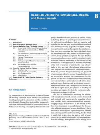
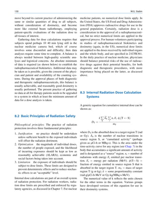
![8 Radiation Dosimetry: Formulations, Models, and Measurements 131
8.3.1 System of the US Society of Nuclear The term x contains several of the terms in our
Medicine’s Medical Internal generic equation above.
Radiation Dose (MIRD) Committee X
x¼ n E f Q i i i i
i
The equation for absorbed dose given in the MIRD
The ICRP developed a system of limitation of
system [1] is:
concentrations in air and water for employees from
X$ this equation and assumptions about the kinetic behav-
Drk ¼ Ah Sðrk rh Þ
ior of radionuclides in the body. These were the well-
h
known maximum permissible concentrations (MPCs).
In this equation, rk represents a target region and rh Employees could be exposed to these concentrations
represents a source region. The use of the subscripts on a continuous basis and not receive an annual dose
“h” and “k” for “source” and “target” is unusual. The rate to the so-called critical organ that exceeded estab-
~
cumulated activity, A, is as defined above; all other lished annual dose limits.
terms were combined in the factor “S”: In the ICRP 30 system, the cumulative dose equiv-
P alent was given as:
k ni Ei fi ðrk rh Þ X
Sðrk rh Þ ¼ i H50;T ¼ 1:6 Â 10 10 US SEEðT SÞ
mrk S
In the MIRD equations, the factor k is 2.13, which In this equation, T represents a target region and S
gives doses in rad, from activity in microcuries, mass represents a source region. The factor SEE is:
in grams, and energy in MeV. The MIRD system, P
which to date, still depends on non-SI units, was ni Ei fi ðT SÞ Qi
i
developed primarily for use in estimating radiation SEE ¼
mT
doses received by patients from administered radio-
pharmaceuticals; it was not intended to be applied to a The factor Us is another symbol for cumulated
system of dose limitation for workers. activity, and the factor 1.6 Â 10 10 is k. Note that
the symbol Q (quality factor), used in some of the
early ICRP manuals, is shown here instead of the
current notation wR (radiation weighting factor). As
8.3.2 The International Commission on in ICRP II, this equation was used to develop a system
Radiological Protection (ICRP) of dose limitation for workers, but unlike the ICRP II
system, limits are placed on activity intake during a
The ICRP has developed two comprehensive internal year, which would prevent cumulative doses (not con-
dosimetry systems intended for use in protecting radia- tinuous dose rates) from exceeding established limits.
tion workers. ICRP publication II [2] was the basis for These quantities of activity were called annual limits
the first set of complete radiation protection regulations on intake (ALIs); derived air concentrations (DACs),
in the USA (Code of Federal Regulations (CFR), Title which are directly analogous to MPCs for air, were
10, Chap. 20, or 10CFR20). These regulations were calculated from ALIs. More recent ICRP documents
replaced (completely) only in 1994 when a revision of (e.g., ICRP 71 [4]) changed the formulation some-
10CFR20 incorporated many of the new procedures what. For example, the equivalent dose at age t in
and results of the ICRP 30 series [3]. Both of these target organ or tissue T due to an intake of a radio-
systems, dealing with occupational exposures, used nuclide at age t0 may be given as:
equivalent dose as their main dose parameter. Á X
In the ICRP II system, the dose equivalent rate was H ðt; t0 Þ ¼ qs ðt; t0 Þ SEEðT S; tÞ
given by the expression: S
51:2 A x qs(t,t0) ¼ the activity of the radionuclide in source
H¼
m organ S at age t after intake at age t0 (Bq), SEE](https://image.slidesharecdn.com/basicsciencesofnuclearmedicine-121209103030-phpapp02/85/CIENCIAS-BASICAS-MN-144-320.jpg)
![132 M.G. Stabin
(T S, t0) = the Specific Effective Energy, as defined in Dose conversion factors (SEEs) may be taken from
Chap. 7, except that the energy is given in J and mass is ICRP 30 (or later documents, but this example is
given in kg, so the units of dose are Gy without the continued as it is still the basis for regulations in the
need for a unit conversion factor. USA), and solve for the H50 values as follows:
The ICRP uses multicompartment models to describe
Sv g transf
the kinetics of the movement of radioactive material H50;gonads ¼ 1:6 Â 10 Â 2:8 Â 105
10
MeV Bq intake
through the respiratory and gastrointestinal tracts,
MeV Sv
which then permit entry of material into the systemic Â9:9 Â 10 6 ¼ 4:4 Â 10 10
circulation, where its movements are treated with indi- g transf Bq
vidual biokinetic models for each element, and some- Sv g transf
times for more than one form of each element. Much H50;breasts ¼ 1:6 Â 10 Â 2:8 Â 105
10
MeV Bq intake
detail about these model systems are not given here, MeV Sv
but are detailed elsewhere [5]. Once the biokinetic Â9:9 Â 10 6 ¼ 4:4 Â 10 10
g transf Bq
model for an element (including the kinetics of the
Sv g transf
intake component (lung or GI)) is characterized, US H50;marrow ¼ 1:6 Â 10 10
 2:8  105
values may be calculated for all of the important MeV Bq intake
source regions, and dose estimates are obtained by MeV Sv g
 9:9  10 6 þ 1:6  10 10
multiplying them by the appropriate dose conversion g transf MeV
factors (SEEs), as shown above. To calculate an SEE, 5 transf
 1:5  10
one needs decay data, data on standard organ masses, Bq intake
and the “absorbed fractions” for photons. In the ICRP MeV Sv
 2:3  10 4 ¼ 6:0  10 9
system, doses are calculated, as are amounts of activity g transf Bq
that would give permissible annual doses to workers.
Sv g transf
The dose conversion factors are usually calculated as H50;lungs ¼ 1:6 Â 10 10
 2:8  105
MeV Bq intake
50-year committed dose (Sv) per Bq of intake (by
6 MeV Sv g
inhalation or ingestion). Knowing our annual dose  9:9  10 þ 1:6  10 10
limits in Sv, we can thus calculate the number of Bq g transf MeV
that are permissible to take in during 1 year of work. transf
 1:8  104
There are two dose limits that must be satisfied at the Bq intake
same time the stochastic limit (50 mSv effective MeV Sv
 6:9  10 4 ¼ 2:4  10 9
whole body) and the nonstochastic limit (500 mSv to g transf Bq
any organ). The permissible amount of activity for any Sv g transf
element that may be taken in during one working year H50;ULI ¼ 1:6 Â 10 10
 2:8  105
MeV Bq intake
(the Annual Limit on Intake, ALI) is that amount that
MeV Sv g
will satisfy both limits and choose the smaller of the  9:9  10 6 þ 1:6  10 10
g transf MeV
two values as our controlling limit.
3 transf
Example  1:4x10
Calculate the ALI and the DAC for inhalation of Bq intake
class D 32P. Solution of the biokinetic model gives the MeV Sv
 1:6  10 3 ¼ 8:0  10 10
following values for the number of disintegrations in g transf Bq
various source organs for a 1 Bq intake: Sv g transf
H50;LLI ¼ 1:6 Â 10 10
 2:8  105
Lungs 1.8 Â 104 MeV Bq intake
ULI contents 1.4 Â 103 MeV Sv g
 9:9  10 6 þ 1:6  10 10
g transf MeV
LLI contents 2.5 Â 103
transf
Cortical bone 1.5 Â 105 Â 2:5 Â 103
Bq intake
Trabecular bone 1.5 Â 105 MeV Sv
 2:6  10 3 ¼ 1:5  10 9
Other tissues 2.8 Â 105 g transf Bq](https://image.slidesharecdn.com/basicsciencesofnuclearmedicine-121209103030-phpapp02/85/CIENCIAS-BASICAS-MN-145-320.jpg)
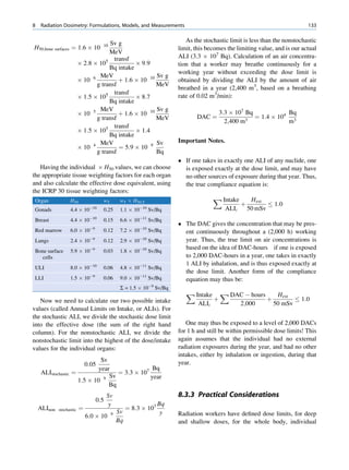
![134 M.G. Stabin
organs, and extremities (hands and feet) [6]. Declared where NS is the number of disintegrations that occur in
pregnant workers adhere to lower dose limits, to pro- a source organ, and DF(T S) is:
tect the unborn child. When working with sources
of radiation, simple procedures such as awareness of P
k ni Ei fi ðT SÞ
time, distance, and shielding (TDS) principles can i
DFðT SÞ ¼
greatly reduce radiation dose. Other references treat mT
these subjects in more depth than is possible here
[7]. Safe administration of radiopharmaceuticals to The DF is conceptually similar to the “S value”
nuclear medicine patients is also a subject of consider- defined in the MIRD system or the “SEE” factor of
able depth, including subjects such as use in children the ICRP system. The number of disintegrations is the
and pregnant women, release of radioactive subjects, integral of a time-activity curve for a source region
communication of risks and benefits, and doses and ~
(AS in the generic equation).
risks from extravasations; many of these topics have RADAR members have produced compendia of
been addressed in other works [7, 35]. decay data, dose conversion factors, and catalogued
standardized dose models for radiation workers and
nuclear medicine patients, among other resources.
They also produced the OLINDA/EXM [9] personal
8.3.4 RADAR
computer software code, which used the equations
shown here and the input data from the RADAR site.
In the early twenty-first century, an electronic resource This code was basically a revised version of the MIR-
was established on the internet to provide rapid, DOSE [10] software, which implemented the MIRD
worldwide dissemination of important dose quantities method for internal dose calculations (but was not in
and data. The RAdiation Dose Assessment Resource any way associated with the MIRD Committee itself).
(RADAR) established a web site (www.doseinfo- The RADAR site and OLINDA/EXM software imple-
radar.com), and provided a number of publications ment all of the most current and widely accepted
on the data and methods used in the system. The models and methods for internal dose calculations,
RADAR system [8] has perhaps the simplest represen- and are constantly updated to reflect changes that
tation of the cumulative dose equation: occur in the science of internal dose assessment. The
X structure of the RADAR schema is summarized in
DT ¼ NS Â DFðT SÞ Fig. 8.1.
S
Internal sources:
Kinetic models– Radionuclide Specific absorbed
radiopharmaceuticals decay data fractions–phantoms
Kinetic models–
occupational radionuclides
Dose conversion
External sources: factors
External source
configuration
Radiation dose
estimates and distributions
Fig. 8.1 An overview of the
Linking of radiation
structure of the RADAR doses to biological effects
schema](https://image.slidesharecdn.com/basicsciencesofnuclearmedicine-121209103030-phpapp02/85/CIENCIAS-BASICAS-MN-147-320.jpg)
![8 Radiation Dosimetry: Formulations, Models, and Measurements 135
8.3.5 Anthropomorphic Models for MIRD 5 Phantom31 NURBS-based adult male
Standardized Dose Calculations model41
The current state of the art in full body anthropomor- Brain
phic phantoms is based on the original Fisher-Snyder
whole body and organ phantom [11]. This phantom
used a combination of geometric shapes spheres,
cylinders, cones, etc. to create a simple representa- Pulmonary
vessels
tion of the body and its internal structures. Monte
Carlo computer programs were used to simulate the
transport of photons through these various structures
in the body, whose atomic composition and density Abdominal
vessels
were based on data provided in the original ICRP
report on Reference Man [12]. This report provided Fig. 8.2 Comparison of the realism of the traditional body
various anatomical data assumed to represent the aver- models with those being used to support current dose modeling
age working adult male in the Western hemisphere, efforts
and has been recently updated [13]. Using this phan-
tom, absorbed fractions for adults based on activity more realistic phantoms using image-based methods
residing in any organ and irradiating any other organ to replace the stylized models of the 1970s with
were calculated and published [11], and dose conver- voxel-based [18 20] or the use of mathematical
sion factors were published [14] in the 1970s for this methods like nonuniform rational B-splines
single model. Later, Cristy and Eckerman [15] mod- (NURBS) [21]. An example of modeling efforts
ified the adult male model somewhat and developed involved with more realistic whole body and organ
models for a series of individuals of different size models is shown in Fig. 8.2.
and age. Six phantoms were developed, which were Spiers et al. at the University of Leeds [22] first
assumed to represent children of ages 0 (newborn), established electron absorbed fractions (AFs) for bone
1-year, 5-year, 10-year, and 15-year-olds, and adults and marrow in a healthy adult male, which were used
of both sexes. in the dose factors (DFs), or S values, in MIRD Pam-
phlet No. 11 [14]. Eckerman re-evaluated this work
and extended the results to derive DFs for fifteen
skeletal regions in six models representing individuals
8.3.6 Absorbed Fractions and Dose
of various ages [23]. The results were used in the
Conversion Factors MIRDOSE 3 software [14] to provide mean marrow
dose, regional marrow dose, and dose-volume histo-
Absorbed fractions for photons at discrete energies grams for different individuals. This model was updated
were published for these phantoms, which contained and improved employing more realistic assumptions
approximately 25 source and target regions. Tables of of energy absorption at low electron energies [24].
S values were never published, but ultimately were For many years, the only source of dose factors for
made available in the MIRDOSE computer software use in practical calculations were found in MIRD
[16]. Stabin et al. developed a series of phantoms for Pamphlet 11 [14], in which factors were given for
the adult female, both nonpregnant and at three stages about 25 organs, but only in the adult male phantom,
of pregnancy [17]. These phantoms modeled the for 117 radionuclides. The MIRDOSE code provided
changes to the uterus, intestines, bladder, and other dose factors for over 240 radionuclides, for about 25
organs that occur during pregnancy, and included organs as well, but in the entire Cristy Eckerman and
specific models for the fetus, fetal soft tissue, fetal Stabin et al. pediatric, adult, and pregnant female
skeleton, and placenta. S values for these phantoms phantoms series (10 phantoms). Siegel and Stabin
were also made available through the MIRDOSE [8] then calculated dose factors for over 800 radio-
software [16]. A number of authors have developed nuclides for:](https://image.slidesharecdn.com/basicsciencesofnuclearmedicine-121209103030-phpapp02/85/CIENCIAS-BASICAS-MN-148-320.jpg)
![136 M.G. Stabin
Fig. 8.3 Frontal views of the
revised ORNL phantoms:
newborn, 1 year, 5 year,
10 year, 15 year, and adult.
Skin and muscle are made
semitransparent for clear
viewing of internal organs and
the skeleton. (From Ref. [51])
Reprinted with permission
from IOP publishing and the
corresponding author
Newborn 1-Year 5 -Year 10 -Year 15 -Year Adult
1. All source and target regions in the six models in 8.4 Doses for Nuclear Medicine Patients
the Cristy/Eckerman phantom series [15] (see
Fig. 8.3)
Dose calculations for nuclear medicine patients
2. All source and target regions in the four models in the
depend on development of a kinetic model, based on
Stabin et al. pregnant female phantoms series [17]
measurements made in animal or human studies, and
3. All target regions in the Watson and Stabin perito-
employment of appropriate dose conversion factors.
neal cavity model [25]
Radionuclides in nuclear medicine are bound to a very
4. All target regions in the Stabin prostate gland
wide variety of compounds, and a separate kinetic
model [26]
model must be developed for each compound; this
5. All source and target regions in the six models of
involves the proper design and execution of experi-
the MIRD head and brain model [27]
ments in either animals or humans to obtain the neces-
6. All source and target regions in the MIRD regional
sary data to build a kinetic model. To design and
kidney model [28]
execute a good kinetic study, one needs to collect the
7. The unit density sphere models of Stabin and
correct data, enough data, and express the data in the
Konijnenberg [29]
proper units. The basic data needed are the fraction (or
These dose factors were based on decay data from percent) of administered activity in important source
the Brookhaven National Laboratory resource (http: organs and excreta samples. We shall discuss later how
//www.nndc.bnl.gov/) [30], and are useful for imple- these data are gathered from an animal or human study.
mentation in the dose equations described above.
These dose conversion factors use the child, adult,
and pregnant woman phantoms and bone and marrow
models described above, and included standard mod- 8.4.1 Data Sampling
eling assumptions, as were described in that paper.
Examples of absorbed fractions and dose factors are It is very important, in either type of study, to take
given in the Stabin and Siegel document [8], and enough samples to characterize both the distribution
on the RADAR internet web site (www.doseinfo- and retention of the radiopharmaceutical over the
radar.com). course of the study. One must gather enough data to](https://image.slidesharecdn.com/basicsciencesofnuclearmedicine-121209103030-phpapp02/85/CIENCIAS-BASICAS-MN-149-320.jpg)
![8 Radiation Dosimetry: Formulations, Models, and Measurements 137
study early and late intake and washout phases, collect proposed in the literature. One method of extrapolat-
data over at least three effective half times of the ing animal data is the % kg/g method [33]. In this
radiopharmaceutical, collect at least two time points method, the animal organ data need to be reported as
per phase of uptake or clearance, account for 100% of percentage of injected activity per gram of tissue, and
the activity at all times, and account for all major paths the animal whole body weight must be known. The
of excretion (urine, feces, exhalation, etc.). Some extrapolation to humans then uses the human organ
knowledge of the expected kinetics of the pharmaceu- and whole body weight, as follows:
tical are needed for a good study design before data ! #
%
collection begins. For example, the spacing of the
 kgTBweight
measurements and the time of the initial measurement gorgan animal
animal
will be greatly different if we are studying a 99mTc- !
labeled renal agent 95% of which is cleared from the gorgan
Â
body in 180 min or an 131I labeled antibody 80% of kgTBweight
human
which clears in the first day and the remaining 20%
%
over the next 2 weeks. The characterization of excre- ¼
organ human
tion is essential to a complete dosimetric evaluation, as
the excretory organs (intestines, urinary bladder) are in
many cases, the organs which receive the highest
absorbed doses, as 100% of the activity (minus
8.4.3 Activity Quantitation
decay) will eventually pass through one or both of
these pathways at different rates. If excretion is not
quantified, the modeler must make the assumption that In human studies, data are collected with a nuclear
the compound is retained in the body and removed medicine gamma camera. Quantification of data gath-
only by radioactive decay. For very short-lived radio- ered with these cameras is rather involved. Projection
nuclides, this may not be a problem and in fact may be images (anterior and posterior, typically) of the patient
quite accurate. For moderately long-lived nuclides, are developed and processed. As this is a projection
this can cause an overestimate of the dose to most image, the actual depth of the object within the patient
body organs and an underestimate of the dose to the is not known. One may draw regions of interest (ROIs)
excretory organs, perhaps significantly. around images of objects that will be recognizable as
internal organs or structures; the number of counts in a
ROI, however, cannot be used directly to calculate
how much activity is in the organ. A number of correc-
8.4.2 Extrapolation of Animal Data tions is needed to the observed number of counts to
obtain a reliable estimate of activity in this object [34].
Data for kinetic studies are generally derived from To remove uncertainties about the depth of the object,
animal studies, usually performed for submission of usually images are taken in front of and behind the
an application for approval to the Food and Drug patient, and a geometric mean of the two values is
Administration in the United States for use of a so- taken. This geometric mean has been shown to be
called Investigational New Drug (IND), and human relatively independent of depth for most radionuclides
studies, usually performed in Phase I, II, or III of of interest, and thus this quantity is thought to be the
approval of a New Drug Application (NDA). In an most reliable for use in quantitation. Other degrading
animal study, the radiopharmaceutical is administered factors such as attenuation and scatter can be tackled
to a number of animals for which organ, whole body, by using a transmission source and an appropriate
and excretion data may be collected. Animal data must scatter correction technique.
be somehow extrapolated to obtain dose estimates for After scatter correction has been applied, the activ-
humans. The extrapolation of animal data to humans ity of the source within the ROI is thus given by:
is not an exact science. Crawford and Richmond r
[31] and Wegst [32] studied some of the strengths IA IP fj
AROI ¼
and weaknesses of various extrapolation methods e me t C](https://image.slidesharecdn.com/basicsciencesofnuclearmedicine-121209103030-phpapp02/85/CIENCIAS-BASICAS-MN-150-320.jpg)
![138 M.G. Stabin
where IA and IP are the Anterior and Posterior counts terize a set of data by a series of exponential terms, as
in the region, me is the effective attenuation coefficient, many systems are well represented by this form, and
t is the average patient thickness over the ROI, fj is the exponential terms are easy to integrate. In general, the
source self-attenuation coefficient (given as [(me t/2)/ approach is to minimize the sum of the squared dis-
sinh(me t/2)], but which is rarely of much impact in the tance of the data points from the fitted curve. The
calculation and so is usually neglected), and C is a curve will have the form:
source calibration factor (cts/s/Bq), obtained by count-
ing a source of known activity in air. Thus, activity in AðtÞ ¼ a1 e b1 t
þ a2 e b2 t
þ ÁÁÁ
identifiable regions of the body, like liver, spleen,
kidneys, etc. may be determined at individual times. The method evaluates the squared difference
ROIs may also be drawn over the entire body, to track between each point and the solution of the fitted curve
the retention and excretion of the compound in the at that point, and minimizes this quantity by taking the
body. Excreta samples may also be taken to study partial derivative of this expression with respect to
excretion pathways. If only a single excretion pathway each of the unknowns ai and bi and setting it equal to
is important, knowledge of whole body clearance may zero. Once the ideal estimates of ai and bi are obtained,
be used to explain excretion. Single Photon Emission the integral of A(t) from zero to infinity is simply:
Computed Tomography (SPECT) or Positron Emis- ð
1
sion Tomography (PET) methods may also be used to a1 a2
AðtÞdt ¼ þ þ :::
obtain quantitative data for dosimetry studies. This b1 b2
0
takes considerably more effort, both in data gathering
and analysis, and is employed by fewer investigators. If the coefficients ai are in units of activity, this inte-
gral represents cumulated activity (the units of the bi
are time 1).
8.4.4 Data Analysis
8.4.4.3 Compartmental Models
Once kinetic data are obtained, analysis of the data
involves estimation of the area under the time-activity The situation frequently arises that you either know
~
curve to obtain the number of disintegrations (N or A) quite a bit about the biological system under investi-
in all important source regions. In general, there are gation or you would like to know in greater detail how
three levels of complexity that our analysis can take: this system is working. In this case, you can describe
the system as a group of compartments linked through
~
transfer rate coefficients. Solving for A of the various
8.4.4.1 Direct Integration
compartments involves solving a system of coupled
differential equations describing transfer of the tracer
One can directly integrate under the actual measured
between compartments and elimination from the sys-
values by a number of methods. This does not give
tem. The solution to the time activity curve for each
much information about your system, but it does allow
compartment will usually be a sum of exponentials,
you to calculate the area under the time-activity curve
but not obtained by least squares fitting each compart-
rather easily. The most common method used is the
ment separately, but by varying the transfer rate coef-
Trapezoidal Method, simply approximating the area
ficients between compartments until the data are well
by a series of trapezoids.
fit by the model.
8.4.4.2 Least Squares Analysis
8.4.4.4 Dose Calculation Steps
In this application, one attempts to fit curves of a given
shape to the data. The curves are represented by math- As outlined with examples by Stabin [35], dose calcu-
ematical expressions which can be directly integrated. lations proceed with the selection of an appropriate
The most common approach is to attempt to charac- standardized anthropomorphic model, extrapolation of](https://image.slidesharecdn.com/basicsciencesofnuclearmedicine-121209103030-phpapp02/85/CIENCIAS-BASICAS-MN-151-320.jpg)
![8 Radiation Dosimetry: Formulations, Models, and Measurements 139
animal data (in preclinical studies), or quantification radionuclides in 33 radiopharmaceuticals, for which
of human imaging data, a thorough kinetic analysis, data for 115 organs were available both for extrapo-
and finally, dose calculations using established, stan- lated animal data and subsequent measurements in
dardized methods, as described above. The reader human subjects. Table 8.1 shows their observations;
is referred to the longer discussion of these topics in they found that animal data generally underpredicted
this publication, with several worked out numerical the actual number of disintegrations (and thus organ
examples. self dose) seen in human subjects, and that the accu-
racy of the extrapolation was not particularly strong.
No strong preference was seen for any particular extra-
polation method, but what was most striking was
8.4.4.5 Uncertainties in Internal Dose
the general lack of accuracy of the extrapolated data.
Calculations
Not until a factor of 10 tolerance was given were the
estimates predictive in close to 90% of the cases.
All dose calculation must begin with an estimation of
In human studies, after administration of the
the amount of activity as a function of time in a source
radiopharmaceutical, a series of images are made
region. As noted above, activity in source regions for
with nuclear medicine imaging systems (gamma
radiopharmaceuticals is generally determined in one
cameras) and counts are extracted from the images
of two ways (a) experiments in animal models with
and related to absolute activity values via calibration
extrapolation of the observed results to humans and
factors. Siegel et al. [37] discuss many of the strengths
(b) measurements in human subjects (volunteers or
and weaknesses of the various data gathering methods
patients) with calculation of dose estimates, either
used in nuclear medicine. Influences on the quality of
general dose estimates for reference individuals or
the data include inherent system energy resolution,
specific dose estimates for the individual patient. In
degradation of this spatial resolution due to collimator
all cases, one begins with a measurement of counts of
septal penetration by high-energy photons (if present
a radioactive source which contains an inherent and
in the decay scheme), data loss due to scatter and
well-known statistical uncertainty.
attenuation, and the inherent statistical variability in
Converting counts to disintegrations requires appli-
any measurement of any radioactive source (see
cation of a calibration factor (e.g., Bq-s/count), which
Chap. 14).
is based on measured counts of a radioactive source in
Stabin [38] performed an analysis of uncertainty in
some experimental situation in which other uncertain-
the various terms of the dose equation shown above,
ties may be introduced but are not often reported (e.g.,
including in activity measurements and integrations.
measurement errors in adding volume of a standard to
The conclusions regarding these other quantities are
a calibration device, errors in making estimates of
summarized in Table 8.2 and discussed here:
distances or time differences, or performance of dose
calibrators). The factor k has zero uncertainty. It is simply an
Sparks and Aydogan [36] discussed the quality analytical quantity based on known conversion
of extrapolation of animal data to humans, for 11 constants. It includes the various factors that are
Table 8.1 Percent of time that different scaling methods were successful in predicting human biokinetics from animal data within
specified uncertainty levels [36]. “Mass” scaling means extrapolation of animal data based on organ concentrations and body mass,
as shown in the Kirschner et al. [33] equation above, “time” scaling means extrapolation of the time scale based on differences in
body mass and assumed metabolic rates
Uncertainty level
Extrapolation type Æ25% Æ50% Æ2X Æ4X Æ10X Æ20X
None 18% 30% 39% 62% 84% 92%
Mass 8% 18% 40% 66% 85% 93%
Time 21% 32% 50% 74% 88% 91%
Mass and time 8% 21% 46% 79% 94% 97%](https://image.slidesharecdn.com/basicsciencesofnuclearmedicine-121209103030-phpapp02/85/CIENCIAS-BASICAS-MN-152-320.jpg)
![140 M.G. Stabin
Table 8.2 Summary of sources of uncertainty in dose estimate
Variable Uncertainty Remarks Publications
k Zero uncertainty An analytical quantity used mainly to Stabin [38]
match among units
ni and Ei Negligible when compared with other Stabin [38]
variables
AF or (SAF) and (~5% or less) uncertainty is reduced by The uncertainty is particularly large when a Stabin [38]
mass terms applying large number of photon patient geometry is significantly
histories in the simulation process different from the assumed reference
(i.e., reduction of statistical error) man/women model.
Biokinetic 10 20% when adjustments are done to Stabin [38]
parameters account for individual kinetics.
~
Inherent in A S
(Fractional
uptake and Tb)
Biokinetics + organ Could result in doubling in 95% Stabin[38]
mass confidence intervals around median
value of dose estimates of most
organ
Effective whole 20 40% Differences in tissue sensitivities (wT) at Stabin [38]
body dose different ages
Data extrapolation to Uncertainty in extrapolation methods Observations suggest considerable Aydogan
human cannot be quantitatively assessed underestimation of human organ uptake et al.
and dose [41]
needed to obtain the dose in the desired units, from organ mass values to individuals who are not well
the units employed for the other variables. represented by the population median value will
The uncertainty in the terms ni and Ei is very low thus be in error by as much as the individual’s
and mostly negligible, in comparison with uncer- body geometry varies from the median, at least
tainties in the other terms in the equation. tens of percent.
The other three terms in the equation, namely f (or The biokinetic parameters inherent in calculation
~
AF), AS and m, contribute the vast majority of the ~
of AS, namely fractional uptake and effective half-
uncertainty to internal dose calculations for radio- time in organs and the body, vary substantially
pharmaceuticals. between individuals. Variability of a factor of 2 or
The absorbed fraction and mass terms used in most more is reasonable to assume for the kinetics of any
internal dose calculations are based on standar- given radiopharmaceutical, if applying a general
dized individuals, i.e., reference man, reference biokinetic model to any individual in the nuclear
woman, reference pediatric individuals, and so medicine patient population. If a specific study
on. When standardized phantoms are used with of an individual is performed to determine the
Monte Carlo radiation transport simulation codes particular kinetics for that individual, and care is
to obtain absorbed fraction (AF) values (or “spe- taken to adjust doses for cases in which organ
cific absorbed fractions,” SAFs, which are equal to volumes are known to vary from those of the refer-
the absorbed fraction divided by the mass of the ence individuals, final doses may have uncertain-
target region), the absolute uncertainty in the cal- ties as low as 10 20%. But in the general case,
culated AFs or SAFs can be limited (to ~5% or less) uptake and clearance of radiopharmaceuticals var-
simply by performing a large number of particle ies substantially between individuals, and although
histories. Thus, the inherent uncertainty is small, the mean or median values may be quite reliable,
and somewhat comparable to that for the decay the uncertainty for any given subject is substantial.
data. However, the application of these SAFs and Two examples from the literature are shown in](https://image.slidesharecdn.com/basicsciencesofnuclearmedicine-121209103030-phpapp02/85/CIENCIAS-BASICAS-MN-153-320.jpg)
![8 Radiation Dosimetry: Formulations, Models, and Measurements 141
100
Fig. 8.4 Uptake and Wb
Heart
retention of 99mTc RP527, a Liver
gastrin releasing peptide Scortum
(GRP) agonist for the Thyroid
Percentage of injected activity (%IA).
10
Breasts
visualization of GRP Blood
receptor expressing
malignancies in various
1
subjects, as reported by Van
200 400 600 800 1,000 1,200 1,400 1,500
de Wiele et al. [39]. Reprinted
with permission of the Society
of Nuclear Medicine 0.1
0.01
0.001
Time (h)
Fig. 8.5 Cumulative
100
excretion of Ho 166 DOTMP
Cummulative % injected
in twelve subjects (six males
and six females) with multiple 80
myeloma. (From Ref. [40])
Reprinted with permission of
Dose
60
the Society of Nuclear
Medicine
40
20
0
0 12 24 36 48
Time (h)
Figs. 8.4 and 8.5 from Van de Wiele [39] and Breitz in mass could account for a doubling in the 95% confi-
et al. [40]. dence intervals around the point (median) dose estimates
Another source of uncertainty in reported estimates for most organs. They stated that absorbed doses that
of effective whole body dose involves changes in any given patient in the broad nuclear medicine popu-
the recommended tissue weighting factors at dif- lation receives may be up to twice as large as reported
ferent times by the ICRP. This may introduce an mean. Figure 8.6 shows their predicted 95% confidence
additional uncertainty of up to 20 40% in reported interval for dose to the liver using their LHS modeling
values of effective dose for diagnostic radiophar- and taking into account all sources of uncertainty.
maceuticals.
Aydogan et al. [41] evaluated the uncertainty in dose
estimates for an 123I labeled brain imaging agent (IPT)
8.5 Image-Based, 3D Patient-Specific,
in seven healthy adult subjects (five males, two
females). They reported uncertainties in the fitted bio- Dosimetry
kinetic coefficients and specific absorbed fractions,
as obtained by first order error propagation (FOEP) Several of the efforts to use image data to perform dose
and Latin Hypercube Sampling (LHS) methods. They calculations, as described above, include the 3D-ID
concluded that variability in individual biokinetics and code from the Memorial Sloan-Kettering Cancer](https://image.slidesharecdn.com/basicsciencesofnuclearmedicine-121209103030-phpapp02/85/CIENCIAS-BASICAS-MN-154-320.jpg)
![142 M.G. Stabin
0.035 867
0.026 650
Frequency
Probability
0.018 433
0.009 216
0.000 0
0.00E+0 1.35E-5 2.70E-5 4.05E-5 5.40E-5
95% Confidence interval is from 7.54E-6 to 5.08E-4 Gy/MBq
Fig. 8.6 An example of absorbed dose distribution (with 95% confidence). It shows the uncertainty in dose estimates for an
123
I labeled brain imaging agent (IPT) in seven healthy adult subjects (five males, two females) (Taken from Ref. [41])
Center [42], the SIMDOS code from the University of
a
Lund [43], the RTDS code at the City of Hope Medical
Center [44], the RMDP code from the Royal Marsden
Hospital [45] and the DOSE3D code [46]. Neither has
a particularly robust and well-supported electron trans-
port code, as is available in EGS [47], MCNP [48], or
GEANT [49]. The PEREGRINE code [50] has also
been proposed for three-dimensional, computational
dosimetry and treatment planning in radioimmunother-
apy. The promise of this type of code is great, in
providing three dimensional, image-based dose calcu-
b lations to the nuclear medicine physician which may be
0.14 used in patient-specific treatment planning in radio-
0.13
0.12 Liver pharmaceutical therapy (see Fig. 8.7). The challenge,
0.11 Tumor as noted above, is finding broad clinical acceptance for
0.10 these approaches and a move away from the approach
0.09
Volume (%)
0.08 to therapy involving a uniform administration of activ-
0.07 ity to all patients, with no consideration of radiation
0.06
0.05
dose. Further discussion about patient-specific dosime-
0.04 try can be found in Chap. 14.
0.03
0.02
0.01
0 References
0% 10% 20% 30% 40% 50% 60% 70% 80% 90% 100%
Dose (%)
1. Loevinger R, Budinger T, Watson E (1988) MIRD primer
Fig. 8.7 Example of 3D dose distributions. (a) Calculated dose for absorbed dose calculations. Society of Nuclear Medi
distribution after administration of 131I MIBG superimposed on cine, New York
axial CT scan. (b) Differential dose volume histogram for nor 2. International Commission on Radiological Protection
mal liver and metastasis. (From Strigari et al. [51]). Reprinted (1960) Report of committee II on permissible dose for
with kind permission from Medical Physics internal radiation. Health Phys. 3](https://image.slidesharecdn.com/basicsciencesofnuclearmedicine-121209103030-phpapp02/85/CIENCIAS-BASICAS-MN-155-320.jpg)
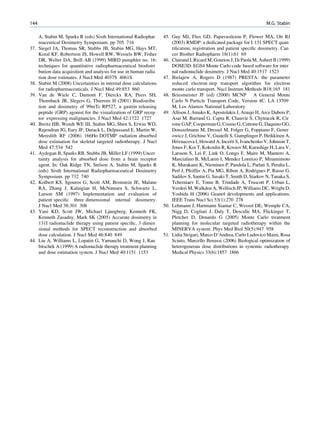
![Radiobiology: Concepts and Basic Principles
9
Michael G. Stabin
Contents blistering in the finger, which was clearly and imme-
9.1 Introduction . . . . . . . . . . . . . . . . . . . . . . . . . . . . . . . . . . . . . . . . 145 diately related to the radiation exposure. William
9.2 Stochastic Versus Nonstochastic Effects . . . . . . . . . . 145 Herbert Rollins (a Boston dentist) demonstrated that
9.2.1 Nonstochastic Effects . . . . . . . . . . . . . . . . . . . . . . . 146 X-rays could kill guinea pigs and result in the death of
9.2.2 Stochastic Effects . . . . . . . . . . . . . . . . . . . . . . . . . . . 146
offspring when guinea pigs were irradiated while preg-
9.3 Cellular Response and Survival . . . . . . . . . . . . . . . . . . . 147
9.3.1 Mechanisms of Radiation Damage nant. Henri Becquerel received a skin burn from a
to Biological Systems . . . . . . . . . . . . . . . . . . . . . . . 148 radium source given to him by the Curies that he
9.3.2 Bystander Effect . . . . . . . . . . . . . . . . . . . . . . . . . . . . 149 carried in a vest pocket at times. He once was reported
9.4 Relevance of Radiation Biology to Radionuclide
to have said: “I love this radium but I have a grudge
Therapy . . . . . . . . . . . . . . . . . . . . . . . . . . . . . . . . . . . . . . . . . . . . 150
9.4.1 Thyroid Disease . . . . . . . . . . . . . . . . . . . . . . . . . . . . . 150 against it!” The first death in an X-ray pioneer attrib-
9.4.2 Other Diseases . . . . . . . . . . . . . . . . . . . . . . . . . . . . . . 151 uted to cumulative overexposure was to C.M. Dally in
9.5 Conclusion . . . . . . . . . . . . . . . . . . . . . . . . . . . . . . . . . . . . . . . . . 151 1904. Radiologists and other physicians who used
References . . . . . . . . . . . . . . . . . . . . . . . . . . . . . . . . . . . . . . . . . . . . . . . . . 151
X-rays in their practices before health physics prac-
tices were common had a significantly higher rate of
leukemia than their colleagues. A particularly tragic
episode in the history of the use of radiation and in the
history of industrialism was the acute and chronic
9.1 Introduction biological damage suffered by the Radium Dial Pain-
ters [1]. Radium was used in luminous paints in the
Very soon after the discovery of radiation and radioac- early 1900s. In factories where luminous dial watches
tivity short- and long-term negative effects of radiation were made, workers (mainly women) would sharpen
on human tissue were observed. Adverse effects of the tips of their paint brushes with their lips, and thus
X-rays were observed by Thomas Edison, William J. ingested large amounts of radium. They had increased
Morton, and Nikola Tesla; they independently reported amounts of bone cancer (carcinomas in the paranasal
eye irritations from experimentation with X-rays and sinuses or the mastoid structures, which are very rare,
fluorescent substances. These effects were thought to and were thus clearly associated with their exposures,
be eye strain, or possibly due to exposure to ultraviolet as well as cancers in other sites) and even spontaneous
radiation. Elihu Thomson (an American physicist) fractures in their jaws and spines from cumulative radi-
deliberately exposed the little finger of his left hand ation injury. Others died of anemia and other causes.
to X-rays for several days, for a short time each day,
and observed pain, swelling, stiffness, erythema, and
9.2 Stochastic Versus Nonstochastic
M.G. Stabin Effects
Department of Radiology and Radiological Sciences, Vanderbilt
University, 1161 21st Avenue South, Nashville, TN 37232
2675, USA There are two broad categories of radiation-related
e mail: michael.g.stabin@vanderbilt.edu effects in humans, stochastic and nonstochastic.
M.M. Khalil (ed.), Basic Sciences of Nuclear Medicine, DOI: 10.1007/978 3 540 85962 8 9, 145
# Springer Verlag Berlin Heidelberg 2011](https://image.slidesharecdn.com/basicsciencesofnuclearmedicine-121209103030-phpapp02/85/CIENCIAS-BASICAS-MN-158-320.jpg)
![146 M.G. Stabin
There are three important characteristics that distin- degree of specialization.” So, rapidly dividing and
guish them. unspecialized cells, as a rule, are the most radiosensi-
tive. Two important examples are cells in the red
marrow and in the developing embryo/fetus. In the
9.2.1 Nonstochastic Effects case of marrow, a number of progenitor cells which,
through many generations of cell division, produce a
variety of different functional cells that are very
Nonstochastic effects (now officially called “deter-
specialized (e.g., red blood cells, lymphocytes, leuko-
ministic effects,” previously also called “acute
cytes, platelets). Some of these functional cells do not
effects”) are effects that are generally observed soon
divide at all, and are thus themselves quite radioresis-
after exposure to radiation. As they are “nonstochas-
tant. However, if the marrow receives a high dose of
tic” in nature, they will always be observed (if the dose
radiation, damage to these progenitor cells is very
threshold is exceeded), and there is generally no doubt
important to the health of the organism. As we will
that they were caused by the radiation exposure. The
see shortly, if these cells are affected, in a short period
major identifying characteristics of nonstochastic
this will be manifested in a measurable decrease in the
effects are:
number of formed elements in the peripheral blood. If
1. There is a threshold of dose below which the the damage is severe enough, the person may not
effects will not be observed. survive. If not, the progenitor cells will eventually
2. Above this threshold, the magnitude of the effect repopulate and begin to replenish the numbers of the
increases with dose. formed elements, and subsequent blood samples will
3. The effect is clearly associated with the radiation show this recovery process.
exposure. In the fetus, organs and systems develop at different
rates. At the moment of conception, of course, we have
Examples include:
one completely undifferentiated cell that becomes two
Erythema (reddening of the skin) cells after one division, then four then eight, and so on.
Epilation (loss of hair) As the rapid cell division proceeds, groups of cells
Depression of bone marrow cell division “receive their assignments” and differentiate to form
(observed in counts of formed elements in organs and organ systems, still with a very rapid rate of
peripheral blood) cell division. At some point, individual organs become
Nausea, vomiting, diarrhea (NVD) often well defined and formed, and cell division slows as the
observed in victims after an acute exposure to fetus simply adds mass. But while differentiation and
radiation early rapid cell division is occurring, these cells are
Central nervous system (CNS) damage quite radiosensitive, and a high dose to the fetus may
Damage to the unborn child (physical deformi- cause fetal death, or damage to individual fetal struc-
ties), microcephaly (small head size at birth), tures. This is discussed further below. On the other
mental retardation hand, in an adult, cells of the CNS (brain tissue, spinal
cord, etc.) are very highly specialized and have very
When discussing nonstochastic effects, it is impor- low, or no, rate of division. The CNS is thus particularly
tant to note that some organs are more radiosensitive radioresistant. One important nonstochastic effect is
than others. The so-called law of Bergonie and death. This results from damage to the bone marrow
Tribondeau [2] states that cells tend to be radiosensi- (first), then to the gastrointestinal tract, then to the
tive if they have three properties: nervous system.
Cells have a high division rate.
Cells have a long dividing future.
Cells are of an unspecialized type. 9.2.2 Stochastic Effects
A concise way of stating the law might be to say
that “the radiosensitivity of a cell type is proportional Stochastic effects are effects that are, as the name
to its rate of division and inversely proportional to its implies, probabilistic. They may or may not occur in](https://image.slidesharecdn.com/basicsciencesofnuclearmedicine-121209103030-phpapp02/85/CIENCIAS-BASICAS-MN-159-320.jpg)
![9 Radiobiology: Concepts and Basic Principles 147
any given exposed individual. These effects generally
manifest many years, even decades, after the radiation
exposure (and were once called “late effects”). Their
major characteristics, in direct contrast with those for
nonstochastic effects, are:
100
1. A threshold may not be observed.
Log cell survival
aD
2. The probability of the effect increases with dose.
3. You cannot definitively associate the effect with b D2
the radiation exposure. 10−1 High LET
Examples include:
Cancer induction
10−2
Genetic effects (offspring of irradiated indivi-
duals) a Low LET
b
10−3
0 4 8 12 16
Dose (Gy)
9.3 Cellular Response and Survival
Fig. 9.1 Typical cell survival curve after exposure to low and
high LET radiation. From Annals of the ICRP, Volume 37,
Much information on the biological effects of radia- Issues 2 4, Pages 1 332 , 2007 with permission
tion has been obtained for many years through the use
of direct experiments on cell cultures. It is of course of low LET will have a more complicated curve, of the
far easier to control the experiment and the variables form:
involved when the radiation source can be carefully
h i
modulated, the system under study can be simple and D=D0 n
S ¼ S0 1 À ð1 À e Þ
uniform, and the results can be evaluated over almost
any period of time desired (days to weeks, or even
over microseconds, such as in the study of free radical Here n is the assumed number of targets that need
formation and reaction [3]). After exposure of a group to be hit in order to inactivate a cell. If n ¼ 1, the
of cells to radiation, the most common concept to equation reduces to the simpler form shown above.
study is that of cell survival. Typically, the natural The usual curve, however, has a “shoulder,” indicating
logarithm of the surviving fraction of irradiated cells that a certain amount of dose must be received before
is plotted against the dose received. any significant effect on cell survival is seen. At higher
As shown in Fig. 9.1, the simplest survival curve is doses, the curve attains the usual linear shape with
a single exponential, and can be formulated as: slope À1/D0. If the linear portion is extrapolated
back to zero dose, it will intercept the y-axis at the
“extrapolation number” n, which is numerically equal
S ¼ S0 e D=D0
to the number of targets assumed to be relevant to the
cells’ survival.
Here S is the surviving fraction, S0 is the original
Several factors affect the shape of the dose
number of cells irradiated, D is the dose received, and
response function other than the LET of the radiation,
D0 is the negative reciprocal of the slope of the curve,
including:
and is called the mean lethal dose. When cells receive
dose D0, the surviving fraction is 0.37, which is 1/e. Dose rate the LD50 of a population of cells will
This dose may also be referred to as the D37 dose, just clearly increase as the dose rate at which a fixed
as we define the LD50, the lethal dose of radiation that dose D is delivered is decreased. Cells have a
will kill half of a population. Generally speaking, considerable capacity to repair radiation damage,
particles with a high linear energy transfer (LET) and if time is allowed for repair, more radiation can
will show this form of a survival curve, while those be tolerated.](https://image.slidesharecdn.com/basicsciencesofnuclearmedicine-121209103030-phpapp02/85/CIENCIAS-BASICAS-MN-160-320.jpg)
![148 M.G. Stabin
Dose fractionation if cells are given a cumulative and for radiopharmaceuticals (including the decay
dose D, but instead of being delivered all at once, it constant, l, for the radionuclide and a term m related
is delivered in N fractions of D/N each, the cell to cellular response rate to radiation damage:
survival curve will show a series of shoulders
linked together, because cellular repair is again DTRT l
BEDTRT ¼ DTRT 1 þ
ongoing between fractions. This is a strategy used ðm þ lÞða=bÞ
in radiation therapy procedures to allow healthy
tissues time for repair while still delivering an It has been shown that that these radiobiological
ultimately lethal dose to the tumor tissues. arguments may be employed to combine radionuclide
Presence of oxygen dissolved oxygen in tissue and external beam radiotherapy [8].
causes the tissue to be sensitive to radiation. Hyp-
oxic cells have been shown to be considerably
9.3.1 Mechanisms of Radiation Damage
more radioresistant. The effect of oxygen is some-
times expressed as the “oxygen enhancement ratio” to Biological Systems
(OER), which is the ratio of the slope of the straight
portion of the cell survival curve with and without Radiation interactions with aqueous systems can be
oxygen present. described as occurring in four principal stages:
Another expression of the standard model of cell 1. Physical
survival gives the fraction of cells surviving the irradi- 2. Prechemical
ation (SF) as a function of the dose delivered (D): 3. Early chemical
4. Late chemical
lnðSFÞ ¼ ÀaD À bD2
In the physical stage of water radiolysis, a primary
where a and b are disease- or even patient-specific charged particle interacts through elastic and inelastic
parameters related to radiosensitivity, and the ratio of collisions. Inelastic collisions result in the ionization
these parameters determines the shape of the cell sur- and excitation of water molecules, leaving behind ion-
vival curve. A dose protraction factor, G, has been ized (H2O+) and excited (H2O*) molecules, and
added to this model [4, 5] to accommodate the effect unbound subexcitation electrons (e sub). A subexcita-
on cell kill by the change in absorbed dose rate: tion electron is one whose energy is not high enough to
produce further electronic transitions. By contrast,
Z1 Zt some electrons produced in the interaction of the pri-
2 _ _ mðt t0 Þ
G¼ 2 DðtÞ Dðt0 Þe dt0 dt mary charged particle with the water molecules may
D have sufficient energy themselves to produce addi-
0 0
tional electronic transitions. These electrons may pro-
where m is the constant of sublethal damage repair and duce secondary track structures (delta rays), beyond
t’ is a time point during the treatment prior to time t. that produced by the primary particle. All charged
The biologically effective dose (BED) [6, 7] has been particles can interact with electrons in the water both
defined as: individually and collectively in the condensed, liquid
phase. The initial passage of the particle, with the
lnðSFÞ production of ionized and excited water molecules
BED ¼ À
a and subexcited electrons in the local track region
(within a few hundred angstroms), occurs within
This quantity is defined for external beam radio- about 10 15 s. From this time until about 10 12 s, in
therapy and therapy with radiopharmaceuticals using the prechemical phase, some initial reactions and rear-
the following equations: rangements of these species occur. If a water molecule
For external beam radiotherapy: is ionized, this results in the creation of an ionized
water molecule and a free electron. The free electron
DEBT =n
BEDEBT ¼ DEBT 1þ rapidly attracts other water molecules, as the slightly
a=b
polar molecule has a positive and negative pole,](https://image.slidesharecdn.com/basicsciencesofnuclearmedicine-121209103030-phpapp02/85/CIENCIAS-BASICAS-MN-161-320.jpg)
![9 Radiobiology: Concepts and Basic Principles 149
and the positive pole is attracted to the electron. A original track structure is lost, and any remaining
group of water molecules thus clusters around the reactive species are so widely separated that further
electron, and it is known as a “hydrated electron” and reactions between individual species are unlikely [5].
is designated as eaq . The water molecule dissociates From 10 6 s onward, referred to as the late chemical
immediately: stage, calculation of further product yields can be
made by using differential rate-equation systems
H2 O ! H2 Oþ þ eaq ! Hþ þ OH Á þeaq which assume uniform distribution of the solutes and
reactions governed by reaction-rate coefficients. Cells
In an excitation event, an electron in the molecule is clearly have mechanisms for repairing DNA damage.
raised to a higher energy level. This electron may If damage occurs to a single strand of DNA, it is
simply return to its original state, or the molecule particularly easy for the cells to repair this damage,
may break up into an H and an OH radical (a radical as information from the complementary chain may be
is a species that has an unpaired electron in one of its used to identify the base pairs needed to complete the
orbitals the species is not necessarily charged, but is damaged area. “Double strand breaks” are more diffi-
highly reactive). cult to repair, but cellular mechanisms do exist that
can affect repair here also.
H2 O ! H Á þ OH Á
The free radical species and the hydrated electron
undergo dozens of other reactions with each other and 9.3.2 Bystander Effect
other molecules in the system. Reactions with other
commonly encountered molecules in aqueous systems Other recent interesting experimental evidence has
are shown in Table 9.1. Reactions with other mole- shown that energy deposition alone cannot always
cules have been studied and modeled by various inves- predict the occurrence of cellular changes, but that in
tigators as well [9 12]. some conditions, cells that have received no direct
The early chemical phase, extending from ~10 12 energy deposition from radiation may demonstrate a
to ~10 6 s, is the time period within which the species biological response, the so-called bystander effect.
can diffuse and react with each other and with other Brooks notes that “[t]he potential for bystander effects
molecules in solution. By about 10 6 s most of the may impact risk from nonuniform distribution of dose
or energy in tissues and raises some very interesting
Table 9.1 Comparison of reaction rate coefficients and reaction
questions as to the validity of such calculations.” Hall
radii for several reactions of importance to radiation biologya [13] notes that “[t]he plethora of data now available
Reaction k (1010 MÀ1 sÀ1) R (nm) concerning the bystander effect fall into two quite
HÁ þ OHÁ ! H2O 2.0 0.43 separate categories, and it is not certain that the two
groups of experiments are addressing the same phe-
eaqÀ þ OH ! OH À
3.0 0.72
nomenon.” Those two categories are:
eaqÀ þ HÁ + H2O ! H2 þ OH À
2.5 0.45
1. Medium transfer experiments a number of inde-
eaqÀ þ H3O ! HÁ þ H2O
+
2.2 0.39 pendent studies [13] have shown that irradiated
HÁ þ HÁ ! H2 1.0 0.23 cells may have secreted some molecule into the
OHÁ þ OHÁ ! H2O2 0.55 0.55 culture medium that was capable of killing cells
when that medium was placed in contact with unir-
2eaqÀ þ 2H2O ! H2 þ 2OHÀ 0.5 0.18
radiated cells. This bystander effect can be seen at
H3Oþ þ OHÀ ! 2H2O 14.3 1.58 radiation doses as low as 0.25 mGy, and does not
eaqÀ À
þ H2O2 ! OH þ OHÁ 1.2 0.57 appear to be significantly increased up to doses of
10 Gy. Medium transfer experiments have shown
OH þ OHÀ ! H2O þ OÀ 1.2 0.36
a
k is the reaction rate constant and R is the “reaction radius” for
an increase in neoplastic transformation as well as
the specified reaction. The use of these concepts is explained in genomic instability in cells that have not them-
the physical chemistry literature selves been irradiated.](https://image.slidesharecdn.com/basicsciencesofnuclearmedicine-121209103030-phpapp02/85/CIENCIAS-BASICAS-MN-162-320.jpg)
![150 M.G. Stabin
2. Microbeam irradiation experiments accurately The advent of multimodality imaging (single photon
directed beams of radiation have facilitated the emission computed tomography (SPECT)/CT or posi-
exposure of only specified cells in a culture tron emission tomography (PET)/CT) and the incor-
medium to radiation, but effects in other, unirradi- poration of patient-specific 3D image in the dosimetric
ated, cells have been observed. Hall discusses a calculations along with radiobiolgical modeling will
striking experiment, in which human fibroblasts lead to a better characterization of radiation dosimetry
were irradiated with microbeams of alpha particles, during therapy applications. There is clear evidence
with cells of one population lightly stained with that dosimetry can prove to be of practical benefit in
cyto-orange, a cytoplasmic vital dye, while cells clinical practice, but many physicians remain skeptical
of another population were lightly stained blue with of the need for dosimetry in routine practice, although
a nuclear vital dye. The two cell populations were it is required in the clinical trial phase of new drug
mixed and allowed to attach to the culture dish, and approval.
the computer controlling the accelerator was pro- Maxon et al. [14, 15] used a dosimetric approach in
grammed to irradiate only blue-stained cells with the treatment of thyroid disease and concluded that a
ten alpha particles directed at the centroid of the reliable response of the diseased tissue is not likely
nucleus. The cells were fixed and stained 48 h later, unless the radiation reaches or exceeds 85 Gy. Kobe
at which time micronuclei and chromosome et al. [16] evaluated the success of the treatment of
bridges were visible in a proportion of the nonhit Graves’ disease in 571 subjects, with the goal of deliv-
(i.e., orange-stained) cells. ering 250 Gy to the thyroid, with the end-point mea-
sure being the elimination of hyperthyroidism,
Other studies have shown interesting effects, such as
evaluated 12 months after the treatment. Relief from
one involving irradiation of the lung base in rats, but in
hyperthyroidism was achieved in 96% of patients
which an increase in the frequency of micronuclei was
who received more than 200 Gy, even for those with
found in the shielded lung apex [10]. On the other hand,
thyroid volumes 40 ml. Individually tailored patient
radiation of the lung apex did not result in an increase in
thyroid dosimetry was made to the targeted total dose,
the chromosome damage in the shielded lung base. This
with ultrasound measurement of subject thyroid mass
suggests that a factor was transferred from the exposed
and adjustment of the procedure to account for differ-
portion of the lung to the shielded part and that this
ences between observed effective retention half-times
transfer has direction from the base to the apex of the
between studies involving the tracer activity and the
lung. In another experiment, exposure of the left lung
therapy administration. Two groups asserted that an
resulted in a marked increase in micronuclei in the
absorbed therapeutic dose can be predicted by a prior
unexposed right lung. Experiments suggest that
tracer administration with a reasonable degree of accu-
bystander effects are limited to the organ irradiated,
racy that would enable patient-specific treatment
and have been demonstrated primarily in experiments
planning [17, 18], and it has further been shown that
with alpha particles. These results challenge the tradi-
the rate of hypothyroidism resulting from the treat-
tional notion of the relationship of dose and effects.
ment of Graves’ disease with radioiodine is correlated
with the absorbed dose [19]. Dorn et al. [20] found that
with lesion doses above 100 Gy were needed to ensure
9.4 Relevance of Radiation Biology a complete response (CR) in their population of over
to Radionuclide Therapy 120 subjects treated for differentiated thyroid cancer.
Jonsson and Mattsson [21] compared theoretical levels
of activity that could be given to patients using patient-
9.4.1 Thyroid Disease specific dose calculations and actual practice in nearly
200 cases of the use of radioiodine to treat Graves’
Clinical applications of dosimetry in radionuclide disease at one institution. They showed that “most of
therapy vary in their application. Standardized meth- the patients were treated with an unnecessarily high
odologies have not been agreed upon, and results of activity, as a mean factor of 2.5 times too high and in
different investigators may be difficult to compare. individual patients up to eight times too high, leading](https://image.slidesharecdn.com/basicsciencesofnuclearmedicine-121209103030-phpapp02/85/CIENCIAS-BASICAS-MN-163-320.jpg)
![9 Radiobiology: Concepts and Basic Principles 151
to an unnecessary radiation exposure both for the 9.5 Conclusion
patient, the family and the public.” Pauwels et al.
found a convincing relationship in their study of 22
Our understanding of radiation biology from internal
patients with 90Y Octreother [22] (an anti-somato-
emitters requires considerable attention in the years to
statin peptide) employing PET imaging with 86Y to
come. Improving this understanding can only come if
characterize the in vivo kinetics of the agent. In the
careful dosimetry is performed with many therapy
treatment of thyroid cancer, treatments are mostly
patients, as dose/effect relationships cannot be studied
based on fixed activities rather than absorbed doses,
at all if there is no calculation of dose. Providing better
although some have employed a dose-based approach
and more durable outcomes for cancer patients requires
[23, 24].
more aggressive and optimized therapy, which again is
not possible without careful and accurate dosimetry. A
recent analysis [34] showed that:
9.4.2 Other Diseases Widely accepted and automated methods, with rel-
atively easy adjustment for patient-specific organ
I-131 meta-iodobenzylguanidine (mIBG) has been masses and use of individual patient kinetic data,
used for many years for the treatment of adult and are of similar cost and difficulty to those used in
pediatric neuroendocrine tumors, including pheochro- other therapeutic modalities.
mocytoma, paraganglioma, and neuroblastoma. In The use of a dosimetry-based approach will result
studies in which dosimetry has been performed it has in better patient outcomes, improving the quality of
been shown that significant variations occur in medical care for patients and reducing costs for the
absorbed doses to the whole body, normal organs, institutions involved.
and tumors if a fixed administered activity approach Careful use of patient-individualized dose calcula-
is used [24, 25]. tions will produce calculated radiation dose esti-
The use of monoclonal antibodies for the treatment mates that correlate well with observed effects.
of cancer, particularly the use of monoclonal anti- Such data, as they accumulate with the increased
bodies (mAbs) against non-Hodgkin’s lymphoma, application of patient-specific techniques, will con-
has been widely studied [26 28]. Two products, tinually improve our understanding of the link
Bexxar and Zevalin, employing an anti-CD20 anti- between radiation dose and effect, and the success
body, have been approved by the United States Food rates of these techniques.
and Drug Administration (US FDA) for the treatment A paradigm shift is needed in the nuclear medicine
of relapsed or refractory B-cell non-Hodgkin’s lym- clinic to accommodate these changes and improve
phoma. Bexxar uses 131I as the radionuclide, and patient therapy. The question is not whether we should
Zevalin employs the longer-range beta emitter 90Y. perform individualized dosimetry for radionuclide
In the treatment with Bexxar, at least whole-body therapy patients, but how we will perform it.
dose is evaluated with a target dose of 0.75 Gy [29]
(which is really to limit marrow toxicity); with Zeva-
lin, unfortunately, no dosimetry is performed [30]. References
Peptide therapy for neuroendocrine tumors has
been investigated, primarily in Europe, including
somatostatin analogues such as the compound DOTA- 1. Mullner R (1989) Deadly glow. The radium dial worker trag
edy. American Public Health Association, Washington DC
DPhe(1)-Tyr(3)-octreotide (DOTATOC). A few dosi- 2. Bergonie J, Tribondeau L (1906) De quelques resultats de la
metric studies have been performed [31 32]. Barone Radiotherapie, et esaie de fixation d’une technique ratio
et al. [33] evaluated kidney dose from administrations nelle. Comptes Rendu des Seances de l’Academie des
of 8.1 GBq 22.9 GBq of 90Y DOTATOC and, cal- Sciences 143:983 985
3. Jonah CD, Miller JR (1977) Yield and decay of the OH
culating the BED instead of just absorbed dose, found radical from 100 ps to 3 ns. J Phys Chem 81:1974 1976
a strong correlation between BED and creatinine 4. Sachs RK, Hahnfeld P, Brenner DJ (1997) The link between
clearance. low LET dose response relations and the underlying](https://image.slidesharecdn.com/basicsciencesofnuclearmedicine-121209103030-phpapp02/85/CIENCIAS-BASICAS-MN-164-320.jpg)
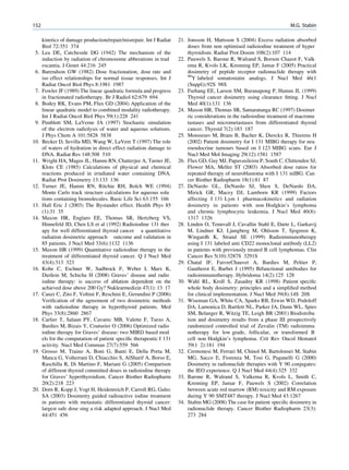


![Elements of Gamma Camera and SPECT
Systems 10
Magdy M. Khalil
Contents radionuclide. Many single-photon and positron emit-
10.1 Introduction . . . . . . . . . . . . . . . . . . . . . . . . . . . . . . . . . . . . . . . . 155 ters are used in the nuclear medicine clinic. The
10.2 The Gamma Camera . . . . . . . . . . . . . . . . . . . . . . . . . . . . . . 156 radiopharmaceuticals are designed for tracing many
10.2.1 Theory . . . . . . . . . . . . . . . . . . . . . . . . . . . . . . . . . . . . . 156 pathophysiologic as well as molecular disorders and
10.2.2 Collimators . . . . . . . . . . . . . . . . . . . . . . . . . . . . . . . . 158
utilize the penetrating capability of gamma rays to
10.2.3 Crystal . . . . . . . . . . . . . . . . . . . . . . . . . . . . . . . . . . . . . 162
10.2.4 Photomultiplier Tube . . . . . . . . . . . . . . . . . . . . . . 164 functionally map the distribution of the administered
10.2.5 Preamplifier . . . . . . . . . . . . . . . . . . . . . . . . . . . . . . . . 165 compound within different biological tissues. The
10.2.6 Amplifier . . . . . . . . . . . . . . . . . . . . . . . . . . . . . . . . . . . 166 imaging systems used for detection of radionuclide-
10.2.7 Pulse Height Analyzer . . . . . . . . . . . . . . . . . . . . . 166
labeled compounds are special devices called scintil-
10.3 Other Photodetectors . . . . . . . . . . . . . . . . . . . . . . . . . . . . . . 167
10.3.1 Position Sensitive PMT . . . . . . . . . . . . . . . . . . . . 167 lation cameras or positron emission tomographic (PET)
10.3.2 Avalanche Photodiode . . . . . . . . . . . . . . . . . . . . . 168 scanners. These devices have passed through a number
10.3.3 Silicon Photomultiplier . . . . . . . . . . . . . . . . . . . . 168 of developments since their introduction in the late
10.4 Semiconductors . . . . . . . . . . . . . . . . . . . . . . . . . . . . . . . . . . . . 169
1960s and have had a significant impact on the prac-
10.5 SPECT/CT . . . . . . . . . . . . . . . . . . . . . . . . . . . . . . . . . . . . . . . . . 170
10.5.1 Levels of Integration . . . . . . . . . . . . . . . . . . . . . . . 171 tice and diagnostic quality of nuclear medicine.
10.5.2 Early and Recent SPECT/CT Systems . . . . . 172 The birth of nuclear medicine instrumentation dates
10.5.3 Temporal Mismatch . . . . . . . . . . . . . . . . . . . . . . . . 173 to 1925; Blumgert and his coworker Otto C. Yens had
10.5.4 Other Artifacts . . . . . . . . . . . . . . . . . . . . . . . . . . . . . 174
modified the cloud chamber to measure the circulation
10.5.5 SPECT/CT Applications . . . . . . . . . . . . . . . . . . . 174
10.6 Conclusions . . . . . . . . . . . . . . . . . . . . . . . . . . . . . . . . . . . . . . . . 175 time using an arm-to-arm method [1]. They used a
References . . . . . . . . . . . . . . . . . . . . . . . . . . . . . . . . . . . . . . . . . . . . . . . . . 175 mixture of radium decay products that emit beta and
gamma rays. Blumgert postulated some assumptions
for designing a detector or measurement technique
that still hold true when compared with requirements
of today: The technique must be objective and nonin-
10.1 Introduction vasive and have the capability to measure the arrival of
the substance automatically [1]. In 1951, Benedict
Nuclear medicine provides noninvasive imaging tools Cassen introduced the rectilinear scanner to register a
to detect a variety of human diseases. The two major distribution of radioactivity accumulated in a human
components of these imaging procedures are radio- body. To scan a given area of interest, the scanner
pharmaceuticals and an imaging system. The latter moves along a straight line collecting activity point
is a position-sensitive detector that relies on detect- by point. The scanner then moves over a predeter-
ing gamma photons emitted from the administered mined distance and moves back in the opposite direc-
tion to span an equivalent length. This process is
continued until the device scans the desired area of
interest. Images are formed by a mechanical relay
M.M. Khalil
Biological Imaging Centre, MRC/CSC, Imperial College printer that prints the acquired events in a dot style.
London, Hammersmith Campus, Du Cane Road, W12 0NN Initially, the scanner was used to scan iodine-131
London, UK in thyroid patients [2]. A major drawback of the
e mail: magdy.khalil@imperial.ac.uk
M.M. Khalil (ed.), Basic Sciences of Nuclear Medicine, DOI: 10.1007/978 3 540 85962 8 10, 155
# Springer Verlag Berlin Heidelberg 2011](https://image.slidesharecdn.com/basicsciencesofnuclearmedicine-121209103030-phpapp02/85/CIENCIAS-BASICAS-MN-168-320.jpg)
![156 M.M. Khalil
rectilinear scanner was the long acquisition time since by the conventional design. Organ-specific or dedi-
radiation events are registered point by point in a cated designs are now well recognized in cardiac as
sequential manner, taking 60 90 min to determine well as scintimammography. Both imaging procedures
the outline of the thyroid gland [2]. Low contrast and were shown to be superior, with such systems realizing
spatial resolution of the formed images were also dis- better imaging quality and enhancement of lesion
advantages of the rectilinear scanner. detectability. When these approaches were implemen-
In 1953, Hal Anger (at the Donner Laboratory of ted and introduced into the clinic, significant changes
the Lawrence Berkeley Laboratory, USA) developed in data interpretation and diagnostic outcome were
the first camera in which a photographic x-ray film achieved.
was in contact with an NaI(Tl) intensifying screen. He For breast imaging, a miniaturized version of the
used a pinhole collimation and small detector size to gamma camera based either on semiconductor technol-
project the distribution of gamma rays onto the scintil- ogy or scintillation detectors has been commercially
lation screen [3]. Initially, the camera was used to scan available. Another example is the cardiac-specific
patients administered therapeutic doses of 131I. A dis- gamma camera, and a growing number of dedicated
advantage of this prototype was the small field of view cardiac systems have been under development (some
of the imaging system (4-in. diameter). Moreover, of them are combined with computed tomography
good image quality was difficult to obtain unless [CT]) to improve the detection task. The developments
high doses were administered along with long expo- in this area are concerned not only with hardware
sure times. In 1958, Anger succeeded in developing components but also some successful approaches to
the first efficient scintillation camera, which was then improve diagnostic images using resolution recovery
called by his name, the Anger camera. Marked prog- and subsequent reduction of imaging time or adminis-
ress in detection efficiency was realized as he used an tered dose. It has become possible that some imaging
NaI(Tl) crystal, photomultiplier tubes (PMTs), and a systems can perform cardiac scanning in a signifi-
larger field of view. cantly shorter scanning time than conventionally
Recent years have witnessed remarkable advances used. The utility of SPECT systems is increasing
in the design of the gamma camera and single-photon with the addition of anatomical imaging devices such
emission computed tomographic (SPECT) systems. as CT and magnetic resonance imaging (MRI), and
These were not only concerned with clinical scanners this is covered in some detail in the last few sections of
but also small-animal SPECT systems, providing a the chapter.
new era of molecular and imaging innovations using Now, there is also interest in using semiconductor
compounds labeled with single-photon emitters. photodetectors to replace the historical PMTs to pro-
Unlike PET, SPECT compounds in theory can provide vide compactness, portability, and reduction of space
images with high spatial resolution and the ability to requirements along with features that allow incorpora-
follow the tracer uptake over more time intervals, tion in multimodality imaging devices. The next gen-
including hours and days. PET imaging lacks such a eration of SPECT and PET devices will exploit these
property due to the short half-lives of most tracers. In characteristics to overcome the current performance
contrast to PET scanners, dual tracer imaging capabil- limitations of both imaging modalities.
ity can be easily achieved using SPECT compounds
and a gamma camera system equipped with a multi-
channel analyzer.
While most of the present and commercially avail- 10.2 The Gamma Camera
able gamma cameras are based largely on the original
design made by Anger using sodium iodide crystal,
successful trials of new imaging systems that suit 10.2.1 Theory
either specific imaging requirements or improved
image quality and enhanced diagnostic accuracy are The gamma camera is a medical radiation detection
emerging. One of these changes is the trend toward device of various hardware components such that
manufacturing a semiconductor gamma camera with each component has a specific role in the photon
performance characteristics superior to those achieved detection process. It is outside the scope of the chapter](https://image.slidesharecdn.com/basicsciencesofnuclearmedicine-121209103030-phpapp02/85/CIENCIAS-BASICAS-MN-169-320.jpg)
![10 Elements of Gamma Camera and SPECT Systems 157
to describe all these elements and their detailed func- by a thresholding process, by which the tube signals
tions. However, the principal components of the imag- below a set value are either ignored or the tubes are
ing system are described in addition to their relative adjusted by a threshold value [5, 6].
contribution to image formation. Generally, these The output of the PMTs is mapped by a network of
components are the collimator, crystal, PMT, pre- electric resistors. The resistors are weighted according
amplifier, amplifier, pulse height analyzer (PHA), to the spatial position of the PMTs in the x- and y-axes
position circuitry, computer system for data correction of the coordinate system of the array. This helps to
and image analysis, and ultimately a monitor for identify the spatial position of an event based on the
image display. relative amount of current received by each resistor.
Briefly, once the patient has been injected and The classical method of event positioning determina-
prepared for imaging, the first hardware component tion is the Anger logic centroid approach, by which
that is met by the incident photons is the collimator. each event results in four signals XÀ, Xþ, YÀ, and
The collimator determines the directionality of the Yþ, and then applying a simple formula per coordinate
incident photons and accordingly forms the hardware to obtain the spatial position within the 2D (two-
role in outlining the activity distribution within differ- dimensional) matrix [1].
ent organs. The accepted photons then interact with In the analog design of the gamma camera, three
the detector crystal to produce scintillation light signals are detected. Two signals indicate the spatial
photons. The photomultiplier converts the light pulse position (X, Y), and one signal indicates the energy of
into an electronic signal. Through a number of steps the incident photon, Z-signal, or pulse height. The
that involve an identification of the photon energy and three signals are a mathematical analysis of the PMT
event positioning, the scintillation site is determined, output identified according to the positive and nega-
which in turn reflects the spatial position of the emitted tive direction of the x (xþ and xÀ) and y (yþ and yÀ)
photons. coordinates. The energy is proportional to the amount
The heart of the scintillation camera is a position- of light produced in the crystal and with the energy
sensitive detector that can localize the interaction sites deposited by the gamma radiation and computed by
where incident photons imparted their energy. The summing all the output signals of the involved PMTs.
conventional design of the gamma camera consists of The position signal is divided by the energy signal so
a continuous structure of a scintillation crystal mapped that the spatial coordinate signals become more inde-
by an array of PMTs. The last component is not a point pendent of the energy signal. However, this simple
or pencil-like photodetector, and size, shape, and the process for event localization suffers from nonuniform
number of PMTs vary among manufacturers. Another spatial behavior, differences in PMT gain, and edge
point that should be mentioned is that the scintillation packing problems, which occurs when the scintillation
light emits a narrow beam of light photons; however, it event becomes close to the crystal edge. In the last
diverges and spreads over the crystal and appears at situation, the positioning algorithm fails to accurately
the PMT end as a cone shape, illuminating more than position the scintillation event as the light distribution
one PMT. Most often, one PMT receives the maxi- from the scintillation site is asymmetric or truncated,
mum amount of light produced by the scintillation leading to improper PMT position weighting and loss
event; therefore, the site of interaction could be deter- of spatial resolution. Furthermore, these problems
mined based on a single PMT signal [4]. The original associated with the detector peripheral regions cause
design proposed by Anger is shown in Fig. 10.1. loss of detection efficiency and a reduction of the
To spatially determine a distribution of radioactiv- imaging field of view. In gamma camera-based
ity inside a human organ, an electronic circuit that is coincidence systems, this phenomenon could result
able to localize the position of the emitted gamma in malfunctioning detector areas corrupting the recon-
radiations must exist in the detection system. The struction process.
inclusion of all PMT signals should theoretically In the digital gamma camera, the signal of the PMT
yield better precision in positioning measurements. is digitized by what is called an analog-to-digital
However, as the distant PMTs receive little light, converter (ADC). Further detector development has
they contribute with less certainty to the identification resulted in a process of digitizing the signal directly
process, leading to increased noise. This can be treated at the preamplifier output to couple each PMT to a](https://image.slidesharecdn.com/basicsciencesofnuclearmedicine-121209103030-phpapp02/85/CIENCIAS-BASICAS-MN-170-320.jpg)
![158 M.M. Khalil
Fig. 10.1 Diagram of the
original Anger logic circuit.
(Reprinted with permission
from [3]. Copyright 1958,
American Institute of Physics)
single ADC. In this situation, position information is acquisition. In the calibration process, the PMT
no longer established using resistors and a summation response is calibrated to a spatial activity distributing
circuit, but the localization circuitry has been totally with a well-defined pattern on the crystal surface [9].
replaced by a dedicated onboard computer [7].
After signal digitization, the determination of pulse
position on the crystal is achieved using the “normal-
ized position-weighted sum” circuit, in a fashion simi-
lar to the analog camera “Anger logic circuit,” or by 10.2.2 Collimators
picking up a correction factor from lookup tables con-
structed previously in a calibration test. This process is Gamma radiation emitted from a radioactive source
implemented through a software program using math- are uniformly distributed over a spherical geometry.
ematical algorithms. Other event-positioning methods They are not like light waves, which can be focused
were developed, such as detailed crystal mapping, into a certain point using optical lenses. Gamma radi-
Gaussian fitting, neural networks, maximum likeli- ation emitted from an administered radionuclide into a
hood estimation, and distance-weighted algorithms patient is detected by allowing those photons that pass
applied in an iterative manner [5, 8]. The lookup through a certain direction to interact with the crystal.
correction tables contain information regarding the By this concept, we can collect and outline the radio-
spatial distortion or camera nonlinearity for accurate activity distributed within different tissues by a multi-
determination of event position during routine patient hole aperture or what is called a collimator.](https://image.slidesharecdn.com/basicsciencesofnuclearmedicine-121209103030-phpapp02/85/CIENCIAS-BASICAS-MN-171-320.jpg)
![10 Elements of Gamma Camera and SPECT Systems 159
The collimator is an array of holes and septa properties and signal-to-noise ratio of the acquired
designed with a specific geometric pattern on a slab scintigraphic images.
of lead. The geometric design of the holes and septa Different types and designs were proposed and
determines the collimator type and function. Unfortu- operating in nuclear medicine laboratories. However,
nately, most gamma radiation emitted from an injected the general types of collimators used in nuclear medi-
radionuclide cannot be detected by the gamma camera cine imaging are parallel hole, converging, diverging,
since many gamma rays do not travel in the directions and a special type known as a pinhole collimator,
provided by collimator holes. This is in great part due which has proved valuable in small animal imaging
to the small solid angle provided by the collimator area with superb intrinsic spatial resolution. The main dif-
in addition to the area occupied by the septal thick- ferences among these types, as mentioned, are their
ness. In the detection process for gamma rays, approx- geometric dimensions, including shape, size, and
imately 1 of every 100,000 photons is detected by the width of the collimator holes. Figures 10.2 10.4
detector system, which is relatively inefficient process show different types of collimators used in nuclear
for gamma ray detection, leading to poor count statis- medicine clinics.
tics [10]. The collimator material is often made of Parallel-hole collimators: The holes and septa of
lead due to its attenuation and absorption properties. parallel-hole collimators are parallel to each other,
Lead has a high atomic number (Z ¼ 82) and high providing a chance for those photons that fall perpen-
density (11.3 g/cm3), providing a mass absorption dicular to the crystal surface to be accepted (Fig.10.2a).
coefficient of 2.2 cm2/g at 140 keV associated with The image size projected by the parallel-hole collimator
Tc-99m emission. onto the crystal is 1:1 since it does not offer any geo-
To obtain a better count rate performance, a reduc- metric magnification to the acquired images. The most
tion of collimator resolution cannot be avoided. On the common types of parallel-hole collimator are
other hand, collimators with better spatial resolution
tend to reduce sensitivity at the cost of improving Low-energy all-purpose (LEAP) (or low-energy
image details. Therefore, there is often a trade-off general- purpose, LEGP) collimator
that should be made to the geometric dimensions Low-energy high-resolution (LEHR) collimator
of the holes and septa. The collimation system has Low-energy high-sensitivity (LEHS) collimator
an important role in spatial resolution, sensitivity, Medium- and high-energy (ME and HE, respec-
and count rate of acquired data; it affects the spatial tively) collimators
a b
Scintillation crystal Scintillation crystal
a
d
s
b
c
a: Hole length Scintillation crystal
Fig. 10.2 Three different d: Hole diameter
types of collimator. (a) Shows s: Septal thickness
the parallel geometry along
b: Source distance
with definition of collimator
holes and septa. (b) Pinhole
geometry. (c) Divergent
collimator Field of view](https://image.slidesharecdn.com/basicsciencesofnuclearmedicine-121209103030-phpapp02/85/CIENCIAS-BASICAS-MN-172-320.jpg)
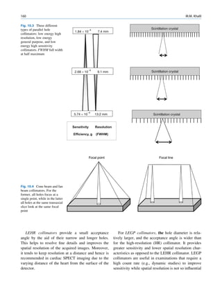
![10 Elements of Gamma Camera and SPECT Systems 161
to the interpretation of the images. Table 10.1 com- were other emissions of higher-energy gamma rays to
pares the general purpose (GP) and HR collimators for lessen septal penetration artifacts and improve quantita-
a commercially available gamma camera system [11]. tive accuracy. Some manufacturers overcame the prob-
The difference in geometric dimensions of the lem by introducing new collimators with a special
hole diameter and length in addition to the septal design that are able to maintain resolution and sensitiv-
thickness of both collimators results in a significant ity of the examination while providing lower septal
improvement of the sensitivity of the GP collimator penetration [13].
over the HR collimator with minimal loss in resolution Converging and diverging collimators have a field
measurements. In Table 10.1, notice that the GP colli- of view different from parallel-hole collimators with
mator provides a count rate efficiency about 50% the same exit plane size. The converging collimators
greater than that of the HR collimator. It should be have a smaller field of view, whereas the diverging
pointed out that manufacturers have their own special ones offer a larger field of view than that provided by
designs and label them differently. Collimator specifi- parallel-hole collimators. In diverging collimators, the
cations given, for example, to a GP collimator might direction of the holes diverges from the point of view
be completely different from those given by another of the back surface of the collimator (the face oppos-
manufacturer to the same type of collimator [12]. ing the crystal). They are used in cameras with a small
LEHS collimators: This type of collimators pro- field of view so that they can lessen large organs to be
vides good count rate capabilities and produces projected onto the camera crystal (Fig. 10.2c).
images with low statistical noise if compared with In converging collimators, the holes are converging
other collimators given the same acquisition time. from the perspective of the back surface of the collima-
However, this is achieved by trading off the resolution tor. Cone beam and fan beam are special types of
properties of the acquired images. In other words, the gamma camera collimators; the former has one focal
improvement in sensitivity is obtained at the cost of point for all collimator holes that lies at a certain dis-
compromising the detectability of fine structures. tance away from the collimator surface and is called
Figure 10.3 shows the trade-off between spatial reso- the focal point. The focal length is minimal at the center
lution and sensitivity in collimator design. of the collimator and increases gradually as it goes to
HE and ME collimators: Medium-energy radionu- the periphery. In the fan beam collimator, each row of
clides in nuclear medicine such as gallium-67 and collimator holes has its own focal point, and all the focal
In-111 and high-energy radionuclides such as I-131 points form a focal line for the entire collimator. As
and Fluorodeoxyglucose-F18 (FDG) have high such, it has parallel collimation along the axial direction
penetrating power and thus could penetrate collimator of the subject and converging collimation within each
septa, causing higher background images and could slice, providing independent and nonoverlapping pro-
adversely affect spatial contrast. ME and HE collimators jection profiles. This geometry allows for a simplified
have increased septal thickness and provide a lower slice-by-slice image reconstruction to be applied. Cone
transparency to high-energy gamma photons than beam geometry, however, complicates image recon-
lower-energy collimators. However, radionuclide stud- struction by involving the holes axially and transaxially.
ies that use ME and HE collimators manifest lower Both collimators are shown in Fig. 10.4.
spatial resolution by degrading small-size lesions. As a A converging collimator provides a magnified view
result, lower quantitative accuracy is achieved by partial of small objects found at locations between the colli-
volume averaging [13]. Nevertheless, ME collimators mator surface and the focal point/line with relative
were recommended over LE collimators in studies that improvement in count sensitivity, which is maximum
required low-energy photons (e.g., iodine-123) if there at the focal site. They are of particular interest in brain
Table 10.1 Characteristics of two different collimators: low energy general purpose (GP) and high resolution (HR) collimatorsa
Hole diameter Septal thickness Hole length Resolution at Relative
Collimator (mm) (mm) (mm) 15 cm (mm) efficiency
GP 1.40 0.18 25.4 11.5 1.0
HR 2.03 0.13 54.0 9.5 0.52
a
Taken from Ref. [11].](https://image.slidesharecdn.com/basicsciencesofnuclearmedicine-121209103030-phpapp02/85/CIENCIAS-BASICAS-MN-174-320.jpg)
![162 M.M. Khalil
tomographic imaging and in some other applications. Table 10.2 Properties of scintillation crystals used in gamma
Resolution of the converging collimator is maximum camera
at the surface and decreases with an increase in source Na(Tl) CsI(Tl) CsI(Na)
distance, while the sensitivity increases gradually Zeff 3.67 54 54
from the collimator surface until the source reaches 3
Density (g/cm ) 50 4.51 4.51
the focal point. This is because the fraction of holes
Decay time (ns) 230 1,000 630
that can see the object at a near distance increases with
a
an increase in object distance; therefore, the object at Photon yield (keV) 38 45 52 39
larger distances is seen by more holes, resulting in Refraction index 1.85 1.8 1.84
improved sensitivity. Magnification takes place only Hygroscopic Yes Slightly yes
in one direction, typically the transverse direction, and
Peak emission (nm) 415 540 420
the cone beam collimator magnifies along the axial a
CsI(Tl) is poorly matched to the response of photomultiplier
direction as well [14]. The drawbacks of converging tube (PMT). However, the scintillation yield is much higher
collimators are reduced field of view and data insuffi- when measured by photodiodes with extended response into
ciency for 3D (three-dimensional) reconstruction. the red region of the spectrum [20].
Pinhole collimator: The pinhole collimator is an
important type that is used frequently in nuclear medi- desired features that make it preferred over alterna-
cine laboratories, especially in small-organ imaging, tives. Table 10.2 lists the most common crystals used
such as thyroid and parathyroid scanning (Fig. 10.2b). in gamma camera design.
Also, it has useful applications in skeletal extremities, The most commonly used detector material for the
bone joints, and the pediatric population. It is a cone- gamma camera is the thallium-activated sodium iodide
shaped structure made of lead, tungsten, and platinum [NaI(Tl)] crystal. It utilizes the scintillation phenome-
and has an aperture of a few millimeters in diameter non to convert gamma rays into light quanta that can be
(2 6 mm). In the small-animal pinhole collimator, the amplified by a PMT to produce a detectable electronic
aperture size can go decrease to 1 2 mm or even less signal. Scintillation occurs when the incident photon
to meet the resolution requirements imposed by small interacts with the crystal material to produce photoelec-
structures and minute tracer uptake [15]. The collima- trons or Compton scattering. The resulting electrons
tor length that extends from the back surface to the from photoelectric or Compton interactions travel
point of the aperture is 20 25 cm. Image formation can short distances within the crystal to produce more inter-
be described using lens equations in the sense that the actions in a form of excitations or ionizations of the
acquired data reveal an inverted and magnified image. crystal atoms and molecules. These excited products
The pinhole collimator provides a magnified image of are deexcited by converting to the ground state by
small objects, yielding an appearance that reflects an releasing scintillation light.
improvement in spatial resolution. A continuous large slab of sodium iodide crystal is
the conventional structure that is used in most gamma
camera designs. The crystal thickness can be 3/8 in.,
5/8 in., or even larger for high detection efficiency
10.2.3 Crystal [16]. A pixilated or segmented version of scintillation
crystals has also been utilized in small SPECT systems
Scintillation crystal is the second component that using photosensitive PMTs as photodetectors and
encounters the incident photons after passing the col- parallel or pinhole imaging geometry, yielding high-
limator holes. There are some favorable properties resolution tomographic images. Segmentation of the
based on which the crystal needs to be selected before scintillation crystal allows the spatial resolution of the
implementation in gamma camera design. Scintillators imaging system to be improved to an extent deter-
of high density, high atomic number, short decay time, mined mainly by the segmentation size. Nevertheless,
high light output, and low cost are desired and allow this comes with a reduction of count sensitivity,
better imaging performance. However, there is no increased costs, and degraded energy resolution [17].
ideal detector material in the field of diagnostic radi- It is often used in small field-of-view, organ specific-
ology; most often, the selected material has some systems or in small-animal scanners. Another option](https://image.slidesharecdn.com/basicsciencesofnuclearmedicine-121209103030-phpapp02/85/CIENCIAS-BASICAS-MN-175-320.jpg)
![10 Elements of Gamma Camera and SPECT Systems 163
that can be considered as an intermediate solution spatial resolution of 2.5 mm, intrinsic energy resolu-
between continuous and segmented or pixilated crys- tion of 35% (at 140 keV), and intrinsic sensitivity of
tals is partially slotted crystals, which have been inves- 42% using an energy width of 35% at 140 keV. Com-
tigated to produce better energy resolution than fully parison of partially slotted CsI(Na) and CsI(Tl) crys-
pixilated crystals and improved detection sensitivity tals revealed the superior performance of the latter in
while maintaining the spatial resolution at better levels terms of detection efficiency and spatial and energy
with an appropriate data analysis [18, 19]. Additional resolution [19].
features provided by such systems are their relative Yttrium aluminum perovskite (YALO3:Ce) is a
ease and cheap manufacturing costs. nonhygroscopic scintillation crystal with the structure
The NaI(Tl) crystal is formed by adding a con- of the perovskite and is called YAP. It has a density of
trolled amount of thallium to a pure sodium iodide 5.37 g/cm3, an effective atomic number of 34 39, and
crystal during growth. The addition of thallium makes light output of 40% relative to NaI(Tl) and is used for
the NaI crystal scintillate at room temperature since gamma as well as annihilation coincidence detection
pure a NaI crystal works at a low temperature under [24]. Lanthanum bromide (LaBr:Ce) is a fast scintilla-
nitrogen cooling [9, 20]. Some precautions are taken tor (short decay time, 16 ns) with high light output.
into account during the design of the NaI(Tl) crystal. It These characteristics are suitable for time-of-flight
must be sealed in an airtight enclosure, usually alumi- applications in PET scanners (see Chap. 11). High
num, to avoid exposure to air owing to its hygroscopic light yield is an important parameter that improves
properties. Exposing the crystal to air can cause yel- the certainty of photon statistics and serves to improve
low spots, which can develop heterogeneous light system spatial and energy resolution. The crystal has
transmission. been evaluated using a flat-panel, photosensitive
Thallium-activated cesium iodide, CsI(Tl), is PMT; it showed good imaging performance for
slightly denser than sodium iodide crystal and better single-photon applications with superior energy reso-
suited if coupled to photodiodes as the emission wave- lution (6 7.5%) and spatial resolution of 0.9 mm. The
length is shifted to higher values, at which the response detection efficiency was also high, yielding 95% at
of PMTs is relatively weaker. It therefore does not 140-keV photon energy [25 27].
properly fit the requirements imposed by PMTs, which While SPECT and PET can be combined into one
need light quanta of shorter wavelengths. A pixilated imaging device known as hybrid SPECT/PET camera,
CsI(Tl) crystal has been tested in a miniaturized gamma there are some modifications that have to be imple-
camera with the collimator matching the detector array mented in electronic circuitry and crystal thickness.
and size suitable for breast imaging. The system showed The latter should be adapted to improve photon detec-
good performance characteristics, such as an improved tion efficiency. The NaI(Tl) crystal is an efficient
spatial resolution, high detection efficiency, and better scintillator for low-energy photons; it is greater than
scatter rejection (~8% full width at half maximum 95% for 140 keV. However, its response against 511
[FWHM] at 140 keV) [21]. The CsI(Tl) crystal has keV is significantly poorer as it shows a coincidence
also been utilized in a pixilated fashion and coupled detection efficiency of about 10%.
with silicon photodiode in a commercial design devel- Some manufacturers have used the traditional scin-
oped as a dedicated cardiac tomograph. Cardius 3 XPO tillation crystal by increasing the thickness up to
is manufactured by Digirad and consists of 768 pixi- 25 mm, while others have modified the crystal so
lated Cs(Tl) crystals coupled to individual silicon that the back surface of the crystal is grooved, allow-
photodiodes and digital Anger electronics for signal ing detection of single-photon emitters to be main-
readout [22]. tained. Meanwhile, the system is efficiently able to
Sodium-activated cesium iodide, CsI(Na), has an image patients injected with medium- and high-energy
emission spectrum similar to NaI(Tl) that suits PMT radiotracers. However, all the major suppliers are cur-
response, with comparable light yield and relatively rently providing PET scanners with a more efficient
slow decay time. It has been utilized in small-animal performance, which have a cylindrical-type design.
SPECT scanners in pixilated 5-mm thickness StarBrite crystal, developed by Bicron Corporation
(21 Â 52 pixels of 2.5 Â 2.5 mm) coupled to photo- (Newbury, OH), is a dual-function 1-in. thick crystal
sensitive PMTs [23]. The system provided an intrinsic that serves to improve the detection efficiency of](https://image.slidesharecdn.com/basicsciencesofnuclearmedicine-121209103030-phpapp02/85/CIENCIAS-BASICAS-MN-176-320.jpg)
![164 M.M. Khalil
medium- and high-energy photons and has been collimator-crystal pair combined with system elec-
incorporated in some commercial designs [16, 28] tronics work together to determine the overall system
(see Fig. 10.5). The slots at the back surface of the spatial resolution and sensitivity given a particular
NaI(Tl) crystal are machined to prevent light diffu- detection task.
sion, reducing the impact of wide-angle reflected light. In humans, visual perception is the ability to inter-
This also maintains a uniform light collection as a pret information and surroundings from visible light
function of position, achieving better intrinsic spatial reaching the eye. In a similar way, the light released
resolution (4 mm) [29, 30]. In performance evaluation from the scintillator after the interaction of the inci-
with other systems, this design provided a good com- dent radiation with the detector system needs an inter-
promise in terms of spatial resolution and sensitivity pretation process. This process in the gamma camera
for 111In ProstaScint1 SPECT imaging. However, the is implemented by the readout component or photo-
detectors that lie in close proximity to the back surface
of the scintillation crystal. A remarkable feature of the
design made by Anger is the photomultiplier mapping
of the detector crystal together with the mathematical
logic he used in event identification.
10.2.4 Photomultiplier Tube
The PMT is an important hardware component in the
detection system of the gamma camera. Its main func-
tion is to convert the scintillation photons to a detect-
able electronic signal. The PMT as shown in Fig. 10.6
is a vacuum tube consisting of an entrance window, a
photocathode, focusing electrodes, electron multiplier
(dynodes), and anode. The PMT has a long history in
many applications, including medical as well as other
Fig. 10.5 StarBrite. (Courtesy of Saint Gobain Crystals) fields such as high-energy physics, spectrophotometry,
PMT
Crystal
Scintillation light
Photoelectron
Semitransparent
photocathode
Focusing
electrodes
Dynodes arranged
in special geometric Voltage
pattern divider
Anode
Optical guide
Fig. 10.6 Photon interaction Output signal to position
logic circuits
and conversion to electronic
signal](https://image.slidesharecdn.com/basicsciencesofnuclearmedicine-121209103030-phpapp02/85/CIENCIAS-BASICAS-MN-177-320.jpg)
![10 Elements of Gamma Camera and SPECT Systems 165
and is widely used in many SPECT and PET imaging 106 108 in addition to low electric noise. However,
systems. the quantum conversion efficiency of PMT is low
since, on average, for every ten scintillation photons
that fall on the photocathode, there is approximately a
release of two electrons, resulting in an efficiency of
10.2.4.1 Photocathode about 20 25% [29]. This in turn has an impact on
intrinsic spatial resolution and energy resolution. The
The photocathode is a photo-emissive surface usually PMT is susceptible to variations in high-voltage power
consisting of bialkali metals with low work functions supply, and its performance is influenced by tempera-
and weakly bound valence electrons. The photocath- ture, humidity, gravity, and magnetic field because of
ode receives the scintillation light from the crystal, the high voltage applied on the dynodes (a limitation
such as an NaI(Tl), by a wavelength of 415 nm at in design of PET/MRI scanners).
maximum. The material for the photocathode that
emits photoelectrons (on an incidence of light quanta)
equivalent to that wavelength will be the material of
choice as it will allow increasing the amount of elec- 10.2.5 Preamplifier
trons emitted and hence will be able to improve the
certainty of the output signal [31]. The shape of the output signal from the PMT is a rapid
The photons emitted from the NaI(Tl) crystal fall rising peak with a slow decaying tail. The rapid peak
on the photocathode of the PMT to eject photoelec- denotes the decay time of the scintillation event within
trons by the physical phenomenon of photoelectric the crystal, and the decaying tail denotes the time
effect. The emitted electrons are amplified through a taken by the electrons to traverse the PMT. The output
series of dynodes placed in a special geometric pattern signal of the PMT cannot be fed directly into the
with high potential difference so that each dynode has amplifier because of the impedance difference
greater voltage (100 300 keV) than the preceding one. between the PMT and the amplifier. The preamplifier
When scintillation light strikes the photocathode, it plays an important role in this regard. It matches the
releases electrons, which are accelerated by the effect impedance between the PMT and the main amplifier
of the high voltage on the first dynode to collide with so that it could be handled by the amplifier and other
it. Electrons emitted from the first dynode are acceler- subsequent electronics. The function of the preampli-
ated to the second dynode to eject more electrons on fier is matching, shaping, and sometimes amplification
collision. The third dynode accelerates the electrons of the signal [9].
from the second dynode toward its face; this multi- Matching: The signal produced from the PMT has
stage electron amplification continues until the elec- a high impedance value, and this requires matching
trons reach the last dynode. At the back end of the with the other electronic circuit components, (i.e.,
PMT, the anode of the PMT collects all the electrons amplifier).
that result from this cascade process. Figure 10.6 illus- Shaping: The signal that is to be fed into a main
trates the process of photon conversion to electronic amplifier needs to have a certain pulse decrease time to
signal after interaction with the crystal. allow proper pole-zero and baseline correction. The
preamplifier works to shape the PMT signal using an
resistor-capacitor circuit (RC) by increasing the time
10.2.4.2 Anode constant, which is then handled by the amplifier.
Amplification: The amplification element of the
The anode of the PMT is an electrode that collects the preamplifier varies in the amount of amplification
amplified electrons through the multistage dynodes according to the type of detector and the magnitude
and outputs the electron current to an external circuit. of the signal. In PMT electronic assembly, the pream-
An adequate potential difference between the anode plifier sometimes has no significant amplification gain
and the last dynodes can be applied to prevent space since the PMT itself provides a considerable amplifi-
charge effects and obtain a large output current [32]. cation through the multistage process of the dynodes.
PMTs have a large electronic gain that can reach The output signal from the preamplifier is a slow](https://image.slidesharecdn.com/basicsciencesofnuclearmedicine-121209103030-phpapp02/85/CIENCIAS-BASICAS-MN-178-320.jpg)
![166 M.M. Khalil
decaying pulse that causes pulse pileup. However, it gamma radiations. One part results from scattering
conveys information regarding the signal amplitude that takes place inside the patient body, and the other
and timing of the scintillation event. This information part results from scattering that occurs in the camera
requires more additional manipulation by an amplifier crystal. The latter is not significant and does not con-
without introducing any type of distortion. The newly tribute to a great extent to the total fraction of scattered
developed SiPM (silicon photomultiplier) photodetec- radiation and the shape of the pulse height spectrum. It
tors (discussed in a separate section of this chapter) is important in medium- and high-energy gamma
provide similar amplification gain as that of PMTs; photons (e.g., In-111 and 131I) and in thick crystals.
hence, preamplification requirements are less demand- The former type of scattering dominates the spectrum
ing in comparison to photodetectors with low gain and results in degraded image quality and contrast
properties. resolution. As a result and for other instrumental rea-
sons (e.g., detector energy resolution), the output sig-
nal is not a sharp peak line on the spectrum; it is a
distribution of pulse heights with one or more photo
10.2.6 Amplifier peaks representing the energies of the administered
radionuclides. The output signal is measured in terms
of voltage or “pulse height,” and every pulse has
The amplifier plays a major role in signal amplifica-
amplitude proportional to the amount of energy depos-
tion and shaping; it is therefore called a shaping ampli-
ited by the gamma ray interactions. A pulse height
fier. Signal amplification is required to permit further
analyzer (PHA), as the name implies, is a device that
processing by the rest of the detector electronics.
that is able to measure the amplitude pulse heights
However, the amplification factor varies greatly with
and compare them to preset values stored within
application and typically is a factor of 100 5,000 [20].
it. There are two types of PHA: single channel and
Shaping of the signal is accomplished by eliminating
multichannel.
the tail from the output signal and giving each signal
its separate width and amplitude without overlapping
with other signals. In summary, the functions of the
10.2.7.1 Single-Channel Analyzer
amplifier are shaping the pulse and decreasing the
resolving time and providing higher gain to drive
The single-channel analyzer records events within a
PHAs, scalars, and so on. It also provides stability to
specified range of pulse amplitude using one channel
maintain proportionality between pulse height and
at a time, applying lower and upper voltage discrimi-
photon energy deposition in the crystal. The amplifier
nators. The output signal of the amplifier has a range
serves to increase the signal-to-noise ratio and main-
of heights (voltages), and selection of the desired
tain proper polarity of the output signal [33].
signals for counting is achieved by setting the lower
discriminator to a level that diminishes all lower
amplitudes, thereby allowing the upper values to be
recorded.
10.2.7 Pulse Height Analyzer
There are different probabilities for the interaction of 10.2.7.2 Multichannel Analyzer
the incident gamma radiations with the crystal. Some
photons impart a fraction or all of their energy into the Acquisition studies that require more energies to be
crystal, whereas other photons impart some of their detected, as in dual-radionuclide acquisitions (e.g., para-
energy and escape the crystal without further interac- thyroid Tl-201 Tc-99m subtraction, meta-iodobenzyl-
tions. Another fraction of photons undergo more than guanidine(MIBG)-131 Tc-99mDTPA [diethylenetria-
one interaction, dissipating all their energies. There- minepentaacetate], and others) and radionuclides with
fore, there are two general forms of scatter that serve more than one energy photo peak (e.g., Ga-67, In-111,
to reduce the capability of the system to accurately and Tl-201), the proper choice for recording these
determine the position and the energy of the incident energies separately and simultaneously is to use a](https://image.slidesharecdn.com/basicsciencesofnuclearmedicine-121209103030-phpapp02/85/CIENCIAS-BASICAS-MN-179-320.jpg)
![10 Elements of Gamma Camera and SPECT Systems 167
multichannel analyzer (MCA). An MCA provides a for an improvement in image spatial resolution.
means of rejecting the scatter region from the spec- The PSPMT-based gamma camera shows the same
trum, allowing the acquired image to be less contami- advantages of a standard gamma camera with the
nated by scattered photons. However, the photo peak additional possibility of utilizing scintillation arrays
region will still contain a significant fraction of photons with a pixel dimension less than 1 mm, thus giving the
that have undergone small-angle scattering. The MCA ability to achieve submillimeter spatial resolution
allows an energy window to be set over the photo peak [37]. Several models have been developed since its
centerline to confine the accepted photons to a certain introduction in 1985 [38]. Three different types can be
energy range. This range is chosen by lower and upper found [39, 40]:
voltage discriminators. The former determines the Proximity mesh dynode: This is the first generation
threshold below which all pulse heights are rejected, and is based on the proximity mesh dynode, by which
and the latter determines the value at which no higher the charge is multiplied around the position of the light
values are accepted. Values that fall between the lower photon striking the photocathode. The charge shower
and upper discriminator are adjusted by a window has a wide intrinsic spread. This model can provide a
width and often are a percentage of the photo peak large active area (e.g., 5 in.) and large number of
energy. For example, a 20% energy window is usually dynodes (e.g., 256) but with large dead space.
selected over the 140-keV photons emitted by Tc-99m. Multichannel dynode: This is a multianode struc-
A 15% window is also used with an improvement in ture that minimizes cross talk between anodes and
contrast and minimal loss in primary photons. More- provides better localization measurements. This type
over, a 10% window is used at the expense of reducing has the following drawbacks: large dead zone, limited
sensitivity [34]. An asymmetric window is another way effective area, and limited number of dynodes.
to reduce the effect of scattered photons as it can Metal channel dynode: Combined with multichan-
improve image contrast, spatial resolution, and clinical nel or crossed-plate anode techniques, this dynode can
impression [35, 36]. provides low cross talk and well-focused charge dis-
A variety of scatter correction techniques has been tribution, reducing the intrinsic spread to 0.5 mm
devised to reduce the adverse effects of scattered radi- FWHM [39]. For SPECT imaging, the use of a
ation. For proper quantitation and absolute measures crossed-wired anode PSPMT is more suitable when
of tracer concentrations, images must be corrected for coupled with a flat crystal because of continuous
scatter and other image-degrading factors. A gamma position linearity requirements. However, this config-
camera with an MCA is necessary when energy-based urations may not allow the best performance of the
scatter corrections are applied to the acquired data. PSPMT; thus, to utilize the full potential of its intrinsic
Advances in semiconductor technology have moti- characteristics, a multicrystal array is needed to attain
vated the development of gamma camera systems high detection efficiency, narrow light spread func-
with significant improvement in energy resolution. tion, and reasonable light output [41].
This in turn improves the spatial contrast and signal- A PSPMT is relatively more expensive than the
to-noise ratio, resulting in improved image quality. conventional type, and most of its use is in preclinical
Chapter 15 has more details about the scatter phenom- small-animal SPECT and PET scanners and portable
enon and its correction techniques. and compact miniaturized gamma cameras, and it is of
interest in producing high-resolution breast scanners
[42, 43]. This type of diagnostic examination requires
compact and flexible systems to fit geometric require-
10.3 Other Photodetectors ments imposed by the female breast. The technologic
advances in the development of a compact flat-panel
PSPMT with less dead space allowed manufacture of a
10.3.1 Position-Sensitive PMT portable gamma camera with a large detection area
and high imaging performance with better spatial res-
The position-sensitive PMT (PSPMT) is a modified olution [44]. It has become possible that an array of
version of the conventional PMT but with compact 256 crystal elements can be coupled directly to one
size and position determination capabilities, allowing PMT, and the crystals can be read out individually [45].](https://image.slidesharecdn.com/basicsciencesofnuclearmedicine-121209103030-phpapp02/85/CIENCIAS-BASICAS-MN-180-320.jpg)
![168 M.M. Khalil
detectors and crystals of longer wavelengths, such as
mentioned for CsI(Tl) crystals. They have higher gain
than silicon photodiodes and better timing character-
istics, on the order of 1 ns [47].
In comparison to the PMT, the APD is sensitive to
temperature variations, can be operated in a lower
voltage than a PMT, and is stable in the presence of
high magnetic fields, like that encountered in MRI
[48]. Thus, the APD was suitable in PET inserts
incorporated into MRI machines, providing compact
and simultaneous hybrid PET/MRI imaging systems.
The semiconductor junction is operated at a high
reverse bias, and the output signal is proportional to
Fig. 10.7 Hamamatsu H8500 flat panel position sensitive the initial number of light photons. They are charac-
photomultiplier tube (PSPMT). (From [100]. # 2006 IEEE terized by low gain and relatively higher quantum
with permission)
efficiency.
The position-sensitive APD (PSAPD) is similar to
the APD but with fewer readout requirements. The
The Hamamatsu H8500 flat-panel PSPMT is shown in back surface is connected to a resistive layer that
Fig. 10.7. allows for multiple contacts to be fabricated. Position
information can then be obtained by the charge sharing
among electrodes that enables the determination of
position of interaction [49]. Consequently, this struc-
10.3.2 Avalanche Photodiode ture allows for simple and compact detector assembly
and enables use of significantly fewer readout chan-
An interesting trend is the development of semicon- nels, resulting in lower manufacturing costs. The
ductor photodetectors to replace the bulky and signifi- alignment of the detector with the crystal array in
cantly large volumetric shape of the PMT. One of the this design is not as important as is the case in the
major drawbacks of PMTs is their sensitivity to mag- individual APD array. This type of photodetector has
netic fields. As the electrons are accelerated within the an attractive interest in small-animal scanners, includ-
PMT by the effect of high potential applied between ing depth of interaction encoding, to improve scanner
photocathode and dynodes and between successive spatial resolution and sensitivity [50 52]. This type
dynodes, any source that influences this process also has applications in high-resolution small-animal
would be an undesired component of noise leading to imaging systems. For example, it has been coupled to
tube performance degradation. A magnetic field more a CsI(Tl) scintillator in a multipinhole small-animal
than 10 mT is able to alter the gain and energy resolu- SPECT system with significant improvement in detec-
tion of a PMT [46]. One of the earliest solutions was to tion efficiency and spatial resolution on the order of
use long cables of fiber optics. The better alternative 1 mm [53].
was then to use semiconductor photodetectors. Efforts
have been made to develop semiconductor detectors
with better detection efficiency and intrinsic spatial
resolution, providing a reliable and robust perfor- 10.3.3 Silicon Photomultiplier
mance compared to PMTs or PSPMTs. Among these
developments are the avalanche photodiodes (APDs), The Geiger-mode APD or silicon photomultiplier
which can be produced in small dimensions, providing (SiPM) is a densely packed matrix of small APDs
an improvement of spatial resolution in addition to (20 Â 20 100 Â 100 mm2) joint together on common
reduced detector volume, and thereby fit space silicon substrate and work in limited Geiger discharge
requirements imposed by multimodality imaging mode. Each cell is an independent Geiger mode detec-
scanners. They are also well suited for pixilated tor, voltage biased, and discharges when it interacts](https://image.slidesharecdn.com/basicsciencesofnuclearmedicine-121209103030-phpapp02/85/CIENCIAS-BASICAS-MN-181-320.jpg)
![10 Elements of Gamma Camera and SPECT Systems 169
process in the vacuum of the PMT to yield an ampli-
fied signal that afterward is used in the calculation of
event position and energy.
The use of semiconductor material in the field of
radiation detection and measurements has been estab-
lished for many years in different areas of science and
engineering. The most commonly used are silicon (Si)
and germanium (Ge). The atomic number of the for-
mer is 14, while for the latter it is 32; incorporating
them in a radiation detector necessitates cooling to
Fig. 10.8 Silicon photomultipliers (SiPMs) from different man liquid nitrogen temperatures to avoid electronic noise
ufacturers. (From [101]). # IEEE with permission)
generated from excessive thermal effects [58]. To
fabricate a gamma camera based on a semiconductor
detector, there has been efforts to search for other
materials of high detection efficiency and amenable
with an incident photon. The scintillation light causes for operation in a room temperature environment.
a breakdown discharge for triggering a cell resulting in Better alternatives of interest in the design of
a fast single photoelectron pulse of very high gain gamma ray detection systems are cadmium telluride
105 107. SiPM produces a standard signal when any (CdTe) and cadmium zinc telluride (CdZnTe), which
of the cells goes to breakdown. When many cells are can be considered promising materials for radiation
fired at the same time, the output is the sum of the detectors with good energy resolution, high detection
standard pulses [54]. Characteristics of SiPM are low efficiency, and room temperature operation. Several
noise factor, photon detection efficiency equivalent to advantages can be obtained when designing a gamma
the standard PMT, and low bias voltage. The higher camera based on semiconductor detectors. One is sys-
gain feature provided by SiPM detector marginalizes tem portability as they enable construction of a com-
the electronic noise in contrast to the standard APDs, pact structure free from the PMT and its bulky volume.
which generally have a gain of 100 200. However, the A semiconductor camera also provides improved
dead space around each pixel and the finite probability image contrast due to its better energy resolution com-
of photons to produce avalanche breakdown reduce pared to a sodium iodide crystal. Besides this last
the detection efficiency of the SiPM, making it signifi- feature, high spatial resolution images can be obtained
cantly lower than for the APD [55]. An SiPM is an owing to the pixilated structure that can be implemen-
interesting alternative to the conventional PMT in ted in the detector design [59].
terms of compactness, same level of electronic gain, More than one commercial SPECT system has been
suitability for PET inserts in magnetic resonance sys- released to the market making use of semiconductor
tems and in detector design based on depth encoding technology for improving imaging quality and diag-
information [56, 57]. Figure 10.8 shows SiPMs from nostic accuracy. D-SPECT (Spectrum Dynamics) is a
different manufacturers. CZT-based semiconductor camera commercialized for
imaging cardiac patients. The system consists of nine
collimated detector columns arranged in a curved con-
figuration to conform to the shape of the left side of a
10.4 Semiconductors patient’s chest. Each individual detector, which con-
sists of 1,024 (16 Â 64) 5-mm thick CZT elements
Instead of detecting the scintillation light on multiple (2.46 Â 2.46 mm), is allowed to translate and rotate
stages, semiconductor detectors provide a detection independently so that a large number of viewing
means by which the incident radiation is converted to angles can be achieved for the region of interest [60].
an electronic signal once it interacts with the detector The improved energy resolution (5.5% at 140 keV)
material. The standard gamma camera, however, relies also allows simultaneous application of a dual-isotope
on converting the incident photon to a scintillation protocol with better identification of lesion defect and
light, which is then amplified through a multistage reduction of photon cross talk [61, 62]. Another design](https://image.slidesharecdn.com/basicsciencesofnuclearmedicine-121209103030-phpapp02/85/CIENCIAS-BASICAS-MN-182-320.jpg)
![170 M.M. Khalil
has been introduced by GE Healthcare (Discovery NM resolving capabilities that are significantly higher
530c) that utilizes an array of pixilated (CZT) semi- than that provided by nuclear SPECT and PET
conductor detectors in fixed positions (without machines. They have the advantages of providing a
motion) that allow acquisition of cardiac projections resolution in the submillimeter range, precise statisti-
in a simultaneous manner. Preliminary evaluations of cal characteristics, and better tissue contrast, espe-
the system demonstrated an improvement in image cially in the presence and use of contrast media [68].
spatial resolution, energy resolution, and sensitivity However, it lacks the property of describing the func-
as opposed to a conventional gamma camera design tional status of a disease. This is important in follow-
[63]. An additional feature was brought about by com- ing up cancer patients postsurgically and in the
bining this system with CT in an integrated SPECT/ presence of fibrotic or necrotic lesions after chemo-
CT (Discovery NM 530c with the LightSpeed VCT or or radiotherapy [69].
Discovery NM/CT 570c) system. The early work of Hasegawa and colleagues opened
A semiconductor camera made of CZT is relatively a gate to many applications in the field of diagnostic
expensive; thus, most of the current commercial ver- radiology and nuclear medicine by coupling two imag-
sions are confined to handheld, miniaturized imaging ing techniques into one operating device: The SPECT
systems or intraoperative small probes. Scintimammo- camera has been merged to x-ray computed tomography
graphy is one of the diagnostic procedures that (CT) in the same device to provide an inherent anato-
requires a dedicated device able to match the position molecular imaging modality able to depict morphologi-
and geometry of the breast. Small and deep lesions are cal as well as functional changes in one imaging session.
a challenging task and require characteristics better While this process of image coregistration can be
than provided by a standard gamma camera. The sen- accomplished by fusing images obtained from two sep-
sitivity of the test is limited by the lesion size, particu- arate modalities, it has been demonstrated that inline
larly for those less than 10 mm [64]. SPECT or PET and CT image acquisition would be
The difficulty in detecting small and deep patho- more advantageous.
logic breast lesions by conventional imaging systems The advantages provided by CT to functional imag-
lies in their limited detection efficiency and relatively ing are not singular. It enables the reading physicians to
poor spatial resolution. However, earlier detection of a precisely localize pathological lesions detected on func-
breast lesion has better diagnostic and prognostic tional images with great confidence. The other advan-
implications. This in turn should be met by instru- tage is the improvement in performing attenuation
ments with high performance characteristics that pro- correction. Again, although this can be performed by
vide better lesion detectability. A breast imager based radionuclide transmission sources, the CT-based
on semiconductor materials such as CZT can provide attenuation correction has been shown to outperform
better spatial and energy resolution, leading to radionuclide-based transmission scanning by providing
improved lesion contrast. This can be achieved using fast and significantly less-noisy attenuation maps.
a narrow energy window, reducing the adverse effects Another benefit provided by CT is the capability of
of scattered photons on image quality. Another prop- deriving dosimetric measurements from the fused
erty of the semiconductor camera is its compactness, images due to accurate outlining of the region of
portability, and smaller dimensions, which fit well interest and thus better volumetric assessment. In the
with breast imaging [65 67]. field of nuclear cardiology, SPECT/CT systems are of
particular importance due to the continuous debate
about attenuation correction [70, 71] and other degrad-
ing factors that reduce diagnostic quality and quantita-
10.5 SPECT/CT tive accuracy, such as partial volume effects [72].
Apart from attenuation correction, calculation of cal-
Nuclear imaging using SPECT or PET techniques cium scoring is also possible with the CT portion of
have well-known capabilities in extracting functional the machine in addition to performing noninvasive
and metabolic information for many human diseases. coronary angiography.
The anatomical details provided by CT and MRI enjoy In some instances, the addition of CT data to
better structural description for human organs by the functional images provides a guiding tool for](https://image.slidesharecdn.com/basicsciencesofnuclearmedicine-121209103030-phpapp02/85/CIENCIAS-BASICAS-MN-183-320.jpg)
![10 Elements of Gamma Camera and SPECT Systems 171
image-based biopsy and thus can play a role in patient and research efforts to use radioactive transmission
management; it also can be of synergistic effect in sources for attenuation correction [77, 78]. The
radiotherapy treatment planning, targeted treatment approaches and different levels of integrating struc-
with brachytherapy, or intensity-modulated radiation tural imaging with the functional data were as follows:
therapy [73]. However, this has become more attrac-
1. The primitive level of integrating functional and
tive with PET imaging than SPECT derived data.
structural images is evidenced by the human
The image of the year for 2006 was an SPECT/CT
brain. This is the traditional way that diagnosticians
image showing both the coronary arteries and blood
used to correlate two images acquired from sepa-
flow to the heart. This took place at the 53rd annual
rate imaging modalities. However, human spatial
meeting of the Society of Nuclear Medicine in San
perception and differences in image textures
Diego, California. The images clearly depicted the ana-
acquired from two different modalities represent a
tomical correlation of the blood flow defects to the
challenge for the reading physicians to mentally
corresponding artery. SPECT/CT devices also are of
correlate the two data sets. This can be more prob-
interest in preclinical and molecular imaging research
lematic in small-size lesions and in identifying the
with similar benefits as those provided to the clinical
spatial extent and spread of a disease.
arena in addition to increased flexibility in performing
2. The second level of image coregistration was
longitudinal research studies.
brought to the clinic by software algorithms. They
On the research level, a tremendous improvement
are relatively simple to apply to rigid structures
in the spatial resolution of micro-CT systems has
such as the brain, where images acquired by CT
enabled the investigators to look at minute structural
or MRI can be merged to functional images
changes that were not possible to see with conven-
acquired and processed on a separate SPECT or
tional systems. Advances in micro-CT systems has
PET machine. This successful implementation is
revealed images of 10 mm and far better, permitting
due to brain rigidity, and variations in the position
subcellular dimensions to be imaged [74].
of internal structures are less likely to occur com-
pared to lung or abdominal image fusion. In a
review [79], localization of brain structures was
10.5.1 Levels of Integration less challenging compared to whole-body applica-
tions, and image fusion may be required for only a
Transmission scanning has been defined as “a useful subgroup of patients. Moreover, in a wide compar-
adjunct to conventional emission scanning for accu- ison of various available software tools for retro-
rately keying isotope deposition to radiographic anat- spective registration of PET/MRI and PET/CT, an
omy” [75]. This article was published by Edward and accuracy of 2 3 mm was consistently achieved,
coworkers in the mid-1960s when they realized the which was below the voxel dimension of PET
importance of combining functional data with their [80] and SPECT as well.
anatomical templates, although they used a simple 3. Use of intrinsic landmarks or extrinsic markers to
approach to obtain the transmission scanning. Investi- accurately fuse the two data sets has been imple-
gators assigned this as the first demonstration of the mented with some impractical drawbacks placed on
feasibility of hybrid imaging [76]. They used an its feasibility in the clinical routine, added technical
Am-241 source located at the center hole of a colli- complexity to the diagnostic procedures, or inaccu-
mated detector; the other detector was adjusted to rate image correlation. This could arise from varia-
acquire the emission and transmission data. This tions that may exist between imaging sessions,
early functional/anatomical study is a clear sign of fixation strategy, or perhaps inability to correlate
the early recognition of combining the two types of internal organs with external markers [69]. Further-
images to strengthen each other and to overcome their more, automatic registration for internal landmarks
inherent weaknesses. However, despite this interesting could also be affected by the limited spatial resolu-
beginning, it was not pursued, and both imaging mod- tion of nuclear images with precise selection of
alities were almost developed independently until the anatomical landmarks; therefore, accurate alignment
early 1990s. Before this time, there was some interest is not guaranteed. These apparent shortcomings of](https://image.slidesharecdn.com/basicsciencesofnuclearmedicine-121209103030-phpapp02/85/CIENCIAS-BASICAS-MN-184-320.jpg)
![172 M.M. Khalil
image coregistration, particularly in nonrigid body dual-modality approaches that drew the attention of
structures, have motivated the development of non- many researchers in the field, and its technical
linear 3D image fusion algorithms able to improve value has been proven in a significant number of
the accuracy of alignment. clinical conditions, leading to a change in patient
4. Ideally, one would like to acquire the structural as management. Although other multimodality imag-
well as the functional data in a simultaneous man- ing approaches have been or are being developed,
ner in the same spatial and temporal domain. This they are aimed most of the time toward preclinical
can be achieved when the detector systems can be imaging, and their clinical counterparts are yet to
incorporated into each other, achieving concurrent be determined.
image acquisition. This multimodality design has
been realized in hybrid PET/MRI systems with
substantial modification to the PET detector system
using APDs instead of the conventional PMTs, as
10.5.2 Early and Recent SPECT/CT
mentioned in this chapter (see [81]).
5. The other alternative is to modify the detector Systems
technology so that a single detector system can
record the two signals with adequate distinction As mentioned, the work of SPECT/CT was launched
for each individual electronic pulse. However, and pioneered by Hasegawa and colleagues at the
x-ray detector systems are energy integrators and University of California, San Francisco, by designing
do not allow individual events to be discriminated the first prototype imaging system able to acquire both
according to their energies. This is in great part due single-photon emitters and x-ray transmission scan-
to the high fluence of the x-ray source compared to ning in one imaging session. The detector material
nuclear photon emission and owing to beam poly- was a high-purity germanium (HPGe) designed for
chromaticity [82]. In other words, the significant simultaneous acquisition of functional and anatomical
differences in photon rates pose challenges to information. The system was a small-bore SPECT/CT
implement multimodality imaging using a “con- system with some technical limitations for direct
ventional” detector for both x-ray and radionuclide application in the clinical setting. The same group
processes; this requires a detector that can switch then developed a clinical prototype SPECT/CT system
between pulse-mode SPECT/PET acquisition and using a GE 9800 CT scanner and GE X/RT as the
charge integration mode [83]. Nevertheless, it has SPECT component mounted in tandem with data
been proposed to simultaneously acquire trimodal- acquired sequentially using the same imaging table.
ity images, namely SPECT/PET/CT, with great The industry recognized the potential of combining
complexities placed on detector design [84]. More- the two imaging techniques, and the first commercial
over, an emerging technology is evolving to record SPECT/CT system was released by GE Healthcare. The
both types of data in a single counting and energy transmission component of the scanner was a one-slice
integrator modes. CT scanner providing a slice thickness of 10 mm. The
6. The other strategy is to place the two detector transmission x-ray source was mounted on a slip-ring
systems side by side such that images can be gantry of the Millennium VG gamma camera (GE
acquired in a sequential manner. This is the way Healthcare). The x-ray detector system was similar to
all clinical SPECT/CT as well PET/CT scanners third-generation CT scanners and was designed to move
are presently manufactured and operated. There- along with the gamma camera detector system. The time
fore, one can simply deduce that the higher the required for one-slice acquisition was about 14 s, and
level of integration, the more demands placed on contiguous slices were taken by moving the patient in
the technologic complexity. The range of multi- the axial direction [86].
modality imaging has been extended to include Coincidence detection was also available since the
functional only, structural only, or a combination crystal thickness was thicker than used in regular
of functional and structural imaging modalities SPECT systems (16.5 mm). The voltage of the x-ray
to yield a variety of integrated or hybrid dia- tube was 140 kVp, and tube current was 2.5 mA,
gnostic options [85] SPECT/CT is among the best leading to a significant reduction of the x-ray absorbed](https://image.slidesharecdn.com/basicsciencesofnuclearmedicine-121209103030-phpapp02/85/CIENCIAS-BASICAS-MN-185-320.jpg)
![10 Elements of Gamma Camera and SPECT Systems 173
dose when compared to standard diagnostic CT as low as 1 2.5 mA or higher, such as 5 80, 20 354,
machines. With these characteristics, the system was or 20 500 mA. The number of slices has also been
not able to reveal useful diagnostic information but increased to 6, 16, 64, or even more, as can be found in
satisfied the requirements of attenuation correction. the Philips BrightView XCT using a flat-panel CT in
The CT subsystem could cover the heart in 13 17 which the number of slices is 140 and slice thickness is
slices in a period of 5 min. 1 mm. However, the slice thickness can be lower than
The same manufacturer released another SPECT/ 1 mm, in some SPECT/CT scanners reaching a value
CT version (GE Infinia Hawkeye-4) that provided a of 0.6 mm. An important feature of the currently
four-slice low-dose CT with fewer demands on room available commercial designs is the high-speed acqui-
space requirements. The slice thickness of this system sitions using spiral CT generation by which the rota-
was fixed to 5 mm. The system was able to provide tion time can be even less than 0.5 s. This allows for
adequate attenuation correction and lesion localization scanning the heart region in one full rotation, mini-
with emphasis placed on limited spatial resolution mizing problems of temporal coverage associated
when compared to fully diagnostic CT machines. Fur- with older CT models. Also, it can help reduce the
thermore, the volumetric dose index in the acquisition likelihood of patient motion and enhance patient
of a head protocol was found to be 8 mGy, in compar- throughput.
ison to 42 mGy as measured by DST, which is a 16- One of the disadvantages provided by fully diag-
slice diagnostic CT used in PET/CT scanners [87]. nostic CT acquisition is the increased absorbed dose
Other vendors have SPECT/CT systems that are able to the subject under examination. As discussed by
to provide diagnosticians high-quality and fully diag- Kauffman and Di Carli [88], cardiac imaging using
nostic CT information; thereby, the utility of the CT hybrid functional and anatomical SPECT/CT is
portion of the hybrid SPECT/CT scanner is no longer challenged by the high radiation dose to the patients.
concerned with attenuation correction and lesion However, recent approaches to minimize high levels
localization, but its clinical benefits have been of absorbed doses have been implemented, such as
extended to other areas of cardiac imaging, such as low-dose CT angiography and a reduced amount of
calculation of calcium scoring and coronary CT angi- injected activity into the patient.
ography. Different SPECT/CT models are shown in
Fig. 10.9.
The tube voltage and current are important para-
meters that determine image quality and levels of the 10.5.3 Temporal Mismatch
absorbed dose. Vendors vary in their CT specification,
such that the tube voltage can be 90 140, 110 130, Another problem also of concern is the differences that
120, or 120 140 kVp. Also, the tube current could be arise from temporal data sampling. The CT scan is
Fig. 10.9 (a) The Siemens Symbia single photon emission (c) BrightView XCT, which integrates the SPECT system with
computed tomography/computed tomography (SPECT/CT) flat detector x ray CT
scanner. (b) Millennium VG Hawkeye SPECT/CT scanner.](https://image.slidesharecdn.com/basicsciencesofnuclearmedicine-121209103030-phpapp02/85/CIENCIAS-BASICAS-MN-186-320.jpg)
![174 M.M. Khalil
acquired in a relatively short duration compared to SPECT image quality could be potentially a candidate
emission acquisition. This actually is a problem espe- to cause undesired errors in the coregistered images.
cially in the thoracic region as the lung and heart are The center of rotation and alignment of the two imag-
moving organs and not fixed in one spatial position. ing modalities is a pertinent factor in the accuracy and
The emission images are a temporal average of the reliability of attenuation correction and image align-
organ motion over the respiratory or cardiac cycles. ment. The existence of a patient’s arms in the field of
This phenomenon has its undesired consequences in view could also introduce attenuation or truncation
corrupting the attenuation correction and leading to artifacts. Metals and high-density materials have the
misregistration errors and false interpretation results. capability of altering the attenuation coefficients and
One of the potential applications of SPECT/CT sys- serve to enhance beam hardening effects.
tems is their utility in cardiac imaging, for which better
attenuation correction can be performed. However, due
to the mentioned limitation, there are recommendations
to check the acquired emission/transmission data for 10.5.5 SPECT/CT Applications
possible artifacts [89]. This issue of data misalignment
between CT and radionuclide emission scanning has The utility of SPECT/CT systems is expanding in
also been found in cardiac PET studies as well as in many areas of research and clinical practice. In addi-
lung FDG examinations. tion to attenuation correction, other image degrading
Kennedy et al. [90] have recently shown that 23% of factors can also be accounted for from the use of the
the patient studied using cardiac SPECT/CT had a mis- CT images. A model-based approach to derive Comp-
registration errors of significant effects and 16% were in ton probabilities or transmission-based scatter correc-
the direction of the most severe artifacts [91]. A compa- tion can be applied using CT images to correct for
rable percentage was also found in Rb-82 cardiac PET/ scattered photons, as shown by Willowson et al. [94].
CT scanning with magnitudes of 24.6% and 29.0% in In the same report, the effect of partial volume also
adenosine stress and rest studies, respectively [92]. Mis- was accounted for using the CT data, and all correc-
registration errors resulted in false-positive results in tions revealed an accurate tracer estimate of 1% with a
40% of patients undergoing dipyridamole stress and precision of Æ7%. In brain studies, correction for
rest Rb-82 cardiac PET/CT [93]. Another study demon- partial volume is critical, and reliable and quantitative
strated 42% errors in cardiac SPECT/CT images, con- outcome cannot be obtained without considerations
cluding that careful inspection of attenuation correction placed on the phenomenon. The binding potential of
maps and registration are needed to avoid reconstruction the striatum can be underestimated by 50% in the
artifacts due to misregistration [94]. Nonetheless, man- absence of partial volume correction. However, in
ual intervention as well as quality control software pro- the presence of anatomically guided partial volume
grams are possible ways to realign the images and correction, substantial improvement can be obtained
improve the correction and imaging task [95]. [97, 98]. In a porcine model, Da Silva et al. [73]
showed that an attenuation correction alone for myo-
cardial perfusion Tc-99m-labeled sestamibi cannot
yield higher quantitative accuracy without proper cor-
rection for partial volume. The combined corrections,
10.5.4 Other Artifacts however, resulted in higher absolute activity concen-
trations within a range of 10%. CT data provided an
One of the potential applications of SPECT/CT sys- accurate template for modeling the subject anatomy
tems is their use in skeletal scintigraphy. Gnanasegaran and for accurate definition of the myocardial bound-
et al. [96] discussed the most common artifacts asso- aries (further discussion can be found in Chap. 15).
ciated with bone SPECT/CT. These are SPECT/CT SPECT/CT systems are also helpful in absorbed
misregistration, respiratory artifacts due to lung dose estimation since the CT images are able to pro-
motion, arms up or down, metal or contrast agent vide a highly accurate assessment of tumor volume.
artifacts, patient size and noise, together with limita- This is an important step in dose calculation schemes
tions arising from CT scanners. Factors that affect required in radioimmunotherapy and other dose](https://image.slidesharecdn.com/basicsciencesofnuclearmedicine-121209103030-phpapp02/85/CIENCIAS-BASICAS-MN-187-320.jpg)
![10 Elements of Gamma Camera and SPECT Systems 175
estimation disciplines. As SPECT data can provide tance dependent weighting. IEEE Nucl Sci Symp Conf
important functional information about drug distribu- Rec 5:2445 2448
6. Kulberg GH, Muehllehner G, van Dijk N (1972) Improved
tion, residence time, and patient follow-up, the addi- resolution of the Anger scintillation camera through
tive value of CT can enhance the quantitative accuracy the use of threshold preamplifiers. J Nucl Med 13
of the measurements for subsequent accurate dose (2):169 171
estimates. In the case of pure beta emitters and 7. Ricard M (2004) Imaging of gamma emitters using scintil
lation cameras. Nucl Instrum Meth Phys Res A 527(1 2):
attempts to image radiotracer distribution based on 124 129, In: Proceedings of the 2nd international confer
bremsstrahlung radiation, x-ray CT provides a good ence on imaging technologies in biomedical sciences
anatomical template to delineate and distinguish vari- 8. Joung J, Miyaoka RS, Kohlmyer S, Lewellen TK (2000)
ous tissue uptakes [99]. Implementation of ML based positioning algorithms for
scintillation cameras. IEEE Trans Nucl Sci 47(3):1104
1111
9. Cherry SR, Sorenson JA, Phelps ME (2003) Physics in
nuclear medicine. Saunders, Philadelphia
10.6 Conclusions 10. Moore SC, Kouris K, Cullum I (1992) Collimator design
for single photon emission tomography. Eur J Nucl Med
19(2):138 150
The gamma camera is a device with multicompart- 11. Lau YH, Hutton BF, Beekman FJ (2001) Choice of colli
ments. Each of these elements plays a role in the mator for cardiac SPET when resolution compensation is
photon detection task and determination of tracer bio- included in iterative reconstruction. Eur J Nucl Med 28
distribution. Digital technology and multihead sys- (1):39 47
12. Murphy PH (1987) Acceptance testing and quality control
tems were key factors that enhanced and motivated of gamma cameras, including SPECT. J Nucl Med 28
the performance of the gamma camera. Implementa- (7):1221 1227
tions of new camera design with different geometry, 13. Inoue Y, Shirouzu I, Machida T, Yoshizawa Y, Akita F,
fast acquisition protocols, and reconstruction algo- Doi I, Watadani T, Noda M, Yoshikawa K, Ohtomo K
(2003) Physical characteristics of low and medium energy
rithms provide relatively new features for the current collimators for 123I imaging and simultaneous dual iso
and future generations of the gamma camera. Semi- tope imaging. Nucl Med Commun 24(11):1195 1202
conductor systems with room temperature materials 14. Accorsi R (2008) Brain single photon emission CT phys
and good performance characteristics are of interest ics principles. Am J Neuroradiol 29(7):1247 1256
15. Peremans K, Cornelissen B, Van Den Bossche B, Aude
among some manufacturers and medical users. Use of naert K, Van de Wiele C (2005) A review of small animal
new photodetectors would help to enhance the diag- imaging planar and pinhole SPECT gamma camera imag
nostic quality of nuclear medicine images. They also ing. Vet Radiol Ultrasound 46(2):162 170
provide compactness, portability, and better imaging 16. Wong TZ, Turkington TG, Polascik TJ, Coleman RE
(2005) ProstaScint (capromab pendetide) imaging using
features. The addition of CT as a structural imaging hybrid gamma camera CT technology. AJR Am J Roent
modality to SPECT systems has found numerous genol 184:676 680
applications in many areas of clinical practice as well 17. Kupinski MA, Barrett HH (2005) Small animal SPECT
as in the research setting. imaging. Spinger Science þ Business Media, New York
18. Giokaris N, Loudos G, Maintas D, Karabarbounis A, Lem
besi M, Spanoudaki V, Stiliaris E, Boukis S et al (2005)
Partially slotted crystals for a high resolution g camera
References based on a position sensitive photomultiplier. Nucl
Instrum Meth Phys Res A 550:305 312
19. Giokaris N, Loudo G, Maintas D, Karabarbounis A, Lem
1. Patton DD (2003) The birth of nuclear medicine instru besi M et al (2006) Comparison of CsI(Tl) and CsI(Na)
mentation: Blumgart and Yens, 1925. J Nucl Med 44(8): partially slotted crystals for high resolution SPECT imag
1362 1365 ing. Nucl Instrum Meth Phys Res A 569:185 187
2. Graham LS, Kereiakes JG, Harris C, Cohen MB (1989) 20. Knoll GF (2000) Radiation detection and measurements,
Nuclear medicine from Becquerel to the present. Radio 3rd edn. Wiley, New York
graphics 9(6):1189 1202 21. Patt BE, Iwanczyk JS, Rossington TC, Wang NW, Tornai
3. Anger H (1985) Scintillation camera. Rev Sci Instrum MP, Hoffman EJ (1998) High resolution CsI(Tl)/Si PIN
29:27 33 detector development for breast imaging. IEEE Trans Nucl
4. Chandra R (1998) Nuclear medicine physics: the basics, Sci 45(4):2126 2131
5th edn. Williams Wilkins, London 22. Garcia EV, Faber TL (2009) Advances in nuclear cardiol
5. Vesel J, Petrillo M (2005) Improved gamma camera per ogy instrumentation: clinical potential of SPECT and PET.
formance using event positioning method based on dis Curr Cardiovasc Imag Rep 2(3):230 237](https://image.slidesharecdn.com/basicsciencesofnuclearmedicine-121209103030-phpapp02/85/CIENCIAS-BASICAS-MN-188-320.jpg)
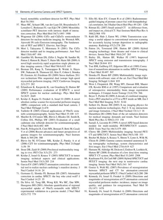
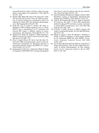
![Positron Emission Tomography (PET): Basic
Principles 11
Magdy M. Khalil
Contents 11.1 Introduction
11.1 Introduction . . . . . . . . . . . . . . . . . . . . . . . . . . . . . . . . . . . . . . . . 179
11.1.1 History . . . . . . . . . . . . . . . . . . . . . . . . . . . . . . . . . . . . . 180 Single-photon emission computed tomography
11.1.2 Positron Physics . . . . . . . . . . . . . . . . . . . . . . . . . . . 180
11.2 PET Scanners: Geometry and Components . . . . . . 182 (SPECT) and positron emission tomography (PET)
11.2.1 PET/CT . . . . . . . . . . . . . . . . . . . . . . . . . . . . . . . . . . . . 184 are three-dimensional (3D) techniques provided by
11.2.2 Scintillation Crystals . . . . . . . . . . . . . . . . . . . . . . . 186 nuclear imaging to functionally map radiotracer
11.2.3 Crystal Photodetector: Light Readout . . . . . . 189 uptake distributed in the human body. SPECT systems
11.3 Events Detected by a PET Scanner . . . . . . . . . . . . . . . 191
11.3.1 True Coincidences . . . . . . . . . . . . . . . . . . . . . . . . . 192 have been described previously; PET imaging is the
11.3.2 Random Coincidences . . . . . . . . . . . . . . . . . . . . . 192 main topic of the present chapter. The radiopharma-
11.3.3 Scatter Coincidences . . . . . . . . . . . . . . . . . . . . . . . 193 ceuticals of PET provide more insights into the meta-
11.4 Scanner Performance . . . . . . . . . . . . . . . . . . . . . . . . . . . . . . 193 bolic and molecular processes of the disease. This in
11.4.1 System Sensitivity . . . . . . . . . . . . . . . . . . . . . . . . . 193
11.4.2 2D Versus 3D . . . . . . . . . . . . . . . . . . . . . . . . . . . . . . 195 turn has made PET a molecular imaging technique that
11.4.3 Noise Equivalent Count Rate . . . . . . . . . . . . . . 195 examines biochemical processes that take place at the
11.4.4 Coincidence Timing Window . . . . . . . . . . . . . . 196 molecular level [1].
11.4.5 Time of Flight . . . . . . . . . . . . . . . . . . . . . . . . . . . . . 196 PET has become a potential “multipurpose” imag-
11.4.6 Spatial Resolution . . . . . . . . . . . . . . . . . . . . . . . . . . 198
11.5 Data Corrections . . . . . . . . . . . . . . . . . . . . . . . . . . . . . . . . . . . 203 ing technique in biomedical, clinical, and research
11.5.1 Normalization . . . . . . . . . . . . . . . . . . . . . . . . . . . . . . 203 arenas. Clinical indications for PET are numerous
11.5.2 Dead Time . . . . . . . . . . . . . . . . . . . . . . . . . . . . . . . . . 204 and include many diseases for which tumor imaging
11.5.3 Attenuation Correction . . . . . . . . . . . . . . . . . . . . . 204 occupies the most important and abundant patients.
11.5.4 Scatter Correction . . . . . . . . . . . . . . . . . . . . . . . . . . 206
11.6 Conclusion . . . . . . . . . . . . . . . . . . . . . . . . . . . . . . . . . . . . . . . . . 208 Thus, it has a well-defined and established role in
References . . . . . . . . . . . . . . . . . . . . . . . . . . . . . . . . . . . . . . . . . . . . . . . . . 209 oncology and diagnosing many types of cancer in
patients besides staging and assessment of response
to therapy. Another important role of PET lies in its
ability to diagnose some neurologic and psychiatric
disorders in addition to noninvasive quantitation of
cerebral blood flow, metabolism, and receptor binding
[2]. Further, PET has a significant role in evaluating
patients with coronary artery disease and detection of
tissue viability, together with its ability to provide an
absolute measure of myocardial blood flow. PET by
nature is “molecular” since its radioligands are accept-
able and familiar to living tissues; therefore, it is
M.M. Khalil considered an indispensible tool in molecular imaging
Biological Imaging Centre, MRC/CSC, Imperial College research. It has several quantitative features as it
London, Hammersmith Campus, Du Cane Road, W12 0NN, can determine with acceptable accuracy the amount
London, UK of tracer transported to or deposited in tissues. In
e mail: magdy.khalil@imperial.ac.uk or magdy khalil@hotmail.
com comparison to other imaging modalities, PET has
M.M. Khalil (ed.), Basic Sciences of Nuclear Medicine, DOI: 10.1007/978 3 540 85962 8 11, 179
# Springer Verlag Berlin Heidelberg 2011](https://image.slidesharecdn.com/basicsciencesofnuclearmedicine-121209103030-phpapp02/85/CIENCIAS-BASICAS-MN-192-320.jpg)
![180 M.M. Khalil
significantly higher detection sensitivity such that tra- a positron would receive much interest or at least
cers with low concentrations, in the nano or pico provide important applications as those who discov-
range, can be detected. ered it were awarded well-ranked and recognized sci-
PET imaging has moved from a useful research tool entific prizes. The positron is one of the antimatter
mainly in neurology (and cardiology) to more specific physical particles that has a short lifetime and decays
tasks in oncology and has become a potential clinical quickly by combining with a surrounding electron to
diagnostic modality. The instrumental aspects were a release two 511-keV photons in a back-to-back colli-
cornerstone in the development of PET imaging and sion process. These two photons are the two arms by
its introduction into the clinic. Furthermore, advances which the physics of PET imaging is rotating.
in PET technology are moving forward and are not
only confined to clinical whole-body scanners but
also are expanding to include organ-dedicated instru-
ments such as positron emission mammography and 11.1.2 Positron Physics
dedicated brain and prostate scanners in addition to
small-animal imaging systems [3]. Now, more atten- There are several positron emitters that are of particu-
tion is paid to the hybrid PET/CT (computed tomo- lar clinical interest. Fluorine-18 is the most widely
graphic) systems, by which the functional information used positron emitter for many reasons. It has well-
obtained by PET imaging and morphologic data defined radiochemistry, and labeling with various bio-
obtained from diagnostic CT are merged into a fused molecules is continuously advancing to suit specific
image to reveal a more accurate and confident anato- target tissues (see Chap. 5). It has a half-life (109.8
molecular diagnosis [4]. This instrumental marriage min) that allows radiochemists to synthesize labeled
has resulted in a significant improvement of patient compounds in a reasonable time and permits other
diagnosis, disease staging, and therapy monitoring. imaging logistics, such as injection, patient prepara-
Moreover, it has had a positive impact on patient tion, and imaging over several time points on either a
comfort, throughput, and more importantly clinical static or dynamic basis, to be carried out. The physical
outcome that has been proven to outperform both characteristics of fluorine are also desirable for pro-
techniques used separately [5]. duction of high-resolution PET images. The maximum
In the next few years, PET/MRI (magnetic reso- energy of the decay of F-18 is 0.650 MeV with a
nance imaging) will prove its clinical usefulness, and small positron range. Other positron emitters, such as
efforts will determine whether it will complement, oxygen-15 (2.03 min), nitrogen-13 (9.97 min), carbon-
replace, or combine PET/CT devices in the routine 11 (20.3 min), and others, have documented clinical
practice of patient diagnosis [6]. The technologic utility in PET imaging (see Table 11.1).
advances in merging the anatomical structures with The physics of PET imaging is based on positron
PET metabolic information besides developments in emission, in which nuclei of an excess amount of
tracer chemistry will definitely improve the radiologic protons exhibit nuclear transition by emitting a posi-
capability of disease detection, and the diagnostic tron particle whose half-life is short. The positron
“world” will be based on the molecular aspects rather travels a short distance based on its kinetic energy
than the traditional methods of detection. and then annihilates by combining with an electron
present in the surrounding medium to give two oppos-
ing 511-keV photons with an angular distribution of
0.5 full width at half maximum (FWHM) at 180 .
11.1.1 History Before annihilation takes place, the positron forms a
new configuration with an electron called positronium,
In 1928, Paul Dirac predicted the existence of he which is a hydrogen-like atom with a short lifetime of
positron, and he shared the Nobel Prize with Erwin about 10 7 s [7]. Because of this unique emission
¨
Schrodinger in 1933. In 1932, Carl Anderson observed process, a single detector system does not provide
positrons in cosmic rays and shared the Nobel Prize appropriate geometry for detection of the two photons
in 1936 with Victor Hess. From the beginning, one as in SPECT imaging; hence, two detectors in coinci-
could expect that the electron antiparticle known as dence are the most convenient way to detect the two](https://image.slidesharecdn.com/basicsciencesofnuclearmedicine-121209103030-phpapp02/85/CIENCIAS-BASICAS-MN-193-320.jpg)
![11 Positron Emission Tomography (PET): Basic Principles 181
Table 11.1 Physical properties of clinically useful PET radionuclides (From [142])
Resolution (mm)
Maximum Average kinetic Effective range
Radionuclide Half life energy (MeV) energy (MeV) FWHM FWTM 2.355 Â rms
Carbon 11 20.3 min 0.96 0.386 0.102 1.03 0.92
Nitrogen 13 9.97 min 1.19 0.492 0.104 1.05 1.4
Oxygen 15 122 s 1.72 0.735 0.188 1.86 2.4
Fluorine 18 109.8 min 0.64 0.250 0.282 2.53 0.54
Copper 64 12.7 h 0.65 0.278 0.501 4.14 0.55
Gallium 68 68.1 min 1.83 0.783 0.58 4.83 2.8
Rubidium 82 1.27 min 3.3 1.475 1.27 10.5 6.1
rms: root mean square
FWTM full width at tenth maximum
FWHM full width at half maximum
events concurrently. Therefore, any two photons that spatial blurring caused by the positron range. In tissues
will reach two opposing detectors (in coincidence) with low density (e.g., lung tissue), the resolution
will be recorded as long as their incidences on the degradation is more pronounced than in tissues with
detector fall within a predefined timing window (2t). high density (e.g., compact bone) (see Fig. 11.2).
The line that connects the two detectors in space is Together with the radionuclide maximum energy and
called the tube or line of response (LOR). The LOR the density of the medium, the relative influence of the
does not necessarily indicate the path along which positron range on the final resolution of the recon-
annihilation took place since one or both photons structed image depends on the spatial resolution of
may have undergone a scattering interaction before the system [9, 10].
reaching the detectors or both may have originated The distribution of the positron trajectories was
from different annihilating sites. Accordingly, there found not to follow a normal Gaussian-like distribu-
are different types of events that can describe the tion and the cusp-shaped pattern was found in a num-
coincidence events detected by a PET scanner. These ber of simulation studies [8, 9]. The effect of positron
are true, scatter, and random coincidences and are range on resolution loss is thus recommended to be
discussed further in this chapter. measured using the positron root mean square range in
the medium of interest or the full width at 20% of
maximum [9]. The characteristic shape of the positron
Positron Range distribution preserves some high frequencies, and the
long tail estimated by full width at tenth maximum
A physical phenomenon reduces the resolution capa- (FWTM) causes severe resolution degradation [8].
bility of the scanner to record events exactly as emitted
from their true original locations (Fig. 11.1). Once the
positron is emitted from the nucleus, it undergoes an Acollinearity
annihilation with an electron to produce the clinically
useful 511-keV photons. However, before this interac- Acollinearity is another physical phenomenon that
tion takes place, the positron travels a distance before is related to the conservation of momentum of the
collision occurs. This distance is a function of the annihilated positron-electron pair. The conservation
kinetic energy of the emitted positrons being higher of mass and energy requires that the two photons are
in positron emitters with high maximum b+ energy, released in 180 ; however, some residual kinetic
such as O-15, Ga-68, and Rb-82 nuclei (maximum energy remains to reduce this angular measure, on
energy 1.72, 1.83, and 3.3 MeV, respectively) [8] average, by Æ 0.25 FWHM as shown in Fig. 11.1.
(Table 11.1). Besides the kinetic energy of the released This in turn leads to a LOR that does not exactly
positron, the surrounding medium contributes to the pass through the annihilation site, resulting in event](https://image.slidesharecdn.com/basicsciencesofnuclearmedicine-121209103030-phpapp02/85/CIENCIAS-BASICAS-MN-194-320.jpg)
![182 M.M. Khalil
Fig. 11.1 (a) Positron range
as determined by the two
headed arrow is the b
perpendicular distance from
the transformed nuclei to the
recorded line of response. In
(b), the recorded line of 180º ± 0.25º a
response due to positron
electron annihilation is not
necessarily 180 yet has an
angular distribution of full
width at half maximum
(FWHM) of 0.25
F-18
mm = 1.76 mm. The constant 0.0022 is derived from
18F
the geometry of the ray and can be calculated as 0.5
tan 0.25 . For a 15-cm detector diameter, resolution
loss would be 0.33 mm, approximately five to six
15O times less than a clinical whole-body scanner. Correc-
tions for acollinearity and positron range are discussed
in the spatial resolution section.
82Rb
11.2 PET Scanners: Geometry
and Components
Compact bone Soft tissue Lung tissue A number of different geometries have been imple-
mented in the design of PET scanners. Starting from
Fig. 11.2 Monte Carlo calculated distribution of annihilation the simple geometry of two opposing detectors as
events around a positron source inserted in different human
tissues (compact bone, soft tissue, and lung tissue). The spread in the conventional dual-head gamma camera to
of the distribution is larger for positron emitters of high maxi more sophisticated full-ring scanners, performance
mum kinetic energy in tissues with low density. (From [9] with characteristics vary from one system to another and
permission from Springer þ Business media) are superior in the latter design. However, full-ring
scanners are more expensive. Other system design
mispositioning and spatial resolution loss. Unlike the geometries are partial ring and polygonal (hexagonal,
positron range, the acollinearity resolution effects are octagonal, . . .). Figures 11.3 and 11.4 illustrate differ-
a function of the detector diameter; thus, a PET scan- ent geometries used in clinical PET systems.
ner of with an 80-cm diameter would suffer a resolu- Hybrid PET/SPECT Camera. As mentioned, the
tion degradation of 0.0022 Â D or 0.0022 Â 800 process of positron-electron annihilation and the](https://image.slidesharecdn.com/basicsciencesofnuclearmedicine-121209103030-phpapp02/85/CIENCIAS-BASICAS-MN-195-320.jpg)
![11 Positron Emission Tomography (PET): Basic Principles 183
Fig. 11.3 Different a b c
geometries used in positron
emission tomographic (PET)
scanners. (a) A dual head
gamma camera with
coincidence circuitry and
sufficient crystal thickness of
about 2.5 cm. (b) Partial ring
detector geometry that
requires rotation around the
patient for data sampling. (c)
Full ring detector geometry
release of two antiparallel photons impose geometric Modifications must be made on the conventional
constraints on PET detector design. This requires that gamma camera to operate in the coincidence mode
the detector pair must be in an opposite direction to [17]. Coincidence triggering is the basic electronic
determine the path traversed by annihilation photons element that needs to be incorporated in the detector
to reach the detectors. Anger proposed the use of two electronics. The count rate capability must be high
opposing gamma cameras at 180 and electronic colli- enough since collimators are unmounted, and the sys-
mation of the 511-keV photons to record coincident tem sensitivity is larger. Crystal thickness (e.g., 16 25
events [11]. This has been employed in longitudinal mm) should be larger than that normally used in
tomography using a dual camera without a rotating SPECT systems to provide a sufficient interaction
system. Further developments resulted in the design of depth to stop a significant portion of the incident
two large field-of-view gamma cameras with a thicker photons, thus improving the detection efficiency.
crystals mounted on a rotating gantry [12]. Dual- and triple-coincidence gamma cameras have
Several limitations faced the early dual opposing been manufactured, and owing to some performance
detector design in providing a convenient measure of limitations, such as spatial resolution, sensitivity, and
activity distribution inside human subjects, such as count rate performance, they are not attractive coinci-
low sensitivity, poor count rate capability, and lack dence PET scanners when compared to the full-ring
of a radionuclide to provide sufficient count statistics design [18 20].
over the time course of the study. These limitations Partial Ring. In PET systems with partial detector
were attributed mainly to the use of NaI(Tl) crystal, rings, the detectors are packed in two opposing detec-
old-fashioned system electronics, and the inherent tor banks arranged in a circular system that continu-
open design. NaI(Tl) crystals have a low stopping ously rotates to capture a complete data set necessary
power against 511-keV photons and a slow decay for image reconstruction [21] (Fig. 11.3b). For exam-
time. The open design, however, allows for more ple, the ECAT ART (Siemens/CTI, Inc., Knoxville,
single events and random coincidences to be accepted, TN) consists of two opposing detectors, each of
especially from regions outside the field of view, 3 Â 11 crystal blocks arranged in axial and tangential
resulting in a low signal-to-noise ratio and reduced direction. The banks are slightly asymmetrically
image contrast [13]. opposed to increase the effective transaxial field of
This system is today named as hybrid SPECT/PET view. The block material is bismuth germanate
camera and is able to provide dual functionality (BGO) and is segmented into 6.75 Â 6.75 Â 20
in imaging single-photon radiopharmaceuticals and mm3 and coupled to four photomultiplier tubes
positron-labeled compounds by switching between (PMTs). There are three blocks in the axial dimension,
SPECT and coincidence detection mode (Fig. 11.3a). giving a 16.2-cm axial field of view and 24 crystal
Using appropriate collimators for 511 keV, this type rings with 242 possible planes of response [21]. The
of imaging system was also used to image patients partial ring design was used in prototyping the first
injected with dual tracers to look at two different PET/CT scanner in which the PET electronics were
metabolic or functional processes that happen simul- placed at the rear portion of the CT gantry housing but
taneously [14 16]. with different operation consoles [22].](https://image.slidesharecdn.com/basicsciencesofnuclearmedicine-121209103030-phpapp02/85/CIENCIAS-BASICAS-MN-196-320.jpg)
![184 M.M. Khalil
Fig. 11.4 Another set of
positron emission
tomographic (PET) scanners a b c
that was proposed in the
literature and implemented in
practice. (a) and
(b) Hexagonal designs using
flat or curved plate detector
arrays, respectively.
(c) Discrete crystals arranged
in modular style in which
modules are adjoined side
to side
Hexagonal geometry. Other systems were also crystals to indentify the depth of interaction (DOI) of
implemented that were based on arrays of six- or the 511-keV photons. The system achieves a spatial
eight-sided geometry. The former design is shown in resolution of 2.3 3.2 mm in the transaxial direction
Fig. 11.4a, b and has more than one variant, such that and a resolution of 2.5 3.4 mm in the axial direction,
the detector assembly can be a single continuous scin- together with a point source system sensitivity of 6%
tillation detector or curved-plate design. However, the and National Electrical Manufacturers Association
number of detectors can be larger, with flat modules (NEMA) NU-2001 line source sensitivity of 3%.
and discrete crystal structure (Fig. 11.4c). The transaxial and axial extents are 31 and 25 cm,
The hexagonal design has some good features as respectively [29].
it is relatively simple and uses fewer PMTs than used Cylindrical design. The most efficient geometries
in a full-ring block detector design. The number of that found wide acceptance among manufacturers and
PMTs may vary from 180 or 288 as in NaI(Tl)-based PET users are those that depend on circular or ring
detector to an even larger number (e.g., 420) as design. The advantage provided by full-ring scanners
in discrete GSO (gadolinium oxyorthosilicate) or is their detection efficiency and count rate capabilities.
LYSO (Cerium doped lutetium yttrium orthosilicate) This in turn provides better system spatial resolution
modular detectors [23 27]. Further, it has a relatively by realizing the statistical requirements imposed by
large axial field of view that can reach 25 cm, such as image reconstruction [30]. The ring design is the
that found, for example, in the curved-plate NaI(Tl) most widely used system geometry and is preferred
design [25]. among manufacturers in clinical and preclinical PET
The use of a large continuous crystal allows only scanners.
3D data acquisitions; to some extent, the large axial
field of view can compensate for the lower sensitivity
of the NaI(Tl) crystal. However, such a detector design
and the use of Anger logic position circuitry led to
an increase in the system dead time. Limiting the light 11.2.1 PET/CT
signal to a finite number of PMTs and optimizing the
light guide for light spread were proposed to mitigate The introduction of PET/CT in the early years of this
these effects [28]. One of the disadvantages of hexag- century was attractive for many practitioners as well as
onal geometry is the intercrystal dead space left by some manufacturers; now, almost all PET scanners are
adjoining the detector modules, a reason that could purchased as hybrid PET/CT systems. As mentioned
cause some reduction of the system sensitivity and in the level of integration (chapter 10), the current
confound the reconstruction process. generation of hybrid PET/CT systems is configured
A hectagonal or eight-sided detector system can be side-by-side such that the CT component is placed in
found in the HRRT system (High Resolution Research the front portion of the scanner while the PET compo-
Tomograph, Siemens Medical Solutions), a dedicated nent is placed at the back end mounted using the same
brain PET scanner that uses two types of scintillation housing over the two gantries or gantries with separate](https://image.slidesharecdn.com/basicsciencesofnuclearmedicine-121209103030-phpapp02/85/CIENCIAS-BASICAS-MN-197-320.jpg)
![11 Positron Emission Tomography (PET): Basic Principles 185
Fig. 11.5 State of the art positron emission tomographic/computed tomographic (PET/CT) scanners. (a) Biograph mCT, Siemens
Medical Solutions. (b) Gemini TF Big Bore system, Philips Company. (c) Discovery PET/CT 600, GE Healthcare
covers and in all cases using the same imaging table. scans. State-of-the-art PET/CT systems are shown in
The axial length traversed by the imaging table varies Fig. 11.5.
among manufacturers, and as the bed travels axially There have been several attempts to improve the
there is more chance for bed deflection, resulting in performance characteristics of PET scanners in terms
misalignment errors. A number of approaches were of spatial resolution, system sensitivity, coincidence
therefore undertaken by scanner manufacturers to timing resolution, and energy resolution while keeping
take into account this deflection of the imaging bed. the cost of the device reasonably affordable. These
Moving from a single to more CT slices (e.g., 4, 6, requirements are as follows [31]:
8, 16, 64, and beyond) offered a great opportunity to
1. High detection sensitivity for the 511-keV photons
enhance the diagnostic speed and possibility of
to improve the detection or areas of low tracer
performing cardiac CT as well as angiography. The
concentration, enhance the statistical quality of
addition of spiral CT technology was remarkable and
dynamic images, and to reduce the injected dose
significantly reduced the acquisition times by many-
2. High resolving capabilities to achieve the maximal
fold together with the advantage of acquiring a large
spatial resolution for volume elements
number of thin slices. In this type of data acquisition,
3. High-energy resolution to minimize the contribu-
the imaging table moves forward in a simultaneous
tion of scattered events
motion with rotation of the x-ray source in a spiral
4. Low dead time to improve count rate performance
path. The delay between interscans can thus be
and high timing resolution to improve random
eliminated (a feature of older CT generations), while
rejections and for time-of-flight (TOF) applications
the z-axis is kept variable across projections. Data
5. Low cost
interpolation is then used to account for these angular
inconsistencies. These fully diagnostic CT systems, Achieving such requirements in one practical
when combined with state-of-the art PET machines, tomograph is difficult from physical and engineering
have made a “one-stop-shop” diagnosis a clinical real- perspectives as well as financial issues. Some perfor-
ity. The radiation dose delivered to the patient using mance parameters are traded off while maintaining the
multislice CT systems has been considered, and solu- quality of others. For instance, reducing the detector
tions such as beam modulations and optimized acqui- diameter is advantageous for improving count sensi-
sition protocols have been used. Low-dose attenuation tivity, while it is not in favor of spatial resolution due
correction using low tube current is another way to to the DOI errors. Small-animal scanners have better
reduce patient dose as long as diagnostic CT is not angular coverage owing to smaller ring diameter but
required. The two major advantages provided by the suffer from depth of interaction. Another factor that is
CT are attenuation correction and anatomical localiza- affected by ring diameter is photon acollinearity,
tion of the PET images; both features have significantly which increases and decreases in a linear fashion
improved the diagnostic performance of the nuclear with the ring diameter. Extension of the axial extent](https://image.slidesharecdn.com/basicsciencesofnuclearmedicine-121209103030-phpapp02/85/CIENCIAS-BASICAS-MN-198-320.jpg)
![186 M.M. Khalil
of the PET scanner can be used to improve scanner 0.5
sensitivity, but this comes with increased cost and 2 Th
sp
degradation of spatial resolution in the axial direction. 0.4 e2 = Bi
s p+s c
0.3 W
(511 keV) Lu
e2
0.2 Gd
11.2.2 Scintillation Crystals Ba
Y BGO
0.1 BaF2
LSO
The type and composition of the scintillation crystal
0.0
are of considerable importance in the detection effi- 30 40 50 60 70 80 90 100
ciency of 511-keV photons. The function of the crystal Atomic number (Z )
is to convert the energy received by the incident
photons into light quanta that are proportional to the Fig. 11.6 The probability that both 511 keV photons interact
via the photoelectric effect as a function of Zeff. There is an
imparted energy. Two major interactions occur when increasing trend in the photofraction probability with the effec
the photon deposits its energy in the crystal: photo- tive atomic number. (From [144] with permission from William
electric and Compton scattering. The electrons Moses, Lawrence Berkeley Lab, USA)
released from either photoelectric or Compton pro-
cesses undergo several interactions within the scintil-
lator material and excite other electrons, which in turn
properties of the scintillation crystals that can signifi-
decay to emanate the scintillation light [32].
cantly improve the scanner performance:
Crystal with a high probability of photoelectric
effect, or photofraction, is preferred since all energy 1. Crystals with high density and high effective atomic
is imparted on the crystal and at one site. However, number Zeff have good intrinsic efficiency. Photo-
Compton interactions reveal more than one interaction electric effect is strongly related but Compton scat-
site and hence degrade the spatial resolution as well as tering is linearly related to the atomic number of
the energy spectrum by broadening the photo peak the material for photons (100 keV). Crystals with
width. Figure 11.6 shows the dependence of the photo- high rZeff4 have good stopping capabilities for 511-
electric effect on the atomic number of various types keV photons [35]. The density and effective atomic
of scintillation crystals. number also determine the attenuation length when
The light emitted by the crystal is viewed and crystals of smaller attenuation length are desired as
detected by photodetectors, where light photons are they serve to reduce resolution degradation caused
converted into an electronic signal for determination by parallax errors.
of event attributes such as energy and position. Many 2. Crystals should have high light output since event
types of the scintillator material have been proposed positioning and energy resolution are related to the
and implemented to improve the detection perfor- amount of light quanta released from the crystal.
mance of PET scanners. Crystals that are able to A sufficient amount of scintillation light can release
provide high photon detection efficiency, short decay a significant number of photoelectrons from the
time, and better energy resolution are highly preferred. photocathode and thus provide better accuracy in
Other optical properties, such as an emission wave- signal measurements and improve energy resolu-
length that matches the photocathode of the PMT, tion. The small size of the crystal elements in the
transparency, and good refractive index, are also block detector design requires a scintillator that can
important factors to ensure maximum transfer of the produce high light output so that position encoding
scintillation light to the PMT. Other manufacturing can be done with good precision. There are a num-
requirements, such as mechanical flexibility, hygro- ber of factors responsible for the statistics of light
scopic properties, ease of handling and fabrication emanating from the scintillator, such as homoge-
into small crystal elements, cost, and availability, are neous distribution of the activation centers, non-
all of concern in the selection of the scintillator mate- proportional response of the crystal, and variation
rial [33, 34]. The following is a list of the major in crystal luminosity [33].](https://image.slidesharecdn.com/basicsciencesofnuclearmedicine-121209103030-phpapp02/85/CIENCIAS-BASICAS-MN-199-320.jpg)
![11 Positron Emission Tomography (PET): Basic Principles 187
3. Crystals with short decay constants or “fast scintil- 11.2.2.2 BGO
lators” serve to improve the timing of the coinci-
dence detection and thus reduce the dead time and The BGO type of crystals has a better detection effi-
random fractions. Scintillators with a short decay ciency and high probability of photoelectric interac-
constant help to improve the count rate perfor- tion (40%) for 511-keV photons. It contains the
mance, especially for scanners operating in 3D bismuth element (Bi), which has a large atomic num-
mode and for clinical studies in which short-lived ber (Z ¼ 83) and its ionic form (Bi3+) is the intrinsic
radionuclides with high activities are used [36]. luminescence center in the crystal [33]. The density of
Another advantage gained by such crystals is their the crystal is approximately two times greater than for
recent introduction in TOF applications, which NaI(Tl), and the attenuation coefficient is three times
require high efficiency and fast scintillators, lead- larger (Table 11.2). These properties have allowed
ing to an improvement in the signal-to-noise ratio. BGO to replace NaI(Tl) crystal, and it was the scintil-
4. Although the selection of the scintillator material lator of choice for many years, until the end of 1990s.
is important and can potentially affect the perfor- However, the low light yield [~15 20% relative to NaI
mance of the scanner, one should realize that (Tl)] and poor energy resolution and response time
the overall system performance is a function of have made it inferior to the other new scintillators,
many components of the imaging system, such as which showed a better response time and better light
electronic circuitry and acquisition and reconstruc- output, such as GSO, LSO (Cerium-doped Lutetium
tion parameters, together with the correction and Orthosilicate) , or LYSO. As mentioned, better
computational techniques [37]. This has been noted response time allows a significant reduction of system
in PET scanners upgraded using better electronic dead time and works to minimize the amount of ran-
circuitry or when new scintillation crystals were dom coincidences that contaminate the prompt events.
introduced in scanners not equipped with appropri- Moreover, it allows a reduction of pulse pile-up in 3D
ate electronic components [38, 39]. mode, in which system sensitivity is high, thereby
resulting in better count rate performance. The first
small-animal PET scanner designed specifically to
image rodents was made of BGO-based block detec-
11.2.2.1 NaI(Tl) tors and was developed in the mid-1990s [40]. When
compared to NaI(Tl), both the nonhygroscopic nature
NaI(Tl) has been extensively used in many nuclear of BGO and its high detection efficiency have permit-
medicine devices, especially in the design of the ted manufacturing a more compact detector assembly
gamma camera. In the early 1970s, it was the most and narrower crystals that improved significantly the
commonly used crystal in PET scanners even though system resolution and sensitivity.
it was an inefficient scintillator for 511-keV photons.
The detection efficiency of NaI(Tl) crystal to Tc-99m
energy and other relatively low-energy gamma emitters 11.2.2.3 Gadolinium Oxyorthosilicate
is reasonably good, and it remains the scintillator
of choice in many SPECT cameras. In comparison Cerium-doped GSO has good properties that have
to other types of scintillation crystals, it has better made it applicable in recent PET scanners (Allegro
energy resolution, poor timing resolution, high dead and Gemini-GXL, Philips Company). Also, it has
time, and low stopping power. The low density and some applications in small-animal scanners and in
moderately low atomic number are responsible for the combination with LSO in systems equipped with par-
poor detection efficiency of NaI(Tl) crystal for 511- allax error correction. The density of this scintillator
keV photons. Another limitation of NaI(Tl) crystal is is comparable to BGO and LSO crystals, and it is
its hygroscopic properties, and careful handling to roughly twice as dense as NaI(Tl). The effective Z is
avoid humidity effects must be taken into account; intermediate between NaI(Tl) and both BGO and
hermetic sealing is required. This in turn led to diffi- LSO. The decay constant (60 ns) is slightly higher
culty in producing small-size elements suitable for than LSO but significantly lower than BGO and NaI
block detector design. (Tl) crystals. An evaluation of a pixilated GSO Anger](https://image.slidesharecdn.com/basicsciencesofnuclearmedicine-121209103030-phpapp02/85/CIENCIAS-BASICAS-MN-200-320.jpg)
![188 M.M. Khalil
Table 11.2 Properties of scintillation crystals used in positron emission tomographic (PET) scanners
NaI(Tl) BGO GSO:Ce LSO:Ce LYSO:Ce LaBr3 BaF2
Density (gm/cm3) 3.67 7.13 6.7 7.4 7.1 5.3 4.89
Effective atomic number (Z) 51 74 59 66 64 47 54
Linear attenuation coefficient (1/cm) 0.34 0.92 0.62 0.87 0.86 0.47 0.44
Light yield (% NaI[Tl]) 100 15 30 75 75 160 5
Decay time (ns) 230 300 65 60 40 41 16 0.8
Emission maximum (nm) 410 480 440 420 420 370 220
Hygroscopic Yes No No No No Yes Slightly
Photoelectric effect (%) 17 40 25 32 33 13 12
Refractive Index 1.85 2.15 1.85 1.82 1.81 1.88 1.56
NaI(Tl) Thallium activated sodium iodide crystal
BGO Bismuth germanate oxyorthosilicate
GSO:Ce Cerium doped gadolinium oxyorthosilicate
LSO:Ce Cerium doped Lutetium orthosilicate
LYSO:Ce Cerium doped lutetium yttrium orthosilicate
LaBr3:Ce Cerium doped lanthanum bromide
BaF2 Barium fluoride
logic-based scanner using NEMA NU 2-2001 proce-
dures revealed acceptable results in terms of energy
resolution, sensitivity, and better image quality 1 Nal(T1)
provided that a patient study can be performed in a
time course of half an hour, including transmission
Intensity
LSO
measurements [24]. That system also demonstrated a
better count rate capability in 3D mode served by the 0.5
fast decay constant of the GSO crystal and optimizing GSO
the light spread detected by PMTs [28]. GSO is not
hygroscopic, absent intrinsic radioactivity, has good BGO
stability versus temperature, and has a good uniform 0
300 400 500 600 700
light output but is not simple to manufacture, and a
Wavelength (nm)
special fabrication procedure is followed to avoid
cracking during crystal design. Another disadvantage Fig. 11.7 LSO is ranked second among other different scin
is its susceptibility to magnetic field effects, rendering tillation crystals in the amount of light yield produced, data
normalized to NaI(Tl) crystal. Further, the peak emission wave
it an unsuitable scintillator for PET/MR hybrid
length does match the bialkali material of the photocathode
systems [41]. of photomultiplier tubes (PMTs). (From [145] # IEEE with
permission)
11.2.2.4 LSO/LYSO
LSO scintillators are characterized by their fast
Unlike BGO, which has no activator, LSO is a cerium- response, represented by short decay time, achieving
activated scintillator and has a large rZeff4, but this is excellent timing resolution. These properties have
lower than BGO. It has good characteristics for dealing made LSO/LYSO an attractive scintillator for PET
with 511-keV photons: The effective Z is 66, density scanners, in particular for TOF applications, and with
(7.4), and m ¼ 0.87 cm 1; thus, it is an efficient scin- its high light output, there is no compromise in detec-
tillator for stopping the annihilation coincidence tor efficiency [42] (see Figs. 11.6 and 11.7). The light
photons. LYSO is similar to LSO and is produced output is approximately three or five times higher than
such that some lutetium is replaced by yttrium atoms. BGO when using an avalanche photodiode (APD) or](https://image.slidesharecdn.com/basicsciencesofnuclearmedicine-121209103030-phpapp02/85/CIENCIAS-BASICAS-MN-201-320.jpg)
![11 Positron Emission Tomography (PET): Basic Principles 189
PMT, respectively. Because of the statistical nature of intrinsic spatial resolution [48]. Following this proce-
the detection process, an LSO-based PET scanner dure in building up a full multiring scanner is neither
should enjoy better energy resolution (~12%), better efficient nor cost-effective as one needs a significantly
event positioning, and a short coincidence timing win- large number of PMTs and crystal material along with
dow. The last is instrumental in establishing the noise electronic channels to implement such a design. Ear-
level resulting from random events [43]. lier efforts in coupling the scintillation detector to
LSO is nonhygroscopic and mechanically rugged, PMTs included four crystals per one or two PMTs.
and simple fabrication is relatively possible to produce However, this strategy was advanced by using more
small and discrete versions of the crystal [34]. However, crystal elements per PMTs. It was a turning point
it has a number of limitations, such as inhomogeneity of when the block detector concept was introduced
light production, high melting point (2,000 C), and in the design of the PET scanners, which is a cost-
cost higher than other crystal types [GSO, BGO, and effective technique in coupling the PMT to the scintil-
NaI(Tl)]. Another limitation of LSO scintillation crys- lation detectors and has also led to an improvement in
tals is their Lu-176 content, which emits low-level spatial resolution [49].
radioactivity; yet, this has been found not to affect the In block detector design, as shown in Fig. 11.8b, c,
extrinsic 511-keV coincidence detection but can affect the scintillation block is segmented into an array of
precise measurements of system sensitivity and scatter small crystals by cutting into precise and well-
fraction using low-level radiation. defined dimensions in the transaxial and axial direc-
LSO crystals are found in many PET systems, such tions. The cutting process leaves a space between the
as high-resolution dedicated brain scanners [44] and small crystals; therefore, a reflective material is used
hybrid multimodality imaging, such as PET/CT, PET/ to optically separate between the neighboring crys-
SPECT, and PET/MRI systems. Other special-purpose tals, defining a path for the light to travel to the PMTs
systems have also been designed using LSO crystals, and to maximize light collection efficiency
such as breast PET scanners and small-animal imaging (Fig. 11.9a). The depth of cutting is empirically
systems [45, 46]. implemented so that the light is distributed in a spa-
One decade ago, the acquisition of whole-body tially linear fashion among the exposed array of
fluorodeoxyglucose-F18 (FDG) studies could take PMTs [48]. In this pseudodiscrete design, a greater
about 1 h, including measurements of transmission depth of cutting is found at the block periphery rather
data [47]. However, these days the same scan can be than at the center. The advantage of the block detec-
acquired in significantly shorter time. This significant tor design is improved spatial resolution by segment-
reduction in the acquisition time is due to many rea- ing the scintillation crystal into smaller elements
sons; one is related to the introduction of efficient and viewed by a finite number of PMTs (usually 4), thus
fast scintillators in the scanner design (in addition to minimizing the high cost required if each crystal
other factors such as CT-based attenuation correction, element is coupled to a single PMT.
fast electronic circuitry and computer processors). In the original design developed by Casey and Nutt,
the block was cut into a 4 Â 8 crystal array, each
5.6 Â 13.5 Â 30 mm3, using BGO detector material.
The crystal block was glued to four square PMTs, each
11.2.3 Crystal Photodetector: Light
25 mm, through a Lucite light guide [49]. Now, the
Readout crystal dimensions vary among PET scanners using
different types of scintillation materials. In commer-
In the conventional gamma camera, the scintillation cial clinical PET/CT scanners, the crystal dimension
crystal is a continuous large slab of NaI(Tl) viewed varies from one system to another (e.g., 4 Â 4,
from its back surface by an array of PMTs that collect 6.4 Â 6.4, etc.), while the crystal thickness ranges
the scintillation light produced from interaction from 20 to 30 mm. Crystal size can be reduced to
of the emitted photons with the detector material 1 2 mm in an array of larger elements, such as
(Fig. 11.8a). In one of the earliest PET detector 20 Â 20 or 13 Â 13, based on design specifications.
designs, the PMT is coupled to a single crystal; thus, However, this arrangement is most common in small-
the cross section of the crystal defines the LOR and animal micro-PET scanners.](https://image.slidesharecdn.com/basicsciencesofnuclearmedicine-121209103030-phpapp02/85/CIENCIAS-BASICAS-MN-202-320.jpg)
![190 M.M. Khalil
Fig. 11.8 (a) Light readout a b
by continuous based
scintillation detector.
(b) Block detector showing
crystal segmentation. Scintillation crystal
(c) Photograph of block
detector coupled to four
photomultiplier tubes (PMTs). Light guide
(Image courtesy of Siemens
Medical Solutions)
PMTs
c
Fig. 11.9 (a) Crystal
segmentation in block detector
design. It allows defining a
path for incident photons and a b c
improved collection
(B + D) − (A + C)
efficiency. (b) Discrete crystal A B x=
A+B+C+D
design, a useful approach in
high resolution PET but with
some limitations (see the text). C D (A + D) − (C + B)
y=
(c) Anger logic positioning A+D+C+B
PMTs
Photosensitive
PMT
Another choice is an assembly of discrete individual crystals provides an improvement in spatial resolution.
scintillation crystals that are packed together yet opti- It can be read out by a position-sensitive or multichan-
cally separated by reflective material to confine the nel photodetector or individual and separate readout
light spread to those crystals that receive the scintilla- channels [50]. Use of semiconductor readout devices
tion light (Fig. 11.9b). This finger-like array of discrete has also been implemented in the last design, in which](https://image.slidesharecdn.com/basicsciencesofnuclearmedicine-121209103030-phpapp02/85/CIENCIAS-BASICAS-MN-203-320.jpg)
![11 Positron Emission Tomography (PET): Basic Principles 191
discrete crystals are read out by an array of elements of In the block detector, four PMTs are often used to
APD [51]. APDs can be produced in dimensions (indi- read out the light emitted from the scintillation material.
vidual elements or array) similar to discrete crystal The centroid Anger logic is used to identify the individ-
dimensions and therefore can overcome size limita- ual crystals from which light photons were emitted
tions encountered in fabricating small PMTs (smaller (Fig. 11.9c). Another approach that improved the encod-
than 10 mm) [52, 53]. They have been successfully ing ratio but suffers from increased dead time is the
introduced as light photodetectors in encoding the quadrant-sharing approach, in which the PMT is shared
position of the light signal and offer a compact volume among four block detectors; the encoding ratio of 16:1
that is useful in PET/MRI systems and small-animal in the conventional block detector design is increased
scanners, including SPECT and PET imagers. by three to four times based on the PMT size and the
Discrete detector design has a number of limita- crystal dimensions [54] (Fig. 11.10). This design was
tions, especially when an individual crystal is read initially proposed to improve the spatial resolution of
out by a single readout channel. Crystal fraction and the PET scanner without increasing the cost and to
packing must match with the size of the available reduce the high price of the scanner by lowering the
photodetectors [50]. Further, it requires extensive use number of PMTs decoding the scintillation detector
of electronic readout channels and thus is an expensive [55]. However, dead time effects and pulse pileup can
and cost-ineffective approach. PET systems based on be treated using recent progress in signal-processing
discrete crystal design are more common in research electronics employing event recovery methods [56, 57].
and small-animal scanners [50, 52]. Alternatively, dis-
crete crystal design can be used in a modular structure
in which the crystal sets are mapped by an array of
PMTs with a continuous light guide adjoining the 11.3 Events Detected by a PET Scanner
detector modules, as can be found, for example, in
the Allegro system (Philips Company). Table 11.3 Singles: PET detectors receive a large amount of
shows the various approaches used to couple the events. A small fraction of these events are detected
PMT and the crystal in PET scanners. as in coincidence, while the remaining are singles that
Table 11.3 Summary of various approaches implemented to couple the photomultiplier tubes (PMTs) and scintillation crystals in
clinical positron emission tomographic (PET) scanners
Scintillation crystals
Continuous Discrete
PMTs 2Â2 Conventional block detector design (e.g., many partial and
full ring scanners)
Array Large crystal coupled to an array of Discrete crystals arranged in large flat module, PIXELAR
PMTs (e.g., curve plate, C PET) Technology (e.g., Allegro and Gemini TF)
Fig. 11.10 Quadrant light
sharing (a) Frontal view.
(b) Lateral view
a b](https://image.slidesharecdn.com/basicsciencesofnuclearmedicine-121209103030-phpapp02/85/CIENCIAS-BASICAS-MN-204-320.jpg)
![192 M.M. Khalil
constitute a large fraction of the detected count rate. background image. True coincidences increase in lin-
Singles can be detected from the coincidence field of ear proportion to the object activity; however, random
view as well as from activity lying outside the field of coincidences are estimated to equal the product of
view. They are a problem in 3D data acquisition, for count rates of the two detectors multiplied by the
which the axial extent of data acquisition is larger and timing window (2t):
extends over the area examined [58].
Rij ¼ 2t Sa Sb
11.3.1 True Coincidences where Sa and Sb are the single-photon event rate of
detectors a and b, respectively. Thus, they are approx-
True coincidences are those photons emitted from a imately proportional to the square of the singles count
single nuclei and detected by the detectors without rate. Random coincidences add false information
interactions in the surrounding medium (Fig. 11.11a). regarding position and serve to reduce image contrast
They are the most preferred type of events not actually and quantitative accuracy as well. They are large in
achievable in practice without contamination with areas with high count rates, such as the abdomen and
other events (scatter and randoms). pelvis, especially if bladder activity is high. A longer
coincidence timing window increases the chance of
accepting more random events, but a shorter timing
window reduces the number of true events. Conse-
11.3.2 Random Coincidences quently, selection of coincidence time is a trade-off
between sensitivity of the scanner and reduction of
Random or accidental coincidences are undesired undesired events such as randoms [59]. In fully 3D
types of events that occur as a result of detecting acquisition mode, the random events are greater than
two coincident events from two different locations for 2D acquisition (with use of septa) since the former
by which their detection timing falls within the coin- provides a larger axial field of view relative to the field
cidence timing window of the system (Fig. 11.11b). of the true events.
Because these two photons are unrelated, they have The formula can be used in estimating the random
nearly equal probability to occur between most decay- events in the acquired images if the activity in the field
ing nuclei in the field of view, resulting in a high of view remains constant during the time course of the
Scatter coincidence
a b c
True coincidence Random coincidence
White circles indicate
scatter
Fig. 11.11 Three different types of coincidences that take is within the acceptance timing window. (c) Scatter events
place during PET imaging. (a) True coincidence occurs due to take place when one or two of the 511 keV photons undergo
decay of a single nuclei and emission of two antiparallel 511 Compton interaction during their travel to the detector pair
keV photons and without undergoing any further interactions. and again with detection time within the specified limit; how
(b) Random coincidence occurs due to decay of two unrelated ever, single photon scattering is more abundant than two photon
nuclei such that their detection by two opposing detector pairs scattering](https://image.slidesharecdn.com/basicsciencesofnuclearmedicine-121209103030-phpapp02/85/CIENCIAS-BASICAS-MN-205-320.jpg)
![11 Positron Emission Tomography (PET): Basic Principles 193
Fig. 11.12 Scatter fractions
0.8 BGO LSO LaBr
simulated for different crystals
as a function of energy GSO
0.7
threshold using a cylinder LYSO
(70 cm long) of diameters 20 0.6
cm (O), 27 cm (■), and 35 cm
Scatter fraction
(~) in a typical 3D scanner. 0.5
Data simulated using a
0.4
scanner of diameter 85 cm and
18 cm axial field of view. The 0.3
gray band is the energy range
threshold used for each 0.2
scintillator. (From [54] with 0.1
kind permission from IOP
Publishing and the 0
corresponding authors) 300 350 400 450 500
Energy threshold (keV)
study. However, in some situations in which the tracer set at a threshold greater than 400 keV, and a signifi-
undergoes some dynamic or fast metabolic processes, cant amount of photons with large-angle scattering can
this method may not be precise enough. Another be rejected. For a scanner with less energy resolution
approach that is frequently applied is the delayed win- (e.g., BGO), the energy width is set wider, and the
dow subtraction method. This method is based on mea- lower bound may go to 300 keV, which results in
suring the random coincidences in a delayed acceptance projection data contaminated by large proportion of
window; then, data are corrected on the fly by subtrac- scattered events. Figure 11.12 shows the scatter frac-
tion or saved for further analysis [60]. The variance tion for a number of scintillators used in commercial
estimate in data collected by the delayed window is PET scanners as a function of the lower energy dis-
high, and applying it directly on the prompts window criminator and different object sizes.
will add noise to the reconstructed images. A variance The contribution of multiple coincidences is
reduction technique can be used to suppress such a also another possible type of events that result from
problem to yield a better signal estimate [61]. three or more photons detected in the coincidence
timing window, leading to detector confusion regard-
ing where the annihilation photons are located.
11.3.3 Scatter Coincidences The detection system does not discriminate
between the various types of the coincidences, and
they are automatically recorded as long as their detec-
Another undesired type of events is scattered coinci- tion time falls within the specified timing limits of the
dence, which results from deflection of one or both scanner. These events (true, random, and scatter coin-
annihilation photons from their true LOR, resulting in cidences) are collectively called prompts, bearing in
a false recorded event regarding position of the anni- mind that the most useful type is the true coincidence.
hilated nuclei and energy of the 511-keV photons
(Fig. 11.11c). Further, scatter coincidences are a func-
tion of the activity distribution, source size, and com- 11.4 Scanner Performance
position of the surrounding media. Scatter events
cause a loss of image contrast and degrade the quanti-
tative accuracy of the measurements. Energy resolu- 11.4.1 System Sensitivity
tion plays an important role in controlling the amount
of scattered photons in the acquired data. PET systems For a given activity distribution inside a human body
with better energy resolution have a greater capability and fixed acquisition time, the signal-to-noise ratio
to reject scattered photons. For a scanner with good and image quality are substantially influenced by the
energy resolution, such as those with LSO/LYSO or sensitivity of the PET scanner. This can be noted in
GSO detector material, the lower energy bound can be dynamic studies in which short time intervals require a](https://image.slidesharecdn.com/basicsciencesofnuclearmedicine-121209103030-phpapp02/85/CIENCIAS-BASICAS-MN-206-320.jpg)
![194 M.M. Khalil
detection system with high sensitivity to collect the discriminate between events based on pulse shape
maximum amount of information. This is particularly discrimination [64]. See the discussion of DOI further
important in areas of poor count uptake or in short in this chapter.
half-life positron emitters (e.g., Rb-82 or O-15). PET The other way is to increase the axial extent of the
systems with high detection efficiency allow for better scanner, an issue that is more related to the cost of the
counting statistics and have a positive impact on scanner as one needs to add more detector material,
image quality in terms of signal-to-noise ratio, spatial including scintillation crystals, readout channels, and
resolution, and other subsequent quantitative analysis. additional electronic circuitry. In 3D acquisition, an
The sensitivity of PET scanners can be classified into increase in the axial extent by 30% can lead to an
two major components: geometric and intrinsic effi- increase in the volume sensitivity by approximately
ciency. The former is related to the detector geometric 80% [65, 66].
design and how its elements are packed closely together](https://image.slidesharecdn.com/basicsciencesofnuclearmedicine-121209103030-phpapp02/85/CIENCIAS-BASICAS-MN-207-320.jpg)
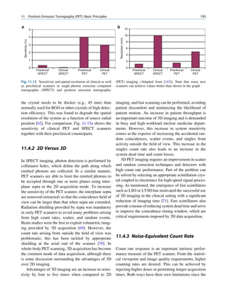
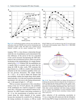
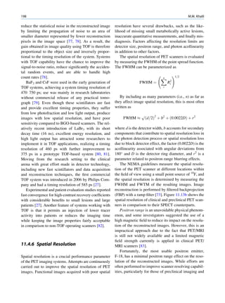
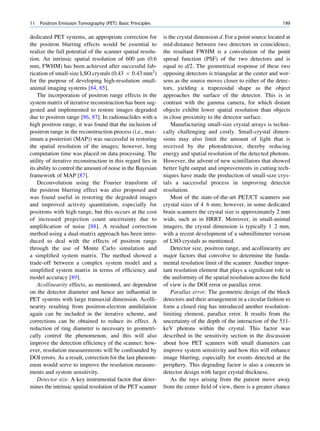
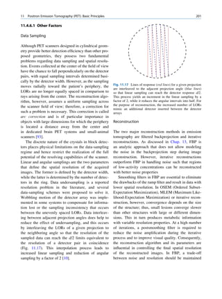
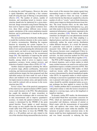
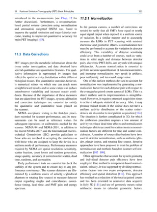
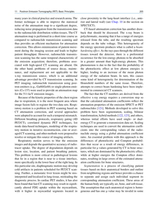
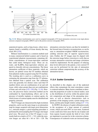
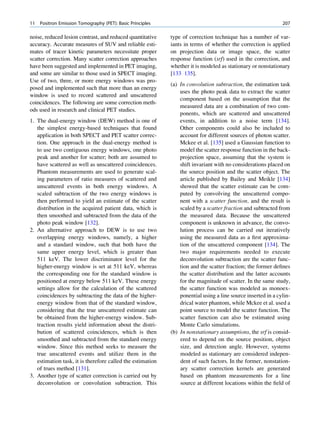
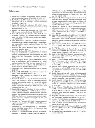
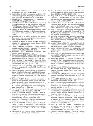
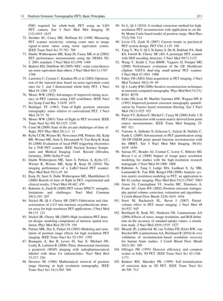
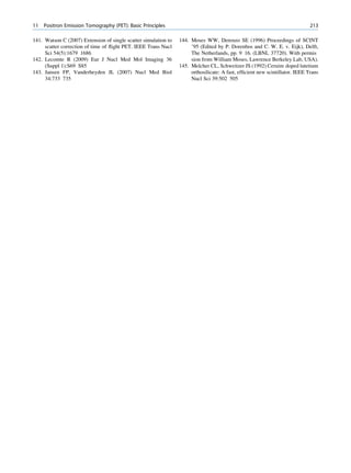


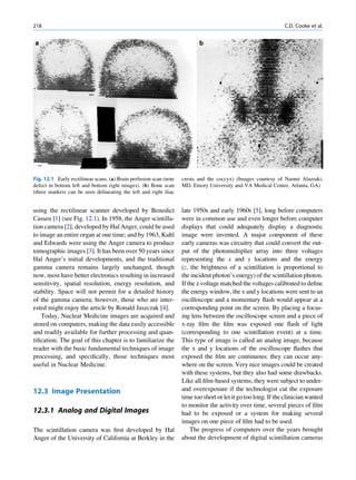
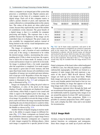
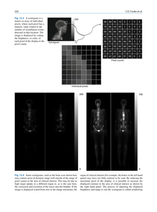
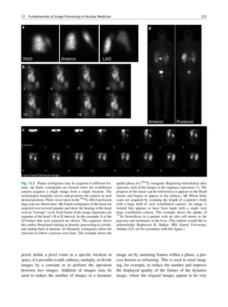
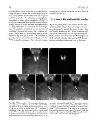
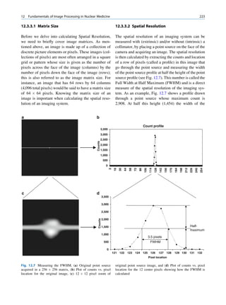
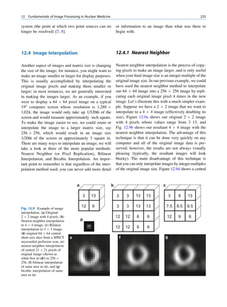
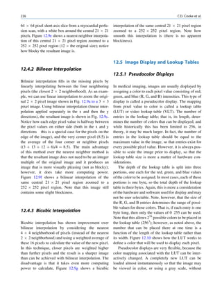
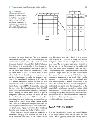
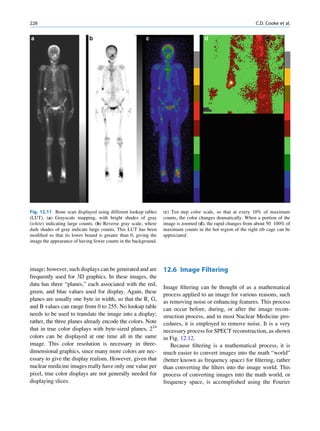
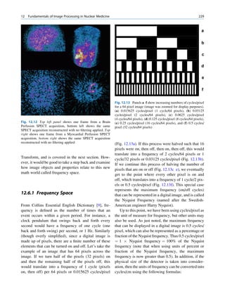
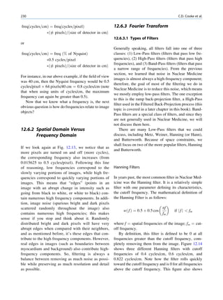
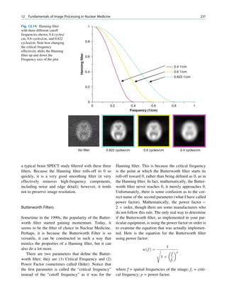
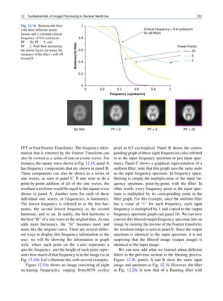
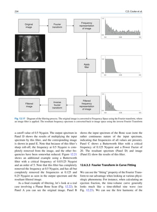
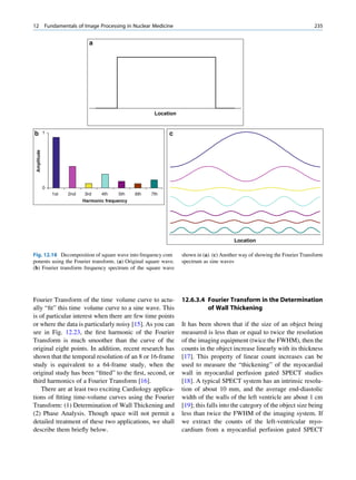
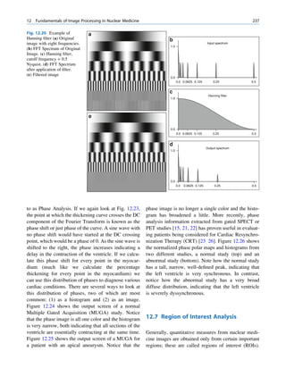
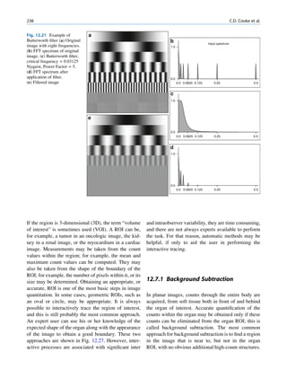
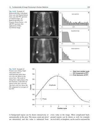
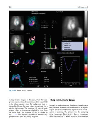
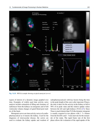
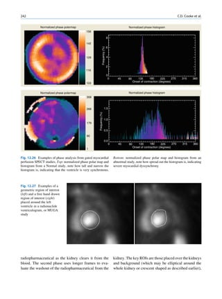
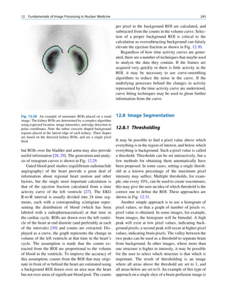

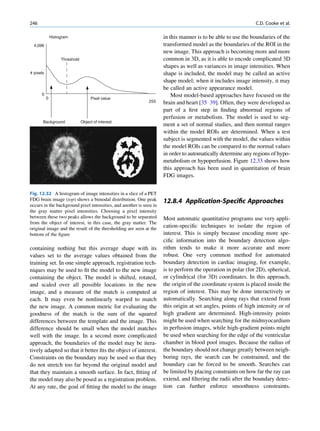
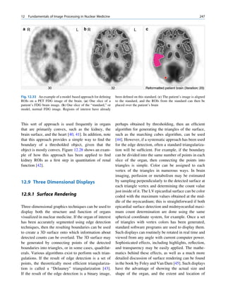
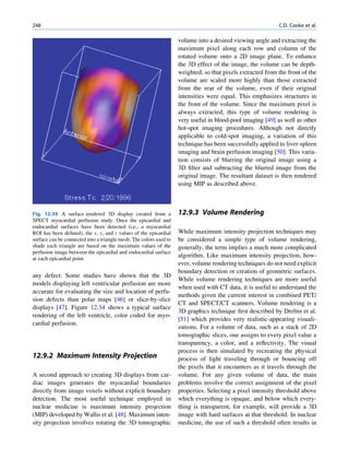
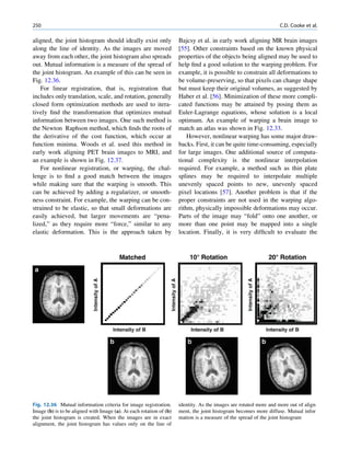
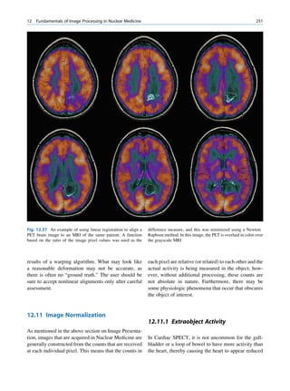
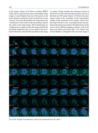
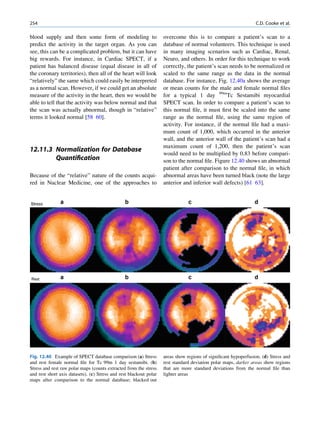
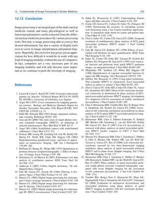
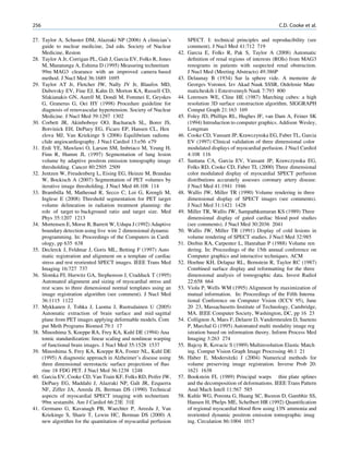


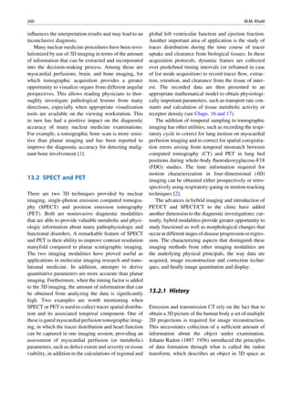
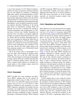
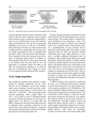
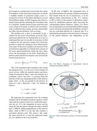
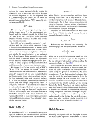
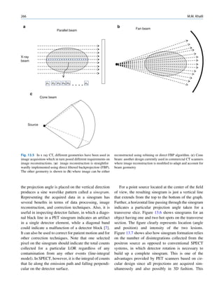
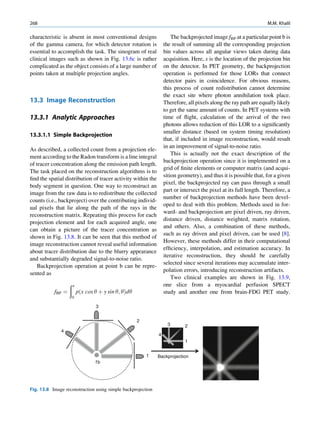
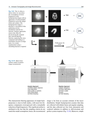
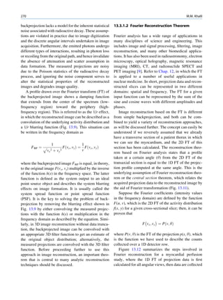
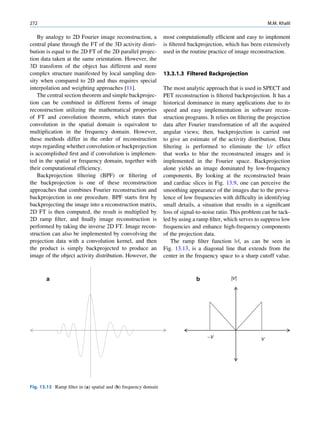
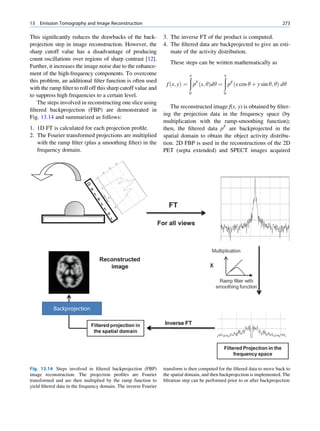
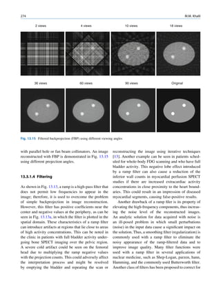
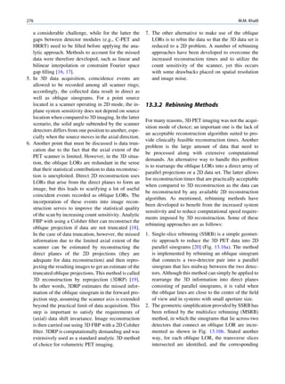
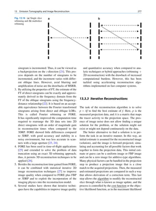
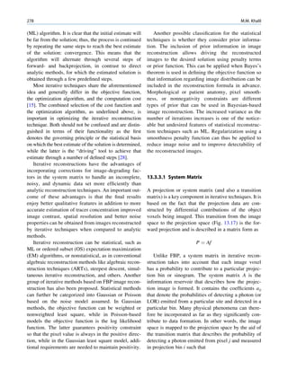
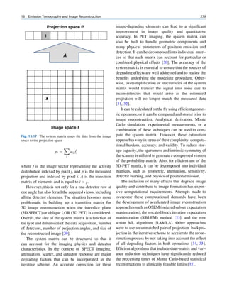
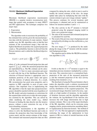
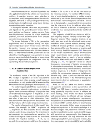
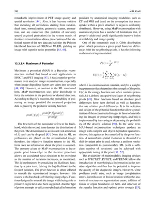
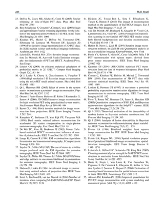
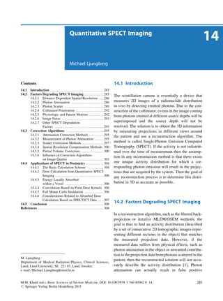
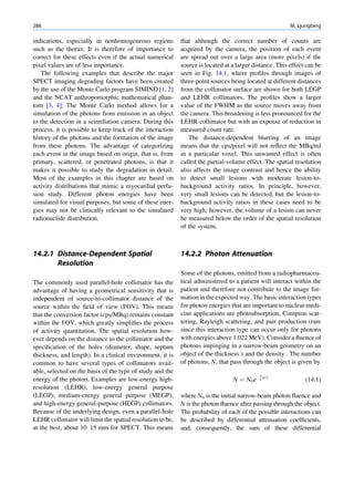
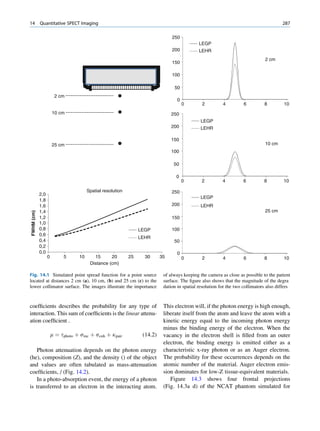
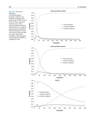
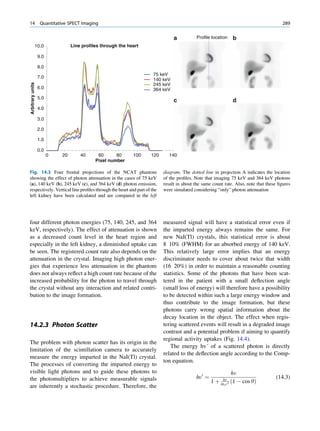
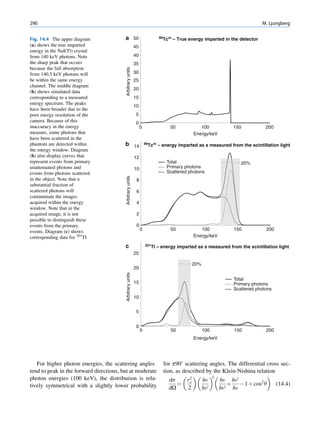
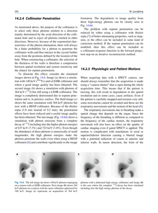
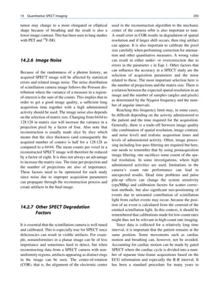
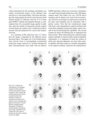
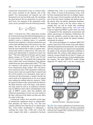
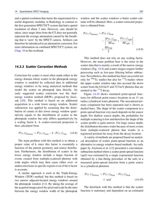
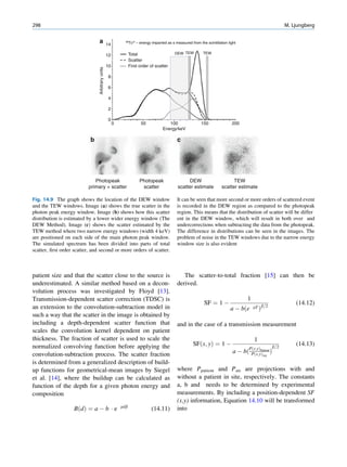
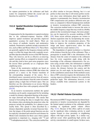
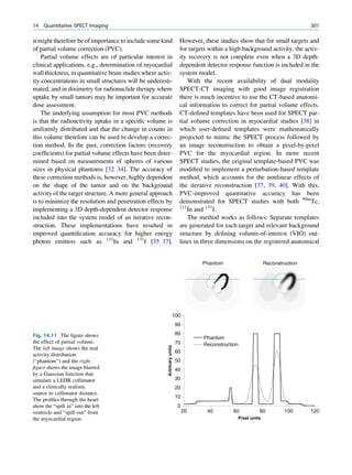
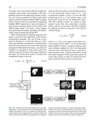
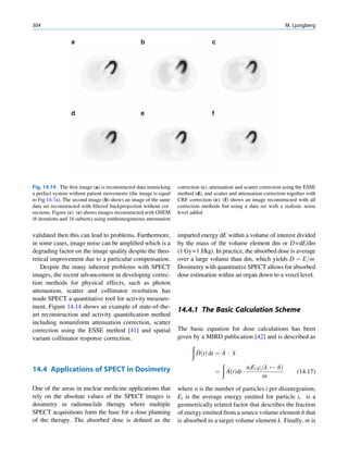
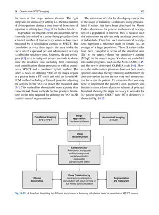
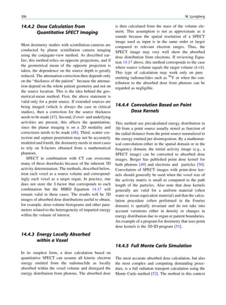
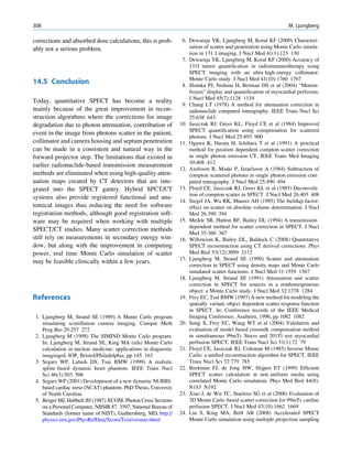
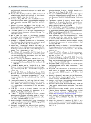

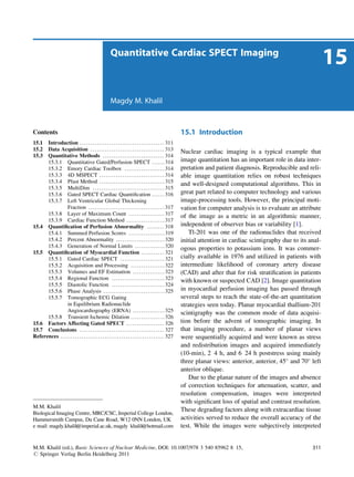
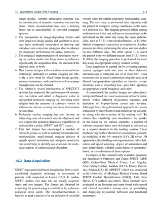
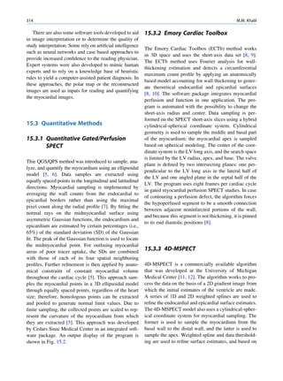
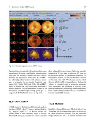
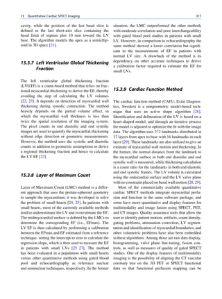
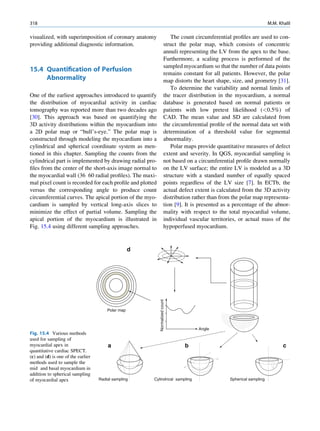
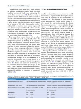
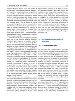
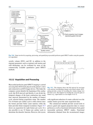
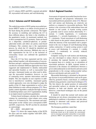
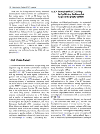
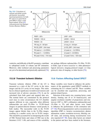
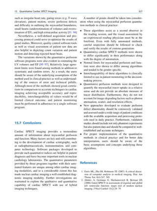
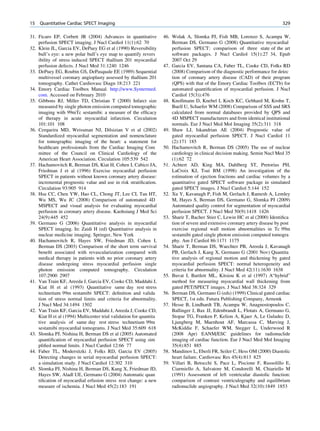
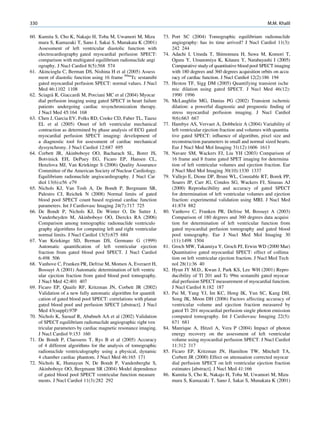
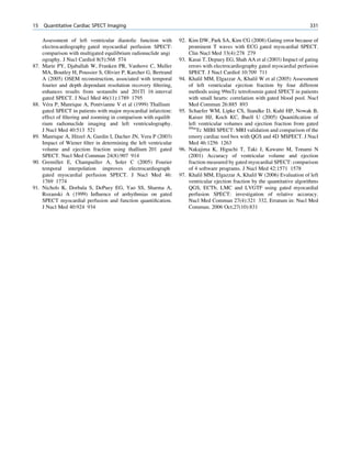


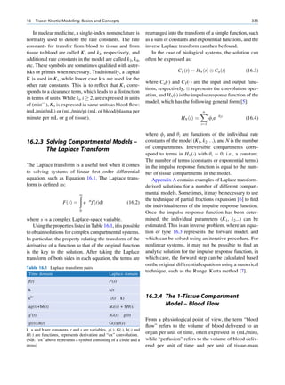
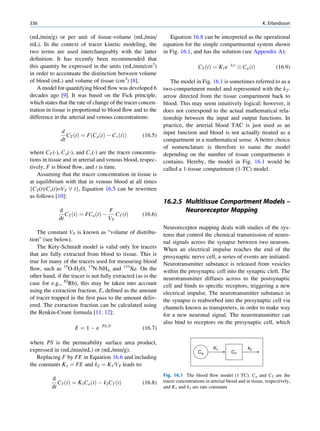
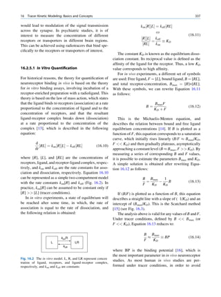
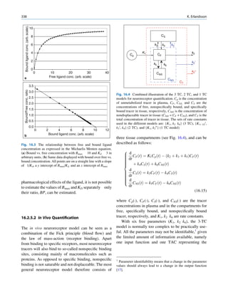
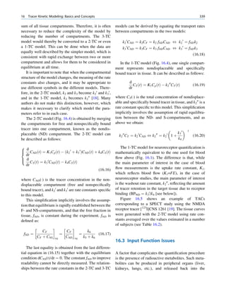
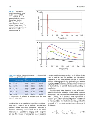
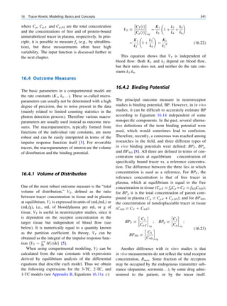
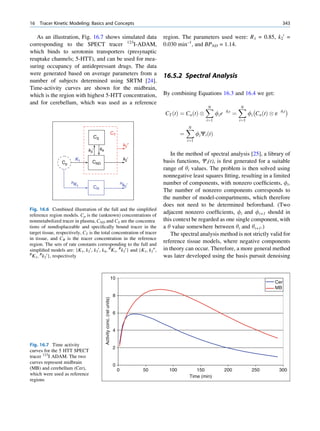
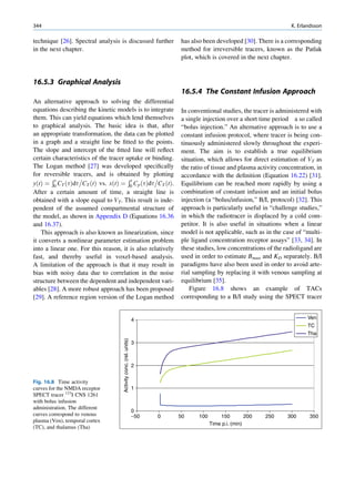
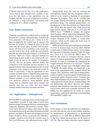


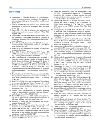
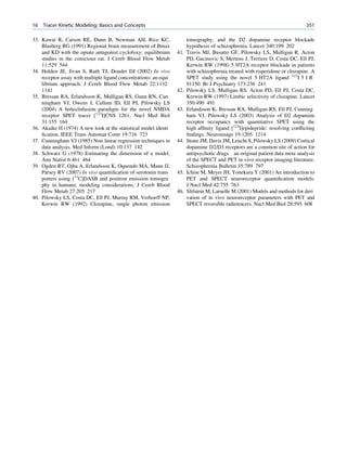

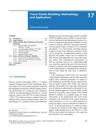
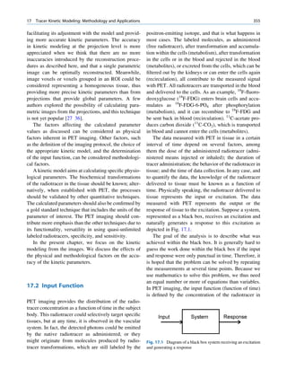
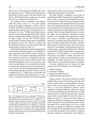
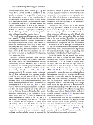
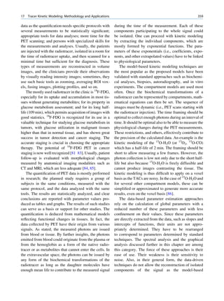
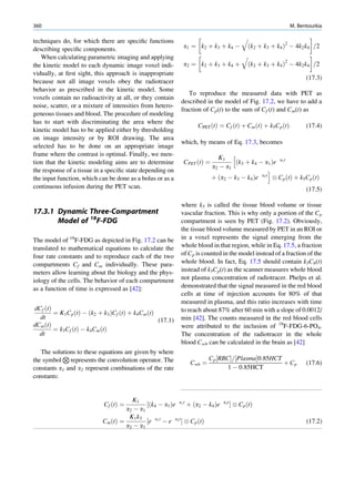
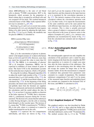
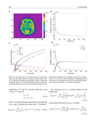
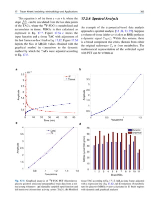
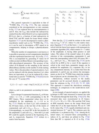
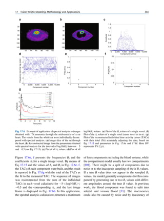
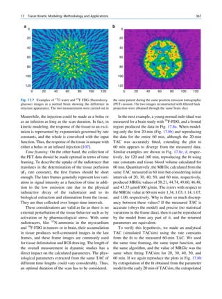
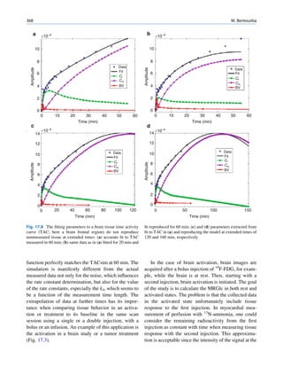
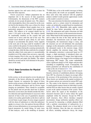
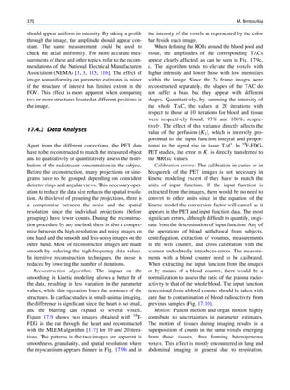
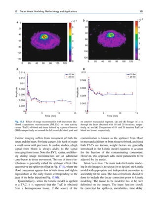
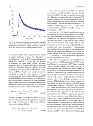
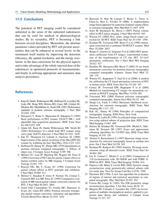
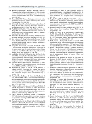
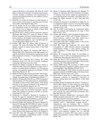


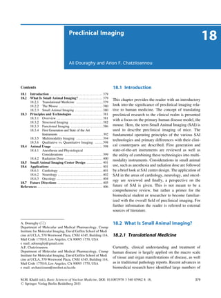
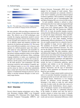
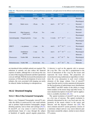
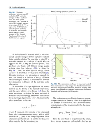
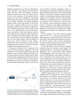
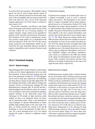
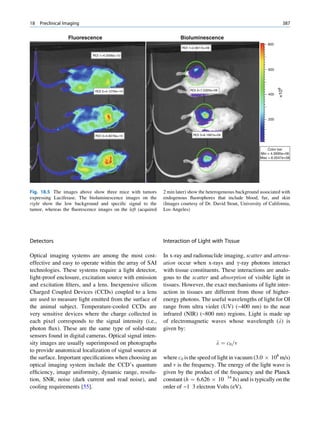
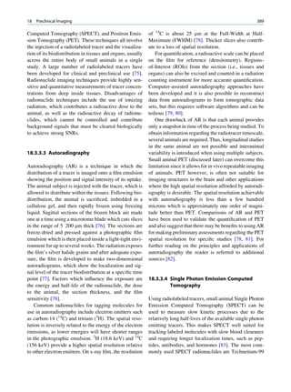
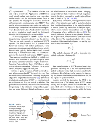
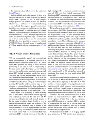
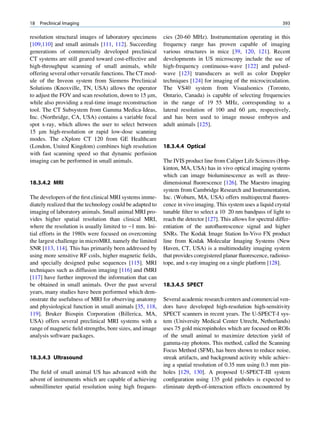
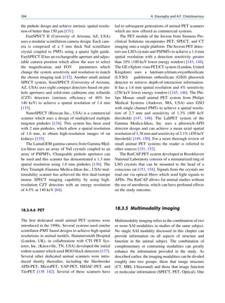
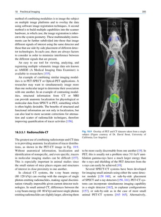
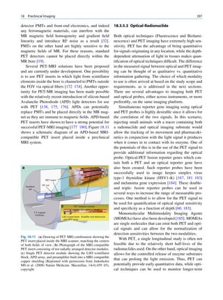
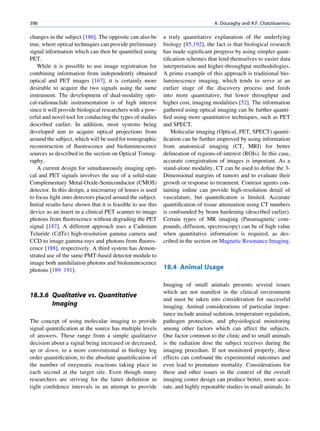
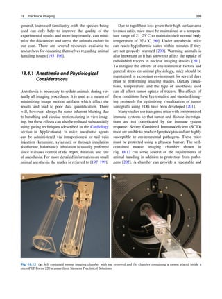
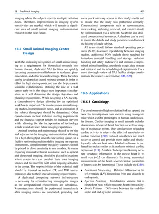
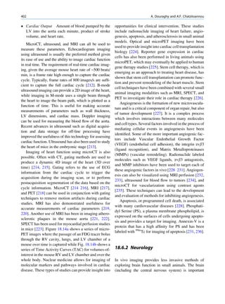
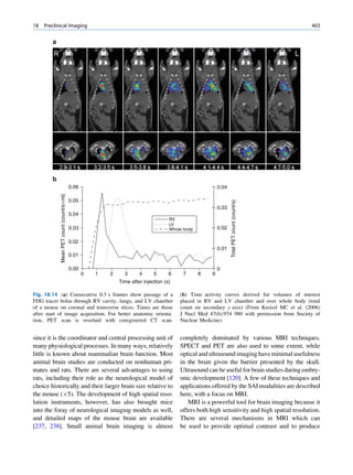
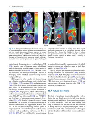
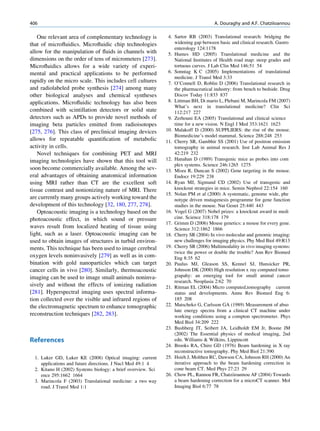
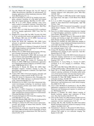
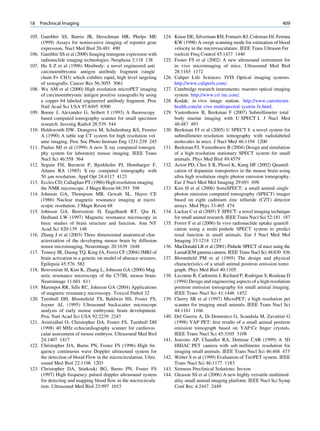
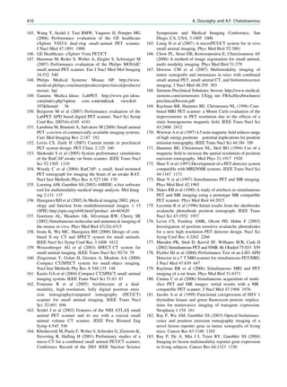
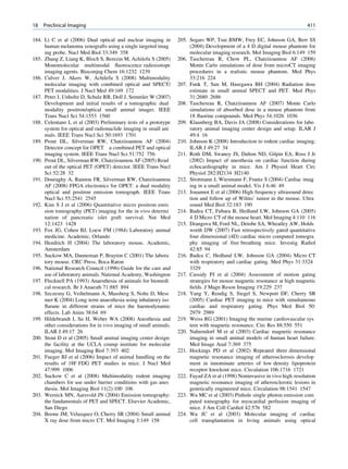
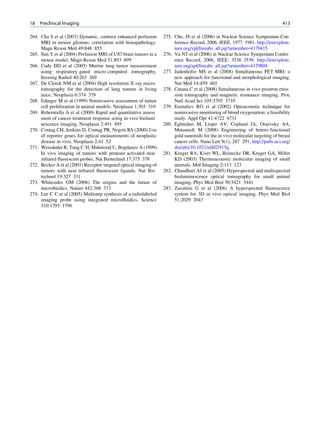

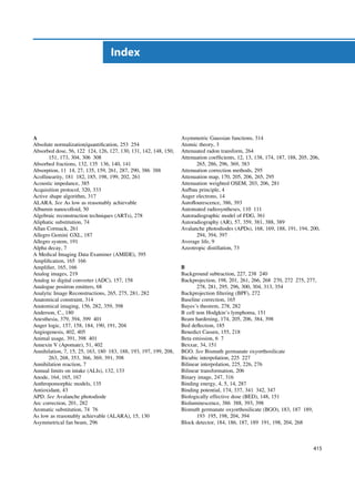
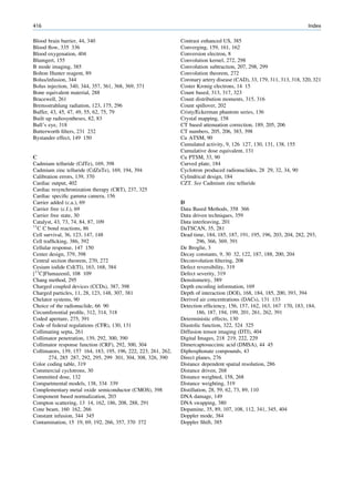
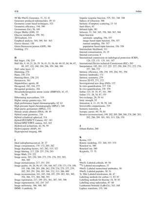
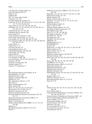
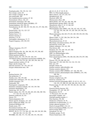
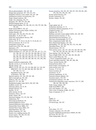
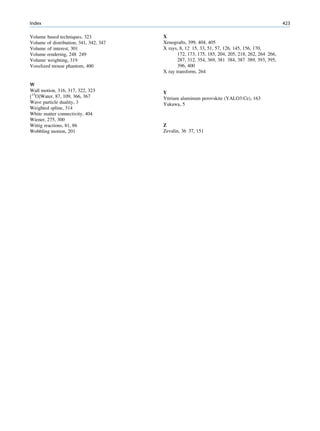

This document provides an overview of basic atomic and nuclear physics concepts relevant to nuclear medicine, including: - Atomic structure is described by quantum mechanics using wave functions and orbitals defined by quantum numbers. Electrons fill orbitals according to the Aufbau principle and Pauli exclusion principle. - Electrons are bound to the nucleus with binding energies. Absorption of sufficient energy can cause ionization or excitation of electrons to higher orbitals. - When electrons drop to lower orbitals to fill vacancies, electromagnetic radiation is emitted with energy equal to the energy difference between the orbitals. This atomic emission is the basis for diagnostic nuclear medicine techniques. - Radioactive decay involves the spontaneous emission of particles

![[2]Radionuclide_Imaging_Surgery](https://cdn.slidesharecdn.com/ss_thumbnails/1664462-thumbnail.jpg?width=640&height=640&fit=bounds)


