![CHAPTER 7 Neuroendocrine Control of the Menstrual Cycle 143
7
regulation.8 Unlike neurons secreting other hypothalamic releas-
ing factors, GnRH neurons do not exist in a defined nucleus but
are scattered throughout the medial basal hypothalamus, with
additional scattered neurons in the preoptic area.9
Over the past three decades, genetic studies in patients with
idiopathic hypogonadotropic hypogonadism (IHH) with con-
comitant disruption of the olfactory system resulting in anosmia
(Kallmann Syndrome; KS) or without anosmia (normosmic idio-
pathic hypogonadotropic hypogonadism; nIHH), have resulted
in unprecedented growth in our understanding of the complex
neuroendocrine control of reproduction. Mutations in well over
50 genes have now been discovered in this rare patient popula-
tion.10 Validation of their function in animal and cell systems
indicates that these can be broadly classified into four groups. A
number of genes have been discovered that are involved in the
early migration and axonal guidance of GnRH neurons on the path
to their eventual home in the hypothalamus, some of which are
associated with other developmental defects. This group of genes
includes Kallmann 1 (KAL1) now known as anosmin 1 (ANOS1),11
chromodomain helicase DNA binding protein 7 (CHG7),12 sex-
determining region of Y-
box 10 (SOX10),13 semaphorin-3A
(SEMA3A),14,15 fasciculation and elongation protein zeta family
zinc finger 1 (FEZF1),16 fibronectin leucine-
rich transmembrane
protein 3 (FLRT3),17 IL-
17 receptor D (IL17RD),17 and structural
maintenance of chromosomes flexible hinge domain-
containing
protein 1 (SMCHD1),7 among others. Genes involved in the con-
trol of GnRH secretion include kisspeptin and its receptor (KISS1/
KISS1R),18–20 tachykinin 3 and its receptor (TAC3/TACR3),21
gonadotropin-
releasing hormone1 (GNRH1),22,23 and dosage-
sensitive sex reversal 1 (DAX1), also known as nuclear receptor
subfamily 0, group B, member 1 (NROB1).24 Genes that appear to
play a role in both GnRH ontogeny and function include fibroblast
growth factor 8 and its receptor fibroblast growth factor receptor
1 (FGF8/FGFR1),25–28 prokineticin 2 and its receptor (PROK2/
PROKR2),29,30 heparin sulfate 6-
O-
sulfotransferase 1 (HS6ST1),31
WD repeat domain 11 (WDR11),32 AXL receptor tyrosine kinase
(AXL),33 NMDA receptor synaptonuclear signaling and neuro-
nal migration factor (NSMF),34 dual specificity phosphatase 6
(DUSP6),17 sprouty homolog 4 (SPRY4),17 and fibroblast growth
factor 17 (FGF17).17 Finally, the genes involved in gonadotrope
stimulation that have been discovered to date in association with
IHH include only DAX1 and gonadotropin-
releasing hormone
receptor (GNRHR).35 As more patients are identified and studied,
this list will undoubtedly continue to grow.
Pulsatile Secretion of GnRH
A prominent feature of the reproductive system is the absolute
requirement for pulsatile secretion of GnRH into the pituitary
portal system for normal gonadotropin secretion. The now clas-
sic studies of Knobil and colleagues in hypothalamic-
lesioned
monkeys receiving GnRH first showed that intermittent stimula-
tion of the pituitary results in secretion of LH and FSH, while
constant GnRH stimulation is associated with suppression of
gonadotropin levels.36 Isolated GnRH neurons exhibit an intrin-
sic pulsatility,37 but there is also a significant body of research
indicating that external influences modify and coordinate the
secretion of GnRH, influencing both the amplitude and fre-
quency of pulsatile GnRH secretion.
Neuromodulators of GnRH Secretion
While a number of neurotransmitters are involved in the control
of GnRH secretion in animal species, only a few have been shown
to have an effect on humans.38 Although there is evidence for
a stimulatory role of the α-
adrenergic system in several animal
models, it is much less likely that it plays a role in the control
of the human menstrual cycle. The role of the dopaminergic
system remains controversial, but studies that have documented
an increase in LH pulse frequency in response to a dopamine
antagonist in women with hypothalamic amenorrhea suggest that
dopamine may inhibit GnRH secretion in women.39,40
Kisspeptin
Knock-
out models suggest that there is considerable redundancy in
the systems that ultimately control GnRH secretion; however, it is
now firmly established that the kisspeptin pathway is a key upstream
modifier of GnRH secretion. As with the genes that are now known
to control the developmental migration of GnRH neurons, a role
for kisspeptin in reproduction was initially discovered by the com-
bination of genetic studies in patients with IHH which identified
mutations in the gene encoding the kisspeptin receptor (KISS1R,
formerly known as G-
protein coupled receptor 54 [GPR54]) and
knock-
out mouse models.18,19 Kisspeptin is an extremely powerful
stimulator of LH, an action that is blocked by a GnRH antagonist,
Ovary
Uterus
GnRH
Estradiol
Estradiol
Inhibin A
Inhibin B
FSH
LH
Estradiol
Pituitary
KISS
NKB
DYN
NKB
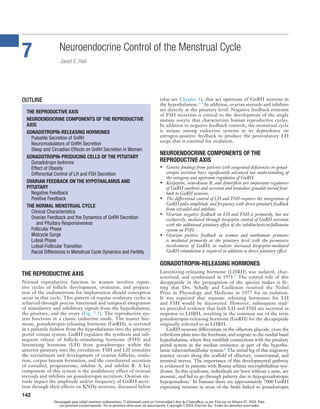
Fig. 7.1 Neuroendocrine control of reproduction requires the pulsatile
secretion of gonadotropin-
releasing hormone (GnRH) released into
the pituitary portal system to stimulate the synthesis and secretion
of luteinizing hormone (LH) and follicle-
stimulating hormone (FSH)
from pituitary gonadotropes. The gonadotropins, in turn, stimulate
follicle development and secretion of gonadal steroids and peptides. As
shown on the right, negative feedback of estradiol and progesterone on
hypothalamic GnRH secretion is mediated through kisspeptin (KISS),
neurokinin B (NKB), and dynorphin (DYN) which are colocalized in the
KNDY neurons of the median eminence. Inhibin A, inhibin B, and estradiol
also exert negative feedback effects directly at the pituitary. As shown on
the left, rising levels of estradiol are responsible for the positive feedback at
the pituitary, which generates the preovulatory gonadotropin surge. (From
Hall JE, Cacciopa P, NIEHS, personal communication.)
Descargado para rafael martinez (rafamartinez.71@hotmail.com) en Universidad Libre de ClinicalKey.es por Elsevier en febrero 07, 2024. Para
uso personal exclusivamente. No se permiten otros usos sin autorización. Copyright ©2024. Elsevier Inc. Todos los derechos reservados.](data:image/gif;base64,R0lGODlhAQABAIAAAAAAAP///yH5BAEAAAAALAAAAAABAAEAAAIBRAA7)
Recommended
More Related Content
Similar to CICLO MENSTRUAL 2024.pdf
Similar to CICLO MENSTRUAL 2024.pdf (20)
More from rafaelmartinez933361
More from rafaelmartinez933361 (11)
Recently uploaded
Recently uploaded (20)
CICLO MENSTRUAL 2024.pdf
- 1. 142 Neuroendocrine Control of the Menstrual Cycle Janet E. Hall 7 THE REPRODUCTIVE AXIS Normal reproductive function in women involves repeti- tive cycles of follicle development, ovulation, and prepara- tion of the endometrium for implantation should conception occur in that cycle. This pattern of regular ovulatory cycles is achieved through precise functional and temporal integration of stimulatory and inhibitory signals from the hypothalamus, the pituitary, and the ovary (Fig. 7.1). The reproductive sys- tem functions in a classic endocrine mode. The master hor- mone, gonadotropin- releasing hormone (GnRH), is secreted in a pulsatile fashion from the hypothalamus into the pituitary portal venous system. GnRH regulates the synthesis and sub- sequent release of follicle- stimulating hormone (FSH) and luteinizing hormone (LH) from gonadotropes within the anterior pituitary into the circulation. FSH and LH stimulate the recruitment and development of ovarian follicles, ovula- tion, corpus luteum formation, and the coordinated secretion of estradiol, progesterone, inhibin A, and inhibin B. A key component of this system is the modulatory effect of ovarian steroids and inhibins on gonadotropin secretion. Ovarian ste- roids impact the amplitude and/or frequency of GnRH secre- tion through their effects on KNDy neurons, discussed below (also see Chapter 1), that act upstream of GnRH neurons in the hypothalamus.1–4 In addition, ovarian steroids and inhibins act directly at the pituitary level. Negative feedback restraint of FSH secretion is critical to the development of the single mature oocyte that characterizes human reproductive cycles. In addition to negative feedback controls, the menstrual cycle is unique among endocrine systems in its dependence on estrogen- positive feedback to produce the preovulatory LH surge that is essential for ovulation. NEUROENDOCRINE COMPONENTS OF THE REPRODUCTIVE AXIS • Genetic findings from patients with congenital deficiencies in gonad- otropin secretion have significantly advanced our understanding of the ontogeny and upstream regulation of GnRH. • Kisspeptin, neurokinin B, and dynorphin are important regulators of GnRH synthesis and secretion and transduce gonadal steroid feed- back to GnRH neurons. • The differential control of LH and FSH requires the integration of GnRH pulse amplitude and frequency with direct pituitary feedback from estradiol and inhibins. • Ovarian negative feedback on LH and FSH is primarily, but not exclusively, mediated through kisspeptin control of GnRH secretion with the additional pituitary effect of the inhibin/activin/follistatin system on FSH. • Ovarian positive feedback in women and nonhuman primates is mediated primarily at the pituitary level with the permissive involvement of GnRH; in rodents increased kisspeptin- mediated GnRH stimulation is required in addition to direct pituitary effect. GONADOTROPIN-RELEASING HORMONES Luteinizing- releasing hormone (LHRH) was isolated, char- acterized, and synthesized in 1971.5 The central role of this decapeptide in the propagation of the species makes it fit- ting that Drs. Schally and Guillemin received the Nobel Prize in Physiology and Medicine in 1977 for its isolation. It was expected that separate releasing hormones for LH and FSH would be discovered. However, subsequent stud- ies provided evidence that both LH and FSH are secreted in response to LHRH, resulting in the common use of the term gonadotropin- releasing hormone (GnRH) for the decapeptide originally referred to as LHRH. GnRH neurons differentiate in the olfactory placode, cross the cribriform plate into the forebrain, and migrate to the medial basal hypothalamus, where they establish connections with the pituitary portal system in the median eminence as part of the hypotha- lamic tuberoinfundibular system.6 The initial leg of this migratory journey occurs along the scaffold of olfactory, vomeronasal, and terminal nerves. The importance of this developmental pathway is evidenced in patients with Bosma arhinia microphthalmia syn- drome. In this syndrome, individuals are born without a nose, are anosmic, and fail to go through puberty due to hypogonadotropic hypogonadism.7 In humans there are approximately 7000 GnRH expressing neurons in areas of the brain linked to gonadotropin OUTLINE THE REPRODUCTIVE AXIS NEUROENDOCRINE COMPONENTS OF THE REPRODUCTIVE AXIS GONADOTROPIN-RELEASING HORMONES Pulsatile Secretion of GnRH Neuromodulators of GnRH Secretion Sleep and Circadian Effects on GnRH Secretion in Women GONADOTROPIN- PRODUCING CELLS OF THE PITUITARY Gonadotropn Isoforms Effect of Obesity Differential Control of LH and FSH Secretion OVARIAN FEEDBACK ON THE HYPOTHALAMUS AND PITUITARY Negative Feedback Positive Feedback THE NORMAL MENSTRUAL CYCLE Clinical Characteristics Ovarian Feedback and the Dynamics of GnRH Secretion and Pituitary Responsiveness Follicular Phase Midcycle Surge Luteal Phase Luteal-Follicular Transition Racial Differences in Menstrual Cycle Dynamics and Fertility Descargado para rafael martinez (rafamartinez.71@hotmail.com) en Universidad Libre de ClinicalKey.es por Elsevier en febrero 07, 2024. Para uso personal exclusivamente. No se permiten otros usos sin autorización. Copyright ©2024. Elsevier Inc. Todos los derechos reservados.
- 2. CHAPTER 7 Neuroendocrine Control of the Menstrual Cycle 143 7 regulation.8 Unlike neurons secreting other hypothalamic releas- ing factors, GnRH neurons do not exist in a defined nucleus but are scattered throughout the medial basal hypothalamus, with additional scattered neurons in the preoptic area.9 Over the past three decades, genetic studies in patients with idiopathic hypogonadotropic hypogonadism (IHH) with con- comitant disruption of the olfactory system resulting in anosmia (Kallmann Syndrome; KS) or without anosmia (normosmic idio- pathic hypogonadotropic hypogonadism; nIHH), have resulted in unprecedented growth in our understanding of the complex neuroendocrine control of reproduction. Mutations in well over 50 genes have now been discovered in this rare patient popula- tion.10 Validation of their function in animal and cell systems indicates that these can be broadly classified into four groups. A number of genes have been discovered that are involved in the early migration and axonal guidance of GnRH neurons on the path to their eventual home in the hypothalamus, some of which are associated with other developmental defects. This group of genes includes Kallmann 1 (KAL1) now known as anosmin 1 (ANOS1),11 chromodomain helicase DNA binding protein 7 (CHG7),12 sex- determining region of Y- box 10 (SOX10),13 semaphorin-3A (SEMA3A),14,15 fasciculation and elongation protein zeta family zinc finger 1 (FEZF1),16 fibronectin leucine- rich transmembrane protein 3 (FLRT3),17 IL- 17 receptor D (IL17RD),17 and structural maintenance of chromosomes flexible hinge domain- containing protein 1 (SMCHD1),7 among others. Genes involved in the con- trol of GnRH secretion include kisspeptin and its receptor (KISS1/ KISS1R),18–20 tachykinin 3 and its receptor (TAC3/TACR3),21 gonadotropin- releasing hormone1 (GNRH1),22,23 and dosage- sensitive sex reversal 1 (DAX1), also known as nuclear receptor subfamily 0, group B, member 1 (NROB1).24 Genes that appear to play a role in both GnRH ontogeny and function include fibroblast growth factor 8 and its receptor fibroblast growth factor receptor 1 (FGF8/FGFR1),25–28 prokineticin 2 and its receptor (PROK2/ PROKR2),29,30 heparin sulfate 6- O- sulfotransferase 1 (HS6ST1),31 WD repeat domain 11 (WDR11),32 AXL receptor tyrosine kinase (AXL),33 NMDA receptor synaptonuclear signaling and neuro- nal migration factor (NSMF),34 dual specificity phosphatase 6 (DUSP6),17 sprouty homolog 4 (SPRY4),17 and fibroblast growth factor 17 (FGF17).17 Finally, the genes involved in gonadotrope stimulation that have been discovered to date in association with IHH include only DAX1 and gonadotropin- releasing hormone receptor (GNRHR).35 As more patients are identified and studied, this list will undoubtedly continue to grow. Pulsatile Secretion of GnRH A prominent feature of the reproductive system is the absolute requirement for pulsatile secretion of GnRH into the pituitary portal system for normal gonadotropin secretion. The now clas- sic studies of Knobil and colleagues in hypothalamic- lesioned monkeys receiving GnRH first showed that intermittent stimula- tion of the pituitary results in secretion of LH and FSH, while constant GnRH stimulation is associated with suppression of gonadotropin levels.36 Isolated GnRH neurons exhibit an intrin- sic pulsatility,37 but there is also a significant body of research indicating that external influences modify and coordinate the secretion of GnRH, influencing both the amplitude and fre- quency of pulsatile GnRH secretion. Neuromodulators of GnRH Secretion While a number of neurotransmitters are involved in the control of GnRH secretion in animal species, only a few have been shown to have an effect on humans.38 Although there is evidence for a stimulatory role of the α- adrenergic system in several animal models, it is much less likely that it plays a role in the control of the human menstrual cycle. The role of the dopaminergic system remains controversial, but studies that have documented an increase in LH pulse frequency in response to a dopamine antagonist in women with hypothalamic amenorrhea suggest that dopamine may inhibit GnRH secretion in women.39,40 Kisspeptin Knock- out models suggest that there is considerable redundancy in the systems that ultimately control GnRH secretion; however, it is now firmly established that the kisspeptin pathway is a key upstream modifier of GnRH secretion. As with the genes that are now known to control the developmental migration of GnRH neurons, a role for kisspeptin in reproduction was initially discovered by the com- bination of genetic studies in patients with IHH which identified mutations in the gene encoding the kisspeptin receptor (KISS1R, formerly known as G- protein coupled receptor 54 [GPR54]) and knock- out mouse models.18,19 Kisspeptin is an extremely powerful stimulator of LH, an action that is blocked by a GnRH antagonist, Ovary Uterus GnRH Estradiol Estradiol Inhibin A Inhibin B FSH LH Estradiol Pituitary KISS NKB DYN NKB Fig. 7.1 Neuroendocrine control of reproduction requires the pulsatile secretion of gonadotropin- releasing hormone (GnRH) released into the pituitary portal system to stimulate the synthesis and secretion of luteinizing hormone (LH) and follicle- stimulating hormone (FSH) from pituitary gonadotropes. The gonadotropins, in turn, stimulate follicle development and secretion of gonadal steroids and peptides. As shown on the right, negative feedback of estradiol and progesterone on hypothalamic GnRH secretion is mediated through kisspeptin (KISS), neurokinin B (NKB), and dynorphin (DYN) which are colocalized in the KNDY neurons of the median eminence. Inhibin A, inhibin B, and estradiol also exert negative feedback effects directly at the pituitary. As shown on the left, rising levels of estradiol are responsible for the positive feedback at the pituitary, which generates the preovulatory gonadotropin surge. (From Hall JE, Cacciopa P, NIEHS, personal communication.) Descargado para rafael martinez (rafamartinez.71@hotmail.com) en Universidad Libre de ClinicalKey.es por Elsevier en febrero 07, 2024. Para uso personal exclusivamente. No se permiten otros usos sin autorización. Copyright ©2024. Elsevier Inc. Todos los derechos reservados.
- 3. PART I The Fundamentals of Reproduction 144 indicating that the effect of kisspeptin on LH is mediated through control of GnRH secretion.41,42 The kisspeptin system is thought to play a dominant role in the onset of puberty and mediates estro- gen and progesterone negative feedback in the median eminence.2 Studies of kisspeptin administration in women, performed using different isoforms and either subcutaneous, intravenous bolus, or intravenous infusion modes of administration, have demonstrated a marked difference in LH response depending on cycle phase and hormonal status.43 The response to kisspeptin is consistently robust in the late follicular, preovulatory, and luteal phases of the menstrual cycle and in postmenopausal women, while there is some inconsistency in the early follicular phase with a lower, and in some cases absent, LH response to kisspeptin44 The LH response to continuous kisspeptin was similarly low in postmenopausal women but increased after two weeks of estrogen replacement in a dose- dependent fashion, an effect that appears to be mediated at both the pituitary and the hypothalamic level.45 In rodents and sheep, there is ample evidence that positive feedback is manifest in the hypothalamus as well as the pituitary with the generation of a marked increase in GnRH at the time of the midcycle gonadotropin surge.46,47 Kisspeptin neurons in the anteroventral periventricular nucleus (AVPV) have now been implicated in this estrogen- positive feedback on GnRH secretion in rodents.1 The relationship of the AVPV to the suprachiasmatic nucleus (SCN) in the rodent provides a potential mechanism for the known circadian timing of the proestrus surge in the rodent.48 Despite compelling evidence in lower animal species, it is likely that kisspeptin control of GnRH does not play a role in the midcycle surge in women; studies in women demonstrate a pau- city of kisspeptin neurons in an analogous hypothalamic region49 and evidence discussed below demonstrates that a GnRH surge is not required for the generation of a midcycle LH surge in GnRH-deficient women50 and further suggests that a GnRH surge is not present in normal women.51 Neurokinin B Neurokinin B (NKB), which is encoded by the tachykinin 3 gene (TAC3) and its cognate receptor, NK3R, encoded by TACR3, have also been implicated in the normal control of GnRH secre- tion through genetic studies in patients with IHH.21 NKB stimu- lates LH secretion, acting upstream of the GnRH neuron.52,53 TAC3 and KISS are colocalized in the human as well as in other species,54 particularly in the arcuate/median eminence. There is significant evidence that the effect of NKB on GnRH is exerted primarily through kisspeptin. NKB agonists stimulate gonado- tropin secretion55 while NKB3 antagonists inhibit LH secretion, but do not appear to do so in the setting of high estrogen, as in the preovulatory phase56 Interestingly, the NKB3/NK3R system also plays a role in hot flashes in estrogen- deficient states.56–58 Endogenous Opioids/Dynorphin There is substantial evidence for the involvement of endorphins in transducing the negative feedback effects of progesterone on pulsa- tile GnRH secretion from studies using the opioid receptor blocker, naloxone, in women.59,60 However, naloxone binds not only to the mu receptor but also to the kappa and gamma receptors and thus these early studies could not provide mechanistic specificity. Dynorphin which binds to the kappa- opioid receptor has now been identified as the key mediator of progesterone negative feedback.61 KNDy Neurons In a variety of animal species and humans it has been shown that kis- speptin, NKB, and dynorphin are coexpressed in cells in the arcuate nucleus/median eminence that are now referred to as KNDy neu- rons. These neurons express estrogen, progesterone, and androgen receptors and mediate gonadal steroid negative feedback on GnRH secretion with increasing evidence that they are also involved in the initiation and termination of GnRH secretion that results in its pul- satile secretion.62 Gamma- amino butyric acid (GABA) may also be involved in mediating estrogen- negative feedback on GnRH secre- tion, particularly in the arcuate/median eminence.63,64 RFamide-Related Peptides RFaimde- related peptides (RFRP) are the mammalian orthologues of gonadotropin inhibitory hormone (GnIH) which was first dis- covered in the hypothalami of the quail.65 In humans, RFRP- 1 and RFRP- 3 neurons send axonal projections to GnRH neurons.66,67 RFRPs are secreted into the pituitary portal system68 and their receptor, G- protein coupled receptor 147 (GPR147), is present on gonadotropes as well as in the hypothalamus66 Taken together with functional data from animal and cellular systems, these find- ings suggest that RFRPs function at both the hypothalamus and pituitary to regulate the secretion of LH and FSH. Interestingly, there is also evidence that RFRPs increase food intake in sheep without reducing energy expenditure.67 There is currently limited data to address the role of these peptides in humans. A three- hour infusion of custom synthesized GnIH resulted in a modest sup- pression of LH secretion in postmenopausal women but failed to inhibit LH secretion in response to pulses of kisspeptin- 10 in men.69 Additional studies will be required to ascertain its physiol- ogy and potential therapeutic role in men and women. Sleep and Circadian Effects on GnRH Secretion in Women Endocrine systems are profoundly influenced by both sleep and endogenous circadian rhythms, which are intrinsic rhythms that persist in the absence of sleep or other environmental cues. Diurnal (day and night) rhythms of LH and gonadal steroids have been well described in men and women. However, studies in which sleep and other environmental cues were controlled have failed to demonstrate an endogenous circadian rhythm of LH or FSH in early follicular phase women (Fig. 7.2) and in postmeno- pausal women, despite the presence of robust circadian rhythms of temperature, cortisol, and TSH.70,71 In contrast, there is compelling evidence that sleep directly affects the pulsatile secretion of LH and presumably that of GnRH. Studies that have separated the effects of sleep from time of day demonstrate that during puberty in boys and girls, pulsatile LH secretion is increased during sleep.72,73 More recent studies indicate that LH pulses are most commonly preceded by slow wave sleep (SWS)74 and further studies have shown that even with repeated sleep interruption, 20 min of accumulated sleep is associated with LH pulse onset in this population.75 These stud- ies suggest that factors associated with SWS stimulate GnRH secretion or that there is an upstream regulator of both GnRH secretion and deep sleep in puberty. Paradoxically, with the maturation of the reproductive system and the onset of ovulatory menstrual cycles, there is a notable slowing of pulsatile LH secretion at night in the early follicu- lar phase of the cycle.76,77 Sleep reversal studies in women have demonstrated that the early follicular phase of nighttime slowing is due to sleep rather than the time of day.78 Importantly, within sleep, brief periods of wakefulness are associated with the onset of LH pulses (Fig. 7.3), while SWS is inhibitory to LH pulses.78 Data relating luteal phase characteristics to early follicular phase nighttime pulse frequency suggests that prior progesterone exposure sensitizes the GnRH pulse generator to the inhibitory effects of sleep in the early follicular phase.79 In this regard, it is of interest that GnRH pulse frequency, as determined by the pulsa- tile secretion of the gonadotropin- free alpha subunit (FAS), is also slower during sleep than wake in postmenopausal women whose gonadal steroid levels are similar to those in early puberty, where sleep is stimulatory.80 Although the effect of sleep is much less than Descargado para rafael martinez (rafamartinez.71@hotmail.com) en Universidad Libre de ClinicalKey.es por Elsevier en febrero 07, 2024. Para uso personal exclusivamente. No se permiten otros usos sin autorización. Copyright ©2024. Elsevier Inc. Todos los derechos reservados.
- 4. CHAPTER 7 Neuroendocrine Control of the Menstrual Cycle 145 7 in normally cycling women, this finding suggests that the inhibi- tory effect of sleep on LH pulse frequency is not only related to prior progesterone exposure but that other factors relating to the development of a mature reproductive axis are involved. GONADOTROPIN- PRODUCING CELLS OF THE PITUITARY LH and FSH are synthesized in gonadotropes, which comprise between 7% and 15% of the cells in the pituitary. Immuno histochemical studies in the rat indicate that approximately 70% of gonadotropes stain for both LH and FSH, while the remainder stains for LH or FSH in approximately equal numbers. As the animals approach the day of the gonadotropin surge in proestrus, monohormonal cells begin to express both βLH and βFSH, while a population of cells that express growth hormone (GH) also express the gonadotropin subunits.81 LH and FSH are glycoprotein hormones whose polypeptide and polysaccharide components are essential for their activity. Biosynthesis of intact gonadotropins involves (1) translation of βLH, βFSH, and the common gonadotropin α- subunit; (2) post- translational modification and folding; (3) combination of the β- and α-subunits; and (4) modification of the oligosaccharide residues on LH and FSH as they traverse the Golgi.82 FSH is synthesized under the dual control of GnRH and the activin/inhibin/follistatin system and is secreted primarily in a constitutive manner with little storage. In contrast, LH is packaged into granules and stored. LH and the gonadotropin- free α subunit (FAS) are then secreted in response to GnRH stimulation of the gonadotrope. Gonadotropn Isoforms Multiple isoforms of LH and FSH, differing in their carbohydrate structure and charge, coexist in both pituitary and serum. When combined with a β-subunit, the α- subunit has two glycosylation sites. FSHβ also has two potential glycosylation sites, while LHβ has a single potential site. This results in the secretion of FSHtri and FSHquatro and LHdi and LHtri. Terminal sulfonated and/ or sialylated residues on these glycoforms add further isoform het- erogeneity to secreted gonadotropins. More basic forms of both LH and FSH yield a greater in vitro potency, but a shorter half- life in the circulation, while the opposite is true for less basic forms.83 The greater number of sialic acid residues on FSH prolongs its half-life,84 whereas the greater number of sulfonated N- acetyl- galactosamine (GalNAc) asparagine- linked oligosaccharides on LH is associated with more rapid clearance due to binding to a specific hepatic receptor.85 Sulfonation and sialylation of LH and FSH vary across the menstrual cycle and in the absence of gonadal steroids; postmenopausal women have a greater preponderance of sialylated forms of both LH and FSH.86 The number of sulfonated and sialylated residues on LH and FSH is tightly linked to hor- mone clearance in women and, by inference, to bioactivity.84 Thus, the disappearance of LH following GnRH receptor blockade with a potent GnRH antagonist is significantly prolonged in post- menopausal women compared to women in the follicular phase and at the midcycle surge (MCS),87 while the disappearance of FAS is unaffected by the absence of gonadal function (Table 7.1). Changes in the isoform composition of FSH in the normal men- strual cycle are likely to augment the effect of the rise in FSH on follicle recruitment and development during the luteal- follicular transition through a decrease in clearance; in contrast isoform changes that increase clearance would curtail the potential effect of the increase in FSH on follicle recruitment at midcycle. Effect of Obesity There is also evidence that gonadotropin secretion is modulated by a factor or factors related to obesity. Serum levels of LH are inversely related to body mass index (BMI) in normal women and in women with polycystic ovary syndrome (PCOS).88 Further studies have indicated that the inhibitory effect of obesity on LH secretion in PCOS is not mediated at the hypothalamus but is associated with a decrease in both the pituitary response to GnRH and the half- life of endogenous, but not exogenous, LH.89,90 The latter finding is consistent with the increase in sul- fonated isoforms of LH and FSH as a function of increasing BMI in women with PCOS.86 Recent studies have shown that in obese men and obese women without PCOS, the combination of an insulin and lipid/heparin infusion which results in increased free fatty acids and triglycerides, suppresses LH and FSH.91 Although the characteristics of pulsatile LH secretion were not measured in this study, the studies described above would suggest that the resultant decrease in LH and FSH is mediated at the pituitary. Fig. 7.2 Mean + sem of temperature, FSH, LH, FAS, and TSH levels in early follicular phase women (n = 11) during a constant routine of light, position, wake, and nutritional intake over a 24- hour period. These studies indicate the constancy of gonadotropin levels across the day and night in the absence of sleep with maintenance of endogenous circadian rhythms of temperature and TSH. Individual studies are aligned to the onset of habitual sleep onset for each subject beginning 8 hours from the onset of sampling, although subjects were awake for the full 24 hours of the study. (Adapted from Klingman KM, Marsh EE, Klerman EB, Anderson EJ, Hall JE. Absence of circadian rhythms of gonadotropin secretion in women. J Clin Endocrinol Metab. 2011;96[5]:1456–1461.) Descargado para rafael martinez (rafamartinez.71@hotmail.com) en Universidad Libre de ClinicalKey.es por Elsevier en febrero 07, 2024. Para uso personal exclusivamente. No se permiten otros usos sin autorización. Copyright ©2024. Elsevier Inc. Todos los derechos reservados.
- 5. PART I The Fundamentals of Reproduction 146 Differential Control of LH and FSH Secretion Although secreted from a common cell type within the gonad- otrope, LH and FSH have markedly different functions in the control of ovarian physiology. These differences in function are reflected in their different patterns of secretion during normal reproductive cycles. The divergent control of LH and FSH is achieved through a combination of differential control of the synthesis and secretion of LH and FSH by the pattern of GnRH stimulation, the preferential control of FSH synthesis by the activin/follistatin system, and differential feedback by ovarian ste- roids and the inhibins at the pituitary. Understanding the control of LH and FSH secretion is critical to our understanding of the dynamics of the menstrual cycle. Gonadotropin-Releasing Hormone FSH is secreted along with LH in response to acute stimulation by GnRH, but the relative role of GnRH in the overall control of FSH synthesis is much less than for LH. Blockade of the GnRH receptor using a specific GnRH receptor antagonist results in 90% inhibition of LH secretion, but only 40% to 60% inhibi- tion of FSH.92 Synthesis and secretion of LH and FSH are dif- ferentially controlled by the amplitude and frequency of GnRH stimulation.93,94 LH is highly responsive to increases in the dose of GnRH, while FSH is relatively insensitive to the GnRH dose. A physiological frequency of GnRH results in the synthe- sis and secretion of FSHβ, LHβ, and free α subunit. However, increases or decreases in the physiological frequency have differ- ential effects on LH and FSH (Table 7.2). Slow frequencies of GnRH stimulation favor synthesis and secretion of FSH in vitro and are associated with an increase in FSH in human studies in settings in which gonadal feedback is low.95 In GnRH- deficient men and women, an increase in the frequency of GnRH stimula- tion increases mean levels of LH with no appreciable change in FSH96,97 (Fig. 7.4), effects that have implications for the patho- physiology of PCOS which is characterized by an increased fre- quency of pulsatile GnRH stimulation of the pituitary.89 The direct effect of GnRH pulse frequency on GnRH receptor num- ber98 underlies the frequency modulation of LH and FSH secre- tion, at least in part. In addition, increased GnRH pulse frequency increases follistatin which would attenuate activin stimulation of FSH synthesis, as discussed below.99 While an increase in GnRH pulse frequency increases the synthesis and mean levels of LH, LH pulse amplitude is inversely related to GnRH pulse frequency.100,101 Studies in GnRH- deficient men and women have shown that slower GnRH pulse frequencies are associated with a higher LH pulse amplitude in Fig. 7.3 As indicated in the left panel, sleep is specifically associated with slowing of luteinizing hormone (LH) pulses. The LH interpulse interval (IPI) is longer and amplitude is higher in normal women during sleep (blue bars) compared with wake (purple bars), whether sleep occurs at night or during the day. As indicated in the right panel, wakefulness is more likely in the 5 to 10 minutes before the onset of an LH pulse (blue bars) than in a similar period indexed to a random LH point that is not associated with an LH pulse (purple bars). (Adapted from Hall JE, Sullivan JP, Richardson GS. Brief wake episodes modulate sleep- inhibited luteinizing hormone secretion in the early follicular phase. J Clin Endocrinol Metab. 2005;90[4]:2050–2055.) –15 to –10 * *** ** Min of wakefulness/5 minutes 3 2 1 0 –10 to –5 –5 to 0 –20 to –15 Minutes from onset * * ** ** ** Wake sleep day Mean LH (IU/L) LH amp (IU/L) LH IPI (minutes) 10 5 0 10 5 0 200 100 0 Wake sleep night TABLE 7.1 The Half- Life of LH, But Not the Gonadotropin Free α Subunit (FAS), Is Influenced by the Gonadal Steroid Milieu LH Gonadotropin Free α Subunit Baseline (IU/L) Mean ± SEM T1/2 (minutes) Mean ± SEM Baseline (pg/mL) Mean ± SEM T1/2 (minutes) Mean ± SEM Postmenopausal 62 ± 3 139 ± 35 774 ± 45 51 ± 26 EFP, LFP, ELP 10 ± 1 57 ± 28 266 ± 44 41 ± 12 MCS 56 ± 11 78 ± 20 627 ± 122 41 ± 19 LH, luteinizing hormone; FSH, follicle- stimulating hormone. (From Sharpless JL, Supko JG, Martin KA, Hall JE. Disappearance of endogenous luteinizing hormone is prolonged in postmenopausal women. J Clin Endocrinol Metab. 1999;84[2]:688–694.) TABLE 7.2 Differential Effects of GnRH Pulse Frequency on LH and FSH LH FSH Increased frequency “In vitro” mRNA ↑ → GnRH-deficient men/women Mean ↑ →↓ Amplitude ↓ Decreased frequency “In vitro” mRNA ↓ ↑ GnRH-deficient men/women Mean ↓ →↑ Amplitude ↑ Descargado para rafael martinez (rafamartinez.71@hotmail.com) en Universidad Libre de ClinicalKey.es por Elsevier en febrero 07, 2024. Para uso personal exclusivamente. No se permiten otros usos sin autorización. Copyright ©2024. Elsevier Inc. Todos los derechos reservados.
- 6. CHAPTER 7 Neuroendocrine Control of the Menstrual Cycle 147 7 response to GnRH, while faster frequencies are associated with a decrease in the LH pulse amplitude response to GnRH. This decrease in LH amplitude seen with pulse frequencies that are faster than those encountered in physiological settings96,97 may be the earliest sign of the pituitary desensitization that is well rec- ognized in association with continuous infusions of GnRH or the use of a GnRH agonist. Autocrine/Paracrine Regulation of Gonadotropins: Activin, Inhibin, and Follistatin Activins, inhibins, and follistatins were first discovered as gonadal factors with preferential actions on FSH secretion from pitu- itary gonadotropes.102 The inhibins, named for their inhibitory effect on FSH secretion in pituitary cell cultures, are heterodi- mers that are composed of one of two β-subunits, βA or βB, and a closely related α- subunit to form inhibin A or inhibin B. In contrast, the activins, which stimulate the synthesis and secretion of FSH, are homodimers of two β- subunits. Activin is secreted by gonadotropes and other pituitary cell populations. Like other members of the TGF- β superfamily of growth and differentia- tion factors, activins exert most of their effects by autocrine or paracrine mechanisms. Activins sequentially interact with one of the two known type- II activin receptors, ActRII or ActRIIB, and the type- 1 receptor, activin receptor- like kinase 4 (ALK4).102 The expression of FSHβ is extremely sensitive to the stimulatory action of activins, which work in concert with and are permissive to the actions of GnRH through the promotion of transcription of both FSHβ and the GnRH receptor.103 The inhibins act as specific antagonists of the FSH stimulatory actions of activin by binding to betaglycan and sequestering the type- II activin receptors. While inhibin is made in the pituitary, circulating inhibin derived from the ovary plays a far greater role in the negative control of FSH. In addition, bone morphogenic proteins, BMP- 6 and BMP- 7, are capable of modulating FSH synthesis in gonadotropes suggesting that other systems may also be involved in the control of FSH.102,104 Follistatin is a monomeric protein that is distinct from the activin and inhibin family and acts as a virtually irreversible bind- ing protein by complexing with activin and masking the binding sites on activin for the type- I and type- II receptors.105 Synthesis of follistatin in pituitary folliculostellate cells and gonadotropes is controlled by activin and GnRH and may be additionally modu- lated by gonadal steroids. The follistatin 288 isoform (FS288) has a high affinity for cell- surface proteoglycans and is presumed to act within the pituitary.104 Activin levels do not appear to vary during female reproductive cycles. However, changes in fol- listatin have been noted during the rat estrus cycle, suggesting that the effects of activin on FSH synthesis and secretion may be modulated through changes in follistatin in addition to effects of inhibin.104 As indicated above, follistatin is increased by fast frequencies of GnRH stimulation and decreased with slower fre- quencies99 compatible with the hypothesis that the activin and follistatin system mediates the effect of GnRH pulse frequency on FSH. Thus, while GnRH appears to be sufficient for control of LH synthesis and secretion, the activin- inhibin- follistatin sys- tem and perhaps BMPs play a significant role in conjunction with GnRH in regulating the synthesis and secretion of FSH. OVARIAN FEEDBACK ON THE HYPOTHALAMUS AND PITUITARY Negative Feedback Estrogen It is well known that low doses of estrogen inhibit gonadotropin secretion. Increased gonadotropin levels in patients with aromatase deficiency provide the most specific evidence of estrogen- negative feedback on the control of both LH and FSH in men and women.106 This is supported by the marked increase in LH and FSH levels in postmenopausal and ovariectomized women.107 The loss of gonadal feedback in ovariectomized rats is associated with increased expression of LHβ, FSHβ, and α subunit mRNA, an increase in the number of cells expressing LHβ accompanied by an increased in cell size and an increase in total expression per cell, and an increase in LH and FSH secretion. Administration of low doses of estradiol reverses these changes,108 as has also been shown in ovariectomized sheep and monkeys. There is considerable evidence that supports a primary hypothalamic site of estrogen- negative feedback on pituitary gonadotropin secretion.38,109 Studies in which GnRH secretion has been measured using a push- pull hypothalamic perfusion technique in rats—or cannulation of the pituitary portal circula- tion in sheep and monkeys—indicate that GnRH rises follow- ing ovariectomy and falls with estrogen replacement. Estradiol administration is associated with a decrease in GnRH expres- sion in rat hypothalamic tissue slices and in a GnRH neuronal Fig. 7.4 The differential effect of increasing gonadotropin- releasing hormone (GnRH) pulse frequency on luteinizing hormone (LH) and follicle- stimulating hormone (FSH) as demonstrated in a GnRH- deficient woman receiving intravenous pulsatile GnRH at intervals indicated by the dotted lines. The study began after 7 days of pulsatile GnRH administration at a dose of 75 ng/kg and a frequency of every 90 minutes. Blood was sampled for 6 hours at dosing intervals of 90 minutes, 60 minutes, and 30 minutes. LH, but not FSH, increased with the shortest dosing interval. The means of LH and FSH, normalized for the overall amount of GnRH administered (nLH and nFSH), suggest that faster GnRH pulse frequencies are associated with early stages of pituitary desensitization. (From Hall JE, Taylor AE, Hayes FJ, Crowley WF Jr. Insights into hypothalamic- pituitary dysfunction in polycystic ovary syndrome. J Endocrinol Invest. 1998;21[9]:602–611.) LH mean nLH mean 10.4 10.4 8.9 9.0 14.3 4.8 FSH mean nFSH mean 20 0 0 1 2 3 4 5 6 7 8 9 10 11 12 13 14 15 16 17 18 LH (IU/L) FSH (IU/L) Time (hour) GnRH dose (75 ng/kg) 10.4 10.4 9.6 6.4 10.7 3.6 15 10 5 0 35 30 25 20 15 10 5 Descargado para rafael martinez (rafamartinez.71@hotmail.com) en Universidad Libre de ClinicalKey.es por Elsevier en febrero 07, 2024. Para uso personal exclusivamente. No se permiten otros usos sin autorización. Copyright ©2024. Elsevier Inc. Todos los derechos reservados.
- 7. PART I The Fundamentals of Reproduction 148 cell line. Studies in estrogen receptor (ER) knock- out animals support an essential role for ERα in estrogen- negative feedback, although both forms of the receptor are present in the hypothal- amus.109 The major hypothalamic estrogen- negative feedback effect is mediated through neurons upstream of GnRH itself, most importantly the KNDY neurons of the arcuate/median eminence.1,2 Estrogen- receptive GABA neurons within the pre- optic area may also play a role in mediating the negative feed- back effects of estrogen on GnRH secretion. Although involved in progesterone negative feedback at the hypothalamus, the opioid system is unlikely to be involved in estrogen- negative feedback.38 In women, the mechanisms underlying estrogen- negative feedback have generally been investigated in postmenopausal or ovariectomized women. The quantity of GnRH, estimated in vivo using submaximal GnRH receptor blockade, is higher in postmenopausal compared with premenopausal women,110 consistent with the increase in GnRH mRNA that has been demonstrated in postmenopausal women studied at autopsy.111 With the administration of low levels of estradiol, the quan- tity of GnRH returned to follicular phase levels in postmeno- pausal women, suggesting that the increase in GnRH in the absence of gonadal feedback can be attributed entirely to the loss of estrogen.112 To further support a hypothalamic site of estrogen- negative feedback in women, neuroimaging studies have now provided evidence of decreased metabolic activity in the medial basal hypothalamus in association with brief expo- sure to relatively low doses of estradiol in postmenopausal women.113 Estradiol administration does not decrease GnRH pulse frequency in the majority of studies in postmenopausal women107 and thus, the negative feedback effect of estradiol on the hypothalamus is likely to be mediated through changes in GnRH pulse amplitude. These results in women are similar to those in which GnRH was measured directly in pituitary por- tal blood in ovariectomized sheep and monkeys and showed a decrease in GnRH pulse amplitude, but not frequency in response to estradiol administration.114,115 Autopsy studies in women are consistent with a role for neurons expressing kisspeptin, NKB, substance P, dynorphin, and ERα in medi- ating the negative feedback of estrogen in the medial basal hypothalamus.49,116,117 While the studies detailed above demonstrate a significant hypothalamic effect of estrogen- negative feedback, they do not exclude a pituitary site of action. In cultured pituitary cells, estradiol transiently reduces the LH response to GnRH,118 and administration of estradiol to hypothalamic- lesioned monkeys receiving pulsatile GnRH decreased LH secretion.119 Both ERα and ERβ are present on gonadotropes109,120 and characterization of the gonadotrope- specific ERα (ESR1) knock- out mouse has now provided definitive evidence of a direct inhibitory effect of estradiol at the pituitary in the rodent.121 Studies in postmeno- pausal women that isolated the pituitary from hypothalamic input have extended these findings from lower animal species to humans by demonstrating that physiological levels of estradiol have a direct pituitary effect on gonadotropin secretion that is greater for FSH than for LH.122 Progesterone Progesterone has a profound effect on gonadotropin secretion that manifests at the hypothalamic level through the slowing of pulsatile GnRH secretion.123,124 This effect requires estrogen priming,125 likely through upregulation of progesterone recep- tors in the hypothalamus.126 In postmenopausal women receiving low doses of estradiol, the addition of progesterone uniformly suppresses GnRH pulse frequency using either LH or FAS as markers of GnRH secretion,112 and administration of proges- terone decreases the overall amount of GnRH secretion.110 It is likely that progesterone exerts its effects on GnRH secretion primarily through its receptors on KNDY neurons.38,127 As indi- cated above, there is ample evidence that the β-endorphin sys- tem plays a critical role in mediating the effects of progesterone on GnRH pulse frequency and studies in sheep indicate that the inhibitory effect of progesterone on GnRH secretion is mediated by dynorphin, a critical component of KNDY neurons.61 Inhibin A and Inhibin B Evidence for a nonsteroidal gonadal factor with feedback effects on the pituitary dates to the early 1900s but it was not until the mid- 1980s that inhibin was isolated and subsequently found to be part of a family of peptides that includes inhibin A, inhibin B, activin, and the functionally related protein, follistatin.104 Inhibin B is present in the circulation in men while both inhibin A and inhibin B are detected in serum and follicular fluid in women dur- ing their reproductive years.128–131 Inhibin A is secreted in both the follicular and luteal phases of the menstrual cycle. During folliculogenesis, peak levels of inhibin A are attained in the preovulatory period and inhibin A is correlated with the size of the dominant follicle in normal cycles,129,130 as is estradiol. Follicular phase inhibin A is primar- ily the product of granulosa cells; however, there is some evi- dence for theca cell production in the mature follicle.132 Studies of βA inhibin subunit expression132 and measurement of dimeric inhibin A in follicular fluid133 are consistent in indicating maximal follicular phase levels in the preovulatory follicle. However, it is now appreciated that inhibin A is also synthesized and secreted at earlier stages of follicular development, beginning in the mid to late follicular phase.134 The peak in inhibin A in the luteal phase and its decline with luteolysis are consistent with production by the corpus luteum, as expected from the high levels of βA inhibin subunit expression in the corpus luteum.132 IncontrasttoinhibinA,thepatternofinhibinBinserumsuggests that it is primarily secreted from small antral follicles. A correlation of inhibin B levels with the size of the dominant follicle is remark- ably absent in women during spontaneous ovulatory cycles,130 sug- gesting that inhibin B is consistent with studies showing that inhibin B levels in follicular fluid do not change as a function of follicle size or maturity.133 They are also consistent with earlier studies demon- strating that expression of βB inhibin subunit mRNA is highest in early antral follicles, with lower levels in the dominant follicle and no evidence of βB- subunit expression in the corpus luteum.132 The latter studies have also shown that inhibin B synthesis is confined to the granulosa cells and is absent in theca cells. Regulation of Inhibin A and Inhibin B by Gonadotropins. A significant body of data has demonstrated an increase in inhibin B secretion in conjunction with early follicular development stimulated by physiological levels of FSH. This was initially suggested by the relationship of the rise in FSH during the luteal- follicular transition to the concomitant increase in inhibin B. In GnRH- deficient women, replacement of pulsatile GnRH at the slower luteal phase frequency of every 4 hours compared with the early follicular phase frequency of every 90 minutes resulted not only in the failure of FSH to rise normally during the luteal- follicular transition but also in the absence of both follicular growth and an increase in the secretion of inhibin B135 (Fig. 7.5). This study demonstrated the remarkable sensitivity of serum inhibin B levels to changes in FSH stimulation within the physiological range during the earliest stages of follicular development. In studies designed to examine the response to differential gonadotropin stimulation at different stages of follicle develop- ment, recombinant human LH (rhLH) or FSH (rhFSH) was administered to women following downregulation of endogenous GnRH secretion using a potent GnRH agonist.136 Consistent Descargado para rafael martinez (rafamartinez.71@hotmail.com) en Universidad Libre de ClinicalKey.es por Elsevier en febrero 07, 2024. Para uso personal exclusivamente. No se permiten otros usos sin autorización. Copyright ©2024. Elsevier Inc. Todos los derechos reservados.
- 8. CHAPTER 7 Neuroendocrine Control of the Menstrual Cycle 149 7 with failure to observe LH receptors in small antral follicles, rhLH alone had no effect on follicle growth or hormone secre- tion when administered daily for up to 7 days. However, daily subcutaneous administration of rhFSH resulted in normal early follicular phase levels of FSH and an increase in inhibin B secre- tion that preceded that of estradiol and inhibin A (Fig. 7.6). Studies using preantral and small antral follicles obtained from ovaries at the time of oophorectomy for nonovarian indications indicated that inhibin B is constitutively secreted from preantral follicles.134 Importantly, FSH does not stimulate the secretion of inhibin B from granulosa cells. Taken together, these studies demonstrate that the increase in inhibin B observed in vivo in response to FSH results from an increased number of granulosa cells rather than direct stimulation of secretion by FSH. At later stages of follicular development, both LH and FSH stimulate secretion of inhibin A and estradiol from the dominant follicle but have no effect on inhibin B.134 Evidence for an Endocrine Negative Feedback Role for Inhibin A or Inhibin B. Inhibin was initially discovered based on its ability to inhibit FSH secretion in pituitary cell cultures.102 Inhibin subunits are expressed in a variety of tissues including the adrenal. However, the only significant source of circulating dimeric inhibins is the gonads and there is compelling evidence that the principal mechanism of action of inhibin in suppressing pituitary FSH secretion is an endocrine action. In addition, FSH levels decrease in response to the administration of pharmacological doses of inhibin A administered in the follicular and luteal phases in the rhesus monkey.137,138 The most compelling line of evidence that inhibin regulates FSH secretion under normal physiological circumstances in women is the failure of physiological levels of gonadal steroids to restore FSH levels to normal in postmenopausal women. The model of reproductive aging has been used by a number of investigators to refine this evidence. FSH levels increase with age, before increases in LH or decreases in estradiol139,140 and a number of studies have demonstrated an inverse relationship between increasing FSH and decreasing inhibin B in association with reproductive aging.139,141,142 Reproductive aging is associated with a decline in fertility that begins in the third decade but accelerates rapidly after age 35 associated with a decrease in the pool of ovarian follicles.143 It is also at age 35 that an increase in follicular phase FSH is first seen. In women 35 or older with regular ovulatory cycles and follicular phase FSH levels still within the normal range, there is a small but significant increase in FSH in the early follicular phase only; decreased inhibin B levels across the entire follicu- lar phase and estradiol levels do not differ from normal women in the early follicular phase but increase in the midfollicular and late follicular phases.130 In the luteal phase, inhibin B, inhibin A, and progesterone are lower in older cycling women while estra- diol levels are preserved. While ovulatory cycles are maintained during the early stages of reproductive aging, inhibins B and A progressively decrease and are associated with a similar progres- sive increase in FSH and maintenance of estradiol.144 These data support an endocrine role for the inhibins independent of estra- diol in the negative feedback control of FSH as well as a key role for FSH in maintaining estradiol levels in the face of a declining pool of ovarian follicles. In young women with regular ovula- tory cycles, it is more difficult to determine whether the inhibins contribute to negative feedback on FSH independent of estra- diol and particularly whether inhibin B and/or inhibin A play a role in the midfollicular phase decline in FSH that is critical to the monofollicular development that characterizes normal repro- ductive cycles in women. While the tools to determine the role of inhibin directly by either administration or blockade are not available, it is possible to investigate the estrogen component of FSH negative feedback and thereby infer the physiological role of inhibin. Studies in which estradiol levels were maintained by estradiol administration during the luteal- follicular transition have been interpreted to suggest that inhibin A is not involved in the negative feedback control of FSH in the luteal- follicular Fig. 7.5 The increased frequency of pulsatile GnRH during the luteal follicular transition facilitates the luteal- follicular rise in FSH as indicated in this study in GnRH- deficient women, each studied twice. The normal rise in FSH in relation to menses is attenuated when the frequency of intravenous GnRH (75 ng/kg) remains at the luteal phase frequency of every 240 minutes (teal circles) compared to the usual increase of every 90 minutes at the time of menses as indicated by the dotted line (purple circles). The attenuated rise in FSH with continuation of the slow luteal phase frequency after menses resulted in absence of the normal follicular phase rise in inhibin B (teal bars, 240 min GnRH pulse interval; purple bars, 90 min GnRH pulse interval). (From Welt CK, Martin KA, Taylor AE, et al. Frequency modulation of follicle- stimulating hormone (FSH) during the luteal- follicular transition: evidence for FSH control of inhibin B in normal women. J Clin Endocrinol Metab. 1997;82[8]:2645–2652.) 15 20 FSH (IU/L) 10 0 3 2 0 1 2 6 0 100 200 3 4 5 5 Inhibin B (pg/mL) 240 minutes 240 minutes GnRH pulse frequency 90 minutes Days from onset of menses Descargado para rafael martinez (rafamartinez.71@hotmail.com) en Universidad Libre de ClinicalKey.es por Elsevier en febrero 07, 2024. Para uso personal exclusivamente. No se permiten otros usos sin autorización. Copyright ©2024. Elsevier Inc. Todos los derechos reservados.
- 9. PART I The Fundamentals of Reproduction 150 transition.145,146 An alternative approach is to block the estrogen receptor. Results of studies using the estrogen receptor blocker, tamoxifen, indicate that the low levels of estradiol in the early fol- licular phase contribute to negative feedback on FSH, mediated at a hypothalamic level as shown in GnRH deficient women.147 However, FSH failed to approach menopausal levels indicating that inhibin B is also critical for FSH regulation during the nor- mal cycle. Thus, both estradiol and the inhibins are required for FSH restraint during the follicular phase.147 Activin/Follistatin The control of FSH is dependent not only on inhibin and estra- diol but also on the activin/follistatin system. Activin acts as a local growth and differentiation factor in the ovary as well as the pituitary.104 During the normal menstrual cycle, total activin A levels are highest at the midcycle and during the luteal follicular transition.148 However, there is no change in activin A in follicu- lar fluid as a function of follicle development,133 no change in free activin across the menstrual cycle,149 and no difference in activin B between the follicular and luteal phases.150 Furthermore, a potential endocrine role of activin can only be considered in the context of follistatin which is synthesized in many tissues as well as the pituitary. While activin has been measured in serum, cir- culating activin is irreversibly bound by the circulating isoform of follistatin, FS315.151 No mechanisms have been identified within tissues that would alter neutralization by follistatin, and therefore it is almost certain that activin acts in an autocrine and paracrine (but not endocrine) fashion in the pituitary to regulate FSH secretion. Gonadotropin Surge Attenuating Factor Gonadotropin Surge Attenuating Factor (GnSAF), also known as gonadotropin surge inhibiting factor (GnSIF), is an ovarian fac- tor that reduces GnRH- induced LH secretion.152,153 Despite many years of investigation, the molecular structure of GnSAF has not been completely characterized,152,154,155 which has hampered efforts to fully understand its regulation and physiological role. Its name derives from the initially hypothesized role of this compound in preventing an early LH surge and premature luteinization of the preovulatory follicle. However, there is now evidence in animal models and in women that there is an inverse relationship between GnSAF bioactivity and follicle size, with the highest concentra- tions in small growing follicles,152 suggesting that its primary role may be during earlier stages of follicle development. Positive Feedback Role of the Pituitary in Positive Feedback and Generation of the Preovulatory Surge In addition to inhibition of gonadotropin secretion, estrogen exerts a stimulatory effect to generate the preovulatory LH surge. This positive feedback effect is seen in multiple animal species,109 and in women. There are two critical questions that form the basis of a mechanistic understanding of gonadotropin surge generation: the first is how estrogen can exert both inhibi- tory and stimulatory effects on LH secretion, and the second is whether the site of estrogen- positive feedback is at the pituitary, the hypothalamus, or both. The direction of estrogen feedback Fig. 7.6 Hormone response to recombinant human follicle- stimulating hormone (FSH; 150 IU daily) for 6 days in normal women after GnRH agonist downregulation of endogenous gonadotropin secretion. (From Welt CK, Schneyer AL. Differential regulation of inhibin B and inhibin a by follicle- stimulating hormone and local growth factors in human granulosa cells from small antral follicles. J Clin Endocrinol Metab. 2001;86[1]:330–336.) FSH (IU/L) 200 400 600 800 500 1000 1500 0 200 400 0 0 1000 2000 3000 0 0 1 2 3 0 150 300 450 1 2 3 4 0 50 100 0 5 10 15 4 5 6 % Change Hormone level Time (days) Inhibin B (pg/mL) Estradiol (pg/mL) Inhibin A (IU/mL) Descargado para rafael martinez (rafamartinez.71@hotmail.com) en Universidad Libre de ClinicalKey.es por Elsevier en febrero 07, 2024. Para uso personal exclusivamente. No se permiten otros usos sin autorización. Copyright ©2024. Elsevier Inc. Todos los derechos reservados.
- 10. CHAPTER 7 Neuroendocrine Control of the Menstrual Cycle 151 7 is dependent on both the degree of estrogen exposure and its duration with low levels of estradiol administration resulting in decreased LH secretion within 12 to 24 hours, while positive feedback requires exposure to higher concentrations over a more prolonged duration.156–158 Pituitary effect of high levels of estrogen. There is ample evidence that high levels of estrogen augment the pituitary response to GnRH across species. Studies in animal models demonstrate an increase in the number of cells expressing the gonadotropin subunits, an increase in GnRH receptor number, an impact on the function of ion channels in the plasma membrane, and regulation of both gene expression and second messenger systems within gonadotropes.98,159,160 Studies in LβT2 pituitary cells indicate that activin may act in concert with estrogen to increase GnRH receptors on pituitary gonadotropes103 while NPY secretion from the median eminence may contribute to pituitary sensitization through changes in the affinity of the GnRH receptor for its ligand.161 Pituitary effect of progesterone. In GnRH- deficient women receiving pulsatile GnRH with or without progesterone indicate that low levels of progesterone as seen in the periovulatory period, increase LH pulse amplitude through a direct pituitary action.162 This effect is independent of the indirect effects of progesterone on the amplitude of LH pulses associated with slowing of GnRH stimulation of the pituitary.100,101 Inhibin A augments the effect of high estrogen levels at the pituitary. Inhibin A is elevated in women before ovulation,130,163 and there are several lines of evidence suggesting that it may play a role in positive feedback at the pituitary level. Inhibin A increases GnRH receptors by three-to sixfold in pituitary cell cultures,164 and this is also true in vivo in the sheep.165 The effects of estradiol and inhibin A on GnRH receptors are additive.166 The effect of inhibin A on the percentage of LH positive cells and the percent of LH positive cells that bind GnRH is greater than the effect of estradiol; however, inhibin A has a less pronounced effect than estradiol on positive feedback effects downstream of the LH receptor.167 Pituitary effects of kisspeptin. An emerging body of data suggests that there may be a pituitary role for kisspeptin in addition to its well- studied hypothalamic role.168 Kisspeptin has been found in the pituitary portal system in rodents and sheep169; however, it does not change dynamically, suggesting that kisspeptin may not be directly involved in pituitary regulation. However, Kiss1 and Kiss1r are expressed on gonadotropes and other pituitary cell types in rodents, and expression of Kiss1 is upregulated at the level of the gonadotrope by a direct action of estrogen acting through the ERα receptor.170 Kisspeptin induces transcription of LHβ and FSHβ gene expression in LβT2 cells171 and increases GnRHR expression in this same cell type.172 Kissr1 in the pituitary is enhanced in female mice during the estradiol- induced LH surge,171 possibly through the effect of the increased secretion of GnRH at midcycle in rodents (see below) and its stimulatory effect on kissr1.172 While kisspeptin positive cells were demonstrated in the anterior pituitary in the monkey, they were not shown to be colocalized to the gonadotrope,173 pointing to potential species differences. KISS1R is present in the human pituitary174; however, further information is not available to determine whether alterations may contribute to pituitary sensitization to GnRH in the setting of high estradiol levels at the time of the midcycle surge. In summary, high levels of estrogen have a profound effect on the pituitary to increase LH secretion through increases in GnRHR as well as its downstream effects. This pituitary effect of high levels of estrogen is likely augmented by elevated inhibin A, the direct pituitary effects of low levels of progesterone, and possibly through a pituitary effect of elevated kisspeptin on LH secretion. Species Differences in Hypothalamic Input to the Preovulatory Surge In the rat and sheep, estrogen- positive feedback on gonadotropin secretion requires an increase in GnRH secretion in addition to pituitary augmentation of the GnRH signal. In rodents, this increase in GnRH secretion is dependent on specific circadian signals.175 In rodents, estrogen- negative feedback occurs in the arcuate nucleus of the medial basal hypothalamus, and kisspeptin neurons in the region coexpress NKB and dynorphin. In contrast, positive feedback occurs in the anteroventral periventricular nucleus (AVPV), where kiss- peptin neurons do not express NKB and dynorphin.1,3 Generation of the preovulatory surge in nonhuman primates appears to be fundamentally different from that in the rodent— while there may be some alteration in GnRH input, this is not controlled in the preoptic area (the site of the AVPV), it is not tied to circadian signals, and the gonadotropin surge does not require an increase in GnRH.47 As in nonhuman primates, an increase in GnRH secretion is not required for the generation of a normal gonadotropin surge in women176,177; moreover, there is no evidence for augmentation of GnRH secretion or even an altered pattern of GnRH stimulation associated with generation of the surge in women.51,178,179 Taken together, these data suggest that the gonadotropin surge in normal women requires ongoing pulsatile GnRH stimulation but is other- wise mediated through the marked increase in pituitary sensitivity to GnRH. Thus, while an increase in GnRH is required for gen- eration of the gonadotropin surge in rodents and sheep, GnRH appears to play a permissive role in generation of the preovulatory LH surge in normal women as it does in nonhuman primates.46,47 THE NORMAL MENSTRUAL CYCLE • Normal reproductive function in women involves repetitive cycles of follicle development, ovulation, and preparation of the endometrium for implantation should conception occur during that cycle. • Increased FSH stimulation during the luteal- follicular transition leads to the recruitment of a cohort of follicles as well as the emer- gence and growth of a dominant follicle. • Secretion of estradiol and inhibin from the ovary is required to limit ongoing FSH stimulation while rising levels of estradiol in combination with other potential factors are essential to the gonadotropin surge. • The corpus luteum secretes progesterone and estradiol to prime the uterus for implantation and its demise allows FSH to rise with the beginning of a new cycle. Clinical Characteristics By convention, the first day of menses is designated “Day 1” and marks the onset of the follicular phase of the menstrual cycle. The follicular phase encompasses the period of recruitment of multiple follicles and the emergence and growth of the dominant follicle (Fig. 7.7). During the follicular phase, rising levels of estradiol are associ- ated with endometrial proliferation. The luteal phase, which begins on the day after the LH surge, is characterized by formation of the corpus luteum, secretion of progesterone, estradiol, and inhibin A, and a coordinated series of changes in the endometrium as it first pre- pares for implantation and then, with a decline of the corpus luteum in the absence of pregnancy, loses its blood supply and is shed. The classic studies of Treloar and colleagues180 reported a median menstrual cycle length of 28 days, with a normal range between 25 and 35 days. More recent studies based on data obtained from mobile tracking apps in large populations of women often followed for extended periods of time, and some with addi- tional urinary ovulation tests, have provided more contemporary data.181–183 In the largest of these studies, data was collected from 1.5 million nonpregnant women across the spectrum of repro- ductive age and BMI.183 Ninety percent of women had a median Descargado para rafael martinez (rafamartinez.71@hotmail.com) en Universidad Libre de ClinicalKey.es por Elsevier en febrero 07, 2024. Para uso personal exclusivamente. No se permiten otros usos sin autorización. Copyright ©2024. Elsevier Inc. Todos los derechos reservados.
- 11. PART I The Fundamentals of Reproduction 152 cycle length between 21 and 35 days. The median cycle length was 28 days in 16% of women, with cycle lengths of 27 and 29 days occurring in 12% each. Cycle length was not influenced by alcohol intake or smoking. A quarter of women had a cycle length variability of 0 to 1.5 days, with over two- thirds of women hav- ing a 6- day variability between cycles. In this study, shorter cycles were associated with greater stress, no exercise, and lighter menses. Cycle variability was highest in women aged 18 to 24 and over 35. Interestingly, women in the highest BMI categories had less cycle variability than their leaner counterparts. Total cycle length is much more highly correlated with follicular than luteal phase length.181 Ovulation occurred between days 13 and 15 in the vast majority of cycles.183 Younger women were more likely to ovulate later and older women to ovulate earlier than this range. Luteal phase duration increased with age and decreased with obesity. In the follicular phase, the progressive increase in diameter of the largest follicle as assessed by ultrasound is highly predictable at approximately 2 mm per day from the time it reaches 11 mm until ovulation (Fig. 7.8). The accompanying rise in estradiol is associ- ated with a progressive increase in the thickness of the endome- trium while the addition of progesterone in the luteal phase results in increased echogenicity of the endometrium (Fig. 7.8). Ovarian Feedback and the Dynamics of GnRH Secretion and Pituitary Responsiveness GnRH secretion can be measured directly in large animal spe- cies, and studies have indicated that under physiological circum- stances, peripheral LH secretion occurs concomitantly with the secretion of GnRH measured in pituitary portal blood.184–186 LH has therefore been used as a marker of GnRH pulse frequency in humans, based on these studies and two additional lines of evidence. The first is that pulsatile secretion of LH is absent in patients with congenital isolated GnRH deficiency and can be restored with pulsatile administration of GnRH.187 The second is that pulsatile secretion of LH in normal subjects is reversibly abolished by the administration of a specific GnRH antagonist.92 Thus, the occurrence of LH pulses can be taken as evidence for the occurrence of a preceding stimulatory GnRH pulse and LH pulse frequency can be used as a peripheral monitor of the fre- quency of pulsatile GnRH secretion. Although the glycoprotein- free alpha subunit (FAS) is secreted from both the gonadotrope and thyrotrope under the control of GnRH and TRH, the pul- satile component of FAS secretion is entirely under the control of GnRH in euthyroid women.92 Thus, FAS can also be used as a surrogate marker of GnRH pulse frequency. As its clearance is faster than that of intact LH, it is a preferable marker when GnRH pulse frequency is rapid or when the clearance of LH is prolonged. The amplitude of the LH or FAS response to GnRH depends on both the amplitude of the GnRH signal and on pitu- itary responsiveness to GnRH. Other techniques must therefore be used to assess the amplitude of GnRH secretion. Results of frequent sampling studies (every 5 or 10 minutes for up to 48 hours) have demonstrated marked variations in the frequency and amplitude of LH pulses across the normal men- strual cycle (Fig. 7.9) and their precise regulation in relation to the preovulatory LH surge.178,187–189 Follicular Phase The early follicular phase is characterized by an initial rise in FSH and recruitment of a new cohort of follicles into the grow- ing pool with increased levels of inhibin B and an early increase in estradiol. In the early follicular phase (days 14 to 9 from the LH surge), the mean interpulse interval of GnRH is approximately 90 to 100 minutes.76,188 The early follicular phase of established reproductive cycles is characterized by a marked slowing of GnRH pulse frequency during sleep78 (Figs. 7.3 and 7.9). Sleep- related slowing of pulsatile GnRH secretion may serve the func- tion of maintaining FSH synthesis during this critical period of follicle recruitment, but this hypothesis has yet to be tested. The midfollicular phase is marked by the emergence of the dominant follicle and a decrease in FSH in response to inhibin B and rising levels of estradiol and a later rise in inhibin A. In the midfollicular phase, GnRH pulse frequency increases and the interpulse interval shortens to approximately 60 minutes. LH pulse amplitude is markedly attenuated, reflecting the nega- tive feedback of estradiol secreted from developing follicles on the amplitude of GnRH pulses and possibly the initial effect of increased GnRH pulse frequency and its effect on gonadotrope responsiveness.96,97 Increased estradiol across the follicular phase results in increasing endometrial proliferation. The late follicular phase is characterized by an exponential rise in estradiol and inhibin A with low levels of inhibin B and FSH. The circhoral frequency of GnRH secretion that began in the midfollicular phase is maintained through the late follicular phase. However, LH pulse amplitude begins to increase due to the stimulatory effects of rising levels of estradiol and possibly Fig. 7.7 The hormonal, follicular, and endometrial dynamics of the normal menstrual cycle from the late luteal phase through menses and the beginning of a new cycle of follicle development, ovulation, and corpus luteum function, as indicated. With the support of the changing frequency of pulsatile gonadotropin- releasing hormone (GnRH) secretion, the integrated actions of follicle- stimulating hormone (FSH; green) and luteinizing hormone (LH; light blue) are responsible for: (1) follicle development with secretion of estradiol (E2; light green), inhibin B (pink) and inhibin A (blue); (2) the preovulatory surge and ovulation; and (3) secretion of progesterone (Prog; purple), estradiol and inhibin A from the corpus luteum. Secretion of estradiol and progesterone results in proliferative and secretory changes in the endometrium (Endo), preparing it for implantation should conception occur. In the absence of conception, endometrial shedding follows the decline in hormone secretion secondary to demise of the corpus luteum. LH FSH Inhibin A Inhibin B E2 Prog Endo GnRH Luteal phase Secretory Menses Proliferative Secretory Luteal phase Follicular phase Descargado para rafael martinez (rafamartinez.71@hotmail.com) en Universidad Libre de ClinicalKey.es por Elsevier en febrero 07, 2024. Para uso personal exclusivamente. No se permiten otros usos sin autorización. Copyright ©2024. Elsevier Inc. Todos los derechos reservados.
- 12. CHAPTER 7 Neuroendocrine Control of the Menstrual Cycle 153 7 –8 –6 –4 –2 0 Follicle diameter (mm) Endometrial thickness (mm) Days from ovulation 30 20 10 0 20 15 10 5 0 Fig. 7.8 Transvaginal ultrasound showing that follicular development is associated with a progressive increase in the diameter of the dominant follicle (marked by arrows, left panels, top to bottom) and an increase in endometrial thickness during the follicular phase (right panel top and middle). The middle panel presents data from studies in 42 women with normal menstrual cycles and demonstrates the changes in follicle diameter (blue lines) and endometrial thickness (brown lines) in relation to the day of ovulation, indicated as 0 on the x axis. In the luteal phase the appearance of the endometrium is characterized by marked echogenicity (single arrow, right panel, bottom). (From Adams JM, Hall JE. Increase in the size of the dominant follicle and endometrial thickness as measured by ultrasound during the follicular phase in 42 normal women, personal communication, 2003.) 0 0 100 200 200 40 20 0 40 20 0 40 20 0 40 20 0 40 20 0 40 20 0 300 40 30 20 10 0 EFP MFP LFP MCS ELP MLP LLP EFP MFP LFP MCS MCS ELP LFP MFP EFP A B MLP LLP ELP MLP LLP Interpulse interval (minutes) Pulse amplitude (IU/L) Fig. 7.9 A. Dynamics of pulsatile luteinizing hormone (LH) secretion in relation to LH (red), FSH (teal), estradiol (green), and progesterone (brown) in the early follicular phase (EFP), midfollicular phase (MFP), and late follicular phase (LFP), during the midcycle surge (MCS) and in the early luteal phase (ELP), midluteal phase (MLP), and late luteal phase (LLP) in normal women. The blue rectangle indicates menses. (Modified from Hall JE, Martin KA, Taylor AE. Body weight and gonadotropin secretion in normal women and women with reproductive abnormalities. In: Hansel W, Bray GA, Ryan DH, eds. Nutrition and reproduction. Louisiana State University Press; 1998:378–393; Pennington Center Nutrition Series.) B. Summary of the dynamic changes in the interpulse interval (top) and amplitude (bottom) of pulsatile LH secretion in relation to the phases of the menstrual cycle. (Modified from and Hall JE. Neuroendocrine Physiology of the early and late menopause. Endocrinol Metab Clin North Am. 2004;33[4]:637–659.) Descargado para rafael martinez (rafamartinez.71@hotmail.com) en Universidad Libre de ClinicalKey.es por Elsevier en febrero 07, 2024. Para uso personal exclusivamente. No se permiten otros usos sin autorización. Copyright ©2024. Elsevier Inc. Todos los derechos reservados.
- 13. PART I The Fundamentals of Reproduction 154 inhibin A on gonadotrope responsiveness to GnRH. The marked increase in estradiol is accompanied by further endometrial proliferation. Midcycle Surge In response to the pituitary actions of the exponential increase in estradiol, and likely inhibin A, progesterone, and kisspeptin secretion in the late follicular phase, LH levels in women increase tenfold over a period of 2 to 3 days while FSH levels increase four- fold (Fig. 7.9). The midcycle surge of LH is absolutely required for final maturation of the oocyte and initiation of follicular rup- ture, which generally occurs 36 hours after the surge. The gonad- otropin surge is essential for normal reproductive cycles. The pattern of estrogen exposure is critical to positive feed- back. Exogenous administration of estradiol to normal women in the early follicular phase156,157 or to postmenopausal women80 induces an increase in both basal and GnRH- stimulated LH secretion that is dependent on the dose and duration of estro- gen exposure. There is further evidence that the surge occurs in response to the increase in estradiol rather than the drop in estradiol that frequently accompanies the onset of the surge.157 In other words, the surge results from positive feedback rather than removal of estrogen- negative feedback. Other evidence indicates that a small increase in progester- one augments this surge. Secretion of progesterone is typically associated with the luteal phase; however, the earliest increase in progesterone is evident in normal women prior to the LH surge. Blockade of progesterone receptors by RU486 delays the surge by up to 3 days despite continued growth of the domi- nant follicle and rising levels of estradiol.190 In GnRH- deficient women, progesterone augments LH pulse amplitude, indicating that in addition to its well- known inhibitory effect on GnRH pulse frequency, progesterone can exert a direct stimulatory effect at the level of the pituitary. In studies in normal early- follicular women in whom a graduated estrogen infusion was initiated in the early follicular phase, it has now been dem- onstrated that progesterone does not appear to influence the height of the LH surge per se, as was initially reported,156 but that it decreases the inter- individual variability in its timing relative to the onset of the infusion.157 Although a gonadotropin surge can be generated in response to a re- creation of normal preovulatory estradiol and proges- terone levels, the amplitude of the LH—but not FSH—surge is less than in normal women. This suggests that there may also be other ovarian factors required for generation of a surge of normal amplitude. As discussed above, there is compelling evidence that inhibin A, which increases dramatically in conjunction with estra- diol in the late follicular phase,130,163 may play such a role, acting at the pituitary level.167,171 A key question is whether estrogen- positive feedback in women is mediated at the hypothalamus, the pituitary, or both. In all species, including women, there is evidence for sensitiza- tion of the pituitary to GnRH stimulation at the time of the preovulatory surge. While a GnRH surge also appears to be present in lower animal species, there is still no evidence that a surge of GnRH is present, or required, in women. Thus, there are important species specificities to the mechanisms underlying this critical process. The classic studies of Yen and colleagues demonstrated that the responses of LH and FSH to exogenous GnRH administra- tion are markedly influenced by the stage of the menstrual cycle, with an exaggerated increase in secretion of both gonadotropins at the time of the midcycle surge,191 confirming the importance of the pituitary as a key site of positive feedback. As reviewed above, animal and in vitro studies demonstrate that estradiol, in conjunction with progesterone, inhibin A, and possibly kis- speptin, acts directly at the pituitary to increase gonadotrope sensitivity to GnRH.98,159,167,171,186 Studies in GnRH- deficient women receiving exogenous GnRH replacement provide the most compelling evidence for the importance of pituitary sensi- tization to GnRH in generation of the midcycle surge in women. When GnRH is administered at a dose and frequency that mim- ics the GnRH pulse frequency in the normal menstrual cycle with development of a single dominant follicle, an abrupt increase in LH and FAS pulse amplitude is observed in the absence of any change in the dose or frequency of GnRH administration (Fig. 7.10), and a normal LH surge is achieved177 (Fig. 7.11). These studies indicate that positive feedback can be achieved in women through pituitary mechanisms alone in the absence of any increase in GnRH input. At the onset of both spontaneous and steroid- induced LH surges in normal women, complete GnRH receptor block- ade results in termination of the surge,179,192,193 indicating that ongoing GnRH secretion is essential for generation of the gonadotropin surge. However, neither the frequency nor the overall amount of GnRH is increased in association with the onset of the gonadotropin surge in normal women. Studies in normal women in which blood samples were drawn every 5 min- utes for up to 36 hours at midcycle indicate a striking increase in LH and FAS pulse amplitude from the late follicular phase to the early and midportions of the surge with no change in pulse frequency during this same period178 (Fig. 7.9). To address the question of whether generation of the surge in women is associated with an increase in the amplitude of GnRH secreted with each bolus, submaximal GnRH receptor blockade with a fixed dose of a GnRH was used allowing competition between endogenous GnRH and the antagonist to provide a semiquan- titative estimate of the overall amount of endogenous GnRH secreted.179 Results of these studies provided no evidence for an increase in the overall amount of GnRH secreted and, in fact, suggested that the amount of GnRH at the surge is less than in the early and late follicular phase. Consistent with this finding, 60 50 40 30 20 10 0 GnRH 75 ng/kg q 60 minutes LH (IU/L) E2 = 131 pg/mL Prog = 0.6 ng/mL 1000 800 600 400 200 0 0 2 4 6 8 10 12 14 16 18 Time (hours) 20 22 24 FAS (IU/L) Fig. 7.10 Luteinizing hormone (LH) and free α-subunit (FAS) sampled every 5 minutes in a gonadotropin- releasing hormone (GnRH)- deficient woman receiving pulsatile GnRH intravenously, demonstrating the abrupt increase in LH and FAS pulse amplitude and mean levels associated with estrogen- positive feedback in the absence of any change in the dose or frequency of exogenous GnRH replacement. (Hall JE, personal communication). E2, estradiol; Prog, progesterone. Descargado para rafael martinez (rafamartinez.71@hotmail.com) en Universidad Libre de ClinicalKey.es por Elsevier en febrero 07, 2024. Para uso personal exclusivamente. No se permiten otros usos sin autorización. Copyright ©2024. Elsevier Inc. Todos los derechos reservados.
- 14. CHAPTER 7 Neuroendocrine Control of the Menstrual Cycle 155 7 further studies have shown that in GnRH- deficient women, the replacement dose of GnRH can be reduced by two- thirds in the late follicular phase from that required for the development of a single dominant follicle without compromising the timing or height of the midcycle surge or the subsequent luteal phase (Fig. 7.12).51 Taken together, these studies indicate that GnRH is absolutely required for generation of the midcycle surge in normal women; however, there is no evidence for an increase in the amplitude or frequency of GnRH secretion and such an increase is not required to generate a surge in GnRH- deficient women receiving pulsatile GnRH replacement. Consistent with this conclusion, neuroimaging studies in postmenopausal women in whom a gonadotropin surge was induced by administration of a graded estradiol infusion show a marked increase in metabolic activity at the pituitary, but not the hypothalamus, in association with estrogen- positive feedback on LH.113 The termination of the LH surge is associated with a dramatic decrease in pulse amplitude accompanied by a decrease in pulse frequency to approximately every 70 minutes178 (Fig. 7.9). The slowing of pulse frequency that51 accompanies the termination of the surge is due, at least in part, to the hypothalamic effects of progesterone. Thus, in lower animal species, in addition to pituitary sensi- tization to GnRH, an increase in GnRH occurs in conjunction with the LH surge. In rodents, rabbits, and sheep, a GnRH surge is necessary to generate the LH surge; in nonhuman primates a GnRH surge may not be necessary.46,47 Importantly, in women a GnRH surge does not appear to occur and is not needed to gen- erate a normal LH surge.51,178,179 Luteal Phase Formation of the corpus luteum after ovulation results in secre- tion of progesterone, estradiol, and inhibin A with inhibitory effects on both LH and FSH secretion. The slowing of pulsa- tile GnRH secretion begins during the termination of the mid- cycle surge and continues through the early, mid, and late luteal phases (Fig. 7.9). In the late luteal phase, interpulse intervals as long as 4 to 8 hours are observed. This slowing of the GnRH pulse generator is due to the effect of progesterone123 but is not expressed without the additional presence of estradiol.125 In the luteal phase, LH pulse amplitudes are significantly higher than in the follicular phase due to progesterone- induced slowing of pulsatile GnRH secretion and the inverse relationship between LH responsiveness to GnRH and GnRH pulse frequency100,101 and possibly due to the direct effect of progesterone at the pitu- itary to increase LH responsiveness to GnRH.162 The corpus luteum has a finite lifespan and in the absence of conception the decrease in progesterone and estradiol results in the shedding of the endometrium. Fig. 7.12 The preovulatory surge in a representative GnRH- deficient woman receiving pulsatile GnRH at a physiologic frequency of every 60 minutes indicating that an LH surge is generated with no change in pulse frequency at a dose of 75 ng/kg, unchanged from the late follicular phase as indicated in the blue horizontal bar (left panel). The height of the LH surge is not diminished despite a reduction in GnRH dose from 75 ng/kg to 25 ng/kg as indicated by the blue horizontal bar (right panel) in a subsequent cycle before the onset of the LH surge. (From Martin KA, Welt CK, Taylor AE, Smith JA, Crowley WF Jr, Hall JE. Is GnRH reduced at the midcycle surge in the human? Evidence from a GnRH- deficient model. Neuroendocrinology. 1998;67[6]:363–369.) 120 140 Control cycle Dose drop cycle GnRH LH (IU/L) 100 80 60 0 15 15 10 10 5 5 10 0 5 0 5 10 15 20 40 Cycle day centered to ovulation 400 75 ng/kg/bolus q 90 q 60 q 90 q 4 hour LH (IU/L) 300 200 100 300 200 100 0 0 2 0 0 5 10 15 20 Days PROG (ng/mL) FSH (IU/L) 25 0 10 20 0 100 200 300 400 E 2 (pg/mL) DF (cm) Fig. 7.11 Administration of intravenous pulsatile gonadotropin- releasing hormone (GnRH) to a GnRH- deficient woman at a physiological frequency with follicle development, ovulation, and normal luteal phase function. Note that the luteinizing hormone (LH) surge is generated in association with an increase in both the size of the dominant follicle (DF) and a marked increase in estradiol (E2), but in the absence of an increase in the dose or frequency of pulsatile GnRH administration. (Adapted from Hall JE, Martin KA, Whitney HA, Landy H, Crowley WF. Potential for fertility with replacement of hypothalamic gonadotropin- releasing hormone in long term female survivors of cranial tumors. J Clin Endocrinol Metab. 1994;79:1166–1172.) FSH, follicle- stimulating hormone. Descargado para rafael martinez (rafamartinez.71@hotmail.com) en Universidad Libre de ClinicalKey.es por Elsevier en febrero 07, 2024. Para uso personal exclusivamente. No se permiten otros usos sin autorización. Copyright ©2024. Elsevier Inc. Todos los derechos reservados.
- 15. PART I The Fundamentals of Reproduction 156 Luteal-Follicular Transition With declining function of the corpus luteum and declining lev- els of progesterone, estradiol, and inhibin A, release from nega- tive feedback permits FSH to rise, an increase that begins before menses and is critical for recruitment of a new cohort of follicles into the developing pool (Fig. 7.7). Maintenance of midluteal phase levels of estradiol prevents this increase in FSH.145,146 Thus, it has been proposed that release from estrogen- negative feedback is the key factor in the luteal- follicular rise in FSH and that other factors such as the decline in inhibin A secretion from the corpus luteum may not play a role. However, studies using tamoxifen to block the estrogen receptor in normal cycles suggest that inhibin A also plays a role in restraining FSH secretion dur- ing the normal luteal phase.147 LH pulse frequency increases before the onset of menses (Fig. 7.13). LH pulse frequency is inversely related to proges- terone levels189 and administration of midluteal phase levels of progesterone189 in conjunction with estradiol prevents the nor- mal luteal- follicular increase in GnRH pulse frequency in normal women.125 As discussed above, there is convincing evidence that the increase in GnRH pulse frequency that occurs between the luteal and follicular phases facilitates the luteal- follicular increase in FSH secretion; FSH is significantly correlated with LH pulse frequency, while the inverse relationship between FSH and estra- diol was not significant.189 Importantly an increase in the fre- quency of exogenous GnRH administration in GnRH- deficient women from the slow luteal phase frequency to the follicular phase frequency is essential to recreate the normal intercycle rise in FSH (Fig. 7.5). Thus, while the slow frequency of GnRH secretion in the luteal phase might be expected to increase FSH synthe- sis either directly or through a decrease in follistatin and a concomitant increase in activin signaling, FSH synthesis and secretion are inhibited by estradiol and inhibin A. With the demise of the corpus luteum, estradiol and inhibin A levels fall, as do those of progesterone. FSH increases with release from negative feedback and with the normal increase in GnRH pulse frequency. There is now evidence that the progressive increase in GnRH pulse frequency from the early to the midfollicular phase represents a gradual loss of the restraining effects of low levels of progesterone on the GnRH pulse generator.194 As the early follicular phase is characterized by sleep- related inhi- bition of pulsatile LH secretion, it is intriguing to speculate that a similar prolonged effect of progesterone is also involved in sensitizing the hypothalamus to the inhibitory effects of sleep during this cycle phase. Menstrual cycle abnormalities or reduced fecundity have been reported in the majority of studies in women exposed to transmeridian travel or rotating shifts.195–200 Given the disruption in sleep architecture with rotating shifts or night work, these studies raise the possibil- ity that slower sleep- related GnRH pulse frequency associated with consolidated sleep is necessary to maintain synthesis of FSH at this critical time. Racial Differences in Menstrual Cycle Dynamics and Fertility Racial disparities between African American and Caucasian women in the incidence of breast cancer, leiomyomas, and osteo- porosis raise the possibility of a greater lifetime exposure to estrogen in African American women. Estradiol levels are 18% higher in the late follicular phase and 40% higher in the midlu- teal and late luteal phases in weight- matched regularly cycling African American women who ovulated a single follicle compared to their Caucasian counterparts.201 In these studies, higher estra- diol in the face of similar androstenedione levels suggested that ovarian aromatase activity is higher in African American women although FSH, the major regulator of ovarian aromatase, was not elevated. Further studies confirmed that increased circulat- ing levels of estradiol in African American women are of ovarian origin with a higher androgen- to- estrogen ratio in follicular fluid and an increase in granulosa cell aromatase expression compared with Caucasian women.202 The reason for this effect may be due to population- specific genetic variation affecting CYP19, which encodes aromatase, as there were no differences in FSH or AMH levels or in FSH receptor expression between African American and Caucasian women.202 There is also evidence that race plays a dramatic role in both infertility and the results of fertility treatment.203–208 The etiol- ogy of these differences is undoubtedly multifactorial. The evi- dence of racial discrepancies in fertility awareness209 and racial differences in pubertal onset210 and reproductive hormones201,202 suggest that differences in fertility and fertility outcome should not be attributed to psychosocial influences without further investigation. TOP REFERENCES Biro FM, Pajak A, Wolff MS, et al. Age of menarche in a longitudinal US Cohort. J Pediatr Adolesc Gynecol. 2018;31(4):339–345. Clarke SA, Dhillo WS. Kisspeptin across the human lifespan: evidence from animal studies and beyond. J Endocrinol. 2016;229(3):R83–98. Gahete MD, Vázquez- Borrego MC, Martínez- Fuentes AJ, Tena- Sempere M, Castaño JP, Luque RM. Role of the Kiss1/Kiss1r sys- tem in the regulation of pituitary cell function. Mol Cell Endocrinol. 2016;438:100–106. Garcia-Galiano D, Pinilla L, Tena- Sempere M. Sex steroids and the control of the Kiss1 system: developmental roles and major regulatory actions. J Neuroendocrinol. 2012;24:22–33. Fig. 7.13 Follicle- stimulating hormone (FSH) and luteinizing hormone (LH) in a normal woman who underwent blood sampling every 10 minutes over a 2- day period during the luteal- follicular transition. This study was conducted beginning 12 days after the subject’s preovulatory LH surge and 3 days before menses. Note the rise in FSH that is evident before menses and is associated with an increase in the frequency of pulsatile LH secretion. The horizontal bars indicate sleep and the inverted triangles indicate statistically identified pulses. (Adapted from Hall JE, Schoenfeld DA, Martin KA, Crowley WF Jr. Hypothalamic gonadotropin- releasing hormone secretion and follicle- stimulating hormone dynamics during the luteal- follicular transition. J Clin Endocrinol Metab. 1992;74[3]:600–607.) 10 15 LH (IU/L) FSH (IU/L) 5 0 30 0 2 0 4 2 4 8 3 8 2 20 12 16 36 40 44 10 20 Time (hours) Descargado para rafael martinez (rafamartinez.71@hotmail.com) en Universidad Libre de ClinicalKey.es por Elsevier en febrero 07, 2024. Para uso personal exclusivamente. No se permiten otros usos sin autorización. Copyright ©2024. Elsevier Inc. Todos los derechos reservados.
- 16. CHAPTER 7 Neuroendocrine Control of the Menstrual Cycle 157 7 Grieger JA, Norman RJ. Menstrual cycle length and patterns in a global cohort of women using a mobile phone app: retrospective cohort study. J Med Internet Res. 2020;22:e17109. Hewitt SC, Korach KS. Estrogen receptors: new directions in the new millennium. Endocr Rev. 2018;39:664–675. Humphries LA, Chang O, Humm K, Sakkas D, Hacker MR. Influence of race and ethnicity on in vitro fertilization outcomes: systematic review. Am J Obstet Gynecol. 2016;214(2):212.e1–212.e17. Li K, Urteaga I, Wiggins CH, et al. Characterizing physiological and symptomatic variation in menstrual cycles using self- tracked mobile- health data. NPJ Digit Med. 2020;3(1):79. Lippincott MF, Chan YM, Rivera Morales D, Seminara SB. Continuous kisspeptin administration in postmenopausal women: impact of estradiol on luteinizing hormone secretion. J Clin Endocrinol Metab. 2017;102(6):2091–2099. Louden ED, Poch A, Kim HG, Ben- Mahmoud A, Kim SH, Layman LC. Genetics of hypogonadotropic Hypogonadism- Human and mouse genes, inheritance, oligogenicity, and genetic counseling. Mol Cell Endocrinol. 2021;534(111334):111334. Mijiddorj T, Kanasaki H, Sukhbaatar U, Oride A, Hara T, Kyo S. Mutual regulation by GnRH and kisspeptin of their receptor expression and its impact on the gene expression of gonadotropin subunits. Gen Comp Endocrinol. 2017;246:382–389. Siegel DR, Sheeder J, Polotsky AJ. Racial and ethnic disparities in fertility awareness among reproductive- aged women. Womens Health Rep (New Rochelle). 2021;2(1):347–354. Skorupskaite K, George JT, Veldhuis JD, Millar RP, Anderson RA. Neurokinin 3 receptor antagonism reveals roles for neurokinin B in the regulation of gonadotropin secretion and hot flashes in postmeno- pausal women. Neuroendocrinology. 2018;106(2):148–157. Sun BZ, Kangarloo T, Adams JM, et al. The relationship between pro- gesterone, sleep, and LH and FSH secretory dynamics in early postme- narchal girls. J Clin Endocrinol Metab. 2019;104(6):2184–2194. Wide L, Naessén T, Sundström- Poromaa I, Eriksson K. Low-and fully N- glycosylated gonadotropins circulating in women with polycystic ovary syndrome. J Endocr Soc. 2021;5(7):bvab080. Visit Elsevier eBooks+ (eBooks.Health.Elsevier.com) for a complete set of references. Descargado para rafael martinez (rafamartinez.71@hotmail.com) en Universidad Libre de ClinicalKey.es por Elsevier en febrero 07, 2024. Para uso personal exclusivamente. No se permiten otros usos sin autorización. Copyright ©2024. Elsevier Inc. Todos los derechos reservados.