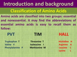
2. Introduction and background
Proteins are polymers of α-amino acids that have unique
three-dimensional structures, making them capable of
performing specific biologic functions.
Proteins are the most abundant and functionally diverse
molecules in living systems. Virtually every life process
depends on this class of molecules. For example, enzymes
and polypeptide hormones direct and regulate metabolism
in the body, whereas contractile proteins in muscles permit
movement. In bone, the protein collagen forms a framework
for the deposition of calcium phosphate crystals. Collagen is
a connective tissue component, is the most abundant
protein by mass in humans.
Proteins
3. Introduction and background
Amino Acids
There are 20 common amino acids that are genetically
encoded and serve as precursors for protein biosynthesis on
ribosomes. Some of these amino acid residues are modified
after biosynthesis (post- translational modification). About
19% of the mass of the human body is protein (about 12kg
protein/ 70 kg man).
4. Introduction and background
The first amino acid be discovered in proteins was asparagine, in
1806. The last of the 20 to be found, threonine, was not identified
until 1938. Their names, in some cases, derived from the source from
which they were first isolated. For example, asparagine and glutamate
were first found in asparagus and gluten, respectivety.
Each amino acid has the following 4 groups
attached to alpha carbon:
1. Amino group: (-NH2 ).
2. Carboxyl group: (-COOH).
3. Hydrogen atom (-H).
4. Side chain (-R).
Structure of Amino Acids
5. Introduction and background
At physiologic pH (approximately pH = 7.4) the carboxyl
group is dissociated, forming the negatively charged
carboxylate ion [-COO-], and the amino group is
protonated [-NH3+]
Structure of Amino Acids
7. Introduction and background
Amino acids, in addition to their role as protein
monomeric units, are energy metabolites and
precursors of many biologically important nitrogen-
containing compounds, such as heme, physiologically
active amines (neurotransmitters), glutathione,
nucleotides, and nucleotide coenzymes.
8. Introduction and background
PVT TIM HALL
Tryptophan T Threonine T Hisitidine H
Valine V Isoleucine I Arginine A
Phenylalanine P Methionine M Leucine L
Lysine L
Amino acids are classified into two groups: essential
and nonessential. it may find the abbreviations of
essential amino acids is easy to recall them as
follow:
Classification of Amino Acids
9. Introduction and background
Classification of Amino Acids
Essential and nonessential amino acids of dietary requirements.
Essential Nonessential
Valine Glycine
Threonine
Alanine
Methionine Serine
Lysine Aspartate
Leucine Asparagine
Isoleucine
Glutamate
Arginine
Glutamine
Histidine
Cysteine
Phenylalanine
Tyrosine
10. Digestive Tract Nitrogen
The digestion of proteins is necessary in order to
convert them into amino acids which can be
absorbed across the intestinal wall. Undigested
proteins are potent antigens and bring about the
production of antibodies (that are themselves
proteins) which produce allergic reactions. These
body defenses against foreign proteins means that
absorption of proteins without digestion would
produce severe allergic shock reactions following
each meal.
11. Digestive Tract Nitrogen
The majority of the amino acids found in human tissues
necessarily come from dietary sources (about 400g of
protein per day), while glutamine, glutamate, and the
remaining nonessential amino acids can be made by
animals. Protein digestion begins in the stomach, where a
proenzyme called pepsinogen (has extra amino acids) is
secreted, autocatalytically converted to pepsin (acid-stable
endopeptidase) or according to the present of HCl, and
used for the first step of proteolysis which is converting
protein to polypeptides and amino acids.
12. Digestive Tract Nitrogen
Most proteolysis takes place in the duodenum as a consequence of
enzyme activities secreted by the pancreas. All of the serine proteases
(trypsin, chymotrypsin, and elastase) and the zinc peptidases
(carboxypeptidase A & B) of pancreatic secretions are produced in the
form of their respective proenzymes.the release and activation of the
pancreatic zymogens is mediated by the secretion of the peptide
hormones cholecystokinin (CCK) and secretin release into the
circulatory system (from special mucosal cells). Together, CCK and
secretin cause the release of protease proenzymes from the pancreas
into the intestine. A second role of CCK is to stimulate adjacent
intestinal cells to secrete enteropeptidase (called enterokinase), a
protease that cleaves trypsinogen to produce trypsin by removal of a
hexapeptide from the NH2-terminus of trypsinogen. Trypsin activates
trypsinogen as well as all the other proenzymes in the pancreatic
secretion, producing the active proteases and peptidases that
hydrolyze dietary polypeptides to oligopeptides and amino acids
14. Absorption and Transport of Amino
Acids
Dipeptides are absorbed with a free amino acids by the
intestinal epithelial cells. But dipeptides are hydrolyzed to
amino acids by dipeptidase in the cytosol of that epithelial
cells before they enter the portal system.
After hydrolysis, amino acids are transferred through
enterocytes to the portal circulation by either active
transport mechanism where energy is required (L- amino
acids) or passive transport mechanism which needs no
energy (D- amino acids).
15. A) Carrier protein transport system
Na+ dependent is the carrier of amino acids and ATP is
hydrolyzed for activation of Na+- K+ ATPase. At high
luminal concentrations Na+ and amino acids are co-
transported down their concentration gradient to the
portal circulation (high concentration in the intestinal
lumen, low in portal circulation). The ATP-dependent
Na+/K+ pump exchanges the accumulated Na+ for
extracellular K+, reducing intracellular Na+ levels and
maintaining the high extracellular Na+ concentration that
is required to drive this transport process. For each amino
acid one ATP molecule is utilized and a specific carrier
protein present in small intestine (similar to that of glucose
absorption). The carreir has one site for the amino acid and
another site for sodium.
17. B) Glutathione transport system (γ-
Glutamyl Cycle)
Three amino acids compose Glutathione which are
glutamate, cystein and glycine. Glutathione transport
system is another mechanism for transport amino acids
across cell membrane of intestine, kidney and brain. The
amino acid reacts with glutamate residue of glutathione at
the surface of the cell to form dipeptide that is transported
across the membrane into the cytosol of the cell. The
enzyme γ- glutamyl transpeptidase catalyzes this reaction
with consuming three molecules of ATP. Subsequent
reactions occur to regenerate glutathione for serving the
cycle.
19. Removal of Nitrogen from Amino
Acids
The catabolism of amino acids generaly involves in two steps
in order to remove nitrogen group and excreted in the form
of urea:
1) Transaminations
2) Oxidative deamination
1) Transaminations
The catabolism of amino acids starts with the reactions
involved in removing amino acid nitrogen from the body
which are known as transaminations,This class of reactions
transfers amino group from all free amino acids into α-
ketoglutarate producing glutamate.
20. Removal of Nitrogen from Amino
Acids
Aminotransferases exist for all amino acids except threonine and
lysine. The most common glutamate and α-ketoglutarate, The two
most impotant aminotransferase reactions are catalyzed by serum
glutamate-pyruvate aminotransferase (SGPT) (also called alanine
aminotransferase, ALT) and serum glutamate-oxaloacetate-
aminotransferase (SGOT) (also called aspartate aminotransferase,
(AST). These two enzymes have been used as clinical markers of
tissue damage, with increasing serum levels indicating an increased
extent of damage. All liver diseases show a high level of both
alanine aminotransferase and aspartate aminotransferase.
C C
O
O NH3
+
R
H
C CH2
O
O CH2
C
O
C O
O
C C
O
O O
R
C CH2
O
O CH2 CH
NH3
+
C O
O
-amino acid -ketoglutrate
+
-keto acid
+
glutamate
E
21. Removal of Nitrogen from Amino
Acids
1- Alanine Aminotransferase (ALT) has an important
function in the delivery of skeletal muscle carbon and
nitrogen (in the form of alanine) to the liver. In skeletal
muscle, pyruvate is transaminated to alanine (in the way of
transporting amino groups to the liver in a nontoxic form),
thus affording an additional route of nitrogen transport.
Then, alanine passes inot the blood and travels to liver. In
the liver, alanine aminotransferase transfers the ammonia
from alanine to α - ketoglutarate and regenerates pyruvate
.The pyruvate can then be diverted into gluconeogenesis.
This process is refered to as the glucose-alanine cycle.
22. Removal of Nitrogen from Amino
Acids
2-Aspartate Aminotransferase (AST) in the amino acid catabolism,
aspartate aminotransferase transfers only the amino groups of
glutamate to oxaloacetate, forming aspartate. This reaction has an
exception to all aminotransferases in transferring amino group of all
amino acids in the form of glutamate to form only aspartate. Aspartate
is used in the urea cycle as a source of nitrogen.
Because of the participation of α–ketoglutarate, as the acceptor of
amino groups, in numerous transaminations, glutamate is a prominent
intermediate in nitrogen elimination as well as in anabolic pathways.
Glutamate produced by transamination can be oxidatively deaminated
or can be used as an amino group donor in the synthesis of
nonoessential amino acids.
23. Removal of Nitrogen from Amino
Acids
2- Oxidative deamination
This reaction liberates the amino group of the amino acids
as a free ammonia that in contrast to the transamination
reactions which transfer amino group to α-keto acid forming
a new amino acid. Oxidatively deamination catalyzed by
glutamate dehydrogenase producing free ammonia.
24. Removal of Nitrogen from Amino
Acids
3- The Glutamine Synthetase Reaction
The glutamine synthetase reaction is also important in
several respects. First it produces glutamine, one of the 20
major amino acids. Second, in animals, glutamine is the
major amino acid found in the circulatory system. Its role is
to carry ammonia to and from various tissues but principally
from peripheral tissues to the kidney.
In kidney, the amide nitrogen of glutamine is hydrolyzed by
the enzyme glutaminase ,this process regenerates glutamate
and free ammonia, which is excreted in the urine.
26. Removal of Nitrogen from Amino
Acids
4- D-Amino acid oxidase
D-Amino acids are not used in the synthesis of mammalian
proteins, but are found in plants and in cell walls of
microorganisms. Since, D-amino acids are containing our
food and are metabolized by the liver. D-Amino acid oxidase
is the enzyme required to oxidative deamination of these
amino acids in the present of FAD. The result is the forming
α-keto acids which can enter the general pathways of amino
acids metabolism and can reaminated to L-isomers, or
catabolized for energy .
28. The Urea (NH2-CO-NH2) Cycle
About 90% of the excreted nitrogen is in the form of urea
which is made in the liver, in a series of reactions that are
distributed between the mitochondrial matrix (the first
two reactions of urea synthesis) and the cytosol (remaining
reactions of urea synthesis). The series of reactions that
form urea is known as the Urea Cycle or the Krebs-
Henseleit Cycle .After urea is produced by the liver, it then
is transported in the blood to the kidneys for excretion in
the urine. Urea is the major disposal form of amino
groups derived from amino acids.
30. Urea Cycle
The overall reaction of urea cycle:
Aspartate + NH3 + CO2 + 3ATP Urea + fumarate +
AMP + PPi + 2ADP + 2Pi + H2O
Three high-energy phosphates (ATP) are consumed in the
synthesis of each molecule of urea. Two ATP for forming
carbamoyl phosphate and one for forming
argininosuccinate. Therefore, the synthesis of urea is
irreversible with a large negative ΔG
31. Urea Cycle Defects (UCDs)
A complete lack of any one of the enzymes of the urea
cycle will result in death shortly after birth. These
disorders are referred to as urea cycle disorders or UCDs.
A common thread to most UCDs is hyperammonemia
leading to ammonia intoxication with the consequences
described below.
Clinical symptoms are most severe when the UCD is at the
level of carbamoyl phosphate synthetase I. Symptoms of
UCDs usually arise at birth and encompass, ataxia,
convulsions, lethargy, poor feeding and eventually coma
and death if not recognized and treated properly.
32. Urea Cycle Defects (UCDs)
In general, the treatment of UCDs has as common
elements the reduction of protein in the diet, removal of
excess ammonia and replacement of intermediates
missing from the urea cycle. Administration of levulose
(suger of fruit and honey) reduces ammonia through its
action of acidifying the colon. Bacteria metabolize
levulose to acidic byproducts which then promotes
excretion of ammonia in the feces as ammonium ions,
NH4+. Antibiotics can be administered to kill intestinal
ammonia producing bacteria.
34. Fate of Urea
By transdeamination of amino acids, the urea synthesized
in the liver can be diffuses from the blood into the kidneys
and also to the intestine where bacterial urease cleaves
urea to CO2 and NH3.
Urea CO2 + NH3
A portion of this ammonia is lost in the feces and other
portion is reabsorbed into the blood. Plasma urea levels
are elevated in patient with kidney failure, enhancing a
large amount of urea to transfer from blood into the gut.
The action of intestinal urease on this urea causes
hyperammonemia in these patients. Giving patient
neomycin orally inhibits the growth of intestinal bacteria
responsible for NH3 production
35.
36. Sources of Ammonia
The sources and metabolism of ammonia are summarized
1. Liver and other tissues: By transdeamination of amino
acids.
2. Kidneys: Form ammonia from glutamine (by
glutaminase enzyme)
37. Sources of Ammonia
3. Intestinal mucosa: Through action of intestinal bacterial
urease on urea present in intestinal fluids.
4. Monoamins Ammonia
5. Catabolism of purines and pyrimidines Ammonia
38. Ammonia Toxicity
Slightly elevated concentration of ammonia are toxic (only
5mg of ammonia per 100ml blood are toxic to humans
central nervous system). Therefore, the level of ammonia
in the blood must be kept low in the range of 1-3 μg per
100ml of human blood. Brain damage is seen in cases of
failure to make urea via the urea cycle or to eliminate urea
through the kidneys. The result of either of these events is
a buildup of circulating levels of ammonium ion which is
called hyperammonemia.
1. Acquired hyperammonemia:
By cirrhosis of liver caused by alcoholism and
bilharziasis
39. Ammonia Toxicity
1. Acquired hyperammonemia:
• By cirrhosis of liver caused by alcoholism and bilharziasis.
• Viral hepatitis or biliary obstruction that result in the formation
of collateral circulation around the liver. The result of that is
shunting the portal blood into blood circulation, so there is no
access to the liver.
• Liver cell failure, as in carcinoma.
2 Inherited hyperammonemia:
If there is any genetic deficiencies of one of the five enzymes of the
urea cycle causes the failure to synthesize urea. This leads to
hyperammonemia during the first week following birth. The reslut of
this kind leads to mental retardation. Therefore, the detoxification
of ammonia is severely impaired leading to the high concentration of
ammonia in the blood.
40. Overall Nitrogen Metabolism
Organisms convert atmospheric nitrogen into forms
available to the human body which humans are totally
dependent on. Nitrogen fixation is carried out by bacterial
nitrogenases forming reduced nitrogen, NH4+ that can then
be used by all organisms to form amino acids, Reduced
nitrogen enters the human body as dietary free amino
acids, protein, and the ammonia produced by intestinal
tract bacteria. A pair of principal enzymes, glutamate
dehydrogenase and glutamine synthatase, are found in all
organisms and effect the conversion of ammonia into the
amino acids glutamate and glutamine, respectively. Amino
and amide groups from these two substances are freely
transferred to other carbon skeletons by transamination
and transamidation reactions.
41. Overall Nitrogen Metabolism
Overview of the flow of nitrogen in the biosphere. Nitrogen, nitrites and nitrates are acted
upon by bacteria (nitrogen fixation) and plants and we digest these compounds as protein in
our diets. Ammonia incorporation in animals occurs through the actions of glutamate
dehydrogenase and glutamine synthase. Glutamate plays the central role in mammalian
nitrogen flow, serving as both a nitrogen donor and nitrogen acceptor