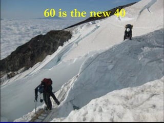
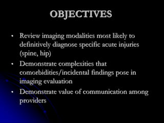
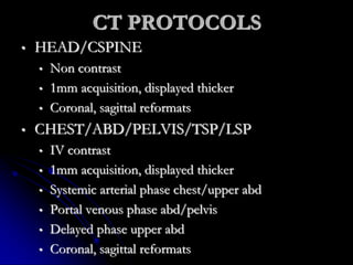
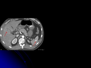
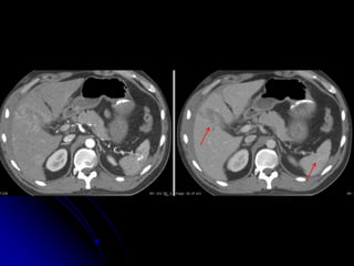
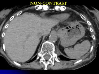
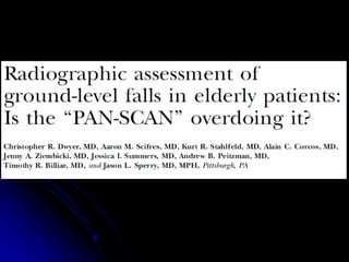
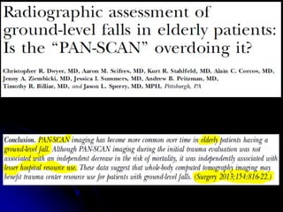
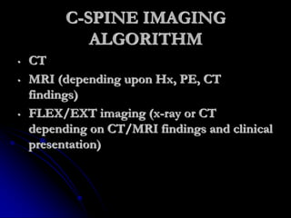
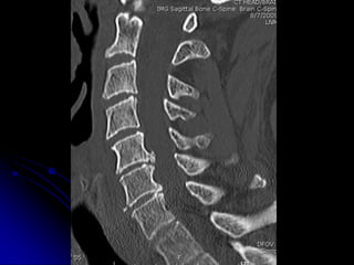
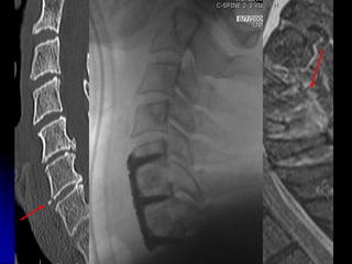
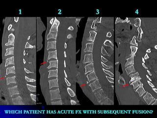
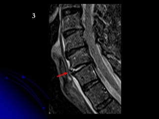
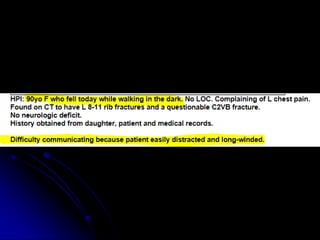
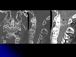
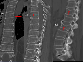
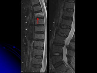
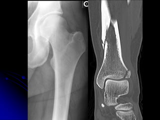
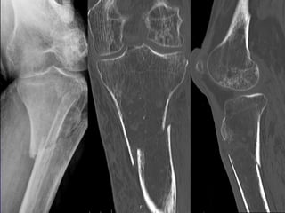
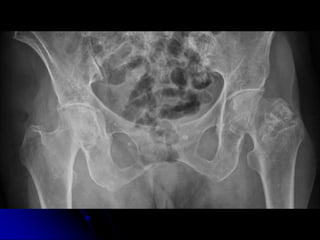
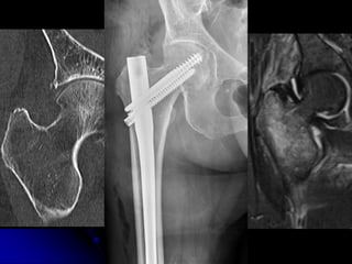
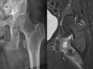
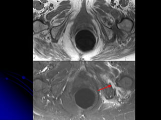
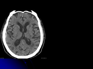
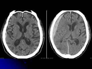
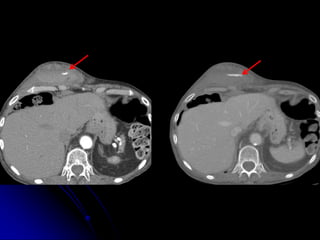
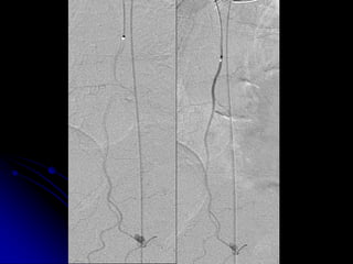
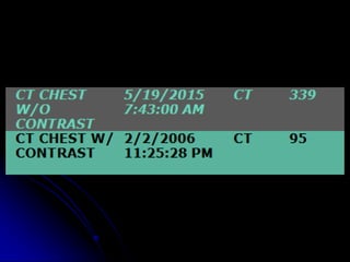
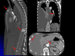
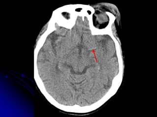
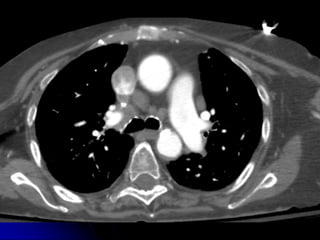
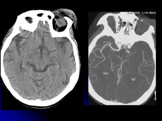
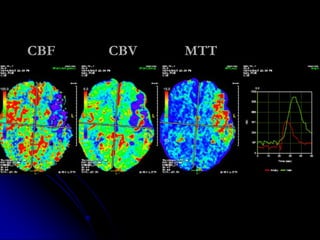
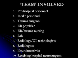
60 is the new 40
70 is the new 50
80
is
the
new
60
CHALLENGES IN
DIAGNOSTIC
IMAGING OF THE
G60 PATIENT
Phillip Moeser, MD
Medical Diagnostic Imaging Group
Medical Director of Radiology QA
John C. Lincoln Medical Center
OBJECTIVES
• Review imaging modalities most likely to
definitively diagnose specific acute injuries
(spine, hip)
• Demonstrate complexities that
comorbidities/incidental findings pose in
imaging evaluation
• Demonstrate value of communication among
providers
TOOLS OF DIAGNOSITIC
AND INTERVENTIONAL
IMAGING
• X-RAY
• FLUOROSCOPY
• NM
• U/S
• CT
• MRI
• IMAGING GUIDED RX/DX
• 3D WORKSTATIONS
THE DIAGNOSTIC
IMAGING ‘TEAM’
1. Technologists
2. Nurses
3. Support personnel (IT/PACS/etc)
4. Radiologists
1972
CORTONA, ITALY JUNE 2005
CT PROTOCOLS
• HEAD/CSPINE
• Non contrast
• 1mm acquisition, displayed thicker
• Coronal, sagittal reformats
• CHEST/ABD/PELVIS/TSP/LSP
• IV contrast
• 1mm acquisition, displayed thicker
• Systemic arterial phase chest/upper abd
• Portal venous phase abd/pelvis
• Delayed phase upper abd
• Coronal, sagittal reformats
NON-CONTRAST
C-SPINE IMAGING
ALGORITHM
• CT
• MRI (depending upon Hx, PE, CT
findings)
• FLEX/EXT imaging (x-ray or CT
depending on CT/MRI findings and clinical
presentation)
1 2 3 4
WHICH PATIENT HAS ACUTE FX WITH SUBSEQUENT FUSION?
3
T6 T6
L2
HIP FRACTURES
INCIDENTAL
FINDINGS &
COMORBIDITIES
COMMUNICATION
MTTCBVCBF
‘TEAM’ INVOLVED
1. Pre-hospital personnel
2. Intake personnel
3. Trauma surgeon
4. ER physician
5. ER/trauma nursing
6. Lab
7. Radiology/CT technologists
8. Radiologists
9. Neurointensivist
10. Receiving hospital neurosurgeon
IT’S MAGIC
EXCEPTIONS
BUT OFFICER,
I THOUGHT 80
IS THE NEW 60
60
Is
The
New
80
THANK YOU

Challenges in Diagnostic Imaging (Dr. Phil Moeser)