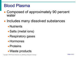
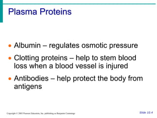
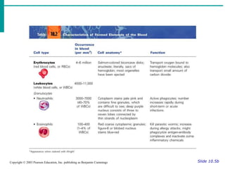
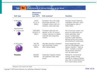
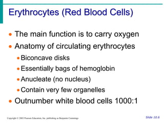
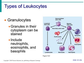
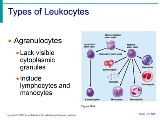
Essentials of Human Anatomy & Physiology
Copyright © 2003 Pearson Education, Inc. publishing as Benjamin Cummings
Slides 10.1 – 10.31
Seventh Edition
Elaine N. Marieb
Chapter 10
Blood
Lecture Slides in PowerPoint by Jerry L. Cook
Blood
Slide 10.1a
Copyright © 2003 Pearson Education, Inc. publishing as Benjamin Cummings
 The only fluid tissue in the human body
 Classified as a connective tissue
Living cells = formed elements
Non-living matrix = plasma
Blood
Slide 10.1b
Copyright © 2003 Pearson Education, Inc. publishing as Benjamin Cummings
Figure 10.1
Physical Characteristics of Blood
Slide 10.2
Copyright © 2003 Pearson Education, Inc. publishing as Benjamin Cummings
 Color range
Oxygen-rich blood is scarlet red
Oxygen-poor blood is dull red
 pH must remain between 7.35–7.45
 Blood temperature is slightly higher than
body temperature
Blood Plasma
Slide 10.3
Copyright © 2003 Pearson Education, Inc. publishing as Benjamin Cummings
 Composed of approximately 90 percent
water
 Includes many dissolved substances
Nutrients
Salts (metal ions)
Respiratory gases
Hormones
Proteins
Waste products
Plasma Proteins
Slide 10.4
Copyright © 2003 Pearson Education, Inc. publishing as Benjamin Cummings
 Albumin – regulates osmotic pressure
 Clotting proteins – help to stem blood
loss when a blood vessel is injured
 Antibodies – help protect the body from
antigens
Formed Elements
Slide 10.5a
Copyright © 2003 Pearson Education, Inc. publishing as Benjamin Cummings
 Erythrocytes = red blood cells
 Leukocytes = white blood cells
 Platelets = cell fragments
Slide 10.5b
Copyright © 2003 Pearson Education, Inc. publishing as Benjamin Cummings
Slide 10.5c
Copyright © 2003 Pearson Education, Inc. publishing as Benjamin Cummings
Erythrocytes (Red Blood Cells)
Slide 10.6
Copyright © 2003 Pearson Education, Inc. publishing as Benjamin Cummings
 The main function is to carry oxygen
 Anatomy of circulating erythrocytes
Biconcave disks
Essentially bags of hemoglobin
Anucleate (no nucleus)
Contain very few organelles
 Outnumber white blood cells 1000:1
Hemoglobin
Slide 10.7
Copyright © 2003 Pearson Education, Inc. publishing as Benjamin Cummings
 Iron-containing protein
 Binds strongly, but reversibly, to oxygen
 Each hemoglobin molecule has four
oxygen binding sites
 Each erythrocyte has 250 million
hemoglobin molecules
Leukocytes (White Blood Cells)
Slide 10.8
Copyright © 2003 Pearson Education, Inc. publishing as Benjamin Cummings
 Crucial in the body’s defense against
disease
 These are complete cells, with a
nucleus and organelles
 Able to move into and out of blood
vessels (diapedesis)
 Can move by ameboid motion
 Can respond to chemicals released by
damaged tissues
Leukocyte Levels in the Blood
Slide 10.9
Copyright © 2003 Pearson Education, Inc. publishing as Benjamin Cummings
 Normal levels are between 4,000 and
11,000 cells per millimeter
 Abnormal leukocyte levels
Leukocytosis
Above 11,000 leukocytes/ml
Generally indicates an infection
Leukopenia
Abnormally low leukocyte level
Commonly caused by certain drugs
Types of Leukocytes
Slide 10.10a
Copyright © 2003 Pearson Education, Inc. publishing as Benjamin Cummings
 Granulocytes
Granules in their
cytoplasm can be
stained
Include
neutrophils,
eosinophils, and
basophils
Figure 10.4
Types of Leukocytes
Slide 10.10b
Copyright © 2003 Pearson Education, Inc. publishing as Benjamin Cummings
 Agranulocytes
Lack visible
cytoplasmic
granules
Include
lymphocytes and
monocytes
Figure 10.4
Granulocytes
Slide 10.11a
Copyright © 2003 Pearson Education, Inc. publishing as Benjamin Cummings
 Neutrophils
Multilobed nucleus with fine granules
Act as phagocytes at active sites of infection
 Eosinophils
Large brick-red cytoplasmic granules
Found in repsonse to allergies and parasitic
worms
Granulocytes
Slide 10.11b
Copyright © 2003 Pearson Education, Inc. publishing as Benjamin Cummings
 Basophils
Have histamine-containing granules
Initiate inflammation
Agranulocytes
Slide 10.12
Copyright © 2003 Pearson Education, Inc. publishing as Benjamin Cummings
 Lymphocytes
Nucleus fills most of the cell
Play an important role in the immune
response
 Monocytes
Largest of the white blood cells
Function as macrophages
Important in fighting chronic infection
Platelets
Slide 10.13
Copyright © 2003 Pearson Education, Inc. publishing as Benjamin Cummings
 Derived from ruptured multinucleate
cells (megakaryocytes)
 Needed for the clotting process
 Normal platelet count = 300,000/mm3
Hematopoiesis
Slide 10.14
Copyright © 2003 Pearson Education, Inc. publishing as Benjamin Cummings
 Blood cell formation
 Occurs in red bone marrow
 All blood cells are derived from a
common stem cell (hemocytoblast)
 Hemocytoblast differentiation
Lymphoid stem cell produces lymphocytes
Myeloid stem cell produces other formed
elements
Fate of Erythrocytes
Slide 10.15
Copyright © 2003 Pearson Education, Inc. publishing as Benjamin Cummings
 Unable to divide, grow, or synthesize
proteins
 Wear out in 100 to 120 days
 When worn out, are eliminated by
phagocytes in the spleen or liver
 Lost cells are replaced by division of
hemocytoblasts
Control of Erythrocyte Production
Slide 10.16
Copyright © 2003 Pearson Education, Inc. publishing as Benjamin Cummings
 Rate is controlled by a hormone
(erythropoietin)
 Kidneys produce most erythropoietin as
a response to reduced oxygen levels in
the blood
 Homeostasis is maintained by negative
feedback from blood oxygen levels
Control of Erythrocyte Production
Slide 10.17
Copyright © 2003 Pearson Education, Inc. publishing as Benjamin Cummings
Figure 10.5
Hemostasis
Slide 10.18
Copyright © 2003 Pearson Education, Inc. publishing as Benjamin Cummings
 Stoppage of blood flow
 Result of a break in a blood vessel
 Hemostasis involves three phases
Platelet plug formation
Vascular spasms
Coagulation
Platelet Plug Formation
Slide 10.19
Copyright © 2003 Pearson Education, Inc. publishing as Benjamin Cummings
 Collagen fibers are exposed by a break
in a blood vessel
 Platelets become “sticky” and cling to
fibers
 Anchored platelets release chemicals to
attract more platelets
 Platelets pile up to form a platelet plug
Vascular Spasms
Slide 10.20
Copyright © 2003 Pearson Education, Inc. publishing as Benjamin Cummings
 Anchored platelets release serotonin
 Serotonin causes blood vessel muscles
to spasm
 Spasms narrow the blood vessel,
decreasing blood loss
Coagulation
Slide 10.21a
Copyright © 2003 Pearson Education, Inc. publishing as Benjamin Cummings
 Injured tissues release thromboplastin
 PF3 (a phospholipid) interacts with
thromboplastin, blood protein clotting
factors, and calcium ions to trigger a
clotting cascade
 Prothrombin activator converts
prothrombin to thrombin (an enzyme)
Coagulation
Slide 10.21b
Copyright © 2003 Pearson Education, Inc. publishing as Benjamin Cummings
 Thrombin joins fibrinogen proteins into
hair-like fibrin
 Fibrin forms a meshwork
(the basis for a clot)
Blood Clotting
Slide 10.22
Copyright © 2003 Pearson Education, Inc. publishing as Benjamin Cummings
 Blood usually clots within 3 to 6 minutes
 The clot remains as endothelium
regenerates
 The clot is broken down after tissue
repair
Undesirable Clotting
Slide 10.23
Copyright © 2003 Pearson Education, Inc. publishing as Benjamin Cummings
 Thrombus
A clot in an unbroken blood vessel
Can be deadly in areas like the heart
 Embolus
A thrombus that breaks away and floats
freely in the bloodstream
Can later clog vessels in critical areas such
as the brain
Bleeding Disorders
Slide 10.24
Copyright © 2003 Pearson Education, Inc. publishing as Benjamin Cummings
 Thrombocytopenia
Platelet deficiency
Even normal movements can cause
bleeding from small blood vessels that
require platelets for clotting
 Hemophilia
Hereditary bleeding disorder
Normal clotting factors are missing
Blood Groups and Transfusions
Slide 10.25
Copyright © 2003 Pearson Education, Inc. publishing as Benjamin Cummings
 Large losses of blood have serious
consequences
Loss of 15 to 30 percent causes weakness
Loss of over 30 percent causes shock,
which can be fatal
 Transfusions are the only way to
replace blood quickly
 Transfused blood must be of the same
blood group
Human Blood Groups
Slide 10.26a
Copyright © 2003 Pearson Education, Inc. publishing as Benjamin Cummings
 Blood contains genetically determined
proteins
 A foreign protein (antigen) may be
attacked by the immune system
 Blood is “typed” by using antibodies that
will cause blood with certain proteins to
clump (agglutination)
Human Blood Groups
Slide 10.26b
Copyright © 2003 Pearson Education, Inc. publishing as Benjamin Cummings
 There are over 30 common red blood
cell antigens
 The most vigorous transfusion reactions
are caused by ABO and Rh blood group
antigens
ABO Blood Groups
Slide 10.27a
Copyright © 2003 Pearson Education, Inc. publishing as Benjamin Cummings
 Based on the presence or absence of two
antigens
Type A
Type B
 The lack of these antigens is called
type O
ABO Blood Groups
Slide 10.27b
Copyright © 2003 Pearson Education, Inc. publishing as Benjamin Cummings
 The presence of both A and B is called
type AB
 The presence of either A or B is called
types A and B, respectively
Rh Blood Groups
Slide 10.28
Copyright © 2003 Pearson Education, Inc. publishing as Benjamin Cummings
 Named because of the presence or
absence of one of eight Rh antigens
(agglutinogen D)
 Most Americans are Rh+
 Problems can occur in mixing Rh+ blood
into a body with Rh– blood
Rh Dangers During Pregnancy
Slide 10.29a
Copyright © 2003 Pearson Education, Inc. publishing as Benjamin Cummings
 Danger is only when the mother is Rh–
and the father is Rh+, and the child
inherits the Rh+ factor
Rh Dangers During Pregnancy
Slide 10.29b
Copyright © 2003 Pearson Education, Inc. publishing as Benjamin Cummings
 The mismatch of an Rh– mother carrying
an Rh+ baby can cause problems for the
unborn child
 The first pregnancy usually proceeds without
problems
 The immune system is sensitized after the first
pregnancy
 In a second pregnancy, the mother’s immune
system produces antibodies to attack the Rh+
blood (hemolytic disease of the newborn)
Blood Typing
Slide 10.30
Copyright © 2003 Pearson Education, Inc. publishing as Benjamin Cummings
 Blood samples are mixed with anti-A and
anti-B serum
 Coagulation or no coagulation leads to
determining blood type
 Typing for ABO and Rh factors is done in
the same manner
 Cross matching – testing for
agglutination of donor RBCs by the
recipient’s serum, and vice versa
Developmental Aspects of Blood
Slide 10.31
Copyright © 2003 Pearson Education, Inc. publishing as Benjamin Cummings
 Sites of blood cell formation
The fetal liver and spleen are early sites of
blood cell formation
Bone marrow takes over hematopoiesis by
the seventh month
 Fetal hemoglobin differs from
hemoglobin produced after birth

Ch 10 Blood.ppt

  • 1.
    Essentials of HumanAnatomy & Physiology Copyright © 2003 Pearson Education, Inc. publishing as Benjamin Cummings Slides 10.1 – 10.31 Seventh Edition Elaine N. Marieb Chapter 10 Blood Lecture Slides in PowerPoint by Jerry L. Cook
  • 2.
    Blood Slide 10.1a Copyright ©2003 Pearson Education, Inc. publishing as Benjamin Cummings  The only fluid tissue in the human body  Classified as a connective tissue Living cells = formed elements Non-living matrix = plasma
  • 3.
    Blood Slide 10.1b Copyright ©2003 Pearson Education, Inc. publishing as Benjamin Cummings Figure 10.1
  • 4.
    Physical Characteristics ofBlood Slide 10.2 Copyright © 2003 Pearson Education, Inc. publishing as Benjamin Cummings  Color range Oxygen-rich blood is scarlet red Oxygen-poor blood is dull red  pH must remain between 7.35–7.45  Blood temperature is slightly higher than body temperature
  • 5.
    Blood Plasma Slide 10.3 Copyright© 2003 Pearson Education, Inc. publishing as Benjamin Cummings  Composed of approximately 90 percent water  Includes many dissolved substances Nutrients Salts (metal ions) Respiratory gases Hormones Proteins Waste products
  • 6.
    Plasma Proteins Slide 10.4 Copyright© 2003 Pearson Education, Inc. publishing as Benjamin Cummings  Albumin – regulates osmotic pressure  Clotting proteins – help to stem blood loss when a blood vessel is injured  Antibodies – help protect the body from antigens
  • 7.
    Formed Elements Slide 10.5a Copyright© 2003 Pearson Education, Inc. publishing as Benjamin Cummings  Erythrocytes = red blood cells  Leukocytes = white blood cells  Platelets = cell fragments
  • 8.
    Slide 10.5b Copyright ©2003 Pearson Education, Inc. publishing as Benjamin Cummings
  • 9.
    Slide 10.5c Copyright ©2003 Pearson Education, Inc. publishing as Benjamin Cummings
  • 10.
    Erythrocytes (Red BloodCells) Slide 10.6 Copyright © 2003 Pearson Education, Inc. publishing as Benjamin Cummings  The main function is to carry oxygen  Anatomy of circulating erythrocytes Biconcave disks Essentially bags of hemoglobin Anucleate (no nucleus) Contain very few organelles  Outnumber white blood cells 1000:1
  • 11.
    Hemoglobin Slide 10.7 Copyright ©2003 Pearson Education, Inc. publishing as Benjamin Cummings  Iron-containing protein  Binds strongly, but reversibly, to oxygen  Each hemoglobin molecule has four oxygen binding sites  Each erythrocyte has 250 million hemoglobin molecules
  • 12.
    Leukocytes (White BloodCells) Slide 10.8 Copyright © 2003 Pearson Education, Inc. publishing as Benjamin Cummings  Crucial in the body’s defense against disease  These are complete cells, with a nucleus and organelles  Able to move into and out of blood vessels (diapedesis)  Can move by ameboid motion  Can respond to chemicals released by damaged tissues
  • 13.
    Leukocyte Levels inthe Blood Slide 10.9 Copyright © 2003 Pearson Education, Inc. publishing as Benjamin Cummings  Normal levels are between 4,000 and 11,000 cells per millimeter  Abnormal leukocyte levels Leukocytosis Above 11,000 leukocytes/ml Generally indicates an infection Leukopenia Abnormally low leukocyte level Commonly caused by certain drugs
  • 14.
    Types of Leukocytes Slide10.10a Copyright © 2003 Pearson Education, Inc. publishing as Benjamin Cummings  Granulocytes Granules in their cytoplasm can be stained Include neutrophils, eosinophils, and basophils Figure 10.4
  • 15.
    Types of Leukocytes Slide10.10b Copyright © 2003 Pearson Education, Inc. publishing as Benjamin Cummings  Agranulocytes Lack visible cytoplasmic granules Include lymphocytes and monocytes Figure 10.4
  • 16.
    Granulocytes Slide 10.11a Copyright ©2003 Pearson Education, Inc. publishing as Benjamin Cummings  Neutrophils Multilobed nucleus with fine granules Act as phagocytes at active sites of infection  Eosinophils Large brick-red cytoplasmic granules Found in repsonse to allergies and parasitic worms
  • 17.
    Granulocytes Slide 10.11b Copyright ©2003 Pearson Education, Inc. publishing as Benjamin Cummings  Basophils Have histamine-containing granules Initiate inflammation
  • 18.
    Agranulocytes Slide 10.12 Copyright ©2003 Pearson Education, Inc. publishing as Benjamin Cummings  Lymphocytes Nucleus fills most of the cell Play an important role in the immune response  Monocytes Largest of the white blood cells Function as macrophages Important in fighting chronic infection
  • 19.
    Platelets Slide 10.13 Copyright ©2003 Pearson Education, Inc. publishing as Benjamin Cummings  Derived from ruptured multinucleate cells (megakaryocytes)  Needed for the clotting process  Normal platelet count = 300,000/mm3
  • 20.
    Hematopoiesis Slide 10.14 Copyright ©2003 Pearson Education, Inc. publishing as Benjamin Cummings  Blood cell formation  Occurs in red bone marrow  All blood cells are derived from a common stem cell (hemocytoblast)  Hemocytoblast differentiation Lymphoid stem cell produces lymphocytes Myeloid stem cell produces other formed elements
  • 21.
    Fate of Erythrocytes Slide10.15 Copyright © 2003 Pearson Education, Inc. publishing as Benjamin Cummings  Unable to divide, grow, or synthesize proteins  Wear out in 100 to 120 days  When worn out, are eliminated by phagocytes in the spleen or liver  Lost cells are replaced by division of hemocytoblasts
  • 22.
    Control of ErythrocyteProduction Slide 10.16 Copyright © 2003 Pearson Education, Inc. publishing as Benjamin Cummings  Rate is controlled by a hormone (erythropoietin)  Kidneys produce most erythropoietin as a response to reduced oxygen levels in the blood  Homeostasis is maintained by negative feedback from blood oxygen levels
  • 23.
    Control of ErythrocyteProduction Slide 10.17 Copyright © 2003 Pearson Education, Inc. publishing as Benjamin Cummings Figure 10.5
  • 24.
    Hemostasis Slide 10.18 Copyright ©2003 Pearson Education, Inc. publishing as Benjamin Cummings  Stoppage of blood flow  Result of a break in a blood vessel  Hemostasis involves three phases Platelet plug formation Vascular spasms Coagulation
  • 25.
    Platelet Plug Formation Slide10.19 Copyright © 2003 Pearson Education, Inc. publishing as Benjamin Cummings  Collagen fibers are exposed by a break in a blood vessel  Platelets become “sticky” and cling to fibers  Anchored platelets release chemicals to attract more platelets  Platelets pile up to form a platelet plug
  • 26.
    Vascular Spasms Slide 10.20 Copyright© 2003 Pearson Education, Inc. publishing as Benjamin Cummings  Anchored platelets release serotonin  Serotonin causes blood vessel muscles to spasm  Spasms narrow the blood vessel, decreasing blood loss
  • 27.
    Coagulation Slide 10.21a Copyright ©2003 Pearson Education, Inc. publishing as Benjamin Cummings  Injured tissues release thromboplastin  PF3 (a phospholipid) interacts with thromboplastin, blood protein clotting factors, and calcium ions to trigger a clotting cascade  Prothrombin activator converts prothrombin to thrombin (an enzyme)
  • 28.
    Coagulation Slide 10.21b Copyright ©2003 Pearson Education, Inc. publishing as Benjamin Cummings  Thrombin joins fibrinogen proteins into hair-like fibrin  Fibrin forms a meshwork (the basis for a clot)
  • 29.
    Blood Clotting Slide 10.22 Copyright© 2003 Pearson Education, Inc. publishing as Benjamin Cummings  Blood usually clots within 3 to 6 minutes  The clot remains as endothelium regenerates  The clot is broken down after tissue repair
  • 30.
    Undesirable Clotting Slide 10.23 Copyright© 2003 Pearson Education, Inc. publishing as Benjamin Cummings  Thrombus A clot in an unbroken blood vessel Can be deadly in areas like the heart  Embolus A thrombus that breaks away and floats freely in the bloodstream Can later clog vessels in critical areas such as the brain
  • 31.
    Bleeding Disorders Slide 10.24 Copyright© 2003 Pearson Education, Inc. publishing as Benjamin Cummings  Thrombocytopenia Platelet deficiency Even normal movements can cause bleeding from small blood vessels that require platelets for clotting  Hemophilia Hereditary bleeding disorder Normal clotting factors are missing
  • 32.
    Blood Groups andTransfusions Slide 10.25 Copyright © 2003 Pearson Education, Inc. publishing as Benjamin Cummings  Large losses of blood have serious consequences Loss of 15 to 30 percent causes weakness Loss of over 30 percent causes shock, which can be fatal  Transfusions are the only way to replace blood quickly  Transfused blood must be of the same blood group
  • 33.
    Human Blood Groups Slide10.26a Copyright © 2003 Pearson Education, Inc. publishing as Benjamin Cummings  Blood contains genetically determined proteins  A foreign protein (antigen) may be attacked by the immune system  Blood is “typed” by using antibodies that will cause blood with certain proteins to clump (agglutination)
  • 34.
    Human Blood Groups Slide10.26b Copyright © 2003 Pearson Education, Inc. publishing as Benjamin Cummings  There are over 30 common red blood cell antigens  The most vigorous transfusion reactions are caused by ABO and Rh blood group antigens
  • 35.
    ABO Blood Groups Slide10.27a Copyright © 2003 Pearson Education, Inc. publishing as Benjamin Cummings  Based on the presence or absence of two antigens Type A Type B  The lack of these antigens is called type O
  • 36.
    ABO Blood Groups Slide10.27b Copyright © 2003 Pearson Education, Inc. publishing as Benjamin Cummings  The presence of both A and B is called type AB  The presence of either A or B is called types A and B, respectively
  • 37.
    Rh Blood Groups Slide10.28 Copyright © 2003 Pearson Education, Inc. publishing as Benjamin Cummings  Named because of the presence or absence of one of eight Rh antigens (agglutinogen D)  Most Americans are Rh+  Problems can occur in mixing Rh+ blood into a body with Rh– blood
  • 38.
    Rh Dangers DuringPregnancy Slide 10.29a Copyright © 2003 Pearson Education, Inc. publishing as Benjamin Cummings  Danger is only when the mother is Rh– and the father is Rh+, and the child inherits the Rh+ factor
  • 39.
    Rh Dangers DuringPregnancy Slide 10.29b Copyright © 2003 Pearson Education, Inc. publishing as Benjamin Cummings  The mismatch of an Rh– mother carrying an Rh+ baby can cause problems for the unborn child  The first pregnancy usually proceeds without problems  The immune system is sensitized after the first pregnancy  In a second pregnancy, the mother’s immune system produces antibodies to attack the Rh+ blood (hemolytic disease of the newborn)
  • 40.
    Blood Typing Slide 10.30 Copyright© 2003 Pearson Education, Inc. publishing as Benjamin Cummings  Blood samples are mixed with anti-A and anti-B serum  Coagulation or no coagulation leads to determining blood type  Typing for ABO and Rh factors is done in the same manner  Cross matching – testing for agglutination of donor RBCs by the recipient’s serum, and vice versa
  • 41.
    Developmental Aspects ofBlood Slide 10.31 Copyright © 2003 Pearson Education, Inc. publishing as Benjamin Cummings  Sites of blood cell formation The fetal liver and spleen are early sites of blood cell formation Bone marrow takes over hematopoiesis by the seventh month  Fetal hemoglobin differs from hemoglobin produced after birth