This study compared pregnancy outcomes of in vitro fertilization (IVF) patients who underwent single embryo transfer where the embryo was selected based on (1) morphology alone or (2) morphology assessed with array comparative genomic hybridization (aCGH). Patients were randomly assigned to one of the two selection methods. The clinical pregnancy and ongoing pregnancy rates were significantly higher in the group where aCGH was used in addition to morphology to select the embryo. No twin pregnancies occurred. The results suggest that aCGH may improve pregnancy outcomes compared to morphology alone by detecting chromosomal abnormalities.

![Yang et al. Molecular Cytogenetics 2012, 5:24 Page 2 of 8
http://www.molecularcytogenetics.org/content/5/1/24
Background
Multiple gestation represents the most significant com-plication
of assisted reproductive treatment (ART). Sin-gle
embryo transfer (SET), either elective or mandatory,
has been advocated as an effective means to avoid mul-tiple
gestation following IVF [1-3]. Despite a welcome
trend in increased acceptance and utilization of elective
SET treatment in some groups [4], most IVF cycles con-tinue
to involve two or more embryos for transfer. When
SET is done, selection of the single embryo or blastocyst
for transfer is typically done on the basis of morphology
[5,6]. However, since acceptable morphology alone can-not
negate the potential for chromosomal error in the
selected embryo, the transfer of one apparently “normal
looking” embryo carries considerable risk [7]. Aneu-ploidy
is the most common abnormality in human
embryos derived from IVF [8-15], a problem that contri-butes
substantially to poor IVF outcomes [16]. As other
investigators have noted, screening embryos by fluores-cence
in situ hybridization (FISH) was a reasonable re-sponse
to this challenge, but the approach was limited
because it failed to screen all chromosomes at the same
time [17-21]. Conventional comparative genomic
hybridization (CGH) has been used for comprehensive
screening of aneuploidy for oocytes and embryos [19,22-
25] with cryopreservation of embryo(s) from which the
biopsy was derived. When results became available, fro-zen
embryo transfer (FET) was subsequently arranged so
that only euploid embryo(s) were transferred.
At present, there is no consensus on the best way to
determine the competency of the embryonic genome
during IVF. Both single nucleotide polymorphism (SNP)
array and array CGH (aCGH) have been validated as ac-curate
methods to achieve comprehensive chromosome
screening when biopsy is performed on d3 for fresh
transfer on d5 [26-30]. The difference in mosaicism be-tween
embryos at d3 and d5 has led to a preference for
biopsy at the blastocyst stage when mosaicism is reduced
[31-33]. When combined with trophectoderm biopsy and
blastocyst vitrification, SNP microarray has resulted in
high implantation rate and low miscarriage rates for
some IVF patients [31]. However, experience is limited
with aCGH to select a single euploid blastocyst for fresh
transfer in the absence of known chromosomal diagno-sis.
In this pilot study, we evaluated a rapid, on-site
aCGH application to select a single euploid blastocyst
for fresh transfer in good prognosis patients <35 yrs of
age, who were undergoing a first IVF attempt.
Methods
Patient sample
Following IRB approval, patients undergoing IVF at our
programs in Beijing and Los Angeles were offered enroll-ment
in this prospective, single-blind, pilot interventional
study to compare embryo assessment by conventional mi-croscopy
alone or with array comparative genomic
hybridization (aCGH) performed on trophectoderm. Writ-ten
informed consent was obtained from all study partici-pants
and all received pre-treatment counseling in
anticipation of possible incorporation of aCGH in their IVF
treatment. Patients were eligible for this study if (female)
age was <35 yrs, if there was a history of regular ovulation,
if etiology of infertility was tubal factor or male factor (or
both), and if no prior IVF treatment had been initiated.
Additionally, all study subjects were required to have a nor-mal
intrauterine contour (confirmed by hysteroscopy), both
ovaries intact, basal serum FSH and estradiol on d2-3 at
<10 IU/l and <60 pg/ml, respectively. IVF patients whose
treatment incorporated donor gametes or frozen/thawed
embryos were excluded. A random number table was used
to determine patients in vitro laboratory management strat-egy
as either (1) traditional morphology assessment plus
aCGH (Group A, n = 55), or (2) conventional morphology
assessment only (Group B, n = 48). Patients (but not labora-tory
or clinical staff) were blinded with regard to their
randomization group. The two cohorts were mutually ex-clusive,
and no study patient had embryos assigned to both
laboratory groups.
Ovarian stimulation and fertilization
Before commencing gonadotropin therapy patients
underwent transvaginal ultrasound evaluation with re-measurement
of serum FSH, LH and estradiol on d3 of
the index cycle. Pituitary downregulation was achieved
with GnRH-agonist administered on d21 of the cycle im-mediately
preceding treatment, as previously described
[33]. Periodic transvaginal ultrasound and serum estra-diol
measurements were used to track follicular growth
and thickness of endometrial lining. When ≥3 follicles
reached 19 mm mean diameter, periovulatory hCG was
administered by subcutaneous injection of recombinant
hCG (250 μg OvidrelW, Merck Serono; Geneva, Switzer-land)
with oocyte retrieval performed under transvaginal
ultrasound guidance 35-36 h later. Following removal of
all cumulus cells, ICSI was performed and normal
fertilization was verified 16-18 h after injection by pres-ence
of two pronuclei and two polar bodies.
Embryo culture and trophectoderm biopsy
All embryos were cultured in sequential media (Vitro-life;
Göteborg, Sweden) to blastocyst stage. On d3 when
embryos were at the 6–8 cell stage, a noncontact 1.48 μ
diode laser (OCTAX Microscience GmbH; Bruckberg,
Germany) was used to create a circular 6-9 μ diameter
opening in the zona pellucida. For embryos randomized
to the aCGH group, this breach enabled biopsy of
trophectoderm (TE) on d5 rapidly. Between 3–5 her-niated
TE cells were gently aspirated by pipette and,](https://image.slidesharecdn.com/cgh-141015065602-conversion-gate02/85/Cgh-2-320.jpg)
![Yang et al. Molecular Cytogenetics 2012, 5:24 Page 3 of 8
http://www.molecularcytogenetics.org/content/5/1/24
when necessary, freed from the blastocyst by applica-tion
of several laser pulses. Harvested TE cells were
washed in PBS and placed within a PCR tube with
2.5 μl 1x PBS as previously described [34]. A uniform
assisted hatching methodology was used for all embryos
irrespective of subsequent TE biopsy or conventional
microscopic assessment alone.
aCGH protocol
Whole genome amplification was performed on-site using
the SurePlex DNA amplification system (BlueGnome Ltd;
Cambridge, UK) in accordance with manufacturer’s guide-lines,
as described elsewhere [34,35]. Briefly, samples and
control DNA (8 μl for each) were labeled with Cy3 and
Cy5 fluorophores (BlueGnome Ltd; Cambridge, UK). La-beling
time was approximately 3 h with DNA resuspended
in dexsulphate hybridization buffer and hybridized over-night
under cover slides. After washing 1x 10 min in sa-line
sodium citrate (SSC)/0.05% Tween-20 at room
temperature, an additional irrigation in SSC 1x 10 min
was completed at room temperature. Slides were washed
in SSC 1x 5 min at 60°C and again for 1 min at room
temperature (in SSC). Vacuum centrifuge was used to
dry microarray slides over 3 min, followed by laser scan-ning
at 10 μm (Agilent Technologies; Santa Clara, USA).
Microarray data were analyzed with BlueFuse software
(BlueGnome, Cambridge, UK) for chromatin loss or gain
across all 24 chromosomes. Aberrations were consid-ered
non-artifact if ≥15 probes deviated from normal
limits as defined by the 24Sure platform. The published
accuracy rate for this aCGH technique when applied to
TE cells is 95% [35].
Blastocyst grading and selection for transfer
In both aCGH and control groups, blastocysts were
graded [36] on a 1 to 6 scale determined by degree of ex-pansion
and hatching status, as follows: Grade 1 (early
blastocyst): blastocoele <1/2 of total embryo volume;
Grade 2 (intermediate blastocyst): blastocoele ≥1/2 of
total embryo volume; Grade 3 (full blastocyst): blasto-coele
fully occupies the embryo; Grade 4 (expanded
blastocyst): blastocoele is larger than early blastocyst and
zona pellucida (ZP) demonstrates thinning; Grade 5
(hatching blastocyst): herniation of trophectoderm cells
from the ZP; and Grade 6 (hatched blastocyst): blastocyst
has escaped the ZP. For blastocysts at Grades 3 to 6, the
inner cell mass (ICM) and trophectoderm (TE) were also
graded. The ICM was graded as follows: A (many ICM
cells packed together tightly); B (several ICM cells
grouped loosely) and C (very few ICM cells). TE was
graded as follows: A (many TE cells forming multiple
epithelial layers); B (few TE cells consisting of a loose
epithelium) and C (very few large TE cells).
Fresh SET was performed on the morning of d6 under
direct ultrasound guidance for all patients. For embryos
in the aCGH group only one euploid blastocyst was
selected for transfer, based on data from the aCGH ana-lysis.
When multiple euploid blastocysts were available
(as determined by aCGH), the best grade euploid blasto-cyst
was selected for transfer. Any surplus euploid blas-tocysts
were vitrified for later use [34]. In the non-aCGH
(control) group, a single blastocyst was selected for fresh
transfer based on morphological criteria only (e.g., no
aCGH evaluation). The surplus blastocysts with good
morphology (grade 3BB or above) were vitrified for fu-ture
FET cycles.
Outcome measures and statistical analysis
Clinical pregnancy rates were tabulated and compared
for IVF patients in both groups. Clinical pregnancy was
defined as an intrauterine gestational sac containing one
embryo which demonstrated cardiac action with rate
≥110/min [37], and pregnancies at ≥20 weeks of gesta-tion
were classified at on-going. Differences between
groups were assessed by Chi-squared and Fisher’s exact
tests. A difference of p<0.05 was considered statistically
significant.
Results
During the four-month study interval, a total of 188 IVF
patients met inclusion criteria and 112 volunteered for
enrollment (59.6%). Fifty six patients were randomized
to each group. Of these, some patients did not initiate
IVF due to failure to complete mandatory pre-IVF test-ing,
they rescheduled their IVF, or they withdrew from
treatment for personal reasons (see Figure 1). For Group
A (morphology + aCGH) and Group B (morphology
only) 55 and 48 IVF patients completed the study, re-spectively.
The clinical and demographic features of the
two groups were similar, as summarized in Table 1.
There were no cancellations or complications for any pa-tient
in either study group.
For patients in Group A, 425 of 457 blastocysts were
biopsied and analyzed via array CGH (7.7 blastocysts/pa-tient).
Biopsy could not be completed for 32 blastocysts
due to poor morphology or because they degenerated
after biopsy. This evaluation revealed aneuploidy in 191/
425 (44.9%) of blastocysts. ‘No signal’ due to amplifica-tion
failure occurred in 8 blastocysts. Among aneuploid
blastocysts, 68/191 (35.6%) had single chromosome loss
(monosomy) and 20.9% displayed single chromosome
gain (trisomy). Approximately 43% of aneuploid blasto-cysts
were chromosomally abnormal due to a severe,
compound genetic defect where two or more chromo-somes
were affected (see Table 2). While chromosomal
abnormalities were detected in all chromosomes, disrup-tions
involving chromosomes 15, 16, 21, 22 and X were](https://image.slidesharecdn.com/cgh-141015065602-conversion-gate02/85/Cgh-3-320.jpg)
![Yang et al. Molecular Cytogenetics 2012, 5:24 Page 4 of 8
http://www.molecularcytogenetics.org/content/5/1/24
1 8
most frequently observed. Errors of chromosomes 4 and
6 were relatively uncommon. All patients in Group A
had at least one euploid blastocyst available for transfer
on d6. For patients in Group B, 389 blastocysts were
microscopically examined (8.1 blastocysts/patient).
A single embryo was selected for transfer to all patients
on d6. As shown in Table 3, the observed ongoing preg-nancy
rate was significantly higher in the morphology +
aCGH group compared to the morphology-only group
(69.1 vs. 41.7%, respectively; p = 0.009). A significant dif-ference
in clinical pregnancy rate was also noted between
the two study groups (70.9 vs. 45.8%, respectively;
p = 0.017). There were no twin pregnancies identified in
either group. A low miscarriage rate was noted for all
study patients, although this was somewhat lower in the
morphology + aCCH group than for the morphology-only
group (2.6 vs. 9.1%, respectively; p = 0.597, by Fisher’s
exact test).
Discussion
Delivery of a healthy singleton live birth is the target out-come
for all infertility treatment. Although elective SET
has emerged as the best answer to reduce the multiple
gestation rate in IVF, uncertainty about the technique it-self,
low patient awareness of the process, lack of a favor-able
reimbursement system, and inferior
cryopreservation success rates have hindered the uptake
of this approach [38]. The value of promoting SET was
recently underscored by a population-based cohort study
of IVF outcomes where cerebral palsy (CP) incidence
was noted among 1042 IVF singletons born after SET in
Denmark [39]. Only one of those children received a CP
diagnosis, compared with 21 CP diagnoses among IVF
singletons born after two or more embryo transfers [39].
In Canada, efforts to mandate SET gained support from
a multi-year review showing how this change in IVF
Table 1 Characteristics of patients whose embryos were
randomized to assessment by morphology with aCGH
(Group A) and blastocyst morphology only (Group B)
Group A (n = 55) Group B (n = 48)
Age (yrs) 31.2 ± 2.5 31.5 ± 2.7
Total oocytes retrieved 19.5 ± 8.2 19.3 ± 8.1
MII (mature) oocytes 16.6 ± 7.8 16.3 ± 7.6
Oocytes fertilized (2pn) 13.1 ± 6.7 12.8 ± 6.4
Day 3 embryos 12.9 ± 1.8 12.6 ± 1.9
Day 5 blastocysts 8.3 ± 2.1 8.1 ± 2.4
Notes: Total number of blastocysts in Group [A] and [B] were 457 and 389,
respectively. aCGH = array comparative genomic hybridization, MII = metaphase
II, 2pn = two pronuclei. All data reported as mean ± SD. There was no
significant difference between groups (p>0.05) in any category.
Table 2 Detail of aCGH results derived from aneuploid
blastocysts (n = 191) in Group A
n (%)
Single chromosome loss (monosomy) 68 (35.6)
Single chromosome gain (trisomy) 40 (20.9)
Dual chromosomal abnormality 55 (28.8)
Complex chromosomal abnormality 28 (14.7)
188
112
56 56
55 48
A B
425 389
Eligible for study entry
Enrolled and randomized
Completed study
Figure 1 Schematic for patients randomized either to embryo
assessment by standard morphology plus aCGH (A) or
morphology alone (B). Withdrawals, deferrals and drop-outs for
each group are circled in red. The total number of blastocysts
associated with each group is circled in blue.
Table 3 Comparison of laboratory findings and clinical
outcome among IVF patients undergoing SET with
embryo assessment by aCGH + morphology (Group A)
and blastocyst morphology alone (Group B)
A B p
Fresh blastocyst transfer according to
morphology assessment:
55 (100) 48 (100)
Grade 5/6 31 (56.4) 28 (58.3)
Grade 4 21 (38.2) 19 (39.6) 0.677a
Grade 3 3 (5.4) 1 (2.1)
Clinical pregnancy 39 (70.9) 22 (45.8) 0.017a
Ongoing pregnancy (≥20wks GA) 38 (69.1) 20 (41.7) 0.009a
Missed abortion 1 (2.6) 2 (9.1) 0.597b
Notes: All data reported as n (%). SET = single embryo transfer; aCGH = array
comparative genomic hybridization; GA = gestational age a by Chi-squared test
b by Fisher’s exact test.](https://image.slidesharecdn.com/cgh-141015065602-conversion-gate02/85/Cgh-4-320.jpg)
![Yang et al. Molecular Cytogenetics 2012, 5:24 Page 5 of 8
http://www.molecularcytogenetics.org/content/5/1/24
practice would prevent infant deaths and reduce serious
complications associated with multiple gestations [40].
Researchers found 17% of all NICU admissions—82
infants from 44 multiple gestations—resulted from
assisted fertility treatments, and most NICU admissions
(75 of 82 infants) were twins or triplets whose mothers
used IVF to become pregnant. Among those 75 babies
there were 6 deaths, and 5 more developed severe intra-ventricular
hemorrhage [40].
Given this background, IVF patients should be encour-aged
to consider elective SET during pre-treatment
counseling. Except for Sweden and Belgium [41,42], all
other jurisdictions allow the decision for number of
embryos for transfer to be made by doctor and patient,
so the role of the reproductive endocrinologist in this
process is vital [38]. How the choice to have elective SET
is communicated has been shown to be an important in-fluencing
factor as this choice is made [43]. Yet in many
clinics, if SET is offered at all, it is the patient herself
who requests this option. Confidence in chance of suc-cess
after SET, younger patient age, and first IVF treat-ment
appear to favor a patient asking for SET [44]. We
support the basic criteria for elective SET as proposed by
others [45], including age <37 yrs, at least two good
quality embryos available (3–5 cells on d2 or 6–9 cells
on d3; <20% fragmentation and no multinucleate blasto-meres),
and no more than one previous failed treatment
cycle. Among Australian IVF patients, preference for a
healthy singleton pregnancy was predictive for elective
SET, but perception of risk of multiple gestation was not
[44]. Reporting on IVF patients in Ireland, Walsh et al.
[46] investigated pre-treatment anxiety about twins and
no association with patient age was observed. When pre-sented
with the option of SET, good prognosis IVF
patients in Ireland agreed with this approach [47].
So why hasn’t elective SET found wider application in
clinical IVF practice? Low pregnancy rates after fresh
SET [48-51] have limited its acceptance, but this criti-cism
of elective SET may be offset when cumulative out-come
with subsequent frozen embryo transfer (FET)
cycles is considered [52-55]. To be sure, more IVF
patients would request elective SET if the success rate
approached that following a two embryo transfer [56]. It
is therefore understandable for both patients and clini-cians
to view elective SET with skepticism unless signifi-cant
refinements in fresh embryo assessment come
forward to facilitate the selection of competent embryos.
The current study extends prior research where aCGH
was used for IVF patients with a known chromosomal
rearrangement [29,35], and is the first to apply this tech-nology
to embryos from young, good prognosis patients
undertaking IVF for the first time. Because SET is more
frequently requested by IVF patients with a favorable
prognosis [47], and since in this setting the clinical
urgency to identify the best single embryo for transfer is
maximal, our hypothesis developed this clinical problem
into a therapeutic solution where aCGH figured promin-ently.
Incorporating aCGH within an IVF clinic not only
promises improved reproductive competency of each
embryo at fresh transfer, it also offers important ploidy in-formation
regarding any supernumary (non-transferred)
embryos which may be cryopreserved for later use. At our
center, integrating aCGH with the clinical IVF program
was associated with the same extra cost typically charged
for the more limited genetic assessment gained from 5-
probe FISH—less than $3000. These considerations should
be particularly welcome among patients and clinicians
contemplating elective SET, but who hesitate to make
decisions without the advantage of comprehensive
chromosomal screening. Moreover, an integrated testing
approach also removed the a priori requirement for mater-ial
to be frozen and shipped off-site for testing, followed
by arranging subsequent FET based on findings from
aCGH performed remotely. We believe that patient stress
was reduced by eliminating FET medications entirely,
while also reducing overall IVF treatment time. How
patients quantify the distinctions between fresh transfer
and FET treatment regimes is the target of ongoing study.
Our research contributes new aCGH data on embryos
from good-prognosis IVF patients, placing the limita-tions
of standard embryo morphology in sharp relief.
The extent of aneuploidy in early human embryos can be
extensive [11,57,58] although this rate is typically lower
in blastocysts [25]. Yet, the current study provides fur-ther
evidence of substantial genetic abnormality in ap-parently
normal blastocysts, including monosomy and
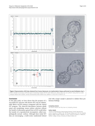
complex aneuploidy [7,25,59]. Our data show conven-tional
morphological criteria alone to be insufficiently ac-curate
even for young, low-risk IVF patients (see
Figure 2). Recent research on thawed blastocysts after
SNP-based comprehensive chromosomal screening and
vitrification has yielded similar results [60].
Several limitations of our investigation should be
acknowledged. First, although elective SET brings dis-tinct
advantages for many IVF patients, the approach is
not for everyone. Indiscriminate use of elective SET for
patients with multiple failed cycles has been criticized as
inferior to a two-embryo strategy [61], and the improved
pregnancy rate noted here may not fully generalize to all
IVF patients. Additionally, this pilot study was designed
to use aCGH for selection of a single blastocyst for fresh
transfer. It is possible that embryo assessment by con-ventional
morphology inappropriately excludes euploid
embryos from transfer although this question was out-side
the scope of our study. Hence, the relation between
chromosomal integrity and morphological grades based
on developmental stage, ICM and TE appearance,
requires further investigation with a larger sample.](https://image.slidesharecdn.com/cgh-141015065602-conversion-gate02/85/Cgh-5-320.jpg)
![Yang et al. Molecular Cytogenetics 2012, 5:24 Page 7 of 8
http://www.molecularcytogenetics.org/content/5/1/24
Authors’ contributions
ZY and JL conceived the research, designed the study, and directed the
aCGH analysis. GSC was the chief statistician in charge of data analysis. SAS,
XL, ACP, ESS, and RDS were the reproductive endocrinologists with oversight
of the clinical program. SSL was the embryologist. ESS edited the manuscript
and organized the revisions. All authors read and approved the final
manuscript.
Received: 3 February 2012 Accepted: 2 May 2012
Published: 2 May 2012
References
1. Coetsier T, Dhont M: Avoiding multiple pregnancies in in-vitro
fertilization: who’s afraid of single embryo transfer? Hum Reprod 1998,
13:2663–2664.
2. Ryan G, Sparks A, Sipe C, Syrop C, Dokras A, Van Voorthis B: A mandatory
single blastocyst transfer policy with educational campaign in a United
States IVF program reduces multiple gestation rates without sacrificing
pregnancy rates. Fertil Steril 2007, 88:354–360.
3. Zander-Fox DL, Tremellen K, Lane M: Single blastocyst embryo transfer
maintains comparable pregnancy rates to double cleavage-stage embryo
transfer but results in healthier pregnancy outcomes. Aust N Z J Obstet
Gynaecol 2011, 51:406–410.
4. Maheshwari A, Griffiths S, Bhattacharya S: Global variations in the uptake of
single embryo transfer. Human Reprod Update 2011, 17:107–120.
5. Gardner DK, Surrey E, Minjarrez D, Leitz A, Stevens J, Schoolcraft WB: Single
blastocyst transfer: a prospective randomized trial. Fertil Steril 2004,
81:551–555.
6. Racowsky C, Ohno-Machado L, Kim J, Biggers JD: Is there an advantage in
scoring early embryos on more than one day? Hum Reprod 2009,
24:2104–2113.
7. Alfarawati S, Fragouli E, Colls P, Stevens J, Gutierrez-Mateo C, Schoolcraft WB,
Wells D: The relationship between blastocyst morphology, chromosomal
abnormality and embryo gender. Fertil Steril 2011, 95:520–524.
8. Hassold T, Hunt P: Maternal age and chromosomally abnormal
pregnancies: what we know and what we knew. Curr Opin Pediatr 2009,
21:703–708.
9. Kuliev A, Cieslak J, Verlinsky Y: Frequency and distribution of chromosome
abnormalities in human oocytes. Cytogenet Genome Res 2005, 111:193–198.
10. Bielanska M, Tan SL, Ao A: Chromosomal mosaicism throughout human
preimplantation development in vitro: incidence, type, and relevance to
embryo outcome. Hum Reprod 2002, 17:413–419.
11. Magli MC, Gianaroli L, Ferraretti AP, Lappi M, Ruberti A, Farfalli V: Embryo
morphology and development are dependent on the chromosomal
complement. Fertil Steril 2007, 87:534–541.
12. Munné S, Alikani M, Tomkin G, Grifo J, Cohen J: Embryo morphology,
developmental rates, and maternal age are correlated with chromosome
abnormalities. Fertil Steril 1995, 64:382–391.
13. Munné S, Sandalinas M, Magli C, Gianaroli L, Cohen J, Warburton D:
Increased rate of aneuploid embryos in young women with previous
aneuploid conceptions. Prenat Diagn 2004, 24:638–643.
14. Munné S, Chen S, Colls P, Garrisi J, Zheng X, Cekleniak N, Lenzi M, Hughes P,
Fischer J, Garrisi M, Tomkin G, Cohen J: Maternal age, morphology,
development and chromosome abnormalities in over 6000 cleavage-stage
embryos. Reprod Biomed Online 2007, 14:628–634.
15. Vanneste E, Voet T, Le Caignec C, Ampe M, Konings P, Melotte C, Debrock S,
Amyere M, Vikkula M, Schuit F, Fryns JP, Verbeke G, D'Hooghe T, Moreau Y,
Vermeesch JR: Chromosome instability is common in human cleavage-stage
embryos. Nat Med 2009, 15:577–583.
16. Wilton L: Preimplantation genetic diagnosis and chromosome analysis of
blastomeres using comparative genomic hybridization. Hum Reprod
Update 2005, 11:33–41.
17. Staessen C, Platteau P, Van Assche E, Michiels A, Tournaye H, Camus M,
Devroey P, Liebaers I, Van Steirteghem A: Comparison of blastocyst
transfer with or without preimplantation genetic diagnosis for
aneuploidy screening in couples with advanced maternal age: a
prospective randomized controlled trial. Hum Reprod 2004, 19:2849–2858.
18. Hardarson T, Hanson C, Lundin K, Hillensjö T, Nilsson L, Stevic J, Reismer E,
Borg K, Wikland M, Bergh C: Preimplantation genetic screening in women
of advanced maternal age caused a decrease in clinical pregnancy rate:
a randomized controlled trial. Hum Reprod 2008, 23:2806–2812.
19. Schoolcraft WB, Katz-Jaffe MG, Stevens J, Rawlins M, Munné S:
Preimplantation aneuploidy testing for infertile patients of advanced
maternal age: a randomized prospective trial. Fertil Steril 2009, 92:157–162.
20. Masternbroek S, Twisk M, van Echten-Arends J, Sikkema-Raddatz B, Korevaar
JC, Verhoever HR: In vitro fertilization with preimplantation genetic
screening. N Engl J Med 2007, 357:9–17.
21. Masternbroek S, Twisk M, van der Veen F, Repping S: Preimplantation
genetic screening: a systematic review and meta-analysis of RCTs. Hum
Reprod Update 2011, 17:454–466.
22. Voullaire L, Wilton L, Slater H, Williamson R: Detection of aneuploidy in
single cells using comparative genomic hybridization. Prenat Diagn 1999,
19:846–851.
23. Wells D, Delhanty JDA: Comprehensive chromosomal analysis of human
preimplantation embryos using whole genome amplification and single
cell comparative genomic hybridization. Mol Hum Reprod 2000, 6:1055–1062.
24. Sher G, Keskintepe L, Keskintepe M, Ginsburg M, Maassarani G, Yakut T,
Baltaci V, Kotze D, Unsal E: Oocyte karyotyping by comparative genomic
hybridization [correction of hybridization] provides a highly reliable
method for selecting “competent” embryos, markedly improving in vitro
fertilization outcome: a multiphase study. Fertil Steril 2007, 87:1033–1040.
25. Fragouli E, Lenzi M, Ross R, Katz-Jaffe M, Schoolcraft WB, Wells D:
Comprehensive molecular cytogenetic analysis of the human blastocyst
stage embryos. Hum Reprod 2008, 23:2596–2608.
26. Hellani A, Abu-Amero K, Azouri J, El-Akoum S: Successful pregnancies after
application of array-comparative genomic hybridization in PGS-aneuploidy
screening. Reprod Biomed Online 2008, 17:814–817.
27. Fishel S, Gordon A, Lynch C, Ndukwe G, Kelada E, Thomton S, Jenner L,
Cater E, Brown A, Garcia-Bernardo J: Live birth after polar body array
comprehensive genomic hybridization prediction of embryo ploidy – the
future of IVF. Fertil Steril 2010, 93:1006.e7–1006.e10.
28. Gutierrez-Mateo C, Colls P, Sanchez-Garcia J, Escudero T, Prates R, Ketterson K,
Wells D, Munné S: Validation of microarray comparative genomic
hybridization for comprehensive chromosome analysis of embryos. Fertil
Steril 2011, 95:953–958.
29. Fioretino F, Spizzichino L, Bono S, Birricik A, Kokkali G, Rienzi L, Ubaldi FM,
Iammarrone E, Gordon A, Pantos K: PGD for reciprocal and Robertsonian
translocation using array comparative genomic hybridization. Hum
Reprod 2011, 26:1925–1935.
30. Handyside AH: PGD and aneuploidy screening for 24 chromosome by
genome-wide SNP analysis: seeing the wood and the trees. Reprod
Biomed Online 2011, 23:686–691.
31. Schoolcraft WB, Fragouli E, Stevens J, Munné S, Katz-faffe MG, Wells D:
Clinical application of comprehensive chromosomal screening at the
blastocyst stage. Fertil Steril 2010, 94:1700–1706.
32. Ly KD, Agarwal A, Nagy ZP: Preimplatation genetic screening: does it help
or hinder IVF treatment and what is the role of the embryo? J Assist
Reprod Genet 2011, 28:833–849.
33. Sills ES, Schattman GL, Veeck LL, Liu HC, Prasad M, Rosenwaks Z:
Characteristics of consecutive in vitro fertilization cycles among patients
treated with follicle-stimulating hormone (FSH) and human menopausal
gonadotropin versus FSH alone. Fertil Steril 1998, 69:831–835.
34. Yang Z, Salem S, Salem-Lyle S, Bayrak A, Salem RD: Trophectoderm cells
derived from blastocyst biopsy are suitable for array CGH analysis of 24
chromosomes. Fertil Steril 2011, 95(Suppl 4):S23.
35. Alfarawati S, Fragouli E, Colls P, Wells D: First births after preimplantation
genetic diagnosis of structural chromosome abnormalities using
comparative genomic hybridization and microarray analysis. Hum Reprod
2011, 26:1560–1574.
36. Sakkas D, Gardner DK: Noninvasive methods to assess embryo quality.
Curr Opin Obstet Gynecol 2005, 17:283–288.
37. Rauch ER, Schattman GL, Christos PJ, Chicketano T, Rosenwaks Z: Embryonic
heart rate as a predictor of first-trimester pregnancy loss in infertility
patients after in vitro fertilization. Fertil Steril 2009, 91:2451–2454.
38. van Peperstraten AM, Nelen WL, Hermens RP, Jansen L, Scheenjes E, Braat DD,
Grol RP, Kremer JA: Why don't we perform elective single embryo transfer?
A qualitative study among IVF patients and professionals. Hum Reprod 2008,
23:2036–2042.
39. Hvidtjørn D, Grove J, Schendel D, Svaerke C, Schieve LA, Uldall P, Ernst E,
Jacobsson B, Thorsen P: Multiplicity and early gestational age contribute
to an increased risk of cerebral palsy from assisted conception: a
population-based cohort study. Hum Reprod 2010, 25:2115–2123.](https://image.slidesharecdn.com/cgh-141015065602-conversion-gate02/85/Cgh-7-320.jpg)