Cell Reprogramming Methods And Protocols Paul J Verma Huseyin Sumer
Cell Reprogramming Methods And Protocols Paul J Verma Huseyin Sumer
Cell Reprogramming Methods And Protocols Paul J Verma Huseyin Sumer
Cell Reprogramming Methods And Protocols Paul J Verma Huseyin Sumer
Cell Reprogramming Methods And Protocols Paul J Verma Huseyin Sumer
1.
Cell Reprogramming MethodsAnd Protocols Paul J
Verma Huseyin Sumer download
https://ebookbell.com/product/cell-reprogramming-methods-and-
protocols-paul-j-verma-huseyin-sumer-5242110
Explore and download more ebooks at ebookbell.com
2.
Here are somerecommended products that we believe you will be
interested in. You can click the link to download.
Cell Reprogramming For Immunotherapy 1st Samuel G Katz Peter M
Rabinovich
https://ebookbell.com/product/cell-reprogramming-for-
immunotherapy-1st-samuel-g-katz-peter-m-rabinovich-47716524
Nuclear Transfer Protocols Cell Reprogramming And Transgenesis 1st
Edition Marie A Di Berardino Auth
https://ebookbell.com/product/nuclear-transfer-protocols-cell-
reprogramming-and-transgenesis-1st-edition-marie-a-di-berardino-
auth-4286992
Nuclear Reprogramming And Stem Cells 1st Edition John Gurdon
https://ebookbell.com/product/nuclear-reprogramming-and-stem-
cells-1st-edition-john-gurdon-2452566
Stem Cells No 265 Nuclear Reprogramming And Therapeutic Applications
Ciba Foundation Symposia Series 1st Edition Novartis Foundation
Symposium
https://ebookbell.com/product/stem-cells-no-265-nuclear-reprogramming-
and-therapeutic-applications-ciba-foundation-symposia-series-1st-
edition-novartis-foundation-symposium-1843012
3.
Cellcycle Synchronization MethodsAnd Protocols Zhixiang Wang
https://ebookbell.com/product/cellcycle-synchronization-methods-and-
protocols-zhixiang-wang-45987910
Cellwide Identification Of Metaboliteprotein Interactions Methods In
Molecular Biology 2554 1st Ed 2023 Aleksandra Skirycz Editor
https://ebookbell.com/product/cellwide-identification-of-
metaboliteprotein-interactions-methods-in-molecular-biology-2554-1st-
ed-2023-aleksandra-skirycz-editor-46502652
Celltocell Communication Cell Atlas Visual Biology In Oral Medicine
Reinhard Gruber
https://ebookbell.com/product/celltocell-communication-cell-atlas-
visual-biology-in-oral-medicine-reinhard-gruber-46507412
Cell Biology And Translational Medicine Volume 17 Stem Cells In Tissue
Differentiation Regulation And Disease Kursad Turksen
https://ebookbell.com/product/cell-biology-and-translational-medicine-
volume-17-stem-cells-in-tissue-differentiation-regulation-and-disease-
kursad-turksen-47172658
Cell And Molecular Biology For Nonbiologists A Short Introduction Into
Key Biological Concepts 1st Edition Lorenz Adlung
https://ebookbell.com/product/cell-and-molecular-biology-for-
nonbiologists-a-short-introduction-into-key-biological-concepts-1st-
edition-lorenz-adlung-47412138
ME T HO D S I N MO L E C U L A R BI O L O G Y
Series Editor
John M. Walker
School of Life and Medical Sciences
University of Hertfordshire
Hatfield, Hertfordshire, AL10 9AB, UK
For further volumes:
http://www.springer.com/series/7651
v
Cell Reprogramming: Methodsand Protocols is a comprehensive review of cellular repro-
gramming technology in vertebrates, aimed at reprogramming differentiated cells and germ
line transmission of pluripotent stem cells. The emphasis here is on providing readily repro-
ducible techniques for inducing pluripotency in somatic cells for disease modeling and the
generation of cloned embryos and animals in a number of key research and commercially
important species. Additional chapters dealing with such reprogramming-related issues
such as analysis of mitochondrial DNA in reprogrammed cells and the isolation of repro-
gramming intermediates are also included. A section providing alternative cutting-edge
methods for nuclear transfer, as well as techniques for the production of germ line chimeras
from embryonic stem cells and induced pluripotent stem cells is also incorporated. This is
complimented with the neonatal care and management of somatic cell nuclear transfer
derived offspring.
Cell Reprogramming also provides an understanding of the factors involved in nuclear
reprogramming, which is imperative for the success of reprogramming. This volume will
prove beneficial to molecular biologists, stem cell biologists, clinicians, biotechnologists,
students, veterinarians, and animal care technicians involved with reprogramming, nuclear
transfer, and transgenesis.
Clayton, VIC, Australia Paul J. Verma
Hawthorn, VIC, Australia Huseyin Sumer
Preface
12.
vii
Preface. . .. . . . . . . . . . . . . . . . . . . . . . . . . . . . . . . . . . . . . . . . . . . . . . . . . . . . . . . v
Contributors. . . . . . . . . . . . . . . . . . . . . . . . . . . . . . . . . . . . . . . . . . . . . . . . . . . . . . ix
PART I BACKGROUND
1 Cellular Reprogramming in Basic and Applied Biomedicine:
The Dawn of Regenerative Medicine. . . . . . . . . . . . . . . . . . . . . . . . . . . . . . . . 3
Wendy Dean
PART II DE NOVO REPROGRAMMING
2 Synthetic mRNA Reprogramming of Human Fibroblast Cells . . . . . . . . . . . . . 17
Jun Liu and Paul J. Verma
3 MicroRNA-Mediated Reprogramming of Somatic Cells into
Induced Pluripotent Stem Cells. . . . . . . . . . . . . . . . . . . . . . . . . . . . . . . . . . . . 29
Shelley E.S. Sandmaier and Bhanu Prakash V.L. Telugu
4 Generation of Footprint-Free Induced Pluripotent Stem Cells
from Human Fibroblasts Using Episomal Plasmid Vectors. . . . . . . . . . . . . . . . 37
Dmitry A. Ovchinnikov, Jane Sun, and Ernst J. Wolvetang
5 Reprogramming of Human Fibroblasts with Non-integrating RNA
Virus on Feeder-Free or Xeno-Free Conditions . . . . . . . . . . . . . . . . . . . . . . . . 47
Pauline T. Lieu
PART III LIVESTOCK, DOMESTIC AND ENDANGERED SPECIES
6 Inducing Pluripotency in Cattle . . . . . . . . . . . . . . . . . . . . . . . . . . . . . . . . . . . 57
Luis F. Malaver-Ortega, Amir Taheri-Ghahfarokhi,
and Huseyin Sumer
7 Generation of Induced Pluripotent Stem Cells (iPSCs) from Adult
Canine Fibroblasts . . . . . . . . . . . . . . . . . . . . . . . . . . . . . . . . . . . . . . . . . . . . . 69
Sehwon Koh and Jorge A. Piedrahita
8 Derivation of Equine-Induced Pluripotent Stem Cell Lines
Using a piggyBac Transposon Delivery System and Temporal
Control of Transgene Expression . . . . . . . . . . . . . . . . . . . . . . . . . . . . . . . . . . 79
Kristina Nagy and Andras Nagy
9 Generation of Avian Induced Pluripotent Stem Cells. . . . . . . . . . . . . . . . . . . . 89
Yangqing Lu, Franklin D. West, Brian J. Jordan, Robert B. Beckstead,
Erin T. Jordan, and Steven L. Stice
Contents
13.
viii
10 Generation ofInduced Pluripotent Stem Cells from Mammalian
Endangered Species . . . . . . . . . . . . . . . . . . . . . . . . . . . . . . . . . . . . . . . . . . . . 101
Inbar Friedrich Ben-Nun, Susanne C. Montague, Marlys L. Houck,
Oliver Ryder, and Jeanne F. Loring
PART IV GERM-LINE TRANSMISSION OF PLURIPOTENT STEM CELLS
11 Generation of Efficient Germ-Line Chimeras Using Embryonic
Stem Cell Injection . . . . . . . . . . . . . . . . . . . . . . . . . . . . . . . . . . . . . . . . . . . . . 113
William A. Ritchie
12 Generation of Viable Mice from Induced Pluripotent
Stem Cells (iPSCs) Through Tetraploid Complementation . . . . . . . . . . . . . . . 125
Lan Kang and Shaorong Gao
13 Cloning Endangered Felids by Interspecies Somatic Cell Nuclear Transfer. . . . 133
Martha C. Gómez and C. Earle Pope
14 Generation of Chimeras from Porcine Induced Pluripotent Stem Cells . . . . . . 153
Franklin D. West, Steve L. Terlouw, John R. Dobrinsky,
Yangqing Lu, Erin T. Jordan, and Steven L. Stice
15 A Novel Method of Somatic Cell Nuclear Transfer
with Minimum Equipment . . . . . . . . . . . . . . . . . . . . . . . . . . . . . . . . . . . . . . . 169
S.M. Hosseini, F. Moulavi, and M.H. Nasr-Esfahani
16 Neonatal Care and Management of Foals Derived by Somatic
Cell Nuclear Transfer . . . . . . . . . . . . . . . . . . . . . . . . . . . . . . . . . . . . . . . . . . . 189
Aime K. Johnson and Katrin Hinrichs
PART V INFLUENCING REPROGRAMMING AND GENOME EDITING
17 Isolation of Reprogramming Intermediates During Generation
of Induced Pluripotent Stem Cells from Mouse Embryonic Fibroblasts . . . . . . 205
Christian M. Nefzger, Sara Alaei, and Jose M. Polo
18 Analysis of Mitochondrial DNA in Induced Pluripotent
and Embryonic Stem Cells . . . . . . . . . . . . . . . . . . . . . . . . . . . . . . . . . . . . . . . 219
William Lee, Richard D.W. Kelly, Ka Yu Yeung, Gael Cagnone,
Matthew McKenzie, and Justin C. St. John
19 Genome Modification of Pluripotent Cells by Using Transcription
Activator-Like Effector Nucleases (TALENs). . . . . . . . . . . . . . . . . . . . . . . . . . 253
Amir Taheri-Ghahfarokhi, Luis F. Malaver-Ortega,
and Huseyin Sumer
Index. . . . . . . . . . . . . . . . . . . . . . . . . . . . . . . . . . . . . . . . . . . . . . . . . . . . . . . . . . . 269
Contents
14.
ix
SARA ALAEI •Department of Anatomy and Developmental Biology,
Australian Regenerative Medicine Institute, Monash University, Clayton,
VIC, Australia
ROBERT B. BECKSTEAD • Department of Animal and Dairy Science,
Regenerative Bioscience Center, University of Georgia, Athens, GA, USA
INBAR FRIEDRICH BEN-NUN • Department of Chemical Physiology, Center for
Regenerative Medicine, The Scripps Research Institute, La Jolla, CA, USA
GAEL CAGNONE • The Mitochondrial Genetics Group, Centre for Genetic Diseases,
Hudson Institute of Medical Research, Monash University, Clayton, VIC, Australia
WENDY DEAN • Epigenetics Programme, The Babraham Institute, Cambridgeshire, UK
JOHN R. DOBRINSKY • JRD Biotechnology, Oregon, WI, USA
SHAORONG GAO • National Institute of Biological Sciences, NIBS, Beijing,
People’s Republic of China; School of Life Sciences and Technology, Tongji University,
Shanghai, People’s Republic of China
MARTHA C. GÓMEZ • Audubon Nature Center for Research of Endangered Species,
New Orleans, LA, USA
KATRIN HINRICHS • Department of Veterinary Physiology and Pharmacology,
College of Veterinary Medicine and Biomedical Sciences, Texas A&M University,
College Station, TX, USA
S.M. HOSSEINI • Department of Reproductive Biotechnology at Reproductive Biomedicine
Research Center, Royan Institute for Biotechnology, ACECR, Isfahan, Iran
MARLYS L. HOUCK • San Diego Zoo Institute for Conservation Research, Escondido,
CA, USA
JUSTIN C. ST. JOHN • The Mitochondrial Genetics Group, Centre for Genetic Diseases,
Hudson Institute of Medical Research, Monash University, Clayton, VIC, Australia
AIME K. JOHNSON • JT Vaughn Large Animal Teaching Hospital, College of Veterinary
Medicine, Auburn University, Auburn, AL, USA
BRIAN J. JORDAN • Department of Animal and Dairy Science, Regenerative Bioscience
Center, University of Georgia, Athens, GA, USA
ERIN T. JORDAN • Department of Animal and Dairy Science, Regenerative Bioscience
Center, University of Georgia, Athens, GA, USA
LAN KANG • Institute of Cancer Stem Cell, Dalian Medical University, Dalian,
People’s Republic of China; National Institute of Biological Sciences, NIBS, Beijing,
People’s Republic of China
RICHARD D.W. KELLY • The Mitochondrial Genetics Group, Centre for Genetic Diseases,
Hudson Institute of Medical Research, Monash University, Clayton, VIC, Australia
SEHWON KOH • Department of Cell Biology, Duke University, Durham, NC, USA;
Duke University Medical Center, Duke University, Durham, NC, USA
WILLIAM LEE • The Mitochondrial Genetics Group, Centre for Genetic Diseases, Hudson
Institute of Medical Research, Monash University, Clayton, VIC, Australia
Contributors
15.
x
PAULINE T. LIEU• Global R&D, Life Technologies Corporation, Carlsbad, CA, USA
JUN LIU • Stem Cell and Genetic Engineering Group, Department of Materials
Engineering, Faculty of Engineering, Monash University—Clayton Campus,
Clayton, VIC, Australia
JEANNE F. LORING • Department of Chemical Physiology, Center for Regenerative Medicine,
The Scripps Research Institute, La Jolla, CA, USA; Department of Reproductive
Medicine, University of California, San Diego, La Jolla, CA, USA
YANGQING LU • Department of Animal and Dairy Science, Regenerative Bioscience Center,
University of Georgia, Athens, GA, USA; JRD Biotechnology, Oregon, WI, USA;
State Key Laboratory for Conservation and Utilization of Subtropical Agro-bioresources,
Guangxi University, Nanning, China
LUIS F. MALAVER-ORTEGA • Monash Institute for Medical Research, Monash University,
Clayton, VIC, Australia; Australian Animal Health Laboratories, CSIRO Biosecurity
Flagship, East Geelong, VIC, Australia
MATTHEW MCKENZIE • The Molecular Basis of Mitochondrial Disease Group,
Centre for Genetic Diseases, Hudson Institute of Medical Research, Monash University,
Clayton, VIC, Australia
SUSANNE C. MONTAGUE • Department of Chemical Physiology, Center for Regenerative
Medicine, The Scripps Research Institute, La Jolla, CA, USA
F. MOULAVI • Department of Reproductive Biotechnology at Reproductive Biomedicine
Research Center, Royan Institute for Biotechnology, ACECR, Isfahan, Iran
ANDRAS NAGY • Lunenfeld-Tanenbaum Research Institute, Mount Sinai Hospital, Toronto,
ON, Canada; Department of Obstetrics and Gynecology, University of Toronto, Toronto,
ON, Canada
KRISTINA NAGY • Lunenfeld-Tanenbaum Research Institute, Mount Sinai Hospital,
Toronto, ON, Canada
M.H. NASR-ESFAHANI • Department of Reproductive Biotechnology at Reproductive
Biomedicine Research Center, Royan Institute for Biotechnology, ACECR, Isfahan, Iran
CHRISTIAN M. NEFZGER • Department of Anatomy and Developmental Biology, Australian
Regenerative Medicine Institute, Monash University, Clayton, VIC, Australia
DMITRY A. OVCHINNIKOV • Stem Cell Engineering Group, Australian Institute for
Bioengineering and Nanotechnology, University of Queensland, Brisbane, QLD,
Australia
JORGE A. PIEDRAHITA • Department of Molecular Biomedical Sciences, College of Veterinary
Medicine, North Carolina State University, Raleigh, NC, USA; Genomics Program,
North Carolina State University, Raleigh, NC, USA; Center for Comparative Medicine
and Translational Research, North Carolina State University, Raleigh, NC, USA
JOSE M. POLO • Department of Anatomy and Developmental Biology, Australian
Regenerative Medicine Institute, Monash University, Clayton, VIC, Australia
C. EARLE POPE • Audubon Nature Center for Research of Endangered Species,
New Orleans, LA, USA
WILLIAM A. RITCHIE • Roslin Embryology Ltd., Macmerry, Tranent, Scotland, UK;
Monash Biomed Private Limited, Delhi, India
OLIVER RYDER • San Diego Zoo Institute for Conservation Research, Escondido, CA, USA
STEVEN L. STICE • Department of Animal and Dairy Science, Regenerative Bioscience
Center, University of Georgia, Athens, GA, USA
Contributors
16.
xi
HUSEYIN SUMER •Department of Chemistry and Biotechnology, Faculty of Science,
Engineering and Technology, Swinburne University of Technology, Hawthorn, VIC,
Australia
JANE SUN • Stem Cell Engineering Group, Australian Institute for Bioengineering and
Nanotechnology, University of Queensland, Brisbane, QLD, Australia
SHELLEY E.S. SANDMAIER • Department of Animal and Avian Sciences, University of
Maryland, College Park, MD, USA; Animal Bioscience and Biotechnology Laboratory,
USDA-ARS, Beltsville, MD, USA
AMIR TAHERI-GHAHFAROKHI • Department of Animal Science, Ferdowsi University of
Mashhad, Mashhad, Iran
BHANU PRAKASH V.L. TELUGU • Department of Animal and Avian Sciences, University of
Maryland, College Park, MD, USA; Animal Bioscience and Biotechnology Laboratory,
USDA-ARS, Beltsville, MD, USA
STEVE L. TERLOUW • Minitube of America, Mt. Horeb, WI, USA
PAUL J. VERMA • Stem Cell and Genetic Engineering Group, Department of Materials
Engineering, Monash University, Clayton, VIC, Australia; South Australian Research
and Development Institute, Turretfield Research Centre, Rosedale, SA, Australia
FRANKLIN D. WEST • Department of Animal and Dairy Science, Regenerative Bioscience
Center, University of Georgia, Athens, GA, USA
ERNST J. WOLVETANG • Stem Cell Engineering Group, Australian Institute for
Bioengineering and Nanotechnology, University of Queensland, Brisbane, QLD,
Australia
KA YU YEUNG • The Mitochondrial Genetics Group, Centre for Genetic Diseases, Hudson
Institute of Medical Research, Monash University, Clayton, VIC, Australia; Molecular
Basis of Metabolic Disease, Division of Metabolic and Vascular Health, Warwick Medical
School, The University of Warwick, Coventry, UK
Contributors
4
But exactly howdoes the forced overexpression of a handful of
transcription factors and chromatin-binding molecules transform
the defined cellular state of a differentiated cell and progress it back
up Waddington’s ascending landscape to assume a pluripotent
phenotype—in essence, a stem cell?
The simple answer is, at present, that we do not know.
However, the full impact of modern genome-wide investigation
and the sheer force of numbers of researchers worldwide leading
this investigation make the prospect of significant mechanistic
understanding only a matter of time and the translation to patient-
specific regenerative medicine a reality in our lifetime. In the course
of these studies there is a real prospect of collateral benefit; much
will be learned about the potential to identify and manipulate
endogenous stem cell populations that function in tissue repair and
replacement throughout life. Indeed the intense study of the pro-
cesses of cellular regression may well hold the key to understanding
healthy aging and offer an explanation for the growing number of
centenarians in our societies, which has seen a fivefold rise over the
last 30 years (Office for National Statistics UK; BBC news, 27th
Sept 2013).
Cellular reprogramming is the conversion of one specific cell
type to another. Arguably, we could well consider that develop-
ment in its usual forward-only direction could constitute a form of
cellular reprogramming. Here, the highly specialized and fully dif-
ferentiated oocyte is reprogrammed on fertilization to restore an
ephemeral totipotent state that is quickly followed by a series of
progressively more differentiated cellular decisions passing through
ever more restricted multipotent junctures to give rise to the fully
formed neonatal animal. In the 1940s Conrad Waddington
described this process in his classical model of the epigenetic land-
scape where one genotype allowed for the generation of multiple
cellular phenotypes [2]. Waddington illustrated the hierarchical
progression of the undifferentiated state by a series of channels
which were progressively more restricted and increasingly sepa-
rated; thus once the cellular state was “fated” thereafter the lineage
was restricted and incapable of either returning to a more undif-
ferentiated state or a different germ cell layer [3]. In the postgen-
omic era, these ideas together with classical developmental and
cellular biology have formed the basis of our understanding of the
field of epigenetics.
However, today cellular reprogramming more often refers to
those landmark methods which included transdifferentiation or
direct cell conversion, somatic cell nuclear transfer (SCNT) and
experimental reprogramming, the basis of the generation of iPS
cells [4]. The chapters which follow outline the details of how to
establish these various models of developmental and cellular pro-
cesses that set the experimental scene for understanding the mech-
anisms underpinning these transitions and serve to allow us
Wendy Dean
22.
5
unprecedented opportunities inbasic, agricultural and biomedical
science to improve health and wellbeing, to enhance food security,
and offer therapeutic solutions to the treatment of chronic disor-
ders in humans.
By way of an introduction to these methods I will outline some
of the origins and common themes which these methods share and
contrast points where they differ. These experimental approaches
have certainly been instrumental in driving a deeper and more
comprehensive understanding of mammalian development and
stem cell biology in general and will undoubtedly continue to drive
fundamental and applied questions in these areas. Perhaps most
exciting, as a result of these experimental systems, fundamentally
held beliefs about the prescriptive nature of developmental pro-
cesses and tissue regeneration upon damage are now being chal-
lenged. The prospect of significant improvement of health span, on
a patient specific basis, is now within sight.
While the focus of this book is the experimental details that
facilitate cellular reprogramming, before embarking on an outline
of these techniques it may be worth touching, if only briefly, on
some processes that occur naturally which are capable of achieving
the same end. Transdifferentiation and cell fusion, much like that
of experimental heterokaryons, do occur naturally [5, 6].
Transdifferentiation constitutes a change in cellular fate, which can
facilitate the transit between lineages in the most extreme case and
between a differentiated cell type and its less differentiated fore-
runner within a given lineage. Here, one distinction that is often
applied is that both of these processes take place by a direct cell
conversion and not via a pluripotent intermediate. In mammals
transdifferentiation can be achieved experimentally by both gain of
function through overexpression and loss of function mechanisms
of one or a few factors and in this way bears some resemblance to
iPS production. Interestingly, these induced transitions can be
studied in vitro using stem cell models such as an ES cell, a proxy
for the inner cell mass of the blastocyst stage in mammals. In what
seems a reversion of the very first cellular decision in development,
ES cells can be driven to acquire trophoblast stem (TS) cell-like
fates [7–9] which implies that the experimental manipulation
endows the cell with permission, and capacity, for the lowering of
the epigenetic barrier that ordinarily separates and defines these
first two cell lineages.
Cell fusion and transdifferentiation have shared a common
past. In 2002 two significant papers identified the potential of ES
cells co-cultured with either neural stem cells or bone marrow cells
to subsequently undergo differentiation to a variety of cell types.
However, this occurred not by dedifferentiation, which was the
first explanation, but by transdifferentiation via spontaneous cell
fusion [10, 11]. At the time this caused a significant rethink in the
field but supplied positive benefit in the greater degrees of vigor
Cellular Reprogramming for Biomedicine
23.
6
that were thereafterrequired of these types of experiments [12].
Perhaps more importantly, this did highlight the fact that these
processes could occur, albeit at a low frequency, establishing the
proof of principle that similar cell–cell fusion events that allow cell
fate transitions may take place in vivo. Thinking along these experi-
mental lines may well be of benefit in particular to the adult stem
cell field.
While SCNT and iPS cell reprogramming are seemingly dia-
metrically opposed they share interesting common origins in the
ferment of mammalian experimental embryology and cell biology
in the 1980s. The premise of SCNT had been based on classical
developmental experiments carried out by Spemann in the 1920s
answering the question of totipotency of nuclei at least early in
development [13]. This was extended by the seminal work of
Briggs and King in the 1950s [14] followed closely by John
Gurdon [15] illustrating that in amphibian models differentiated
nuclei could be transplanted to the enucleated oocyte and give rise
to an adult organism. While this confirmed nuclear conservation,
they also showed that the regenerative potency with nuclear donors
isolated from more advanced, and hence more differentiated tis-
sues, was progressively restricted [16]. As a whole this progressive
restriction, i.e., the very idea that Waddington described as canali-
zation, seemed to be holding up.
In 1983, McGrath and Solter published a method of nuclear
transplantation in mammals using a fusogenic virus [17]. This laid
the ground work for the flurry of reports of “cloning” in mammals
from embryonic cells in the sheep by Steen Willadsen [18] to the
landmark achievement of Campbell and Wilmut in 1996 of the
generation of a live cloned sheep, Dolly, from an adult, fully dif-
ferentiated, mammary cell nucleus [19]. To date cloning has been
successful in more than 15 mammalian species including the extinct
Pyrenean ibex and a handful of other endangered species [20].
While cloning in most species has been a success, among endan-
gered species cloning has been more difficult. Of these only the
mouflon sheep survived for more than a few days after birth [21].
Clearly the oocyte, in conjunction with modulation of widespread
chromatin remodeling, can reinstruct a terminal program to relive
its developmental past; something once thought to be unachiev-
able under any circumstance [22].
The induction of stem cells starting from differentiated fibro-
blasts is an extreme form of cell fate conversion and hence may
constitute an extreme form of transdifferentiation. Here, the con-
trast to the reprogramming in SCNT is stark. The cellular as well as
the nuclear status of the fibroblast must be dedifferentiated and
ultimately progressed to the pinnacle of the canalized landscape in
order to form pluripotent stem cells. In this form of reprogram-
ming the cell is a most unsuitable environment with little of its own
capacity to direct retrograde dedifferentiation unto pluripotency.
Wendy Dean
24.
7
The earliest incarnationsof this process were first described in
Lasser et al. [23] where overexpression of a defined transcription
factor (TF), MyoD, was able to drive fibroblasts toward a muscle
cell fate. While this worked best in mesodermally derived cells,
similar results were also obtained in ectodermal and endodermal
derivatives hinting at the now familiar concept that forced over-
expression of TFs, defining for a given cell type, greatly assists in
the transdifferentiation toward that cell type [24]. In practice this
is but a short step away in taking this idea forward toward a des-
tination in stem cell populations—in essence the seed of the
“four-factor cocktail” had been planted. Over the intervening
years intra-germ layer conversion was demonstrated for a vast
number of TF combinations. Interestingly, the dynamics of the
transition were highly variable with both the starting cell type
and the order of expression of the TF cocktail able to influence
the cellular outcome. In fact, only relatively recently has this
approach succeeded in “long distance” direct conversion; start-
ing with fibroblasts a “three-factor” cocktail was able to generate
functional neurons [25].
Induced pluripotent stems cells have changed the way we think
about cellular differentiation, cell fate commitment, and the unidi-
rectional nature of development [26]. Beyond that, the very nature
of the stably differentiated cell has been challenged along with the
ideas of the epigenome that serve to reinforce and fix that state.
While remarkable in the insights that derived from conversion of
cell types both within and across germ layer boundaries, direct cell
conversion has significant limitations. Ideally, and in keeping with
the need to be able to supply adequate numbers of any cell type in
any lineage, stem cells seem like the best option and those equiva-
lent to embryonic stem cells would allow unrestricted and ethically
uncomplicated extension to therapeutic applications in the treat-
ment of disease.
Applying the lessons of intra-lineage conversion, Takahashi
and Yamanaka focused their attention on transcription factor net-
works associated with pluripotency and self-renewal, both hall-
marks of pluripotent embryonic stem (ES) cells. Distilling the list
to the now well known “four-factor cocktail,” of Oct3/4, Sox2,
Klf4 and c-Myc (OSKM), and transfecting them into either fetal or
adult mouse, and later human, fibroblasts lead eventually to the
generation of the first iPS cells [1]. Remarkably, in mouse and
human, expression from the delivery systems is eventually taken
over by the endogenous loci thereby supplying a continuous source
of the essential factors characteristic of the target ES cells. Although
highly inefficient, these cells fulfilled their potential being able to
differentiate into all three germ layers and in the generation of
both chimeric animals and entirely iPS-derived mice by tetraploid
complementation, the gold standard for demonstrating pluripo-
tency. Interestingly, a large proportion of the domestic animal iPS
Cellular Reprogramming for Biomedicine
25.
8
systems fail toeither activate the endogenous loci or silence the
transgenes in the course of iPS reprogramming.
Better and more efficacious delivery systems that did not
involve viral vectors, requisite for use in humans, have now been
achieved. Many iterations and reiterations of the “essential factors”
have also taken place with replacements now in common use. In
this respect it is remarkable that the “four factors” have been found
to be so broadly able to direct iPS cell generation across such a
wide cross section of mammalian species. In a few cases, in bovine
[27] and the endangered class of Felids [28] is an additional factor,
namely Nanog, required for iPS cell reprogramming. In the goat
and sheep, eight factors have been reported to be required to
reprogram primary ear fibroblasts [29, 30].
Second- and third-generation reprogramming approaches to
iPS cells now exist which employ either small molecule inhibitors
or transfection of families of microRNAs alone or in combination
with the Yamanaka factors [31, 32]. MicroRNAs are particularly
abundant in pluripotent ES cells; among the most abundant, the
miR301/367 in humans and the miR290 cluster in the mouse, are
themselves up-regulated by the OSKM quartet and mutually rein-
force the pluripotent state thereby driving cells toward this termi-
nus. Coupled to their ability to down-regulate de novo methylation
the up-regulation of the miR290 cluster also enhances, among
other functions, the kinetics of the mesenchymal to epithelial tran-
sition (MET) requisite for reprogramming to iPS status [33–35].
Incidentally, alteration of the culture environment has also proven
to enhance iPS cell reprogramming.
The ability to generate ES cells in mouse and human has been
a breakthrough in pioneering the idea of replacement therapies for
faulty genes together with functional and mechanistic studies in all
biological disciplines, which ultimately underpin applied research.
In domestic species of agricultural and veterinary importance,
while some species have been amenable to the generation of
embryonic-like stem cells especially in light of improvements trans-
lated from the mouse, many have yet to achieve the same unre-
stricted claims to pluripotency. Here, iPS cell generation may prove
to be the solution as is the case in the equine system. Equine
ES-like cells possess only some of the full repertoire of the pluripo-
tent spectrum while equine iPS cells seem to be fully functional
and able to contribute to teratomas in engraftment experiments
[36]. Targeting of iPS cells once established may not prove univer-
sally simple. For example, human ES cells are refractory to conven-
tional genome editing via homologous recombination achieving
only very low efficiencies compared to the mouse and hence other
targeted methodologies such as zinc finger proteins, TALENs and
CRISPR are required [37].
The development of SNCT has long been regarded as a means
by which rare and endangered species might be rescued from
Wendy Dean
26.
9
impending extinction. Indeed,even some now extinct species have
been reanimated by NT where appropriate recipient species
hybrids still survive. It would now seem possible that iPS genera-
tion may provide additional avenues to help in supporting efforts
to save endangered species offering prospects of generation of
gametes in vitro from iPS cells as has been achieved with ES cells
[38–40]. Despite the relative ease in which the iPS generation has
been successful across a very wide swath of mammalian species, the
generation of gametes may not prove as simple; nonetheless, there
is reason for great optimism that the species variation among germ
cell maturation can be overcome and functional gametes gener-
ated across the diverse class of Mammalia. Failing the ability to
generate full maturation of gametes, iPS cells may well allow for
unprecedented mechanistic studies into germ cell development
across a wide selection of species many of whom may offer better
and closer physiological comparisons to humans without serious
ethical limitations [38, 41].
2 The Epigenome and Life in Culture
With the unparalleled promise of personalized medicine and gen-
eration of patient-specific tissue by stem cell therapies, replacement
and renewal no longer seems like a distant prospect. Less ambi-
tious but potentially more beneficial is the ability to test patient-
specific matching of drug treatment by using iPS cells either
directly or on tissue-specific differentiation. Veterinary drug test-
ing and biopharmaceutical companies may well screen and develop
treatments tailored by genetically typing patient groups to offer
the best fit for regulation of metabolic disorders using iPS cells
derived from specifically defined allelic profiling.
However, the question remains about the role of the epig-
enome and the influence of culture-based rearing of cells and tis-
sues especially where tissue engraftment is required. Here, lessons
from ES cells as a proxy for iPS cells will be highly informative. It
has long been recognized that cells in culture, including embry-
onic stem cells acquire increasing levels of DNA methylation, as a
function of the duration of life in culture, a significant barrier to
both dedifferentiation via SCNT and iPS reprogramming.
Recent evaluation of the DNA methylation profile of primed
vs naïve ES cells has shed light on this question. Small molecule
inhibitors (aka 2i) that both enhance ES cell derivation and reduce
their heterogeneity in culture have focused attention on the role of
the composition of the culture media and the DNA methylome in
mouse [42–45] and in human ES cells [46]. Thus the presence of
conventional serum can affect the pluripotential capacity of ES
cells by significant modulation of DNA methylation, notably by
increasing methylation and decreasing naïve pluripotency. In as
Cellular Reprogramming for Biomedicine
27.
10
much as microRNAfamilies that are associated with iPS cell repro-
gramming negatively regulate DNA methyltransferases and hence
DNA methylation, these two common components (i.e., serum
and microRNAs) seem to be at odds with one another for the
reprogramming process. Loss of DNA methylation, especially tied
to natural reprogramming, has been a dominant interest in the
field of epigenetics. The discovery of another significant pathway
able to down-regulate DNA methylation by methylcytosine
oxidation-coupled to repair pathways may be able to offer some
answers [47, 48]. A family of three enzymes, the ten-eleven-
translocation or TETS, iteratively oxidizing the methyl group on
cytosine to hydroxymethyl cytosine (5hmC) eventually leads to
this loss of DNA methylation via the return to the cytosine group.
Enzymatically, this reaction requires reduced Fe2+
and
α-ketoglutarate as cofactors and is hence very sensitive to the media
conditions and gaseous environment during culture.
Ascorbic acid, Vitamin C (VitC), has been known to enhance
iPS generation in mouse and humans for some time. Here acting
not via the 2i pathway but rather by alleviating the senescence road-
block, in the presence of VitC the histone demethylases Jhdm1a/1b
are stimulated [49]. Interestingly, TET1 is involved via its func-
tional domain in the formation of 5hmc at loci critical for MET in
a VitC-dependent manner [50]. In a systematic screen, the absence
of all H3K9me2 and me3 histone methylases, which include
Suv39H1 and 2, G9A and SetDB1, were found to work synergisti-
cally with VitC to enhance iPS cell reprogramming [50]. The mod-
ulation of H3K9me2/me3 is mechanistically linked to loss of DNA
methylation [51]. As such the presence of VitC in somatic cell
reprogramming is tied to loss of DNA methylation likely via repli-
cation-dependent passive mechanisms that involve loss of H3K9
methylation as well.
Whether or not the acquisition of DNA methylation during
culture of iPS cells will constitute a barrier to their widespread
application is not yet clear. In mouse ES cells maintained in stan-
dard serum-based culture conditions CpG methylation is high.
However, what happens to this hypermethylation once it is intro-
duced into a cellular context in vivo or upon tissue derivation has
not been systematically explored. In a simple but elegant test of
this question the results of a recent experiment gives us cause for
optimism. ES cells carrying a GFP reporter were used to make
chimeric animals by the classical blastocyst injection method. These
chimeric embryos were collected at E17.5 and the GFP-positive
cells isolated by flow cytometry and subsequently evaluated for lev-
els of DNA methylation. While the original ES cells were heavily
methylated, those GFP-positive cells isolated from tissues of these
embryos showed reduced levels of DNA methylation that were not
significantly different from the GFP-negative host cells. In essence,
in dividing cells within an in vivo environment, the DNA
Wendy Dean
28.
11
methylation levels hadbeen returned to normal [52]. Whether this
is universally true in other species needs to be proven.
Collectively, we are closing in on solutions to overcome many
of the barriers that currently limit unbridled enthusiasm and realis-
tic optimism for the promise of iPS cell-based application to regen-
erative medicine. The regulation of the epigenome is amongst one
of the most complicated barriers which unify the challenges of both
SCNT and iPS cell reprogramming irrespective of the application
[53]. At present the incredible rate of research output in this area is
rivaled only by that of the stem cell biology (which is overlapping
with iPS cells). Lessons learned in driving the program back to the
top of the Waddington landscape have revealed that pathways at
intermediate heights may well provide equally good or better van-
tage points for obtaining multipotent stem cell populations both
in vitro and that are resident in vivo, that might offer solutions to
contemporary obstacles. Indeed, direct cell conversion has chal-
lenged our belief about the distance between differentiated lineages
and the depth of the canalization. Late in 2014, the direct conver-
sion of fibroblasts into thymic epithelial-like cells giving rise to a
functional thymus-like organ on transplantation of aggregates
together with T-cell precursors and support cells was reported [54].
The chapters that follow offer practical solutions and guide-
lines on how to overcome the obstacles that currently impede our
progress in experimental reprogramming. Innovation will come
when we challenge the dogma and invite fresh eyes to use our
methods and supply their own new questions. The 2012 Nobel
Prize for Medicine and Physiology to Dr. Shinya Yamananka and
Sir John Gurdon acknowledged the start of exciting and indeed
remarkable discoveries in reprogramming. No doubt the first of
very many!
References
1. Takahashi K, Yamanaka S (2006) Induction of
pluripotent stem cells from mouse embryonic
and adult fibroblast cultures by defined factors.
Cell 126:663–676
2. Waddington CH (1942) Canalization of devel-
opment and the inheritance of acquired charac-
ters. Nature 150:563–565
3. Waddington CH (1957) The strategy of the
genes. George Allen & Unwin, London, UK
4. Jopling C, Boue S, Izpisua Belmonte JC
(2011) Dedifferentiation, transdifferentiation
and reprogramming: three routes to regenera-
tion. Nat Rev Mol Cell Biol 12:79–89
5. Nygren JM, Jovinge S, Breitbach M et al
(2004) Bone marrow-derived hematopoietic
cells generate cardiomyocytes at a low fre-
quency through cell fusion, but not transdif-
ferentiation. Nat Med 10:494–501
6. Orlic D, Kajstura J, Chimenti S et al (2001)
Bone marrow cells regenerate infarcted myo-
cardium. Nature 410:701–705
7. Niwa H, Miyazaki J, Smith AG (2000)
Quantitative expression of Oct-3/4 defines
differentiation, dedifferentiation or self-
renewal of ES cells. Nat Genet 24:372–376
8. Niwa H, Toyooka Y, Shimosato D et al (2005)
Interaction between Oct3/4 and Cdx2 deter-
mines trophectoderm differentiation. Cell
123:917–929
9. Lu CW, Yabuuchi A, Chen L et al (2008) Ras-
MAPK signaling promotes trophectoderm for-
mation from embryonic stem cells and mouse
embryos. Nat Genet 40:921–926
10. Ying QL, Nichols J, Evans EP et al (2002)
Changing potency by spontaneous fusion.
Nature 416:545–548
Cellular Reprogramming for Biomedicine
29.
12
11. Terada N,Hamazaki T, Oka M et al (2002)
Bone marrow cells adopt the phenotype of
other cells by spontaneous cell fusion. Nature
416:542–545
12. Wells WA (2002) Is transdifferentiation in
trouble? J Cell Biol 157:15–18
13. Spemann H (1938) Embryonic development
and induction. Hafner, New York, NY
14. Briggs R, King TJ (1952) Transplantation of
living nuclei from blastula cells into enucleated
frogs’ eggs. Proc Natl Acad Sci U S A
38:455–463
15. Gurdon JB (1962) Adult frogs derived from
the nuclei of single somatic cells. Dev Biol
4:256–273
16. Gurdon JB (1962) The developmental capacity
of nuclei taken from intestinal epithelium cells
of feeding tadpoles. J Embryol Exp Morphol
10:622–640
17. McGrath J, Solter D (1983) Nuclear transplan-
tation in the mouse embryo by microsurgery
and cell fusion. Science 220:1300–1302
18. Willadsen SM (1986) Nuclear transplantation
in sheep embryos. Nature 320:63–65
19. Wilmut I, Schnieke AE, McWhir J et al (1997)
Viable offspring derived from fetal and adult
mammalian cells. Nature 385:810–813
20. Lanza RP, Cibelli JB, Diaz F et al (2000)
Cloning of an endangered species (Bos gaurus)
using interspecies nuclear transfer. Cloning
2:79–90
21. Loi P, Ptak G, Barboni B et al (2001) Genetic
rescue of an endangered mammal by cross-
species nuclear transfer using post-mortem
somatic cells. Nat Biotechnol 19:962–964
22. McGrath J, Solter D (1984) Inability of mouse
blastomere nuclei transferred to enucleated
zygotes to support development in vitro.
Science 226:1317–1319
23. Lassar AB, Paterson BM, Weintraub H (1986)
Transfection of a DNA locus that mediates the
conversion of 10 T1/2 fibroblasts to myo-
blasts. Cell 47:649–656
24. Weintraub H, Tapscott SJ, Davis RL et al
(1989) Activation of muscle-specific genes in
pigment, nerve, fat, liver, and fibroblast cell
lines by forced expression of MyoD. Proc Natl
Acad Sci U S A 86:5434–5438
25. Lujan E, Chanda S, Ahlenius H et al (2012)
Direct conversion of mouse fibroblasts to self-
renewing, tripotent neural precursor cells. Proc
Natl Acad Sci U S A 109:2527–2532
26. Ladewig J, Koch P, Brustle O (2013) Leveling
Waddington: the emergence of direct pro-
gramming and the loss of cell fate hierarchies.
Nat Rev Mol Cell Biol 14:225–236
27. Sumer H, Liu J, Malaver-Ortega LF et al
(2011) NANOG is a key factor for induction of
pluripotency in bovine adult fibroblasts. J Anim
Sci 89:2708–2716
28. Verma R, Liu J, Holland MK et al (2013)
Nanog is an essential factor for induction of
pluripotency in somatic cells from endangered
felids. Biores Open Access 2:72–76
29. Ren J, Pak Y, He L et al (2011) Generation of
hircine-induced pluripotent stem cells by somatic
cell reprogramming. Cell Res 21:849–853
30. Sartori C, DiDomenico AI, Thomson AJ et al
(2012) Ovine-induced pluripotent stem cells
can contribute to chimeric lambs. Cell
Reprogram 14:8–19
31. Judson RL, Babiarz JE, Venere M et al (2009)
Embryonic stem cell-specific microRNAs pro-
mote induced pluripotency. Nat Biotechnol
27:459–461
32. Mikkelsen TS, Hanna J, Zhang X et al (2008)
Dissecting direct reprogramming through inte-
grative genomic analysis. Nature 454:49–55
33. Benetti R, Gonzalo S, Jaco I et al (2008) A
mammalian microRNA cluster controls DNA
methylation and telomere recombination via
Rbl2-dependent regulation of DNA methyl-
transferases. Nat Struct Mol Biol 15:268–279
34. Sinkkonen L, Hugenschmidt T, Berninger P
et al (2008) MicroRNAs control de novo DNA
methylation through regulation of transcrip-
tional repressors in mouse embryonic stem
cells. Nat Struct Mol Biol 15:259–267
35. Subramanyam D, Lamouille S, Judson RL et al
(2011) Multiple targets of miR-302 and miR-
372 promote reprogramming of human fibro-
blasts to induced pluripotent stem cells. Nat
Biotechnol 29:443–448
36. Nagy K, Sung HK, Zhang P et al (2011)
Induced pluripotent stem cell lines derived
from equine fibroblasts. Stem Cell Rev
7:693–702
37. Liu Y, Rao M (2011) Gene targeting in human
pluripotent stem cells. Methods Mol Biol
767:355–367
38. Hayashi K, Ohta H, Kurimoto K et al (2011)
Reconstitution of the mouse germ cell specifi-
cation pathway in culture by pluripotent stem
cells. Cell 146:519–532
39. Hayashi K, Saitou M (2013) Generation of
eggs from mouse embryonic stem cells and
induced pluripotent stem cells. Nat Protoc
8:1513–1524
40. Nayernia K, Nolte J, Michelmann HW et al
(2006) In vitro-differentiated embryonic stem
cells give rise to male gametes that can generate
offspring mice. Dev Cell 11:125–132
Wendy Dean
30.
13
41. Imamura M,Hikabe O, Lin ZY et al (2014)
Generation of germ cells in vitro in the era of
induced pluripotent stem cells. Mol Reprod
Dev 81:2–19
42. Ficz G, Hore TA, Santos F et al (2013) FGF
signaling inhibition in ESCs drives rapid
genome-wide demethylation to the epigenetic
ground state of pluripotency. Cell Stem Cell
13:351–359
43. Habibi E, Brinkman AB, Arand J et al (2013)
Whole-genome bisulfite sequencing of two dis-
tinct interconvertible DNA methylomes of
mouse embryonic stem cells. Cell Stem Cell
13:360–369
44. Leitch HG, McEwen KR, Turp A et al (2013)
Naive pluripotency is associated with global
DNA hypomethylation. Nat Struct Mol Biol
20:311–316
45. Yamaji M, Ueda J, Hayashi K et al (2013)
PRDM14 ensures naive pluripotency through
dual regulation of signaling and epigenetic
pathways in mouse embryonic stem cells. Cell
Stem Cell 12:368–382
46. Takashima Y, Guo G, Loos R et al (2014)
Resetting transcription factor control circuitry
toward ground-state pluripotency in human.
Cell 158:1254–1269
47. Kriaucionis S, Heintz N (2009) The nuclear
DNA base 5-hydroxymethylcytosine is present
in Purkinje neurons and the brain. Science
324:929–930
48. Tahiliani M, Koh KP, Shen Y et al (2009)
Conversion of 5-methylcytosine to
5-hydroxymethylcytosine in mammalian DNA
by MLL partner TET1. Science 324:930–935
49. Wang T, Chen K, Zeng X et al (2011) The his-
tone demethylases Jhdm1a/1b enhance
somatic cell reprogramming in a vitamin-C-
dependent manner. Cell Stem Cell 9:575–587
50. Chen J, Guo L, Zhang L et al (2013) Vitamin C
modulates TET1 function during somatic cell
reprogramming. Nat Genet 45:1504–1509
51. Lehnertz B, Ueda Y, Derijck AA et al (2003)
Suv39h-mediated histone H3 lysine 9 methyla-
tion directs DNA methylation to major satellite
repeats at pericentric heterochromatin. Curr
Biol 13:1192–1200
52. Ludwig G, Nejman D, Hecht M et al (2014)
Aberrant DNA methylation in ES cells. PLoS
One 9:e96090
53. Hemberger M, Dean W, Reik W (2009)
Epigenetic dynamics of stem cells and cell lin-
eage commitment: digging Waddington’s
canal. Nat Rev Mol Cell Biol 10:526–537
54. Bredenkamp N, Ulyanchenko S, O’Neill KE
et al (2014) An organized and functional thy-
mus generated from FOXN1-reprogrammed
fibroblasts. Nat Cell Biol 16:902–908
Cellular Reprogramming for Biomedicine
18
integration and mutagenesisinherent to DNA and viral-based
technologies. Moreover, the mRNA reprogramming approach
offers a robust and dose-titratable of multiple different mRNA
expression, which allows for stoichiometry control of individual
factors required during reprogramming.
We have efficiently generated iPSCs from the skin fibroblasts of
a type 1 diabetes patient using a Stemgent®
mRNA reprogram-
ming system. Here, we describe a stepwise protocol for the genera-
tion of mRNA-derived iPSCs from primary human fibroblasts
using a Stemgent®
synthetic modified mRNA, focusing on material
preparation (including primary human fibroblasts, feeder cells,
inducing medium, and conditioned medium), plating cells, trans-
fecting cells, identifying iPSC colonies, picking and passaging iPSC
colonies. The protocol described here is for reprogramming of
human fibroblasts to pluripotency, however, which has broad
applicability in other species.
2 Materials
This protocol describes the use of the Stemgent® mRNA repro-
gramming system to reprogram four wells of human skin dermal
fibroblasts at one time in a 6-well plate format. Material prepara-
tion should begin 1 week prior to starting the experiment. All
materials should be prepared under sterile conditions in a biologi-
cal safety cabinet.
1. Pluriton medium. Thaw the 500 mL bottle of Pluriton medium
completely at 4 °C (see Note 1). Once the medium bottle has
thawed completely, add 5 mL of penicillin/streptomycin
(100×) to the bottle. Pipet thoroughly to mix. Pipet 40 mL
aliquots of the medium into seven 50 mL conical tubes
(280 mL total). Freeze the seven medium aliquots at
−20 °C. Store the remaining 220 mL of medium at 4 °C for
use during the first week for generating NuFF-conditioned
Pluriton medium.
2. Pluriton supplement. Thaw the 200 μL vial of supplement on
ice (see Note 2). Pipet 4 μL of supplement directly into the
bottom of 50 sterile, low protein-binding microcentrifuge
tubes. Freeze and store the supplement aliquots at −70 °C for
up to 3 months.
3. B18R Recombinant protein. Thaw the 40 μg vial of B18R pro-
tein (eBioscience, #34-8185-85; 0.5 mg/mL stock concentra-
tion, 80 μL total volume) on ice (see Note 3). Pipet 4 μL of the
B18R protein directly into the bottom of 20 sterile, low
protein-binding microcentrifuge tubes. Freeze and store the
protein aliquots at −70 °C for up to 3 months.
2.1 Tissue and Cell
Culture Reagents
Jun Liu and Paul J. Verma
36.
19
4. mRNA cocktail.Thaw the individual vials containing each
mRNA reprogramming factor on ice. Keep mRNA vials on ice
at all times (see Note 4). Using RNase-free aerosol-barrier tips,
combine the mRNA factors according to the table below in a
sterile, 1.5 mL RNase-free microcentrifuge tube on ice.
Oct4 mRNA 385.1 μl
Sox2 mRNA 119.2 μl
Klf4 mRNA 155.9 μl
c-Myc mRNA 147.7 μl
Lin28 mRNA 82.5 μl
nGFP mRNA 110.6 μl
mRNA cocktail mix 1000 μl
Pipet the contents of the tube to mix thoroughly. Aliquot
50 μL of the mRNA cocktail into 20 individual sterile, 1.5 mL
RNase-free microcentrifuge tubes. Freeze and store the ali-
quots at −70 °C.
5. Human fibroblast medium: 10 % serum (fetal bovine/calf
serum), DMEM—high glucose with sodium pyruvate and
L-glutamine added and 1 % penicillin–streptomycin. Filter-
sterilize medium using a 0.22 μm pore size, low protein-
binding filter. Store at 4 °C for up to 2 weeks.
6. Human iPSC culture medium: 20 % Knockout serum replace-
ment, DMEM/F-12, 1 % Non-essential amino acids, 1 %
L-glutamine, 0.1 % β-mercaptoethanol, 8 ng/mL basic fibro-
blast growth factor, and 1 % penicillin-streptomycin. Filter-
sterilize medium using a 0.22 μm pore size, low protein-binding
filter. Store at 4 °C for up to 2 weeks.
7. MEF culture medium: 10 % serum (fetal bovine/calf serum),
DMEM—high glucose with sodium pyruvate and L-glutamine
added and 1 % penicillin–streptomycin. Filter-sterilize medium
using a 0.22 μm pore size, low protein-binding filter. Store at
4 °C for up to 2 weeks.
3 Methods
1. Thaw one vial of inactivated NuFF cells containing approxi-
mately 4×10⁶
cells.
2. Incubate the cells in the T75 flask using human fibroblast
medium at 37 °C and 5 % CO2 for overnight.
3. Aspirate the NuFF culture medium from the T75 tissue cul-
ture flask.
3.1 Generating
NuFF-Conditioned
Pluriton Medium
Synthetic mRNA Reprogramming of Human Fibroblast Cells
37.
20
4. Add 10mL of PBS to the cells to wash.
5. Add 25 mL of Pluriton medium supplemented with 25 μL of
bFGF (to a final bFGF concentration of 4 ng/mL) to the T75
flask (see Note 5).
6. Incubate the cells overnight at 37 °C and 5 % CO2.
7. After 24 h incubation, the medium in the T75 flask can be col-
lected as NuFF-conditioned Pluriton medium and be frozen at
−20 °C, and replaced with 25 mL fresh Pluriton medium sup-
plemented with bFGF to a final concentration of 4 ng/mL.
8. Repeat the collection and exchange of medium daily through
day 6.
9. Thaw all aliquots of previously collected NuFF-conditioned
Plurito medium at 4 °C.
10. Collect final 25 mL of NuFF-conditioned Pluriton medium
from the NuFF cells in the T75 flask.
11. Pool all thawed NuFF-conditioned Pluriton medium and filter
using a 0.22 μm pore size, low protein-binding filter.
12. Dispense filtered NuFF-conditioned Pluriton medium into
40 mL aliquots and re-freeze at −20 °C until use.
1. Punch biopsies are obtained from volunteer’s non-sun exposed
buttock skin with ethics approval and patient consent (see
Note 6). Punch biopsy size is about 6–8 mm in diameter.
2. In sterile hood transfer the skin sample to a 100-mm sterile
dish containing 10 mL of PBS.
3. Dissect the dermis from the rest of the skin (epidermis and
subcutaneous tissue) using scalpel and forceps.
4. Mince the dermis into small pieces (~1 mm3
) and place about
three or four fragments on the bottom of a well of 6-well
plates, separated from one another.
5. Allow explants to air-dry for 15 min.
6. Gently add 2 mL of fibroblast medium to cover each tissue
piece. Place the plates in the 5 % CO2 incubator at 37 °C.
7. Incubate for 7 days without touching the flask to allow cells to
migrate out of tissue fragments.
8. Change the medium once per week, until substantial number
of fibroblasts is observed.
9. When 80 % confluent, passage 1:3 using 0.25 % trypsin/
EDTA. A small aliquot should be taken for mycoplasma testing
by PCR.
10. Begin reprogramming at passage 3 and freeze down backup
vials in liquid nitrogen for storage.
3.2 Human Dermal
Fibroblast Isolation
Jun Liu and Paul J. Verma
38.
21
1. Add 1mL of sterile 0.2 % gelatin (in ddH2O) in each of 4 wells
of a 6-well tissue culture plate. Incubate the plate for a mini-
mum of 30 min at 37 °C and 5 % CO2.
2. Thaw 1×10⁶
inactivated NuFF cells in a 37 °C waterbath until
only a small ice crystal remains (see Note 7).
3. Transfer the NuFF cells to a 15 mL conical tube and add 5 mL
of human fibroblast medium to the cells while gently agitating
the contents of the tube.
4. Centrifuge the cells for 4 min at 200×g.
5. Aspirate the supernatant and resuspend the cell pellet in 8 mL
of human fibroblast medium.
6. Aspirate the gelatin solution from the four wells of the pre-
pared 6-well plate and add 2 mL of NuFF cell suspension to
each of the four wells.
7. Incubate the cells overnight at 37 °C and 5 % CO2.
The procedure is appropriate for dermal fibroblasts in culture in a
T75 flask and may not be applicable to all target cell types. For
target cells other than fibroblasts, harvest the cells according to an
appropriate protocol and plate in the format described below.
1. Remove the culture medium from the T75 flask of cells to be
harvested.
2. Wash the cells with 10 mL of PBS in the flask.
3. Add 3 mL of 0.05 % Trypsin/EDTA to the flask and incubate
for 5 min at 37 °C and 5 % CO2.
4. Add 6 mL of human fibroblast medium (or appropriate target
cell medium containing serum) to the flask to neutralize the
Trypsin/EDTA.
5. Transfer the cell suspension to a 15 mL conical tube and cen-
trifuge for 5 min at 200×g.
6. Remove the supernatant and resuspend the pellet in 5 mL of
human fibroblast medium.
7. Count the cells in solution and calculate the live cell density.
8. Aspirate the culture medium from NuFF feeder cells and plate
the target cells in three independent wells of the NuFF feeder
plate at densities of 5×10³
, 1×10⁴
, 2.5×10⁴
cells per well in
2 mL total volume per well. Plate human BJ fibroblasts in a
well with NuFF feeder cells at density of 1×10⁴
as control.
9. Incubate the cells at 37 °C and 5 % CO2.
At day 1 of transfection, the cells must be cultured in the medium
with 200 ng/mL B18R for 2 h before the first transfection
with mRNA.
3.3 NuFF Feeder
Cells Plating
3.4 Target Cell
Plating
3.5 Transfection
3.5.1 Day 1 Transfection
Synthetic mRNA Reprogramming of Human Fibroblast Cells
39.
22
1. Add 10mL of Pluriton medium to a sterile 100 mm dish.
2. Incubate the medium for 2 h at 37 °C and 5 % CO2 to equili-
brate the medium (see Note 8).
3. Thaw one vial of Pluriton supplement and one vial of B18R
protein on ice.
4. Add 4 μl of the supplement and 4 μl of the B18R protein to the
medium to generate Pluriton reprogramming medium (with
B18R protein).
5. Aspirate the target cell medium from each of the 4 wells to be
transfected.
6. Add 2 mL of Pluriton reprogramming medium (with B18R
protein) to each of the four wells.
7. Incubate the cells for 2 h at 37 °C and 5 % CO2 prior to
transfecting.
8. Thaw one 50 μL aliquot of the mRNA cocktail on ice
(Tube 1).
9. Using RNase-free, aerosol-barrier pipette tips, add 200 μL of
Opti-MEM to the tube containing the mRNA cocktail and
pipet gently to mix (Tube 1).
10. In a second sterile, RNase-free 1.5 mL microcentrifuge tube,
add 225 μl of Opti-MEM and 25 μL of RNAiMAX, mix gently
(Tube 2).
11. Transfer the entire contents of Tube 2 to the mRNA cocktail
solution in Tube 1 to generate the mRNA transfection com-
plex and pipet gently 3–5 times.
12. Incubate the mRNA transfection complex at room tempera-
ture for 15 min to allow the mRNA to properly complex with
the transfection reagent.
13. In a dropwise manner, add 120 μL of the mRNA transfection
complex to each of the four wells to be transfected.
14. Gently rock the 6-well plate from side to side and front back to
distribute the mRNA transfection complex evenly across the
wells.
15. Incubate the cells for 4 h at 37 °C and 5 % CO2.
16. Add 10 mL of medium to a sterile 100 mm dish and incubate
the medium for at least 2 h at 37 °C and 5 % CO2 to equilibrate
the medium.
17. Just prior to use, add 4 μL of supplement and 4 μL of the
B18R protein to the equilibrated medium to generate Pluriton
reprogramming medium (with B18R protein).
18. After the target cells have been transfected for 4 h, aspirate the
medium containing the mRNA transfection complex from
each well (see Note 9).
Jun Liu and Paul J. Verma
40.
23
19. Add 2mL of the equilibrated Pluriton reprogramming medium
(with B18R protein) to each well.
20. Incubate the cells overnight at 37 °C and 5 % CO2.
The transfection procedure must be repeated each day from Day 2
to Day 6 exactly as done on Day 1. Monitor the cell cultures daily,
observing cell proliferation rates, morphology changes, and nGFP
expression in each well (Fig. 1).
1. Prepare the mRNA transfection complex as described for
Day 1 (see Note 10).
2. Transfect cells as described for Day 1.
3. Equilibrate Pluriton medium and prepare Pluriton reprogram-
ming medium (with B18R protein) as described for Day 1.
4. Change medium after 4 h of transfection and incubate the cells
overnight at 37 °C and 5 % CO2.
Starting at Day 7, NuFF-conditioned Pluriton reprogramming
medium must be used in place of Pluriton reprogramming medium.
Transfection of the target cells must be continued as done previ-
ously from Day 1 to Day 6. The protocol for generating and pre-
paring NuFF-conditioned Pluriton reprogramming medium is
detailed in Subheading 3.1. Continue to monitor the cell cultures
3.5.2 Day 2–6
Transfection
3.5.3 Day 7–18
Transfection
Fig. 1 Observation of target cells during day 1 to day 5. Transfected cells will begin to appear in small clusters
with a more compacted morphology compared with the fibroblasts at day 5. The nGFP expression should
appear in the transfected cells
Synthetic mRNA Reprogramming of Human Fibroblast Cells
41.
24
daily, as morphologicalchanges become more pronounced between
Day 7 and Day 18 (Fig. 2).
1. Prepare the mRNA transfection complex as described for Day
1 (see Note 10).
2. Transfect cells as described for Day 1.
3. Equilibrate NuFF-conditioned Pluriton medium and prepare
NuFF-conditioned Pluriton reprogramming medium (with
B18R protein) as described for Day 1.
4. After 4 h of transfection, remove the medium containing the
transfection reagent and add 2 mL of equilibrated NuFF-
conditioned Pluriton reprogramming medium (with B18R
protein) to each well, as described for Day 1.
5. Incubate the cells overnight at 37 °C and 5 % CO2.
1. Prepare MEF feeder cells in 12-well plates 1 day before iPSC
colony pickup.
2. Thaw one aliquot of Pluriton supplement on ice and add 4 μL
of the supplement to 10 mL of Pluriton medium to generate
Pluriton reprogramming medium.
3. Aspirate the MEF culture medium from 12-well MEF feeder
plates.
4. Add 1 mL of PBS to each well to rinse and aspirate the PBS.
3.6 Pickup and
Culture of iPSC
Colonies
Fig. 2 Morphological changes of an emerging colony and colony pickup. (a, b, c) Morphological changes char-
acteristic of an iPSC cluster marked with a yellow dashed circles. (c) The iPSC colony was manually cut into
eight pieces, which should be transferred to an individual well of a 12-well plate with a newly pated feeder
layer. (d, e, f) Health human iPSC colonies with defined colony edges and the uniform and compact iPSC within
the colonies
Jun Liu and Paul J. Verma
42.
25
5. Add 1mL of human iPSC culture medium to each of the
rinsed wells.
6. Aspirate the medium from the well of the 6-well plate that the
primary iPSCs will be picked from.
7. Add 2 mL of Pluriton reprogramming medium to the well of
iPSCs to be picked.
8. Using a stereo microscope, locate iPSC colonies based on
morphology. Using a glass picking tool or 1 mL insulin syringe,
gently divide the colony into approximately 4–9 pieces
(see Note 11).
9. Using a pipettor with a sterile 10 μL pipet tip, transfer the
detached colony pieces out of the reprogramming well and into
an individual well of the prepared 12-well plate (see Note 12).
10. Repeat the picking and replating process for the next iPSC
colonies. Pick one colony at a time and transfer the cell aggre-
gates of each to a new well of the prepared 12-well inactivated
MEF feeder plate.
11. After six iPSC colonies have been picked and replated, return
both the 12-well plate and the primary reprogrammed colo-
nies on the 6-well plate to the incubator at 37 °C and 5 % CO2.
After allowing the cells to incubate for at least 30 min, an addi-
tional six primary iPSC colonies can be picked and replated on
a new prepared 12-well MEF feeder plate. Repeat this process
(steps 10–12) in increments of six iPSC colonies at a time
until a sufficient number of colonies have been picked.
12. The iPSCs are cultured in human iPSC culture medium, or
adapted to other proven ESC culture conditions. The cell cul-
ture medium must be changed every day to provide necessary
nutrients and growth factors for the maintenance of human
iPSCs (see Note 13).
4 Notes
1. The 500 mL bottle of Pluriton medium may take up to 2 days
to thaw completely at 4 °C. Approximately 220 mL of Pluriton
medium will be used during the first week of the protocol and
for generating NuFF-conditioned Pluriton medium. The
remaining medium should be aliquoted and stored at −20 °C
until use. After thawing, the shelf-life of Pluriton medium is 2
weeks when stored at 4 °C.
2. The 200 μL vial of Pluriton supplement must be aliquoted in
single-use vials and frozen at −70 °C until use in order to mini-
mize degradation of components in the supplement. One 4 μL
aliquot will be used for each daily 10 mL medium preparation.
Synthetic mRNA Reprogramming of Human Fibroblast Cells
43.
26
Once the single-usealiquots have been thawed they must be
used immediately and cannot be re-frozen.
3. The B18R protein must be aliquoted into single-use vials and
frozen at −70 °C until use. All vials of the B18R protein must
be kept on ice at all times in order to minimize degradation of
the protein. One aliquot will be used for each day of transfec-
tion. Once the single-use aliquots have been thawed they must
be used immediately and cannot be re-frozen.
4. Create a master mRNA cocktail and aliquot the mix into
single-use volumes. This can be done up prior to beginning
the reprogramming experiment. Combine all mRNA factors
according to the volumes in the table below. When reprogram-
ming 4 wells at a time, aliquot the mRNA cocktail into 20
single-use vials, one of which will be used for each day of trans-
fection. The mRNA cocktail, as prepared below, has a molar
stoichiometry of 3:1:1:1:1:1 for the Oct4, Sox2, Klf4, c-Myc,
Lin28 and nGFP mRNAs, respectively. Each mRNA factor is
supplied at a concentration of 100 ng/L. Once the single-use
aliquots have been thawed they cannot be re-frozen.
5. The total number of cells plated in the flask will determine the
volume of Pluriton medium that can be effectively conditioned
each day. If 3×10⁶
to 4×10⁶
NuFF cells have been plated in
the T75 flask, 25 mL of Pluriton medium can be conditioned
each day. If less than 3×10⁶
cells were plated in the flask, add
2 mL of Pluriton medium per 2.5×10⁵
cells plated. A mini-
mum of 2.25×10⁶
NuFF cells (18 mL medium) should be
used in one T75 flask.
6. Before any material can be collected from a human volunteer,
ethical approval for the research must be obtained form the
local institutional ethics committee. Only trained and autho-
rized personnel should perform skin biopsies, and every sub-
ject for whom skin is taken must give written informed consent.
It is essential that the designation of the cell strain is unam-
biguous. It should be unique and maintain donor anonymity.
7. Inactivated NuFF cells should be evenly plated at a density of
2.5×10⁵
cells per well of a 6-well plate in a total volume of
2 mL of human fibroblast medium per well. If one vial of NuFF
cells contains more than 1×10⁶
cells, the remainder of the
NuFF cells should be plated in a separate T75 flask to be used
to generate NuFF-conditioned Pluriton medium (see
Subheading 3.1 “Generating conditioned Pluriton medium”).
8. If reprogramming under low oxygen conditions, the medium
should be equilibrated at low O2 tensions.
9. Do not leave the mRNA transfection complex in the culture
medium for longer than 4 h, as prolonged exposure to the
Jun Liu and Paul J. Verma
44.
27
RNAiMAX transfection reagentwill result in increased cellular
toxicity.
10. Cells undergoing reprogramming must be transfected with the
mRNA reprogramming factor cocktail every day. It is impor-
tant to transfect the cells at the same time each day in order to
maintain sufficient levels of mRNA transcripts to allow for con-
tinual expression of the mRNA factors.
11. It is important to break up the colony into smaller cell aggre-
gates, but not into single cells.
12. Transfer all of the pieces from one colony into a single well of
the 12-well plate.
13. For the first few passages after a picking from the reprogrammed
cultures, the cells should be passaged manually (without
enzymes or centrifugation) at low split ratios to build robust,
dense cultures. The cells can be split using an enzymatic proto-
col for routine culture once there are a large number of human
iPSC colonies in the well(s). Human iPSCs are generally pas-
saged every 4–7 days in culture, but the actual passaging sched-
ule and split ratio for each passage will vary depending on the
cell culture’s quality and growth. It is important to passage the
cells before the culture becomes overgrown.
Acknowledgement
This work was supported by the Victorian Government’s
Infrastructure Operational Program and collaboration with
Stemgent, Inc.
References
1. Takahashi K, Yamanaka S (2006) Induction of
pluripotent stem cells from mouse embryonic
and adult fibroblast cultures by defined factors.
Cell 126:663–676
2. Takahashi K, Tanabe K, Ohnuki M et al (2007)
Induction of pluripotent stem cells from adult
human fibroblasts by defined factors. Cell
131:861–872
3. Stadtfeld M, Nagaya M, Utikal J et al (2008)
Induced pluripotent stem cells generated with-
out viral integration. Science 322:945–949
4. Yu J, Hu K, Smuga-Otto K et al (2009) Human
induced pluripotent stem cells free of vector and
transgene sequences. Science 324:797–801
5. Jia F, Wilson KD, Sun N et al (2010) A nonvi-
ral minicircle vector for deriving human iPS
cells. Nat Methods 7:197–199
6. Okita K, Matsumura Y, Sato Y et al (2011) A
more efficient method to generate integration-
free human iPS cells. Nat Methods 8:409–412
7. Okita K, Nakagawa M, Hyenjong H et al
(2008) Generation of mouse induced pluripo-
tent stem cells without viral vectors. Science
322:949–953
8. Woltjen K, Michael IP, Mohseni P et al (2009)
piggyBac transposition reprograms fibroblasts
to induced pluripotent stem cells. Nature
458:766–770
9. Yusa K, Rad R, Takeda J et al (2009) Generation
of transgene-free induced pluripotent mouse
stem cells by the piggyBac transposon. Nat
Methods 6:363–369
10. Kim D, Kim CH, Moon JI et al (2009)
Generation of human induced pluripotent
Synthetic mRNA Reprogramming of Human Fibroblast Cells
45.
28
stem cells bydirect delivery of reprogramming
proteins. Cell Stem Cell 4:472–476
11. Zhou H, Wu S, Joo JY et al (2009) Generation
of induced pluripotent stem cells using recom-
binant proteins. Cell Stem Cell 4:381–384
12. Fusaki N, Ban H, Nishiyama A et al (2009)
Efficient induction of transgene-free human
pluripotent stem cells using a vector based on
Sendai virus, an RNA virus that does not inte-
grate into the host genome. Proc Jpn Acad Ser
B Phys Biol Sci 85:348–362
13. Ban H, Nishishita N, Fusaki N et al (2011)
Efficient generation of transgene-free human
induced pluripotent stem cells (iPSCs) by
temperature-sensitive Sendai virus vectors.
Proc Natl Acad Sci U S A 108:14234–14239
14. Judson RL, Babiarz JE, Venere M et al (2009)
Embryonic stem cell-specific microRNAs pro-
mote induced pluripotency. Nat Biotechnol
27:459–461
15. Miyoshi N, Ishii H, Nagano H et al (2011)
Reprogramming of mouse and human cells to
pluripotency using mature microRNAs. Cell
Stem Cell 8:633–638
16. Warren L, Manos PD, Ahfeldt T et al (2010)
Highly efficient reprogramming to pluripo-
tency and directed differentiation of human
cells with synthetic modified mRNA. Cell Stem
Cell 7:618–630
17. Mandal PK, Rossi DJ (2013) Reprogramming
human fibroblasts to pluripotency using modi-
fied mRNA. Nat Protoc 8:568–582
Jun Liu and Paul J. Verma
30
processed by Droshaand Dicer into mature miRNAs of 18–23
nucleotides in length [3, 4]. The mature miRNAs are characterized
by a “seed sequence” at the 5′-end between nucleotides 2–8, exhib-
iting perfect complementarity with the target gene [2]. After a
miRNA recognizes and binds to the target mRNA, it inhibits trans-
lation in either of the two ways: (1) targeting the mRNA for cleav-
age if the miRNA shares perfect complementarity with the sequence
or (2) in the case of partial complementarity prevents assembly of a
ribosome initiation complex and initiation of translation [3]. Due
to the ability of miRNAs to bind to target sequences, albeit with
poor complementarity, one miRNA is often capable of binding to a
cohort of mRNAs and inhibiting translation. Accordingly, many
genes can often be regulated by a candidate miRNA [1, 2].
In embryonic stem cells (ESC), several classes of miRNAs have
been identified to be specifically enriched, indicating a possible role
in maintaining pluripotency [5]. Potentially several more exist
based on the putative ability of certain transcripts to form hairpin
miRNA precursors [5]. A much stronger evidence for the role of
miRNAs in maintaining pluripotency comes from the discovery
that several of the miRNA genes have binding sites for core pluri-
potency genes Oct4, Sox2, and Nanog in their promoters [6]. In
ESC, miRNAs specifically target genes which affect varying prop-
erties of pluripotency such as transcription factors, cell cycle genes,
and genes involved in epigenetic regulation. Regulation of pluripo-
tency by such diverse cellular mechanisms is necessary to ensure
greater stability of ESC [2].
Considering the abundance of miRNAs in ESC, and their
putative role in regulating pluripotency, the ability of miRNAs to
aid in the production and maintenance of induced pluripotent
stem cells (iPSCs) has been increasingly studied. iPSCs have tradi-
tionally been generated from somatic cells via retroviral delivery
of Oct4, Sox2, Klf4, and c-myc (OSKM) reprogramming factors,
as was first reported by Takahashi and Yamanaka [7]. However,
the induction of pluripotency by OSKM is rather inefficient
(0.001–0.01 %), yielding very few colonies per million cells
infected with retrovirus [7, 8]. Recently, iPSCs have been
produced with greater efficiency by incorporating specific miRNA
clusters shown to be involved in regulating the pluripotent state
[9, 10]. Specifically, a well-studied mir-302/367 cluster, which has
been shown to play a role in regulating cell cycle, and is regulated
by the core pluripotency factors Oct4, Sox2, Nanog, and Tcf3, has
been utilized in the reprogramming efforts [6, 11]. Mir-302/367
cluster comprises of five miRNAs, mir-302a, -b, -c, -d, and
mir-367, expressed as a polycistronic construct and located within
intron 8 of the LARP7 gene in humans, with homologs in several
species including cattle, pigs, and mice [6, 9]. Interestingly, the
seed sequences of the four miRNAs (302a/b/c/d) are
identical, and share high degree of conservation across species.
When used in combination with the traditional OSKM factors in
Shelley E.S. Sandmaier and Bhanu Prakash V.L. Telugu
48.
31
reprogramming experiments, thenumber of iPSC colonies has
been shown to be enhanced by at least two orders of magnitude
(0.1–0.8 %) [9, 10]. In fact, cells can be reprogrammed to pluri-
potency with mir-302/367 and a histone deacetylase (HDAC)
inhibitor, valproic acid, alone, and show ESC-like morphology
sooner than cells reprogrammed with OSKM. Moreover, these
iPSCs are capable of contributing to all three germ layers as well
as giving rise to germ-line chimeras in mice [9]. Human iPSCs
have also been generated using miRNAs with or without the addi-
tion of OSKM into the genome [9, 12–14]. Therefore, our and
several other laboratories have adopted miRNAs as a standard fac-
tor in reprogramming iPSC (Manuscript in preparation). The use
of miRNA is especially important in reprogramming somatic cells
from livestock species, where the efficiencies of reprogramming
are even lower, and the conditions for optimal culture not com-
pletely understood. In this manuscript, the procedures for making
and using miRNA-based vectors for reprogramming somatic cells
from the domestic animal species, pig, are discussed. However,
the methods discussed below can easily be adopted for other
model organisms.
2 Materials
Store all reagents and media at 4 °C unless otherwise noted.
1. Complete media: 440 mL HyClone High Glucose DMEM
(ThermoScientific), 50 mL 10 % fetal calf serum (FCS), 2.5 mL
100× GlutaMAX (Gibco), 5 mL 100× nonessential amino
acids, and 5 mL 100× sodium pyruvate (see Note 1).
2. iPSC media: 382.5 mL HyClone DMEM F12
(ThermoScientific), 100 mL knockout serum replacer (KSR)
(Gibco), 2.5 mL GlutaMAX, 5 mL nonessential amino acids,
10 mL sodium bicarbonate solution 7.5 %, and 8 ng/mL
FGF2 (R&D Systems).
3. 0.25 % trypsin–EDTA for dissociation and harvesting of cells.
4. Dimethyl sulfoxide (DMSO) for cryopreservation.
5. CF-1 mice OR irradiated mouse embryonic fibroblasts (MEFs).
6. Phosphate-buffered saline (PBS).
7. T-75 flasks.
8. Cryovial freezing container filled with 2-propanol.
1. 293-FT cells (Life Technologies) viral packaging cells.
2. Geneticin (G418).
3. Gelatin.
4. Polyjet (Signagen).
2.1 Cell Culture
2.2 Lentivirus
Production
miRNA Mediated Reprogramming of Somatic Cells
49.
32
5. Polybrene.
6. Packagingplasmids and vectors with genes of interest,
maxiprepped.
7. Valproic acid (Stemgent), store at −20 °C.
3 Methods
Cells should always be incubated at 37 °C in 5 % CO2 unless
otherwise noted. Passaging of cells is always done with 0.25 %
trypsin–EDTA unless otherwise noted.
As an alternative to generating your own MEFs for use as feeder
cells, irradiated MEFs from this mouse strain are available for
purchase.
1. Set up a mating by placing one 8-week-old CF-1 female in
a cage with one CF-1 male. Check daily in the morning to
determine the presence of a copulatory plug. The first sighting
of a plug will be considered day 0.5 of gestation.
2. On day 13.5 of gestation, sacrifice pregnant females by cervical
dislocation. Remove the uterus, isolate the embryos, and per-
form the following steps in a laminar flow hood. Remove limbs
and internal organs of the fetuses, and mince the remainder of
the fetuses in 3 mL of 0.25 % trypsin–EDTA using a sterile
scalpel blade (see Note 2). Allow cells to digest for 30–60 min
in a 37 °C incubator with 5 % CO2. Halt the reaction with
6 mL of complete media.
3. Centrifuge cells at 800×g for 10 min and aspirate supernatant.
Wash cells twice more with 6 mL complete media and centrifu-
gation. Plate cells in 150 mm dishes.
4. After 1–2 days of culture, trypsinize cells and freeze in
92 % complete media+8 % DMSO in liquid nitrogen
(see Notes 3 and 4). In order to irradiate, thaw the frozen vials
and grow in T-75 flasks (see Note 5). Passage cells 2–5 times at
a ratio of 1:5. Remove media and add PBS to irradiate. After
irradiation, count cells and freeze as before with a density of
5–10×106
cells per vial.
1. Thaw one vial of 293-FT cells and put into a T-75 flask with
17 mL complete media. On day 2, feed cells with complete
media containing 500 μg/mL of G418.
2. On day 3, passage cells using 3 mL of 0.25 % trypsin–EDTA at
1:4 using complete media+G418. Keep the passaged cells in a
T-75 flask.
3. On day 4, passage cells as before and count using a hemocy-
tometer. Seed 4×10⁶
cells per 100 mm dish (see Note 6) in
complete media which does not contain G418.
3.1 Production
of Mouse Embryonic
Fibroblasts
3.2 Production
of Lentivirus
for Transduction
Shelley E.S. Sandmaier and Bhanu Prakash V.L. Telugu
50.
33
4. The nextday (day 0), transfect cells. Two hours before
transfection, refresh the media with 5 mL of complete media
per 100 mm dish. Because the lentivirus is divided into multi-
ple parts to ensure safety, each plasmid will be infected into an
individual 100 mm dish to produce lentivirus of one type only.
Mix the following amounts of DNA in 250 μL of plain DMEM
per single reaction (see Note 7):
(a) pMD2.G (VSV-G): 3.15 μg per dish.
(b) psPAX2: 5.85 μg per dish.
(c) Plasmid containing OSK: 6 μg per dish.
(d) Plasmid containing MLN: 6 μg per dish.
(e) Plasmid containing miR-302/367: 6 μg per dish.
5. In a separate mixture, add 30 μL of Polyjet to 220 μL of plain
DMEM per single reaction. Mix gently. Add 250 μL Polyjet
mixture to each DNA solution dropwise and gently finger flick
to mix. Incubate for 15 min at room temperature (see Note 8).
6. Add mixture dropwise to prepared 293-FT cells. Gently shake
dish left and right, and then forward and backward to mix, and
incubate.
1. On day 0 (the same day you transfect 293-FT cells), thaw the
frozen porcine fibroblasts and seed one vial per T-75 flask
(see Note 9). One day later, trypsinize the cells and count; each
well of a 6-well plate will need 1×10⁵
cells.
1. 18 h post-transfection (day 1), aspirate and discard the
supernatant and feed 293-FT cells with 9 mL per 100 mm dish
of complete media containing only 2 % FCS (see Note 1).
2. On day 2 (48 h post-transfection) collect the supernatant and
centrifuge at 200×g for 10 min to separate any cells collected.
Add 1.2 μL Polybrene per 0.5 mL of complete media per well
of a 6-well plate containing porcine fibroblasts. Then add
1.5 mL per well of combined transfected supernate. Incubate
cells with lentivirus for 6 h only, then aspirate, and feed with
2 % FCS media. Repeat transduction of target cells as above
24 h later, on day 3.
1. Feed each well of the 6-well plate with 1.5 mL of complete
media. When cells become confluent, passage with .05 %
trypsin at a ratio of 1:6 onto two 100 mm dishes containing
MEF feeder cells. Feed each dish with 7.5 mL of complete
media for 1 more day.
2. The next day, switch to feeding with iPSC media which con-
tains 0.5 μM valproic acid. Feed daily with 7.5 mL for 7 days.
3. After the 7 days on valproic acid, continue to feed daily with
iPSC media alone. When fibroblasts begin to overgrow, split
3.3 Preparation
of Porcine Fibroblasts
3.4 Transduction
of Porcine Fibroblasts
3.5 Reprogramming
of Fibroblasts to iPSCs
miRNA Mediated Reprogramming of Somatic Cells
51.
34
plates with trypsinand plate on fresh MEFs at a ratio of 1:4 or
greater once or twice during the initial reprogramming period
(see Note 10). Continue feeding cells with 7.5 mL iPSC media
and check for colonies daily.
4. After colonies begin to appear, they are manually picked with
pulled Pasteur pipettes and moved individually to a single well
of a 24-well plate, with a layer of MEFs (see Note 11).
5. As necessary, colonies can gradually be moved up to a 12-well
and then a 6-well plate.
6. Once colonies have appeared, there are several things to do
right away to establish good (or eliminate poor) cell lines.
These include AP staining (see Note 12), PCR amplification
for pluripotency genes (Oct4, Nanog), and analysis of morpho-
logical characteristics of iPSCs.
4 Notes
1. FCS should be stored at −20 °C. Avoid multiple freeze-thaw
cycles by aliquoting serum and thawing single aliquots for
storage at 4 °C. GlutaMAX can be stored at room temperature
or 4 °C. Even though several of these reagents are shipped
sterile, filter sterilize the complete mixture to ensure sterility.
For complete media containing only 2 % FCS, add 10 mL FCS
to 480 mL DMEM. Keep all other additive amounts the same.
2. Use each fetus as a separate replicate. That is, each fetus should
require 3 mL trypsin and should be performed in a separate tube.
3. We use a dry trypsinization throughout. To do this, add the
appropriate amount of trypsin solution to the flask or well and
immediately remove the excess. Allow cells to incubate for
5 min, and then add complete media back to the well for pas-
saging. This minimizes the amount of stress on cells by provid-
ing a bare minimum of trypsin while also allowing for single-cell
passage.
4. Freezing should be done slowly (−1 °C/min); we use Mr.
Frosty freezing containers filled with 2-propanol that allow for
slow freezing. Put vials of cells into freezing container, and
place into −80 °C freezer. Keep in freezer overnight and trans-
fer to liquid nitrogen the next day for long-term storage.
5. When thawing cells, do so quickly to avoid damage. Thaw at
37 °C, and immediately add 1 mL complete media drop by
drop. Pipette gently up and down a few times and transfer to
15 mL tube containing 3 mL media. Centrifuge cells at 200×g
for 5 min. Remove supernatant, and add complete media to
resuspend cells.
Shelley E.S. Sandmaier and Bhanu Prakash V.L. Telugu
52.
35
6. 100 mmdishes should be pretreated with gelatin. Add 5 mL
gelatin and incubate for 3–5 h. After this time, aspirate gelatin
and seed cells as indicated.
7. Because you will most likely be performing multiple replicates
of transfections, make a master mix of DNA and
DMEM. Remember to keep different plasmids in separate
master mixes.
8. Again, make a master mix of Polyjet and DMEM to account
for the multiple dishes you will be transfecting. Do not add
DNA to Polyjet solution—always add Polyjet to DNA.
9. If cells are sparsely populated, thaw the cells a few days earlier
and passage them once using trypsin and complete media.
10. Usually, passaging during this period is done around day 10
and again around day 20. Be vigilant about noticing any
changes in cell population as soon as they occur, as overgrowth
of feeders can inhibit generation of iPSCs.
11. Pulled Pasteur pipettes are made by heating the glass over
a flame near the end of the pipette and, once warm, bending
the glass to create an L-shape at the end. When manually pas-
saging, use the bent end of the pipette to scrape colonies off
the plate, then collect the media containing cells, and transfer
or split onto the new plate.
12. When performing an AP stain, always move cells to a different
plate from those you want to continue culturing. The fixative
used in AP staining can kill cells in adjacent wells if you decide
to stain in the same plate you are keeping colonies you want to
maintain.
Acknowledgements
This work was supported in part by funds from Maryland
Agriculture Experimental Station (MAES) Seed Grant, Maryland
Stem Cell Research Fund (MSCRF) Exploratory Grant, and the
Department of Animal and Avian Sciences, University of Maryland.
References
1. Ying SY, Chang DC, Lin SL (2008) The
microRNA (miRNA): overview of the RNA
genes that modulate gene function. Mol
Biotechnol 38:257–268
2. Huang XA, Lin H (2012) The miRNA regula-
tion of stem cells. Wiley Interdiscip Rev Membr
Transp Signal 1:83–95
3. Cullen BR (2013) MicroRNAs as mediators of
viral evasion of the immune system. Nat
Immunol 14:205–210
4. Lee Y, Kim M, Han J, Yeom KH, Lee S, Baek
SH, Kim VN (2004) MicroRNA genes are
transcribedbyRNApolymeraseII.EMBOJ23:4051–
4060
5. Houbaviy HB, Murray MF, Sharp PA (2003)
Embryonic stem cell-specific MicroRNAs. Dev
Cell 5:351–358
6. Card DA, Hebbar PB, Li L, Trotter KW,
Komatsu Y, Mishina Y, Archer TK (2008)
Oct4/Sox2-regulated miR-302 targets cyclin
miRNA Mediated Reprogramming of Somatic Cells
53.
36
D1 in humanembryonic stem cells. Mol Cell
Biol 28:6426–6438
7. Takahashi K, Yamanaka S (2006) Induction of
pluripotent stem cells from mouse embryonic
and adult fibroblast cultures by defined factors.
Cell 126:663–676
8. Di Stefano B, Maffioletti SM, Gentner B, Ungaro
F, Schira G, Naldini L, Broccoli V (2011) A
microRNA-based system for selecting and main-
taining the pluripotent state in human induced
pluripotent stem cells. Stem Cells 29:1684–1695
9. Anokye-Danso F, Trivedi CM, Juhr D, Gupta
M, Cui Z, Tian Y, Zhang Y, Yang W, Gruber
PJ, Epstein JA, Morrisey EE (2011) Highly
efficient miRNA-mediated reprogramming of
mouse and human somatic cells to pluripotency.
Cell Stem Cell 8:376–388
10. Judson RL, Babiarz JE, Venere M, Blelloch R
(2009) Embryonic stem cell-specific microR-
NAs promote induced pluripotency. Nat
Biotechnol 27:459–461
11. Marson A, Levine SS, Cole MF, Frampton
GM, Brambrink T, Johnstone S, Guenther
MG, Johnston WK, Wernig M, Newman J,
Calabrese JM, Dennis LM, Volkert TL, Gupta
S, Love J, Hannett N, Sharp PA, Bartel DP,
Jaenisch R, Young RA (2008) Connecting
microRNA genes to the core transcriptional
regulatory circuitry of embryonic stem cells.
Cell 134:521–533
12. Miyoshi N, Ishii H, Nagano H, Haraguchi N,
Dewi DL, Kano Y, Nishikawa S, Tanemura M,
Mimori K, Tanaka F, Saito T, Nishimura J,
Takemasa I, Mizushima T, Ikeda M, Yamamoto
H, Sekimoto M, Doki Y, Mori M (2011)
Reprogramming of mouse and human cells to
pluripotency using mature microRNAs. Cell
Stem Cell 8:633–638
13. Zhang Z, Wu WS (2013) Sodium butyrate
promotes generation of human iPS cells
through induction of the miR302/367 cluster.
Stem Cells Dev 22(16):2268–2277
14. Lin SL, Chang DC, Chang-Lin S, Lin CH, Wu
DT, Chen DT, Ying SY (2008) Mir-302 repro-
grams human skin cancer cells into a pluripo-
tent ES-cell-like state. RNA 14:2115–2124
Shelley E.S. Sandmaier and Bhanu Prakash V.L. Telugu
Tammany, a Republicanmay be pardoned for suggesting that the
wisdom of Tammany is due to the wisdom of the Old Serpent.
Certainly, many innocent persons have been accused of dalliance
with the foul fiend on much worse primâ facie evidence than that
which is furnished by the universal admission that Tammany, out of
the most uncompromising materials, has succeeded in achieving
exploits which antecedently would have been absolutely impossible.
For Tammany, although preserving and maintaining from first to last
a discipline which is the despair of all the other political machines in
the country, has never been without fierce internecine fights. It has
cast out leader after leader, and the ferocity of the feuds within
Tammany has exceeded that of any of the combats which have been
waged against the common enemy. Nevertheless, notwithstanding
all schisms, all reverses, all exposures, Tammany remains to this day
the strongest, the best disciplined, and the most feared political
organisation in the world.
57.
TAMMANY HALL, OPENED1860.
Mr. Croker, in the series of interviews which I reported in the October
number of the Review of Reviews, argued with much force and
plausibility that it was contrary to the law of human nature that an
organisation could live and last so long if it were composed of Thugs
and desperados, and that witness no doubt is true. Even so stout
and stalwart an opponent of Tammany as Dr. Albert Shaw has
frequently felt himself constrained to admit that the insane fashion in
which New York has been governed rendered even the rule of
Tammany preferable to the constitutional and legal chaos which was
the only substitute. Dr. Shaw, speaking of the system under which
New York has hitherto been governed, said:—
58.
To know itsins and outs is not so much like knowing the parts and
the workings of a finely adjusted machine as it is like knowing the
obscure topography of the great Dismal Swamp considered as a
place of refuge for criminals.
Again he wrote:—
In New York, the absurdly disjointed and hopelessly complex array
of separate boards, functions, and administrative powers, first makes
it impossible for the community to focalise responsibility anywhere in
the formal mechanism of municipal government, and then makes it
possible for an irresponsible self-centred political and mercenary
society like Tammany to gain for itself the real control, and thus to
assume a domination that ought to be centred in some body or
functionary directly accountable to the people. Government by a
secret society like Tammany is better than the chaos of a disjointed
government for which there can be no possible location of central
responsibility.
It is not for me to dogmatise where experts, native to New York,
hopelessly disagree. But viewed from the outside the secret of
Tammany’s success seems to lie chiefly in the fact that Tammany has
from the first been really a democratic organisation. No one was too
poor, too wicked, or too ignorant to be treated by Tammany as a
man and a brother if he would stand in with the machine and join
the brotherhood.
This secret of Tammany—the open secret—was explained to me in
Chicago by a saloon-keeper of more than dubious morals who had
been a Tammany captain in New York. I saw him the night after Dr.
Parkhurst had scored his first great success over the politicians of
New York. The ex-Tammany Captain shook his head when I asked
him what he thought of Dr. Parkhurst’s campaign. He had no use for
Dr. Parkhurst. For a time, he thought, he might advertise himself,
which was no doubt his object, but after that everything would go
on as before. The one permanent institution in New York was
Tammany.
59.
I asked himto explain his secret. “Suppose,” said I, “that I am a
newly arrived citizen in your precinct, and come to you and wish to
join Tammany, what would be required of me?”
“Sir,” said he, “before anything would be required of you we would
find out all about you. I would size you up myself, and then after I
had formed my own judgment I would send two or three trusty men
to find out all about you. Find out, for instance, whether you really
meant to work and serve Tammany, or whether you were only
getting in to find out all about it. If the inquiries were satisfactory
then you would be admitted to the ranks of Tammany, and you
would stand in with the rest.”
“What should I have to do?”
“Your first duty,” said he, “would be to vote the Tammany ticket
whenever an election was on, and then to hustle around and make
every other person whom you could get hold of vote the same
ticket.”
“And what would I get for my trouble?” I asked.
“Nothing,” said he, “unless you needed it. I was twenty years captain
and I never got anything for myself, but if you needed anything you
would get whatever was going. It might be a job that would give
you employment under the city, it might be a pull that you might
have with the alderman in case you got into trouble, whatever it was
you would be entitled to your share. If you get into trouble,
Tammany will help you out. If you are out of a job Tammany will see
that you have the first chance of whatever is going. It is a great
power, is Tammany. Whether it is with the police, or in the court, or
in the City Hall, you will find Tammany men everywhere, and they all
stick together. There is nothing sticks so tight as Tammany.”
Therein, no doubt, this worthy ex-captain revealed the great secret,
of Tammany’s success. Tammany is a brotherhood. Tammany men
stick together, and help each other.
60.
The record ofTammany, however, hardly bears out the claim made
for it by Mr. Croker as to the honesty and purity of its administration.
From its very early days Tammany has had a bad record for
dishonesty and utter lack of scruple. As early as 1837, two Tammany
leaders, who had held the federal offices of Collector of the Port of
New York, and of United States District Attorney for the Southern
district of New York, skipped to Europe after embezzling, the one
£250,000, the other £15,000. About twenty years later, another
Tammany leader, who was appointed Postmaster for New York,
advanced £50,000 of post-office money in order to carry
Pennsylvania for Buchanan. These, however, were but bagatelles
compared with the carnival of plunder which was established when
Tweed was Tammany Boss.
It was not until about the middle of the century that Tammany laid
the hand upon the agency which for nearly fifty years has been the
sceptre of its power. A certain Southerner, rejoicing in the name of
Rynders, who was a leading man in Tammany in the Forties,
organised as a kind of affiliated institution the Empire Club, whose
members were too disreputable even for Tammany. These men,
largely composed of roughs and rowdies, who rejoiced in the
expressive title of the Bowery Plug Uglies, were the first to lay their
hand upon the immigrant and utilise him for the purpose of carrying
elections. Mr. Edwards, writing in McClure’s Magazine, says:—
It was the Empire Club, indeed, which taught the political value of
the newly-arrived foreigner. Its members approached the immigrants
at the piers on the arrival of every steamship or packet; conducted
them into congenial districts; found them employment in the city
works, or perhaps helped them to set up in business as keepers of
grog-shops.
“Politics in Louisiana,” General Grant is reported to have said on one
occasion, “are Hell.” They seem to have been very much like hell in
the days when the Plug Uglies with Rynders at their head ruled the
roast at Tammany. Mr. Edwards tells a story which sheds a lurid ray
of light on the man and manners of that time. Mr. Godwin, who
61.
preceded Mr. Godkinin the incessant warfare which the Evening Post
has waged against Tammany, had given more than usual offence to
Rynders. That worthy, therefore, decided to assassinate the editor as
he was taking his lunch at the hotel. Mike Walsh, however, a plucky
Irishman, interfered, and enabled Godwin to make his escape. When
the intended victim had gone out—
Rynders stepped up to Walsh and said: “What do you mean by
interfering in this matter? It is none of your affair.”
“Well, Godwin did me a good turn once, and I don’t propose to see
him stabbed in the back. You were going to do a sneaking thing; you
were going to assassinate him, and any man who will do that is a
coward.”
“No man ever called me a coward, Mike Walsh, and you can’t.”
“But I do, and I will prove that you are a coward. If you are not one,
come upstairs with me now. We will lock ourselves into a room; I will
take a knife and you take one; and the man who is alive after we
have got through, will unlock the door and go out.”
Rynders accepted the challenge. They went to an upper room. Walsh
locked the door, gave Rynders a large bowie-knife, took one himself,
and said: “You stand in that corner, and I’ll stand in this. Then we
will walk towards the centre of the room, and we won’t stop until
one or the other of us is finished.”
Each took his corner. Then Walsh turned and approached the centre
of the room. But Rynders did not stir. “Why don’t you come out?”
said Walsh. Rynders, turning in his corner, faced his antagonist, and
said: “Mike, you and I have always been friends; what is the use of
our fighting now? If we get at it, we shall both be killed, and there is
no good in that.” Walsh for a moment said not a word; but his lip
curled, and he looked upon Rynders with an expression of utter
contempt. Then he said: “I told you you were a coward, and now I
prove it. Never speak to me again.”
62.
Mike Walsh, thehero of this episode of the bowie-knife, is notable as
having been the first man to publicly accuse Tammany of tampering
with the ballot-box. He was not the last by any means; but Tammany
seems to have begun well, for, says Mr. Edwards:—
Roscoe Conkling once said, chatting with a group of friends, that
Governor Seward had told him that the Tammany frauds committed
by the Empire Club in New York City in 1844 unquestionably gave
Polk the meagre majority of five thousand which he obtained in New
York State, and by which he was brought to the Presidency.
FERNANDO WOOD.
It is not surprising that with this beginning things went on from bad
to worse until Mike Walsh, a few years before the War, publicly
declared in a great Democratic meeting in the city:—
63.
“I tell younow, and I say it boldly, that in this body politic of New
York there is not political or personal honesty enough left to drive a
nail into to hang a hat upon.”
There is a fine picturesqueness about this phrase which enables it to
stick like a burr to the memory. It was not, however, until the Irish
emigration began in good earnest that Tammany found its vocation.
Fernando Wood was first elected to the Mayoralty in 1854. Fernando
Wood was a ward politician who first became known to the public by
a prosecution in which it was proved that he had cheated his partner
by altering the figures in accounts. He did not deny the charge, but
pleaded statutory limitation. Having thus succeeded in avoiding gaol,
he promptly ran for the Mayoralty, and was duly elected. With him
came what Mr. Godkin calls “the organisation of New York politics on
a criminal basis.” The exploits of Fernando Wood, however, were
thrown entirely into the shade by the lurid splendour of his
successor.
This was William M. Tweed, the famous “Boss” Tweed, who began
his life as a journeyman, and ended it in Ludley Street Gaol, after
having ruled New York for years, as if he were a Turkish Pasha. After
serving apprenticeship as a Member of the New York Senate, Deputy
Street Commissioner, and President of the Board of Supervisors, he
gradually made his way upwards until he was recognised as Boss of
Tammany. It was not, however, until the year 1868 that he
succeeded in giving the public a true taste of his quality. Even
hardened Tammany politicians were aghast at the colossal frauds
which he practised at the polls—frauds not only unique in their
dimensions, but in the exceeding variety and multiplicity of their
methods. On January 1st, 1869, Tweed and his allies began to
plunder the city in a fashion which might have made the mouth of a
Roman proconsul water. His ally, Connolly, was made Comptroller,
while Tweed himself found ample scope for his fraudulent genius in
the posts of Deputy Street Commissioner and Supervisor. In the first
year he issued fraudulent warrants for £750,000. The money was
spent fast and furiously. Tweed was a fellow of infinite variety, and
64.
he seemed almostto revel in the diversity of methods by which he
could plunder the public. One very ingenious and simple fraud was
his securing an Act of the Legislature, making a little paper which he
owned the official organ of the City Government. In that capacity he
drew £200,000 a year from the rates and taxes, as compensation for
printing the report of the proceedings of the Common Council. Mr.
Edwards says:—
He established a printing company, whose main business was the
printing of blank forms and vouchers, for which in one year two
million eight hundred thousand dollars was charged. Another item
was a stationer’s company, which furnished all the stationery used in
the public institutions and departments, and this company alone
received some three millions a year. On an order for six reams of cap
paper, the same amount of letter paper, two reams of notepaper, two
dozen pen-holders, four small ink-bottles, and a few other articles,
all worth not more than fifty dollars, a bill of ten thousand dollars
was rendered and paid.
The frauds upon which the conviction of Tweed was obtained
consisted in the payment of enormously increased bills to
mechanics, architects, furniture-makers, and, in some instances, to
unknown persons, for supplies and services. It was the expectation
that an honest bill would be raised all the way from sixty to ninety
per cent. In the first months of the ring’s stealing the increase was
about sixty per cent. Some of the bills were increased by as much as
ninety per cent., but the average increase was such as to make it
possible to give sixty-seven per cent. to the ring, the confederates
being allowed to keep thirty-three per cent.; and of that thirty-three
per cent. probably at least one-half was a fraudulent increase.
After a time the outrageous nature of his stealings provoked a revolt
in Tammany itself. It is to this which Mr. Croker looks back with such
proud complacency as marking the advent of reformed Tammany.
Tweed was beaten at the elections, and his opponents secured a
majority on the Board of Aldermen. Thereupon the resourceful rascal
promptly went down to Albany, bought up a sufficient number of
65.
Congressmen and senatorsto give him control of the Legislature,
and so secured a new Charter for New York, which legislated his
opponents out of office. By this Charter a board of audit was created
which consisted of Tweed, Connolly and Mayor Hall. What followed is
thus described by the Nation:—
The “Board” met once for but ten minutes, and turned the whole
“auditing” business over to Tweed. This sounds like a joke, but is
true. Tweed then went to work, and “audited” as hard as he could,
Garvey and other scamps bringing in the raw material in the shape
of “claims,” and he never stopped till he had “audited” about
6,000,000 dols. worth. Connolly’s part in the little game then came
in, and that worthy citizen drew his warrants for the money, which
that simple-minded “scholar and gentleman” the Mayor endorsed,
without having the least idea what was going on. Tweed’s share of
the plunder amounted to about 1,000,000 dols. in all. The Joint
Committee, reporting on the condition of the city’s finances, declared
that the discoverable stealings of three years are 19,000,000 dols.,
which is probably only half the real total.
Never was a more unblushing rascal, as Mr. Tilden said in his
account of Tweed’s sovereignty. The Tammany Ring
controlled the State Legislature, the police, and every department or
functionary of the law; several of the judges on the bench were its
servile instruments, and issued decrees at its command; it secured
the management of the election “machine,” and “ran” it at its own
free will and pleasure; a large part of the press was absolutely at its
disposal. In the course of three years it had paid to eleven
newspapers the sum of 2,329,482 dols. (about £466,000) nominally
for advertisements, most of which were never even published, or
never seen. Not only the City government, but the lion’s share of the
State government also had fallen into the hands of “Boss” Tweed
and his confederates. Millions of dollars were stolen by the
conspirators by means of “street openings,” “improvements,” new
pavements, and other frauds. The Ring took from the public treasury
a sum amounting to over £1,500,000 for furnishing and “repairing” a
66.
new Court-house. Thecharges for plastering alone came to about
£366,000. For carpets, warrants were drawn for £120,000, although
there were scarcely any carpets in the building. The floors were
either bare, or covered with oil-cloth. Nearly £100,000 was alleged
to have been paid for iron safes, and over £8,200 for “articles” not
defined and never found. The total sum stolen was over £4,000,000.
WILLIAM M. TWEED.
Tweed’s brief but dazzling career—for he was indeed a hero clad in
Hell-fire—is said by President Andrews to have cost the City of New
York 160,000,000 dols. The fine levied by Germany on the City of
Paris after the War of 1870-1 was only one-fourth that amount.
Fraud may be more costly than War. The total direct property loss
occasioned by the great fire at Chicago in 1871, when three square
miles of buildings were burned down, and 98,500 persons rendered
67.
homeless, was only30,000,000 dols. above the plunder of Tweed
and his gang. Thus Fraud can be almost as ruinous as Fire.
MR. TILDEN.
Tweed was a fellow, if not of infinite jest like poor Yorick, at least of
infinite insolent humour. In 1871 he boasted that he had amassed a
fortune of 20,000,000 dols. Nor did he in the least scruple to avow
the means by which he acquired it. President Andrews, of Brown
University, in telling the history of the last quarter century, says, “He
used gleefully to show his friends the safe where he kept money for
bribing legislators, finding those of the Tammany-Republican stripe
easiest game. Of the contractor who was decorating his country
place at Greenwich he inquired, pointing to a statue, ‘Who the hell is
that?’ ‘That is Mercury, the god of merchants and thieves,’ was the
reply. ‘That’s bully,’ said Tweed; ‘put him over the front door.’”
68.
Tweed was tothe last popular with the masses of the people. Even
when the whole town was ringing with proofs of his guilt, he stood
as candidate for the Senate of New York State, and was elected. He
had distributed in the poorer districts some £10,000 worth of coal
and flour, and one of his champions brought down the house by
declaring that “Tweed’s heart has always been in the right place,
and, even if he is a thief, there is more blood in his little finger and
more marrow in his big toe than the men who are abusing him have
in their whole bodies.”
This man, with this excessive development of marrow in his big toe,
was ultimately run down by Mr. Tilden and the Committee of
Seventy. Connolly, the Comptroller, weakened and made terms with
his opponents by appointing Mr. Green as Deputy-Comptroller. Mr.
Green had little difficulty in laying hands upon all that was necessary
in order to secure the prosecution and conviction of Tweed. Tweed’s
two infamous judges were driven from the bench, and he himself
was clapped into gaol. He made his escape, and sought refuge in
Spain. He was, however, delivered up to the American authorities,
and reconducted to prison, where he died. To the last Tweed
retained possession of much of his ill-gotten wealth. An offer which
was made to surrender the residue of his millions in return for his
liberty was rejected.
Tweed thought himself on the whole, an ill-used man. The judge
who tried Tweed declared that he had perverted the “power with
which he was clothed in a manner more infamous, more outrageous,
than any instance of a like character which the history of the civilised
world afforded.” But Tweed himself declared that he believed he had
done right, and was willing to “submit himself to the just criticism of
any and all honest men.” From this it would seem that Mr. Croker is
not alone in his imperturbable consciousness of public rectitude.
Tweed on one occasion admitted that he had perhaps erred, but he
explained he was not to blame. The fault lay with human nature in
the first place, and with the system under which New York was
governed in the second. Therein, no doubt, he was right. “Human
69.
nature,” he said,“could not resist such temptations as were offered
to men who were in power in New York, so long as the disposition of
the offices of the city was at their command.”
The most outrageous thing that Tweed ever did was to pass a bill
through the State Legislature at Albany, giving the judges unlimited
power to punish summarily whatever they chose to consider to be
contempt. By this law, which was fortunately vetoed by the
Governor, every newspaper in New York would have been gagged as
effectually as the press of Constantinople.
After Tweed fell, Tammany was reorganised under Honest John Kelly
and Richard Croker. Mr. Godkin declares that Honest John Kelly was
only honest in name. He says:—
John Kelly practised the great Greek maxim “not too much of
anything,” simply made every candidate pay handsomely for his
nomination, pocketed the money himself, and, whether he rendered
any account of it or not, died in possession of a handsome fortune.
His policy was the very safe one of making the city money go as far
as possible among the workers by compelling every office-holder to
divide his salary and perquisites with a number of other persons.
The same system had prevailed down to the year 1894, when
Tammany, for the first time in many years, was driven from power.
Just before the upset, the New York Evening Post published the
records of the twenty-eight men who now or recently composed the
Executive Committee of Tammany. It showed that they were all
professional politicians, and that among them were one convicted
murderer, three men who had been indicted for murder, felonious
assault, and bribery, respectively, four professional gamblers, five ex-
keepers of gambling houses, nine who either now or formerly sold
liquor, three whose fathers did, three former pugilists, four former
rowdies, and six members of the famous Tweed gang. Seventeen of
these held office, seven formerly did, and two were favoured
contractors.
70.
By these menNew York was governed down to the year 1894. All
the efforts of the reformers seemed in vain. Mr. Godkin reluctantly
confessed:—
The power of the semi-criminal organisation known as Tammany Hall
not only remains unshaken, but grows stronger from year to year.
Every year its management descends, with perfect impunity, into the
hands of a more and more degraded class.
But it is ever the darkest hour before the dawn. Although on the
very eve of the November election of 1894 it was declared that “Mr.
Croker held almost as despotic a sway over New York as an Oriental
potentate over his kingdom,” one month after that statement had
been made he was hurled from power by a great outburst of popular
indignation. How that was brought about I will now proceed to tell.
MR. E. L. GODKIN, EDITOR OF THE “EVENING POST,” NEW YORK.
The sworn foe of Tammany.
71.
CHAPTER IV.
THE LEXOWSEARCHLIGHT.
Mr. Lowell good-humouredly chaffed John Bull when he declared that
He detests the same faults in himself he neglected,
When he sees them again in his child’s glass reflected,
and we only need to glance at current English criticisms upon
American affairs to justify the poet’s remark. Especially is this the
case with a vice which of all others is regarded as distinctively
English. John Bull has plenty of faults, but of those which render him
odious to his neighbours there is none which is quite so loathsome
as his “unctuous rectitude.” That phrase, coined by Mr. Rhodes to
express the contempt which he and every one who knew the facts
felt on contemplating the hypocrisy and Pharisaism displayed in
connection with the Jameson Raid, is likely to live long after Mr.
Rhodes has vanished from this mortal scene. This tendency to
Pharisaism and self-righteous complacency, which thanks God that it
is not as other men are, is one of those vices which John Bull’s
children seem to have inherited in full measure. We are pretty good
at Pharisaism in the Old Country, but we are “not a circumstance,” to
use the familiar slang, when we compare ourselves to some of the
Pharisees reared across the Atlantic. This has nowhere been brought
into such strong relief as when on the very eve of the exposure and
discomfiture of Tammany their spokesmen took the stump and
talked like very Pecksniffs concerning the immaculate purity of
Tammany Hall.
72.
The same characteristicis observable in all of them. Whether it is
Boss Tweed, appealing confidently to the verdict of honest men
upon a career of colossal theft and almost inconceivable fraud; or
Mr. Croker, who, after surveying his whole life, declares that he has
not discovered a single action which he has reason to regret, for he
has not done anything but good all his life; or Bourke Cochran, who
was at one time the Apollo and the Demosthenes of Tammany, the
same unctuous rectitude oozes out of every pore. When Tammany
was at its heyday of prosperity and power in 1889, it assembled in
its thousands to cheer enthusiastically the impassioned oratory of Mr.
Cochran, who declared, as among the self-evident truths which
found an echo in every breast, that “if corruption prevails among the
people, liberty will become a blighting curse, subversive of order.
Corruption once begun, decay is inevitable and irresistible; the
destruction of the Republic is immediate, immeasurable,
irredeemable; since history does not record a case of a popular
government which has been arrested in its downward course.”
Tammany listened to this with ecstatic admiration, cheered to the
echo their eloquent oracle, and then went on using the proceeds of
a system of blackmail for the perfecting of an engine of corruption to
which it is difficult to discover a parallel in the annals of mankind.
In Mr. Croker’s case, his calm consciousness of incorruptible virtue
seems to be based upon a curious inversion of a belief in a Divine
Providence. Tammany is not strong in theology, but Mr. Croker, in
talking to me, based his argument in favour of the excellence of
Tammany on the postulate that the government of the universe was
founded on the law of righteousness. This being the case, it was
only possible to reconcile the continued existence of Tammany on
one of two hypotheses. Either the domination of evil was permitted
for a season for some sufficient cause hidden in the inscrutable
mysteries of the Divine councils, or we must boldly assert that, all
evidence to the contrary notwithstanding, Tammany rule was in
accordance with the eternal law, Credo quia impossibile, rather than
admit that so great an anomaly as a terrestrial Inferno could be
permitted to exist by the good government of God. Mr. Croker, of
73.
course, adopted thelatter hypothesis. There is much in it, no doubt,
especially to those in Mr. Croker’s position. It is, however, open to
the fatal objection that the same process of logic would à fortiori
secure a certificate of good conduct for the Great Assassin of
Stamboul himself. The Ottoman Empire has lasted even longer than
Tammany Hall, but even Mr. Croker would shrink from maintaining
that Abdul Hamid was on that account the exemplary vicegerent of
the Almighty.
This Pharisaic panoply in which Tammany was clad, as in a coat of
mail, was no small element of its strength. The consciousness of
wrong-doing is always an element of weakness. Not until a man can
do evil and persuade himself that he is doing good can he silence
that conscience which makes cowards of us all. Probably this
unctuous rectitude on the part of Tammany and its Boss should be
estimated as one of the chief obstacles in the way of the scattered
and despairing band of reformers who, five or six years ago,
confronted the stronghold of iniquity entrenched in their midst.
Its position, indeed, appeared almost impregnable. Tammany Hall
commanded an annual revenue large enough to equip and maintain
a small army. It had under its orders the whole of the executive
force in its police—a body of men practically above the law, armed
with powers hardly inferior to those of the police of St. Petersburg.
Besides the police, all the persons on the pay-rolls of the City and
County were under the thumb of the Boss. There was hardly a city
official, from the highest to the lowest, who did not hold office by
the sovereign will and pleasure of Tammany. As there are 27,000
names on those pay-rolls, all of whom were voters and were taxable
to an almost unlimited extent whenever the Tammany exchequer
needed to be replenished, it is obvious how enormous were the odds
against the assailants of Tammany.
74.
Photo by TomReveley, Wantage.
RICHARD CROKER IN HIS GARDEN AT WANTAGE, BERKSHIRE.
But the unctuous rectitude of its leaders, the prompt obedience of
the police Janissaries, and the discipline of the standing army of the
twenty-seven thousand Pretorians on the city pay-rolls, were by no
means the only difficulties which had to be overcome. Tammany Hall
itself might be compared to a central citadel or keep of a Norman
fortress. The outworks consisted of all the saloons, gaming hells,
and houses of ill-fame in the City of New York. Some of these, no
doubt, were by no means enthusiastic in support of the powers that
be, but they resembled tribes which, having been subdued by force
of arms, are compelled to pay tribute and use their weapons in
75.
support of theirconquerors. In New York, just before the revolt
against Tammany, the number of licences for the sale of intoxicants
in New York City was over 6,000. The number of unlicensed drinking
places was estimated at from 2,000 to 3,000. Each of these saloons
might be regarded as a detached outwork, holding a position in
advance of the main citadel, and covering it from the attack of its
foes.
In those days it used to be said that licences were granted by the
Excise Board to anybody who had not served a term in a
penitentiary. One indignant divine declared that it was perfectly safe
to say that, if the Devil himself should apply to the Excise Board for
a licence to set up a branch establishment on the children’s
playground in the Central Park, it would be granted. As to the other
establishments of even worse fame than the saloon, there was an
unwritten contract by which, in return for tribute paid directly or
indirectly, they were shielded by the strong arm of Tammany from
the enforcement of the law. It was calculated that if all the saloons
in New York were placed side by side, averaging them at only twenty
feet frontage each, they would form a line of circumvallation twenty
miles long. To put it in another way, there was on an average one
saloon for every thirty voters.
In addition to its control of the saloon, Tammany had two extremely
important financial resources which have not yet been mentioned.
The first was the control of the city contracts. A great city like New
York, with an expenditure that exceeded that of the whole Federal
Government of the United States fifty years ago, had an enormous
means of influence at its disposal in the mere granting of contracts.
But even this was a comparatively trivial element in the financial
strength of Tammany. There existed in New York, as in almost every
city, great corporations representing enormous capital, and dividing
gigantic dividends, which, in the Tammany scheme of the universe,
might have been created for the express purpose of furnishing an
unfailing supply of revenue to the party chest. The corporations
which enjoyed franchises from the city, giving them control of the
76.
streets, whether forthe purpose of traction, of lighting, or of
electrical communication, were Tammany’s milch cows. They all
possess monopolies, granted to them in the first instance either by
corruption or by negligence, which enable them to plunder the
public. These monopolies can only be terminated or modified by the
Legislature, and the Legislature can only act in obedience to the
party machine. All that needs to be done when the campaign fund
runs low is for the Boss to intimate to the various corporations that
milking time has come, and that if they do not contribute liberally of
their substance to the party treasury, Tammany will no longer be
able to give them protection when the usual attack is made next
session upon their monopoly or their franchise. Money is the sinews
of war, and as the Tammany war chest was always full, Tammany
snapped its fingers at all its enemies, and contemptuously declared
that the reformers did not amount to a row of pins.
THE CHILDREN’S PLAYGROUND, CENTRAL PARK, NEW YORK.
77.
The outlook undoubtedlywas very gloomy. From the point of view of
practical politics it was simply hopeless; nevertheless, in a couple of
years the fortress was stormed, and the government of New York
placed in the hands of the Reformers. The story of the way in which
this was brought about should never be forgotten by all those who
are called upon to lead forlorn hopes against immense odds. As long
as the world lasts, such narratives are among the most precious
cordials which in times of danger and distress restore the courage
and revive the faith of man. Dr. Parkhurst’s attack on Tammany is
one of the latest of a long series of victories achieved by the leader
of an outnumbered handful. When Gideon went forth against the
hosts of Midian with only three hundred followers, he left a leading
case on record for the encouragement of all who should come after.
How many reformers and revolutionists who have helped the world
forward in the path of progress have been cheered by the dream in
which the Midianitish soldier saw a cake of barley bread smite and
overturn the multitudinous camp of the conqueror, history does not
record! But if ever a man needed the inspiration of that barley cake
it was Dr. Parkhurst, when in 1892 he set himself to the desperate
task of wresting New York City from the grasp of Tammany.
Dr. Parkhurst was a Massachusetts minister of Puritan ancestry, who,
in 1880, at the age of thirty-eight, had been called to Madison
Square Church, in New York. For ten years he went in and out
among the people, quietly building up his church, ministering to his
congregation, and learning at first-hand the real difficulties which
offered almost insuperable obstacles to right living in New York. In
1890, on the eve of the November election, he preached a sermon
on municipal politics, which, although it failed in influencing the
polls, nevertheless marked Dr. Parkhurst out as the man to succeed
Dr. Howard Crosby as President of the Society for the Prevention of
Crime. He took office in 1891. In less than twelve months he began
the campaign from which he never withdrew his hand until the
government of the city was wrested from the control of Tammany.
78.
Nothing is morecharacteristic, both of the state of things in New
York and the uncompromising directness of Dr. Parkhurst, than the
fact that he had no sooner assumed the control of the Society for
the Prevention of Crime than he adopted as his motto the significant
watchword, “Down with the Police!” That fact alone speaks volumes
as to how utterly New York City had fallen under the control of the
Evil One. For a society for the prevention of crime to adopt “Down
with the Police!” as its watchword, seems to us of the Old World
absolutely inconceivable. The police exist for the prevention of
crime, yet here was a society of leading citizens, presided over by a
doctor of divinity, putting in the forefront of its programme the
formula “Down with the Police!”
Strange though it may seem to us, the best people of New York
understood and appreciated what Dr. Parkhurst was after. But it was
not till the 14th of February, 1892, that he put the trumpet to his lips
and blew a blast the echoes of which are still sounding through the
world. His sermon was an impeachment of the Government of New
York, the like of which had seldom been heard before in a Christian
pulpit. If any one questions the justice of the title of this volume, let
him read what Dr. Parkhurst said in the sermon, of which the
following sentence is a fair sample:—
There is not a form under which the Devil disguises himself that so
perplexes us in our efforts, or so bewilders us in the devising of our
schemes, as the polluted harpies that, under the pretext of
governing this city, are feeding day and night on its quivering vitals.
They are a lying, perjured, rum-soaked and libidinous lot.
That was plain speaking in honest, ringing Saxon, for Dr. Parkhurst
knew that there was no better way of spoiling the trump card of the
Devil’s game than to refuse to let him keep things mixed. He
maintained that the district attorney, or, as we should say, the public
prosecutor, was guilty of complicity with vice and crime: that “every
effort to make men respectable, honest, temperate, and sexually
clean was a direct blow between the eyes of the mayor and his
whole gang of drunken and lecherous subordinates, who shielded
79.
and patronised iniquity.”Criminals and officials, he declared, were
hand-and-glove, and he summed up the whole matter in the
following concise exposition of the status quo in “Satan’s Invisible
World” in New York, 1892:—“It is simply one solid gang of rascals,
half of the gang in office and the other half out, and the two halves
steadily catering to each other across the official line.”
From Frank Leslie’s Weekly.
REV. C. H. PARKHURST, D.D., DENOUNCING TAMMANY’S
GOVERNMENT OF NEW YORK.
Of course there was a great outcry. Some good people were
scandalised, while as for the bad ones, they were simply outraged at
such “violent and intemperate utterances in the pulpit.” One of the
police captains declared “it was a shame for a minister of the Gospel
to disgrace the pulpit by such utterances.” Dr. Parkhurst was
summoned before the Grand Jury, and solemnly reproved for making
80.
statements which hecould not for the moment substantiate with
chapter and verse. When the Grand Jury condemned him and the
judge rebuked him, Tammany was in high glee; but Dr. Parkhurst
bided his time. He was not a man to be “downed” by censure.
Finding that his general statements were scouted because he could
not produce first hand evidence as to the literal accuracy of each
particular instance on which he built up his general finding, he took
the bold and courageous step of going himself through the houses
of ill-fame, gaming hells, and other resorts which were running open
under the protection of the police. He was accompanied in his
pilgrimage by a detective and a lawyer, and for three weeks every
night Dr. Parkhurst, to use his own phrase, “traversed the avenues of
our municipal hell.” They entered into no houses not easy of access,
went into no places which were not recognised as notorious, and
were perfectly well known by the constable on the beat. In one case
they succeeded in proving police collusion by getting the policeman
on beat to stand guard while they visited the house, ostensibly for
an immoral purpose, in order to warn them against any signs of a
possible raid.
Having thus mastered his facts and obtained incontrovertible
evidence at first hand as to the fact of police complicity in the
wholesale violation of the law, Dr. Parkhurst stood up in his pulpit on
the morning of March 13th, 1892, and once more arraigned the city
authorities. This time, however, he was armed with a mass of facts
ascertained at first hand, and supported by unimpeachable,
independent testimony. He brought forward no fewer than two
hundred and eighty-four cases in which the law was flagrantly
violated under the noses of the police, who, he maintained, were
guilty of corrupt complicity in the violation of the law they were
appointed to enforce.
It was a great sermon, and one that shook the city to its centre.
Some idea of its drift and spirit may be gained from this extract:—
There is little advantage in preaching the Gospel to a young fellow
on Sunday, if he is going to be sitting on the edge of a Tammany-
81.
maintained hell therest of the week. Don’t tell me that I don’t know
what I am talking about. Many a long, dismal, heart-sickening night,
in company with two trusted friends, have I spent since I spoke on
this matter before, going down into the disgusting depths of this
Tammany-debauched town; and it is rotten with a rottenness that is
unspeakable and indescribable, and a rottenness that would be
absolutely impossible except by the connivance, not to say the
purchased sympathy, of the men whose one obligation before God,
men, their own consciences, is to shield virtue and make vice
difficult. Now, that I stand by, because before Almighty God I know
it, and I will stand by it though buried beneath presentments as
thick as autumn leaves in Vallombrosa, or snowflakes in a March
blizzard.
And stand by it Dr. Parkhurst did. He was promptly summoned again
before the Grand Jury, and this time he had his facts at command.
Instead of being rebuked, the Grand Jury reported emphatically that
it was impossible to reconcile the facts presented by Dr. Parkhurst
with any other theory than that of wholesale police corruption.
The following month various keepers of disreputable houses were
prosecuted upon Dr. Parkhurst’s evidence, when every effort was
made to damage Dr. Parkhurst by representing him as the vicious
criminal who was responsible for the very evils which he had brought
to light.
It is the old, old story. As long as you sit still and say nothing you
are all right, but the moment you call attention to a hideous wrong
or a shameful crime, all those whose iniquities you have disclosed
combine with your enemies in order to make a busy public believe
that it is you who have exposed the crime who is the real criminal,
while they, poor innocents, are the injured parties, for whom a
respectable public should have nothing but sympathy, and
commiseration.
The ferocity of the attacks upon Dr. Parkhurst provoked a reaction in
his favour. The City Vigilance Society was formed by the association
82.
of forty religiousand secular societies of the city. The work of
sapping and mining went steadily on. In order to bring odium upon
Dr. Parkhurst, the police suddenly decided to close up several houses
of ill-fame, so as to turn their unfortunate occupants into the streets
on one of the coldest nights of the winter of 1892. Dr. Parkhurst met
this by promptly providing homes for all the dispossessed women.
Foiled in this cruel manœuvre, the police prosecuted Dr. Parkhurst’s
detective for an alleged attempt to levy blackmail. This was Satan
reproving sin with a vengeance, and for the moment it had a
temporary success. The detective was convicted, in the first
instance, but on appeal the verdict was set aside. Undaunted,
however, by this reverse, Dr. Parkhurst began to carry the war into
the enemy’s camp. He got up cases against forty-five of the sixty-
four gambling and disorderly houses which were allowed to run by
the police captain of a single precinct. The trials followed with
varying results. It was evident that the difficulties in the way of
obtaining a full disclosure of police corruption could only be
overcome by special measures. Public opinion was now deeply
stirred, and the Chamber of Commerce memorialised the Senate of
New York City to hold an inquiry into the Police Department of New
York.
The Senate appointed a Committee of Investigation, and passed a
bill providing for the payment of its expenses. This bill was vetoed
by Governor Flower, himself a Democrat, whose veto elicited another
illustration, if it were wanted, of the marvellous Pharisaism of
Tammany and its friends.
83.
Welcome to ourwebsite – the perfect destination for book lovers and
knowledge seekers. We believe that every book holds a new world,
offering opportunities for learning, discovery, and personal growth.
That’s why we are dedicated to bringing you a diverse collection of
books, ranging from classic literature and specialized publications to
self-development guides and children's books.
More than just a book-buying platform, we strive to be a bridge
connecting you with timeless cultural and intellectual values. With an
elegant, user-friendly interface and a smart search system, you can
quickly find the books that best suit your interests. Additionally,
our special promotions and home delivery services help you save time
and fully enjoy the joy of reading.
Join us on a journey of knowledge exploration, passion nurturing, and
personal growth every day!
ebookbell.com


![3
Paul J. Verma and Huseyin Sumer (eds.), Cell Reprogramming: Methods and Protocols, Methods in Molecular Biology,
vol. 1330, DOI 10.1007/978-1-4939-2848-4_1, © Springer Science+Business Media New York 2015
Chapter 1
Cellular Reprogramming in Basic and Applied Biomedicine:
The Dawn of Regenerative Medicine
Wendy Dean
Abstract
Fertilization triggers a cascade of cellular and molecular events restoring the totipotent state and the
potential for all cell types. However, the program quickly directs differentiation and cellular commitment.
Under the genetic and epigenetic control of this process, Waddington likened this to a three-dimensional
landscape where cells could not ascend the slope or traverse once canalized thus leading to cell fate deci-
sions and the progressive restriction of cellular potency. But this is not the only possible outcome at least
experimentally. Somatic cell nuclear transfer and overexpression of key transcription factors to generate
induced pluripotent cells have challenged this notion. The return to pluripotency and the reinstatement of
plasticity and heterogeneity once thought to be the exclusive remit of the developing embryo can now be
replicated in vitro. The following chapter introduces some of these ideas and suggests that the fundamental
principles learned may constitute the first step toward the opportunity for specific tissue renewal and
replacement in healthy aging and the treatment of chronic diseases—the age of regenerative medicine.
Key words Cellular reprogramming, Regenerative medicine, Induced pluripotent cells, Healthy aging
1 Introduction—Filling in Waddington’s Canal
Cellular reprogramming entered the realm of our imagination in
2006 when Shinya Yamamaka announced that a “four-factor cock-
tail” could transform differentiated fibroblasts into induced, plu-
ripotent stem cells [1]. This inspired a more prescriptive and
defined way of achieving the alchemic transmogrification of defined
cellular states. The significance of the breakthrough discovery of
reprogramming fully differentiated cells back to a pluripotent state
in what developmentally constitutes a retrograde cellular transition
was quickly acknowledged with the joint awarding of a Nobel Prize
in Physiology or Medicine in 2012 to Profs Shinya Yamanaka and
Sir John Gurdon for their complementary work in nuclear repro-
gramming. Their discoveries had laid much of the groundwork for
the concept of experimentally induced retrograde progression to
induced pluripotent stem (iPS) cells.](https://image.slidesharecdn.com/2621055-250607150208-b98c9c02/75/Cell-Reprogramming-Methods-And-Protocols-Paul-J-Verma-Huseyin-Sumer-20-2048.jpg)
![4
But exactly how does the forced overexpression of a handful of
transcription factors and chromatin-binding molecules transform
the defined cellular state of a differentiated cell and progress it back
up Waddington’s ascending landscape to assume a pluripotent
phenotype—in essence, a stem cell?
The simple answer is, at present, that we do not know.
However, the full impact of modern genome-wide investigation
and the sheer force of numbers of researchers worldwide leading
this investigation make the prospect of significant mechanistic
understanding only a matter of time and the translation to patient-
specific regenerative medicine a reality in our lifetime. In the course
of these studies there is a real prospect of collateral benefit; much
will be learned about the potential to identify and manipulate
endogenous stem cell populations that function in tissue repair and
replacement throughout life. Indeed the intense study of the pro-
cesses of cellular regression may well hold the key to understanding
healthy aging and offer an explanation for the growing number of
centenarians in our societies, which has seen a fivefold rise over the
last 30 years (Office for National Statistics UK; BBC news, 27th
Sept 2013).
Cellular reprogramming is the conversion of one specific cell
type to another. Arguably, we could well consider that develop-
ment in its usual forward-only direction could constitute a form of
cellular reprogramming. Here, the highly specialized and fully dif-
ferentiated oocyte is reprogrammed on fertilization to restore an
ephemeral totipotent state that is quickly followed by a series of
progressively more differentiated cellular decisions passing through
ever more restricted multipotent junctures to give rise to the fully
formed neonatal animal. In the 1940s Conrad Waddington
described this process in his classical model of the epigenetic land-
scape where one genotype allowed for the generation of multiple
cellular phenotypes [2]. Waddington illustrated the hierarchical
progression of the undifferentiated state by a series of channels
which were progressively more restricted and increasingly sepa-
rated; thus once the cellular state was “fated” thereafter the lineage
was restricted and incapable of either returning to a more undif-
ferentiated state or a different germ cell layer [3]. In the postgen-
omic era, these ideas together with classical developmental and
cellular biology have formed the basis of our understanding of the
field of epigenetics.
However, today cellular reprogramming more often refers to
those landmark methods which included transdifferentiation or
direct cell conversion, somatic cell nuclear transfer (SCNT) and
experimental reprogramming, the basis of the generation of iPS
cells [4]. The chapters which follow outline the details of how to
establish these various models of developmental and cellular pro-
cesses that set the experimental scene for understanding the mech-
anisms underpinning these transitions and serve to allow us
Wendy Dean](https://image.slidesharecdn.com/2621055-250607150208-b98c9c02/75/Cell-Reprogramming-Methods-And-Protocols-Paul-J-Verma-Huseyin-Sumer-21-2048.jpg)
![5
unprecedented opportunities in basic, agricultural and biomedical
science to improve health and wellbeing, to enhance food security,
and offer therapeutic solutions to the treatment of chronic disor-
ders in humans.
By way of an introduction to these methods I will outline some
of the origins and common themes which these methods share and
contrast points where they differ. These experimental approaches
have certainly been instrumental in driving a deeper and more
comprehensive understanding of mammalian development and
stem cell biology in general and will undoubtedly continue to drive
fundamental and applied questions in these areas. Perhaps most
exciting, as a result of these experimental systems, fundamentally
held beliefs about the prescriptive nature of developmental pro-
cesses and tissue regeneration upon damage are now being chal-
lenged. The prospect of significant improvement of health span, on
a patient specific basis, is now within sight.
While the focus of this book is the experimental details that
facilitate cellular reprogramming, before embarking on an outline
of these techniques it may be worth touching, if only briefly, on
some processes that occur naturally which are capable of achieving
the same end. Transdifferentiation and cell fusion, much like that
of experimental heterokaryons, do occur naturally [5, 6].
Transdifferentiation constitutes a change in cellular fate, which can
facilitate the transit between lineages in the most extreme case and
between a differentiated cell type and its less differentiated fore-
runner within a given lineage. Here, one distinction that is often
applied is that both of these processes take place by a direct cell
conversion and not via a pluripotent intermediate. In mammals
transdifferentiation can be achieved experimentally by both gain of
function through overexpression and loss of function mechanisms
of one or a few factors and in this way bears some resemblance to
iPS production. Interestingly, these induced transitions can be
studied in vitro using stem cell models such as an ES cell, a proxy
for the inner cell mass of the blastocyst stage in mammals. In what
seems a reversion of the very first cellular decision in development,
ES cells can be driven to acquire trophoblast stem (TS) cell-like
fates [7–9] which implies that the experimental manipulation
endows the cell with permission, and capacity, for the lowering of
the epigenetic barrier that ordinarily separates and defines these
first two cell lineages.
Cell fusion and transdifferentiation have shared a common
past. In 2002 two significant papers identified the potential of ES
cells co-cultured with either neural stem cells or bone marrow cells
to subsequently undergo differentiation to a variety of cell types.
However, this occurred not by dedifferentiation, which was the
first explanation, but by transdifferentiation via spontaneous cell
fusion [10, 11]. At the time this caused a significant rethink in the
field but supplied positive benefit in the greater degrees of vigor
Cellular Reprogramming for Biomedicine](https://image.slidesharecdn.com/2621055-250607150208-b98c9c02/75/Cell-Reprogramming-Methods-And-Protocols-Paul-J-Verma-Huseyin-Sumer-22-2048.jpg)
![6
that were thereafter required of these types of experiments [12].
Perhaps more importantly, this did highlight the fact that these
processes could occur, albeit at a low frequency, establishing the
proof of principle that similar cell–cell fusion events that allow cell
fate transitions may take place in vivo. Thinking along these experi-
mental lines may well be of benefit in particular to the adult stem
cell field.
While SCNT and iPS cell reprogramming are seemingly dia-
metrically opposed they share interesting common origins in the
ferment of mammalian experimental embryology and cell biology
in the 1980s. The premise of SCNT had been based on classical
developmental experiments carried out by Spemann in the 1920s
answering the question of totipotency of nuclei at least early in
development [13]. This was extended by the seminal work of
Briggs and King in the 1950s [14] followed closely by John
Gurdon [15] illustrating that in amphibian models differentiated
nuclei could be transplanted to the enucleated oocyte and give rise
to an adult organism. While this confirmed nuclear conservation,
they also showed that the regenerative potency with nuclear donors
isolated from more advanced, and hence more differentiated tis-
sues, was progressively restricted [16]. As a whole this progressive
restriction, i.e., the very idea that Waddington described as canali-
zation, seemed to be holding up.
In 1983, McGrath and Solter published a method of nuclear
transplantation in mammals using a fusogenic virus [17]. This laid
the ground work for the flurry of reports of “cloning” in mammals
from embryonic cells in the sheep by Steen Willadsen [18] to the
landmark achievement of Campbell and Wilmut in 1996 of the
generation of a live cloned sheep, Dolly, from an adult, fully dif-
ferentiated, mammary cell nucleus [19]. To date cloning has been
successful in more than 15 mammalian species including the extinct
Pyrenean ibex and a handful of other endangered species [20].
While cloning in most species has been a success, among endan-
gered species cloning has been more difficult. Of these only the
mouflon sheep survived for more than a few days after birth [21].
Clearly the oocyte, in conjunction with modulation of widespread
chromatin remodeling, can reinstruct a terminal program to relive
its developmental past; something once thought to be unachiev-
able under any circumstance [22].
The induction of stem cells starting from differentiated fibro-
blasts is an extreme form of cell fate conversion and hence may
constitute an extreme form of transdifferentiation. Here, the con-
trast to the reprogramming in SCNT is stark. The cellular as well as
the nuclear status of the fibroblast must be dedifferentiated and
ultimately progressed to the pinnacle of the canalized landscape in
order to form pluripotent stem cells. In this form of reprogram-
ming the cell is a most unsuitable environment with little of its own
capacity to direct retrograde dedifferentiation unto pluripotency.
Wendy Dean](https://image.slidesharecdn.com/2621055-250607150208-b98c9c02/75/Cell-Reprogramming-Methods-And-Protocols-Paul-J-Verma-Huseyin-Sumer-23-2048.jpg)
![7
The earliest incarnations of this process were first described in
Lasser et al. [23] where overexpression of a defined transcription
factor (TF), MyoD, was able to drive fibroblasts toward a muscle
cell fate. While this worked best in mesodermally derived cells,
similar results were also obtained in ectodermal and endodermal
derivatives hinting at the now familiar concept that forced over-
expression of TFs, defining for a given cell type, greatly assists in
the transdifferentiation toward that cell type [24]. In practice this
is but a short step away in taking this idea forward toward a des-
tination in stem cell populations—in essence the seed of the
“four-factor cocktail” had been planted. Over the intervening
years intra-germ layer conversion was demonstrated for a vast
number of TF combinations. Interestingly, the dynamics of the
transition were highly variable with both the starting cell type
and the order of expression of the TF cocktail able to influence
the cellular outcome. In fact, only relatively recently has this
approach succeeded in “long distance” direct conversion; start-
ing with fibroblasts a “three-factor” cocktail was able to generate
functional neurons [25].
Induced pluripotent stems cells have changed the way we think
about cellular differentiation, cell fate commitment, and the unidi-
rectional nature of development [26]. Beyond that, the very nature
of the stably differentiated cell has been challenged along with the
ideas of the epigenome that serve to reinforce and fix that state.
While remarkable in the insights that derived from conversion of
cell types both within and across germ layer boundaries, direct cell
conversion has significant limitations. Ideally, and in keeping with
the need to be able to supply adequate numbers of any cell type in
any lineage, stem cells seem like the best option and those equiva-
lent to embryonic stem cells would allow unrestricted and ethically
uncomplicated extension to therapeutic applications in the treat-
ment of disease.
Applying the lessons of intra-lineage conversion, Takahashi
and Yamanaka focused their attention on transcription factor net-
works associated with pluripotency and self-renewal, both hall-
marks of pluripotent embryonic stem (ES) cells. Distilling the list
to the now well known “four-factor cocktail,” of Oct3/4, Sox2,
Klf4 and c-Myc (OSKM), and transfecting them into either fetal or
adult mouse, and later human, fibroblasts lead eventually to the
generation of the first iPS cells [1]. Remarkably, in mouse and
human, expression from the delivery systems is eventually taken
over by the endogenous loci thereby supplying a continuous source
of the essential factors characteristic of the target ES cells. Although
highly inefficient, these cells fulfilled their potential being able to
differentiate into all three germ layers and in the generation of
both chimeric animals and entirely iPS-derived mice by tetraploid
complementation, the gold standard for demonstrating pluripo-
tency. Interestingly, a large proportion of the domestic animal iPS
Cellular Reprogramming for Biomedicine](https://image.slidesharecdn.com/2621055-250607150208-b98c9c02/75/Cell-Reprogramming-Methods-And-Protocols-Paul-J-Verma-Huseyin-Sumer-24-2048.jpg)
![8
systems fail to either activate the endogenous loci or silence the
transgenes in the course of iPS reprogramming.
Better and more efficacious delivery systems that did not
involve viral vectors, requisite for use in humans, have now been
achieved. Many iterations and reiterations of the “essential factors”
have also taken place with replacements now in common use. In
this respect it is remarkable that the “four factors” have been found
to be so broadly able to direct iPS cell generation across such a
wide cross section of mammalian species. In a few cases, in bovine
[27] and the endangered class of Felids [28] is an additional factor,
namely Nanog, required for iPS cell reprogramming. In the goat
and sheep, eight factors have been reported to be required to
reprogram primary ear fibroblasts [29, 30].
Second- and third-generation reprogramming approaches to
iPS cells now exist which employ either small molecule inhibitors
or transfection of families of microRNAs alone or in combination
with the Yamanaka factors [31, 32]. MicroRNAs are particularly
abundant in pluripotent ES cells; among the most abundant, the
miR301/367 in humans and the miR290 cluster in the mouse, are
themselves up-regulated by the OSKM quartet and mutually rein-
force the pluripotent state thereby driving cells toward this termi-
nus. Coupled to their ability to down-regulate de novo methylation
the up-regulation of the miR290 cluster also enhances, among
other functions, the kinetics of the mesenchymal to epithelial tran-
sition (MET) requisite for reprogramming to iPS status [33–35].
Incidentally, alteration of the culture environment has also proven
to enhance iPS cell reprogramming.
The ability to generate ES cells in mouse and human has been
a breakthrough in pioneering the idea of replacement therapies for
faulty genes together with functional and mechanistic studies in all
biological disciplines, which ultimately underpin applied research.
In domestic species of agricultural and veterinary importance,
while some species have been amenable to the generation of
embryonic-like stem cells especially in light of improvements trans-
lated from the mouse, many have yet to achieve the same unre-
stricted claims to pluripotency. Here, iPS cell generation may prove
to be the solution as is the case in the equine system. Equine
ES-like cells possess only some of the full repertoire of the pluripo-
tent spectrum while equine iPS cells seem to be fully functional
and able to contribute to teratomas in engraftment experiments
[36]. Targeting of iPS cells once established may not prove univer-
sally simple. For example, human ES cells are refractory to conven-
tional genome editing via homologous recombination achieving
only very low efficiencies compared to the mouse and hence other
targeted methodologies such as zinc finger proteins, TALENs and
CRISPR are required [37].
The development of SNCT has long been regarded as a means
by which rare and endangered species might be rescued from
Wendy Dean](https://image.slidesharecdn.com/2621055-250607150208-b98c9c02/75/Cell-Reprogramming-Methods-And-Protocols-Paul-J-Verma-Huseyin-Sumer-25-2048.jpg)
![9
impending extinction. Indeed, even some now extinct species have
been reanimated by NT where appropriate recipient species
hybrids still survive. It would now seem possible that iPS genera-
tion may provide additional avenues to help in supporting efforts
to save endangered species offering prospects of generation of
gametes in vitro from iPS cells as has been achieved with ES cells
[38–40]. Despite the relative ease in which the iPS generation has
been successful across a very wide swath of mammalian species, the
generation of gametes may not prove as simple; nonetheless, there
is reason for great optimism that the species variation among germ
cell maturation can be overcome and functional gametes gener-
ated across the diverse class of Mammalia. Failing the ability to
generate full maturation of gametes, iPS cells may well allow for
unprecedented mechanistic studies into germ cell development
across a wide selection of species many of whom may offer better
and closer physiological comparisons to humans without serious
ethical limitations [38, 41].
2 The Epigenome and Life in Culture
With the unparalleled promise of personalized medicine and gen-
eration of patient-specific tissue by stem cell therapies, replacement
and renewal no longer seems like a distant prospect. Less ambi-
tious but potentially more beneficial is the ability to test patient-
specific matching of drug treatment by using iPS cells either
directly or on tissue-specific differentiation. Veterinary drug test-
ing and biopharmaceutical companies may well screen and develop
treatments tailored by genetically typing patient groups to offer
the best fit for regulation of metabolic disorders using iPS cells
derived from specifically defined allelic profiling.
However, the question remains about the role of the epig-
enome and the influence of culture-based rearing of cells and tis-
sues especially where tissue engraftment is required. Here, lessons
from ES cells as a proxy for iPS cells will be highly informative. It
has long been recognized that cells in culture, including embry-
onic stem cells acquire increasing levels of DNA methylation, as a
function of the duration of life in culture, a significant barrier to
both dedifferentiation via SCNT and iPS reprogramming.
Recent evaluation of the DNA methylation profile of primed
vs naïve ES cells has shed light on this question. Small molecule
inhibitors (aka 2i) that both enhance ES cell derivation and reduce
their heterogeneity in culture have focused attention on the role of
the composition of the culture media and the DNA methylome in
mouse [42–45] and in human ES cells [46]. Thus the presence of
conventional serum can affect the pluripotential capacity of ES
cells by significant modulation of DNA methylation, notably by
increasing methylation and decreasing naïve pluripotency. In as
Cellular Reprogramming for Biomedicine](https://image.slidesharecdn.com/2621055-250607150208-b98c9c02/75/Cell-Reprogramming-Methods-And-Protocols-Paul-J-Verma-Huseyin-Sumer-26-2048.jpg)
![10
much as microRNA families that are associated with iPS cell repro-
gramming negatively regulate DNA methyltransferases and hence
DNA methylation, these two common components (i.e., serum
and microRNAs) seem to be at odds with one another for the
reprogramming process. Loss of DNA methylation, especially tied
to natural reprogramming, has been a dominant interest in the
field of epigenetics. The discovery of another significant pathway
able to down-regulate DNA methylation by methylcytosine
oxidation-coupled to repair pathways may be able to offer some
answers [47, 48]. A family of three enzymes, the ten-eleven-
translocation or TETS, iteratively oxidizing the methyl group on
cytosine to hydroxymethyl cytosine (5hmC) eventually leads to
this loss of DNA methylation via the return to the cytosine group.
Enzymatically, this reaction requires reduced Fe2+
and
α-ketoglutarate as cofactors and is hence very sensitive to the media
conditions and gaseous environment during culture.
Ascorbic acid, Vitamin C (VitC), has been known to enhance
iPS generation in mouse and humans for some time. Here acting
not via the 2i pathway but rather by alleviating the senescence road-
block, in the presence of VitC the histone demethylases Jhdm1a/1b
are stimulated [49]. Interestingly, TET1 is involved via its func-
tional domain in the formation of 5hmc at loci critical for MET in
a VitC-dependent manner [50]. In a systematic screen, the absence
of all H3K9me2 and me3 histone methylases, which include
Suv39H1 and 2, G9A and SetDB1, were found to work synergisti-
cally with VitC to enhance iPS cell reprogramming [50]. The mod-
ulation of H3K9me2/me3 is mechanistically linked to loss of DNA
methylation [51]. As such the presence of VitC in somatic cell
reprogramming is tied to loss of DNA methylation likely via repli-
cation-dependent passive mechanisms that involve loss of H3K9
methylation as well.
Whether or not the acquisition of DNA methylation during
culture of iPS cells will constitute a barrier to their widespread
application is not yet clear. In mouse ES cells maintained in stan-
dard serum-based culture conditions CpG methylation is high.
However, what happens to this hypermethylation once it is intro-
duced into a cellular context in vivo or upon tissue derivation has
not been systematically explored. In a simple but elegant test of
this question the results of a recent experiment gives us cause for
optimism. ES cells carrying a GFP reporter were used to make
chimeric animals by the classical blastocyst injection method. These
chimeric embryos were collected at E17.5 and the GFP-positive
cells isolated by flow cytometry and subsequently evaluated for lev-
els of DNA methylation. While the original ES cells were heavily
methylated, those GFP-positive cells isolated from tissues of these
embryos showed reduced levels of DNA methylation that were not
significantly different from the GFP-negative host cells. In essence,
in dividing cells within an in vivo environment, the DNA
Wendy Dean](https://image.slidesharecdn.com/2621055-250607150208-b98c9c02/75/Cell-Reprogramming-Methods-And-Protocols-Paul-J-Verma-Huseyin-Sumer-27-2048.jpg)
![11
methylation levels had been returned to normal [52]. Whether this
is universally true in other species needs to be proven.
Collectively, we are closing in on solutions to overcome many
of the barriers that currently limit unbridled enthusiasm and realis-
tic optimism for the promise of iPS cell-based application to regen-
erative medicine. The regulation of the epigenome is amongst one
of the most complicated barriers which unify the challenges of both
SCNT and iPS cell reprogramming irrespective of the application
[53]. At present the incredible rate of research output in this area is
rivaled only by that of the stem cell biology (which is overlapping
with iPS cells). Lessons learned in driving the program back to the
top of the Waddington landscape have revealed that pathways at
intermediate heights may well provide equally good or better van-
tage points for obtaining multipotent stem cell populations both
in vitro and that are resident in vivo, that might offer solutions to
contemporary obstacles. Indeed, direct cell conversion has chal-
lenged our belief about the distance between differentiated lineages
and the depth of the canalization. Late in 2014, the direct conver-
sion of fibroblasts into thymic epithelial-like cells giving rise to a
functional thymus-like organ on transplantation of aggregates
together with T-cell precursors and support cells was reported [54].
The chapters that follow offer practical solutions and guide-
lines on how to overcome the obstacles that currently impede our
progress in experimental reprogramming. Innovation will come
when we challenge the dogma and invite fresh eyes to use our
methods and supply their own new questions. The 2012 Nobel
Prize for Medicine and Physiology to Dr. Shinya Yamananka and
Sir John Gurdon acknowledged the start of exciting and indeed
remarkable discoveries in reprogramming. No doubt the first of
very many!
References
1. Takahashi K, Yamanaka S (2006) Induction of
pluripotent stem cells from mouse embryonic
and adult fibroblast cultures by defined factors.
Cell 126:663–676
2. Waddington CH (1942) Canalization of devel-
opment and the inheritance of acquired charac-
ters. Nature 150:563–565
3. Waddington CH (1957) The strategy of the
genes. George Allen & Unwin, London, UK
4. Jopling C, Boue S, Izpisua Belmonte JC
(2011) Dedifferentiation, transdifferentiation
and reprogramming: three routes to regenera-
tion. Nat Rev Mol Cell Biol 12:79–89
5. Nygren JM, Jovinge S, Breitbach M et al
(2004) Bone marrow-derived hematopoietic
cells generate cardiomyocytes at a low fre-
quency through cell fusion, but not transdif-
ferentiation. Nat Med 10:494–501
6. Orlic D, Kajstura J, Chimenti S et al (2001)
Bone marrow cells regenerate infarcted myo-
cardium. Nature 410:701–705
7. Niwa H, Miyazaki J, Smith AG (2000)
Quantitative expression of Oct-3/4 defines
differentiation, dedifferentiation or self-
renewal of ES cells. Nat Genet 24:372–376
8. Niwa H, Toyooka Y, Shimosato D et al (2005)
Interaction between Oct3/4 and Cdx2 deter-
mines trophectoderm differentiation. Cell
123:917–929
9. Lu CW, Yabuuchi A, Chen L et al (2008) Ras-
MAPK signaling promotes trophectoderm for-
mation from embryonic stem cells and mouse
embryos. Nat Genet 40:921–926
10. Ying QL, Nichols J, Evans EP et al (2002)
Changing potency by spontaneous fusion.
Nature 416:545–548
Cellular Reprogramming for Biomedicine](https://image.slidesharecdn.com/2621055-250607150208-b98c9c02/75/Cell-Reprogramming-Methods-And-Protocols-Paul-J-Verma-Huseyin-Sumer-28-2048.jpg)


![17
Paul J. Verma and Huseyin Sumer (eds.), Cell Reprogramming: Methods and Protocols, Methods in Molecular Biology,
vol. 1330, DOI 10.1007/978-1-4939-2848-4_2, © Springer Science+Business Media New York 2015
Chapter 2
Synthetic mRNA Reprogramming of Human Fibroblast Cells
Jun Liu and Paul J. Verma
Abstract
Reprogramming of somatic cells, such as skin fibroblasts, to pluripotency was first achieved by forced
expression of four transcription factors using integrating retroviral or lentiviral vectors, which result in
integration of exogenous DNA into cellular genome and present a formidable barrier to therapeutic appli-
cation of induced pluripotent stem cells (iPSCs). To facilitate the translation of iPSC technology to clinical
practice, mRNA reprogramming method that generates transgene-free iPSCs is a safe and efficient method,
eliminating bio-containment concerns associated with viral vectors, as well as the need for weeks of screen-
ing of cells to confirm that viral material has been completely eliminated during cell passaging.
Key words Reprogramming, Transgene-free, Induced pluripotent stem cells, Modified mRNA,
Transfection
1 Introduction
The discovery that induced pluripotent stem cells (iPSCs) can be
generated from differentiated cell types, e.g., skin fibroblasts,
through the overexpression of a set of defined transcription fac-
tors holds the promise for regenerative medicine and cell-based
autologous therapies [1, 2]. The initial approach utilized retrovi-
ral vectors to deliver OCT4, KLF4, SOX2, and c-MYC to repro-
gram mouse and human fibroblasts to iPSCs. However, this
approach carries the risk associated with integration of exotic
transgene sequences into the genome and therefore is precluded
for cell-based therapeutic applications in patients. A variety of
technologies have been developed for transgene integration-free
pluripotency reprogramming, such as using adenoviral vectors [3,
4], non-integrating DNA plasmid-based vectors [5–9], protein
transduction [10, 11], Sendai viral vectors [12, 13], microRNA-
based reprogramming [14, 15], and modified mRNA-based
reprogramming approach [16, 17]. The modified mRNA technol-
ogy is a non-viral, non-integrating, clinically relevant reprogram-
ming method, and completely eliminates the risk of genomic](https://image.slidesharecdn.com/2621055-250607150208-b98c9c02/75/Cell-Reprogramming-Methods-And-Protocols-Paul-J-Verma-Huseyin-Sumer-34-2048.jpg)


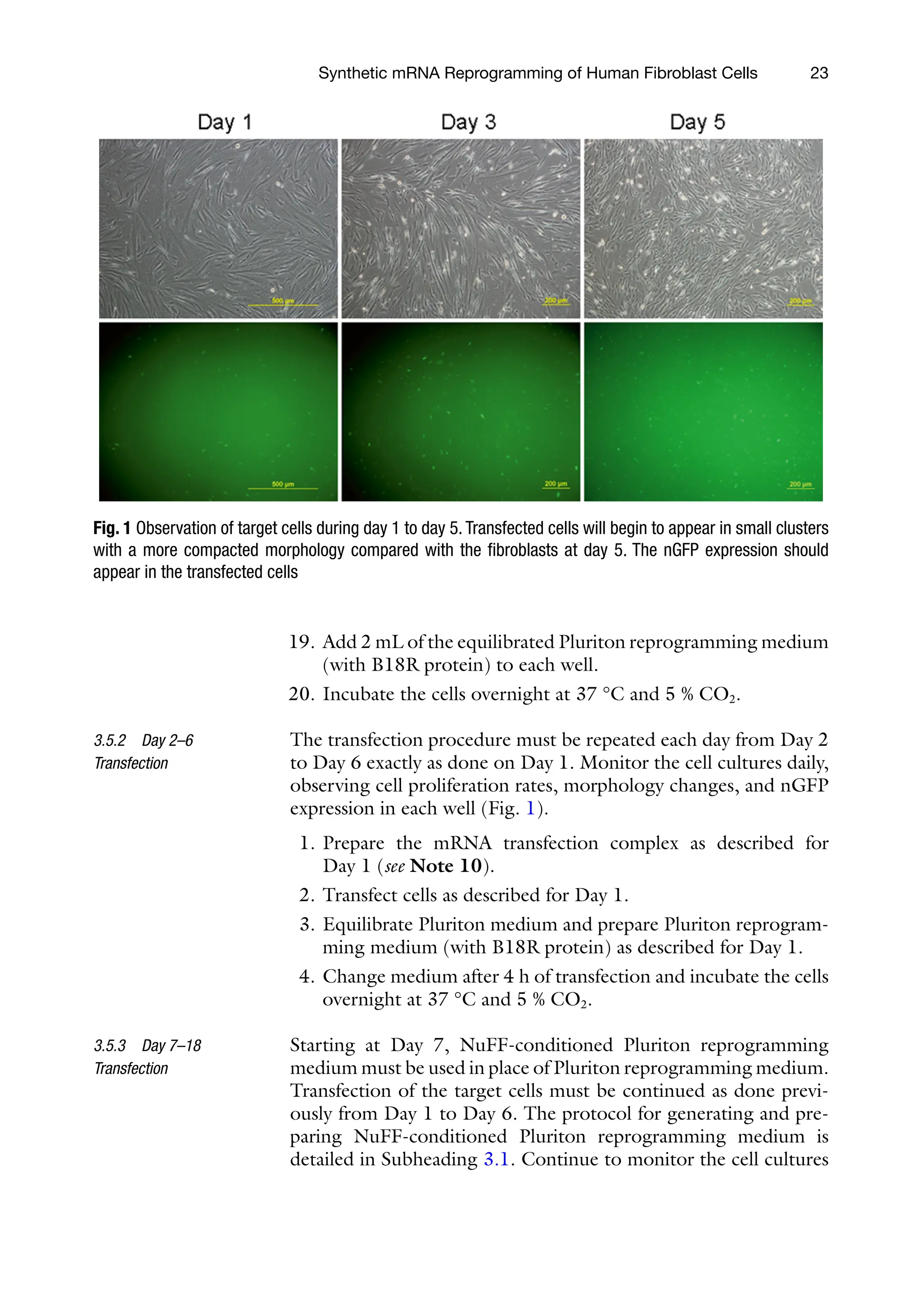
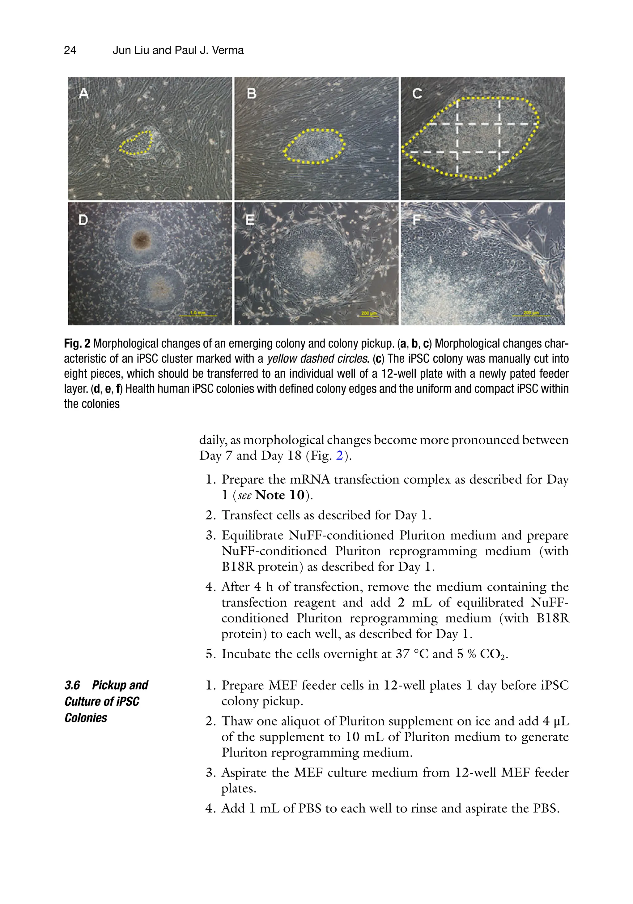

![29
Paul J. Verma and Huseyin Sumer (eds.), Cell Reprogramming: Methods and Protocols, Methods in Molecular Biology,
vol. 1330, DOI 10.1007/978-1-4939-2848-4_3, © Springer Science+Business Media New York 2015
Chapter 3
MicroRNA-Mediated Reprogramming of Somatic Cells
into Induced Pluripotent Stem Cells
Shelley E.S. Sandmaier and Bhanu Prakash V.L. Telugu
Abstract
MicroRNAs or miRNAs belong to a class of small noncoding RNAs that play a crucial role in posttran-
scriptional regulation of gene expression. Nascent miRNAs are expressed as a longer transcript, which are
then processed into a smaller 18–23-nucleotide mature miRNAs that bind to the target transcripts and
induce cleavage or inhibit translation. MiRNAs therefore represent another key regulator of gene expres-
sion in establishing and maintaining unique cellular fate. Several classes of miRNAs have been identified to
be uniquely expressed in embryonic stem cells (ESC) and regulated by the core transcription factors Oct4,
Sox2, and Klf4. One such class of miRNAs is the mir-302/367 cluster that is enriched in pluripotent cells
in vivo and in vitro. Using the mir-302/367 either by themselves or in combination with the Yamanaka
reprogramming factors (Oct4, Sox2, c-Myc, and Klf4) has resulted in the establishment of induced plu-
ripotent stem cells (iPSC) with high efficiencies. In this chapter, we outline the methodologies for estab-
lishing and utilizing the miRNA-based tools for reprogramming somatic cells into iPSC.
Key words ESC, IPSC, miRNA, Pluripotency, Reprogramming
Abbreviations
iPSC Induced pluripotent stem cells
ESC Embryonic stem cells
TALENS Transcription activator-like effector nucleases
CRISPR Clustered regularly interspaced short palindromic repeat
ZFN Zinc finger nucleases
1 Introduction
MicroRNAs (miRNAs) are short noncoding RNAs that bind target
mRNAs via complete or incomplete sequence complementarity and
regulate stability and translatability of the message [1–3]. Nascent
miRNAs are transcribed from endogenous loci via Pol II RNA
polymerase as 85–100 base pair nascent transcripts, which are then](https://image.slidesharecdn.com/2621055-250607150208-b98c9c02/75/Cell-Reprogramming-Methods-And-Protocols-Paul-J-Verma-Huseyin-Sumer-46-2048.jpg)
![30
processed by Drosha and Dicer into mature miRNAs of 18–23
nucleotides in length [3, 4]. The mature miRNAs are characterized
by a “seed sequence” at the 5′-end between nucleotides 2–8, exhib-
iting perfect complementarity with the target gene [2]. After a
miRNA recognizes and binds to the target mRNA, it inhibits trans-
lation in either of the two ways: (1) targeting the mRNA for cleav-
age if the miRNA shares perfect complementarity with the sequence
or (2) in the case of partial complementarity prevents assembly of a
ribosome initiation complex and initiation of translation [3]. Due
to the ability of miRNAs to bind to target sequences, albeit with
poor complementarity, one miRNA is often capable of binding to a
cohort of mRNAs and inhibiting translation. Accordingly, many
genes can often be regulated by a candidate miRNA [1, 2].
In embryonic stem cells (ESC), several classes of miRNAs have
been identified to be specifically enriched, indicating a possible role
in maintaining pluripotency [5]. Potentially several more exist
based on the putative ability of certain transcripts to form hairpin
miRNA precursors [5]. A much stronger evidence for the role of
miRNAs in maintaining pluripotency comes from the discovery
that several of the miRNA genes have binding sites for core pluri-
potency genes Oct4, Sox2, and Nanog in their promoters [6]. In
ESC, miRNAs specifically target genes which affect varying prop-
erties of pluripotency such as transcription factors, cell cycle genes,
and genes involved in epigenetic regulation. Regulation of pluripo-
tency by such diverse cellular mechanisms is necessary to ensure
greater stability of ESC [2].
Considering the abundance of miRNAs in ESC, and their
putative role in regulating pluripotency, the ability of miRNAs to
aid in the production and maintenance of induced pluripotent
stem cells (iPSCs) has been increasingly studied. iPSCs have tradi-
tionally been generated from somatic cells via retroviral delivery
of Oct4, Sox2, Klf4, and c-myc (OSKM) reprogramming factors,
as was first reported by Takahashi and Yamanaka [7]. However,
the induction of pluripotency by OSKM is rather inefficient
(0.001–0.01 %), yielding very few colonies per million cells
infected with retrovirus [7, 8]. Recently, iPSCs have been
produced with greater efficiency by incorporating specific miRNA
clusters shown to be involved in regulating the pluripotent state
[9, 10]. Specifically, a well-studied mir-302/367 cluster, which has
been shown to play a role in regulating cell cycle, and is regulated
by the core pluripotency factors Oct4, Sox2, Nanog, and Tcf3, has
been utilized in the reprogramming efforts [6, 11]. Mir-302/367
cluster comprises of five miRNAs, mir-302a, -b, -c, -d, and
mir-367, expressed as a polycistronic construct and located within
intron 8 of the LARP7 gene in humans, with homologs in several
species including cattle, pigs, and mice [6, 9]. Interestingly, the
seed sequences of the four miRNAs (302a/b/c/d) are
identical, and share high degree of conservation across species.
When used in combination with the traditional OSKM factors in
Shelley E.S. Sandmaier and Bhanu Prakash V.L. Telugu](https://image.slidesharecdn.com/2621055-250607150208-b98c9c02/75/Cell-Reprogramming-Methods-And-Protocols-Paul-J-Verma-Huseyin-Sumer-47-2048.jpg)
![31
reprogramming experiments, the number of iPSC colonies has
been shown to be enhanced by at least two orders of magnitude
(0.1–0.8 %) [9, 10]. In fact, cells can be reprogrammed to pluri-
potency with mir-302/367 and a histone deacetylase (HDAC)
inhibitor, valproic acid, alone, and show ESC-like morphology
sooner than cells reprogrammed with OSKM. Moreover, these
iPSCs are capable of contributing to all three germ layers as well
as giving rise to germ-line chimeras in mice [9]. Human iPSCs
have also been generated using miRNAs with or without the addi-
tion of OSKM into the genome [9, 12–14]. Therefore, our and
several other laboratories have adopted miRNAs as a standard fac-
tor in reprogramming iPSC (Manuscript in preparation). The use
of miRNA is especially important in reprogramming somatic cells
from livestock species, where the efficiencies of reprogramming
are even lower, and the conditions for optimal culture not com-
pletely understood. In this manuscript, the procedures for making
and using miRNA-based vectors for reprogramming somatic cells
from the domestic animal species, pig, are discussed. However,
the methods discussed below can easily be adopted for other
model organisms.
2 Materials
Store all reagents and media at 4 °C unless otherwise noted.
1. Complete media: 440 mL HyClone High Glucose DMEM
(ThermoScientific), 50 mL 10 % fetal calf serum (FCS), 2.5 mL
100× GlutaMAX (Gibco), 5 mL 100× nonessential amino
acids, and 5 mL 100× sodium pyruvate (see Note 1).
2. iPSC media: 382.5 mL HyClone DMEM F12
(ThermoScientific), 100 mL knockout serum replacer (KSR)
(Gibco), 2.5 mL GlutaMAX, 5 mL nonessential amino acids,
10 mL sodium bicarbonate solution 7.5 %, and 8 ng/mL
FGF2 (R&D Systems).
3. 0.25 % trypsin–EDTA for dissociation and harvesting of cells.
4. Dimethyl sulfoxide (DMSO) for cryopreservation.
5. CF-1 mice OR irradiated mouse embryonic fibroblasts (MEFs).
6. Phosphate-buffered saline (PBS).
7. T-75 flasks.
8. Cryovial freezing container filled with 2-propanol.
1. 293-FT cells (Life Technologies) viral packaging cells.
2. Geneticin (G418).
3. Gelatin.
4. Polyjet (Signagen).
2.1 Cell Culture
2.2 Lentivirus
Production
miRNA Mediated Reprogramming of Somatic Cells](https://image.slidesharecdn.com/2621055-250607150208-b98c9c02/75/Cell-Reprogramming-Methods-And-Protocols-Paul-J-Verma-Huseyin-Sumer-48-2048.jpg)
![37
Paul J. Verma and Huseyin Sumer (eds.), Cell Reprogramming: Methods and Protocols, Methods in Molecular Biology,
vol. 1330, DOI 10.1007/978-1-4939-2848-4_4, © Springer Science+Business Media New York 2015
Chapter 4
Generation of Footprint-Free Induced Pluripotent Stem
Cells from Human Fibroblasts Using Episomal Plasmid
Vectors
Dmitry A. Ovchinnikov, Jane Sun, and Ernst J. Wolvetang
Abstract
Human induced pluripotent stem cells (hiPSCs) have provided novel insights into the etiology of disease
and are set to transform regenerative medicine and drug screening over the next decade. The generation
of human iPSCs free of a genetic footprint of the reprogramming process is crucial for the realization of
these potential uses. Here we describe in detail the generation of human iPSC from control and disease-
carrying individuals’ fibroblasts using episomal plasmids.
Key words Human induced pluripotent stem cells, Reprogramming, Episomal plasmid vectors,
Fibroblasts, Transfection, Genomic integration
1 Introduction
Lentiviral or retroviral delivery of reprogramming factors has been
a powerful tool in pioneering the field of cell reprogramming.
However, the concerns associated with the disruption of the
genome at the viral integration sites, number and position and the
unpredictable nature of transgene silencing, as well as their
potential reactivation following differentiation have made
integration-dependent methods unsuitable for clinical applica-
tions. Indeed, with the wealth of currently available alternative
technologies there is really no need to modify the genomic DNA
of the target cell when generating induced pluripotent stem cells.
Researchers have a number of options ranging from piggybac or
sleeping beauty transposon-based or Cre recombinase-aided meth-
ods to excise integrated reprogramming cassettes, or to avoid
DNA-integrating methods altogether and use mRNA, helper-
dependent adenoviral, Sendai virus-derived or episomal vector-
based methods [1–6]. Here we describe a protocol for the
generation of human iPSCs from fibroblasts using episomal plasmid](https://image.slidesharecdn.com/2621055-250607150208-b98c9c02/75/Cell-Reprogramming-Methods-And-Protocols-Paul-J-Verma-Huseyin-Sumer-54-2048.jpg)