Download to read offline
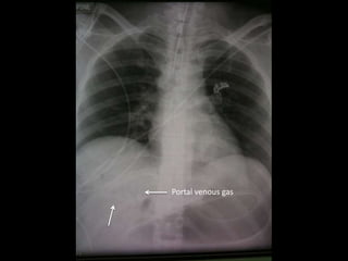


This document discusses portal venous gas seen on chest x-rays. Portal venous gas occurs when air enters the portal vein, usually due to necrosis of the stomach or gut wall, and is a very serious sign. It needs to be differentiated from pneumobilia, where air is in the bile ducts. Portal venous gas has a peripheral distribution of air in the liver, while pneumobilia is more centrally located. The document also mentions pneumatosis intestinalis, where air is found in the gut wall, which can indicate necrotizing enterocolitis or gut infarction.


