SlideShare a Scribd company logo
1 of 123
Download to read offline
2

                            CASE OF THE WEEK
                         PROFESSOR YASSER METWALLY
CLINICAL PICTURE

CLINICAL PICTURE:

11 years old female patient presented with congenital left sided hemiaplasia and Lennox Gastaut syndrome. The patient's family
gave a history of west syndrome during the first year of the patient's life. Fundus examination revealed chorioretinal lacunae.
EEG examination revealed 1.5 C/S slow spike/slow wave discharge of Lennox Gastaut syndrome. The patient's scholastic
achievement was very poor.

RADIOLOGICAL FINDINGS

RADIOLOGICAL FINDINGS:


Figure 1. Precontrast MR T1 images showing a huge right sided intraventricular /parenchymal cyst associated with agenesis of
the septum pellucidum. The corpus callosum is markedly hypoplastic and deficient. The cerebral cortex is lissencephalic. Notice
the right sided hemimegalencephaly and the subependymal nodular heterotopia. Subcortical band heterotopia can also be
appreciated.


Figure 2. Precontrast MR T1 images showing a huge right sided intraventricular /parenchymal cyst associated with agenesis of
the septum pellucidum. The corpus callosum is markedly hypoplastic and deficient. The cerebral cortex is lissencephalic. Notice
the right sided hemimegalencephaly and the subependymal nodular heterotopia.


                                              www.yassermetwally.com
3


Figure 3. MR T2 images showing a huge right sided intraventricular /parenchymal cyst associated with agenesis of the septum
pellucidum. The corpus callosum is markedly hypoplastic and deficient. The cerebral cortex is lissencephalic. Notice the right
sided hemimegalencephaly and the subependymal nodular heterotopia. Subcortical band heterotopia can also be appreciated.


Figure 4. Precontrast MR T1 images showing A huge right sided intraventricular /parenchymal cyst. The cerebral cortex is
lissencephalic. Notice the right sided hemimegalencephaly and the subependymal nodular heterotopia. Subcortical band
heterotopia can also be appreciated.


                                             www.yassermetwally.com
4


Figure 5. Precontrast MR T1 images showing a huge right sided intraventricular /parenchymal cyst. The corpus callosum is
markedly hypoplastic and deficient. The cerebral cortex is lissencephalic. Notice the subependymal nodular heterotopia and
hypoplasia of the optic nerve. The cerebellum and brain stem are also hypoplastic.


                                                                                  Figure 6. Precontrast MR T1 images
                                                                                  showing      a    huge     right    sided
                                                                                  intraventricular /parenchymal cyst. The
                                                                                  cerebral cortex is lissencephalic. Notice
                                                                                  the right sided hemimegalencephaly and
                                                                                  the subependymal nodular heterotopia.


                                                                              Figure 7. Precontrast MR T1 images showing
                                                                              marked hypoplasia of the cerebellum and
                                                                              brain stem.


                                            www.yassermetwally.com
5


                                                                            Figure 8. Chorioretinal lacunae


      Criteria that are highly suggestive of Aicardi syndrome

             Partial or complete callosal agenesis

             Cortical dysplasia

             Gross asymmetry of the hemispheres

             Periventricular or subcortical heterotopias

             Cysts of the choroid plexus or around the third ventricle is highly suggestive of AS

DIAGNOSIS:

DIAGNOSIS: AICARDI SYNDROME ASSOCIATED WITH MULTIPLE CORTICAL DYSPLASIAS THAT INCLUDE
HEMIMEGALENCEPHALY, LISSENCEPHALY, HETEROTOPIAS, AND SEPTO-OPTIC DYSPLASIA.

DISCUSSION

DISCUSSION:

The Aicardi syndrome (AS) is classically defined as a triad of abnormalities that includes agenesis of the corpus callosum,
infantile spasms, and chorioretinal lacunae (1,2). Other eye defects and costovertebral and other malformations occur frequently.
The syndrome has been observed exclusively in individuals with two X chromosomes, and only one familial case is known (3,4).

Progress in neuroimaging has revealed that the central nervous system malformation in AS is not limited to agenesis of the
corpus callosum but consists of a complex of abnormalities characterized by severe neuronal migration defects with
periventricular and subcortical heterotopias, cortical polymicrogyria, and a tendency toward the development of cystic
formations in the choroid plexuses and in other parts of the brain (3-5). The migration anomalies may even be more important
for the definition of AS than callosal agenesis, which is a non- specific finding. Similarly, the eye abnormalities are often complex,
and defects of closure of the primitive cupula are commonly present (6-8).

The incidence of AS is unknown. More than 300 cases are known to this writer, and at least 170 cases have been published. In
series of infantile spasms, AS may account for up to 4% of cases (9), but a selection bias is probable in series originating from
tertiary referral centers.

CLASSICAL FINDINGS IN AICARDI SYNDROME

The classical picture of AS has been outlined in several articles (3,6, 1 0, I 1). The seizures are typically infantile spasms. These
have been the only seizure type in 86 of 184 patients (47%) and have been associated with other seizures, especially partial motor
attacks, in 65 of 184 patients (35%) (6). The partial seizures often begin before the infantile spasms, at times as early as the first
few days of life (6,12). The age at first seizure was less than 3 months in 68% of 146 patients and less than I month in 23%. The
age at the first spasm in 137 patients was less than I month in 18% and less than 3 months in 56%. Typical tonic spasms have
been reported even during the neonatal period, and AS is one possible cause of the "early infantile epileptic encephalopathy"
described by Ohtahara et al. (13). Partial seizures and infantile spasms often occur in association, a focal or lateralized tonic or
clonic seizure being followed by a similarly lateralized cluster of spasms. The focal seizure is manifested on the EEG by a
localized discharge of repetitive spikes lasting 10-30 s. The spasms follow immediately after the spikes and are associated with a
series of slow complexes, often with a superimposed fast rhythm that is usually of higher amplitude on the side of the initial


                                                  www.yassermetwally.com
6
partial discharge. The complexes occur 6- 20 s apart, and no paroxysmal activity appears between them. The initial partial
seizure and the spasms that follow appear to represent a single attack that usually occurs on awakening or on changing from slow
wave to REM sleep (12,14). Such periodic spasms are also associated with other brain malformations (14).

Typical hypsarrhythmia is rare in AS (18% of 137 cases). The most common interictal EEG abnormality is an asymmetrical
pseudo-periodic tracing with bursts of paroxysmal irregular slow and sharp waves of 3- to 6-s duration, separated by a relatively
flat EEG for 5-20 s. Such bursts may occur independently over each hemisphere or may remain unilateral, with various
abnormalities over the other hemisphere, the so-called "split-brain" EEG (6). The tracings more commonly evolve into multifocal
paroxysmal abnormalities than into typical hypsarrhythmia. At a late stage, spike-and- wave complexes are unusual (6,9).

The choroidal lacunae are multiple rounded whitish or pinkish areas ranging in size from one-tenth to several disc diameters.
They are in the same plane as the retina, so that vessels do not bend on crossing their border. Pigment deposits may be visible at
their periphery and may increase with age (6,15). The largest lacunae tend to cluster around the disc, whereas small pinkish
lesions tend to be more peripheral. They are usually bilateral, but unilateral lacunae can be seen even when the opposite eye is
not microphthalmic. Typical lacunae are probably pathognomonic of AS. Colobomata are a very common finding and are of
significance for the diagnosis, especially when lacunae are few or atypical. Unlike the lacunae, they are frequently unilateral.
Colobomatous discs are surrounded by pigmented rings in many cases. Other eye abnormalities, including persistence of the
primary vitreous, anterior synechiae, and microphthalmia, are not uncommon. In one patient, the appearance was that of retinal
detachment suggestive of the Walker- Warburg syndrome. Interestingly, despite the very abnormal ophthalmoscopic aspect,
useful vision is often preserved as far as can be assessed. The ERG has been found normal or only mildly altered in several
patients. The visual evoked potentials are usually present but abnormal, probably as a result of the cortical abnormalities. The
fundoscopic appearance results from thinning of the choroid and sclera in the areas corresponding to the lacunae where the
pigment epithelium is depigmented or hypopigmented (7), with degeneration of the rods and cones (8). There is no evidence of
inflammatory lesions, and the appearance is suggestive of an early developmental disorder.

Agenesis of the corpus callosum was complete in 137 of 152 patients for whom the information was available and was partial in
15 (10%). Partial agenesis is usually posterior (6), but cases of agenesis of both the genu and the splenium, with preservation of
the body of the callosum, are known (16). The ventricular contour is not smooth as it is in most cases of callosal agenesis and the
ventricles, including the third ventricle, have markedly irregular contours. In most cases there is also marked asymmetry
between the two hemispheres, the larger one commonly but not consistently contralateral to a hemiplegia when present or to the
side most involved by the spasms. Various abnormalities of the posterior fossa have also been reported (6).

The neurologic and mental impairment in AS is almost always very marked. The estimated survival rate in one study (17) was
75% at 6 years and 40% at 15 years. Epilepsy persists in most patients as infantile spasms, which is unusual with other causes.
Approximately one-third of patients are unable to feed themselves and only one-quarter are able to walk. Only rare patients can
use two-word sentences (3). One patient had relatively well-developed language at age 14 years (17). According to McGregor et
al. (17), a worse prognosis was correlated with larger lacunar size. Hemiplegia is commonly present but it is seldom isolated, and
some degree of contralateral involvement is the rule. Mild microcephaly is common but is never present at birth. Indeed, some
infants have large heads that may result from hydrocephalus or from the development of large intracranial cysts. Although an
occasional patient has been operated on for shunting of hydrocephalus due to aqueductal stenosis or for drainage of a cyst, this is
seldom justified because the cysts do not usually continue to increase in postnatal life and ventricular dilatation usually remains
static even when marked.

NEW FINDINGS IN AICARDI SYNDROME

Over the past few years the diagnosis of AS has been facilitated by modern imaging techniques. These have shown that agenesis
of the corpus callosum is virtually never an isolated finding but is part of a complex of developmental abnormalities. Migration
anomalies are probably present in all cases. They include periventricular and/or subcortical nodular heterotopias, which are
responsible for the irregular contours of the lateral ventricles, and cortical dysplasia with thickening of the cortical plate and
abnormal rectilinear or blurred interface between gray and white matter involving one or both hemispheres to a variable degree.
These dysplastic areas probably correspond pathologically to polymicrogyria (3, 5,15,18,19). Periventricular heterotopias are
easily detectable, but the diagnosis of subcortical heterotopias and cortical dysplasia requires high-quality MRI. Cysts are
frequently revealed on MRI. They can involve the glomus of the choroid plexus on one or both sides and/or the region of the third
ventricle and pineal gland, where they may be single or multiple and may be quite large (Fig. 1). The cysts give a slightly more
intense signal than CSF and their walls may enhance with gadolinium contrast. Cysts around the third ventricle were probably
responsible for the distorted appearance of this ventricle on MRI or CT scan, which could not result from heterotopias. Cysts of
the cerebral hemispheres are uncommon. Cysts of the posterior fossa have been reported (3,6). Most of them correspond to
partial agenesis of the cerebellar vermis, to a megacisterna magna, or to arachnoid cysts. True intraparenchymal cysts are
uncommon. The combination of cysts of the choroid plexus in association with agenesis of the corpus callosum permits the
antenatal diagnosis of AS (20). Solid tumors, especially papillomas of the choroid plexus, have been reported in several patients
(3,6) and may be multiple (21). Peripheral embryonic tumors have also been described (22,23).


                                               www.yassermetwally.com
7


                                                     Figure 1. Huge cyst in a case of Aicardi syndrome. The cyst probably arises
                                                     from the pineal area and develops into the left parietooccipital lobe, displacing
                                                     and compressing the lateral ventricle. Note also agenesis of the corpus
                                                     callosum and abnormal appearance of the posterior cortex on both sides,
                                                     suggestive of migration disorder.


The combination of partial or complete callosal agenesis, cortical dysplasia, gross asymmetry of the hemispheres, periventricular
or subcortical heterotopias, and cysts of the choroid plexus or around the third ventricle is highly suggestive of AS (3).
Incomplete forms in which one or several components are lacking are not uncommon. Such forms are confirmed as AS by the
presence of other typical manifestations, especially choroidal lacunae. There is probably no good reason to single out one
component of the malformation complex so that cases without callosal agenesis but with heterotopias and/or cortical dysplasia
can be diagnosed when other cardinal features are present. Two such cases (both with lacunae and one with vertebral
abnormalities) are known to this author.

Criteria that are highly suggestive of Aicardi syndrome

      Partial or complete callosal agenesis

      Cortical dysplasia

      Gross asymmetry of the hemispheres

      Periventricular or subcortical heterotopias

      Cysts of the choroid plexus or around the third ventricle is highly suggestive of AS


                               Figure 2. Image shows a cross-section of an eye in a patient with Aicardi syndrome. The arrow
                               indicates chorioretinal lacunae.


PATHOLOGY

Pathologic data on AS are scanty, and detailed microscopic examination of the brain is available for only two cases (5,18). Total
or partial absence of the corpus callosum was found in all verified cases, usually with the presence of Probst bundles. Other
structures may be lacking, such as the first cranial nerve and the mammary bodies. Other commissures, e.g., the fornix or the
anterior commissure (6), may be absent, but this is inconstant.

Abnormalities of gyration were found in all studied cases (5-7,15,18). Their macroscopic aspect is variable, but microgyria is
found microscopically and is of the unlayered type (18), taking the form of a thin, undulating cellular ribbon without any laminar
organization. Fusion of the molecular layers of facing convolutions may result in the appearance of a pachygyric cortex (5).
Heterotopias include subcortical neurons scattered in the white matter. Cysts have been reported in several cases (3,18,24) and
were of ependymal origin.

Pathologic findings are consistent with an etiologic factor acting before the end of the migration period and the development of
the corpus callosum, which is complete by 14 weeks of gestational age (25).

NOSOLOGIC LIMITS OF AS: THE PROBLEM OF INCOMPLETE FORMS


                                                 www.yassermetwally.com
8
Because there is no laboratory marker specific for AS, the classical triad remains the cornerstone of diagnosis. The existence of
incomplete forms, however, seems likely, and cases without callosal agenesis have been already discussed. In addition to the two
cases above, six cases of possible AS without callosal agenesis are known (6). All six had infantile spasms and choroidal lacunae,
and four had irregular ventricular contours on CT. None of these patients underwent an MRI. Cases without infantile spasms
have been reported (6) and may not be rare. Recognition of the spasms may be difficult because they are often quite
asymmetrical and atypical and are frequently associated with partial seizures, which may be the predominant seizure type.
Isolated partial seizures are not unusual at onset or late in the course, but they may be absent altogether.

The existence of AS cases without lacunae is particularly difficult to accept, because these are considered pathognomonic for the
syndrome. There are, however, female patients with a suggestive brain malformation,, infantile spasms, and other abnormalities
who might represent atypical forms. I know of three such patients who presented with infantile spasms and agenesis of the corpus
callosum. Two of these had periventricular heterotopias on CT scan and an asymmetrical burst-suppression pattern on EEG.
One girl had bilateral colobomata of the disk and another a small pigmented retinal area on one side. None of them had
undergone MRI.

GENETIC AND CYTOGENETIC DATA

The original hypothesis that AS is an X-linked dominant disorder that is lethal at an early stage of gestation for affected
hemizygous male conceptuses appears most compatible with the observed data (2,3,6). Only females are affected, with the
exception of two phenotypical males with two X chromosomes (26). The male child with an XY karyotype reported by Curatolo
et al. (27) is too atypical to be included (6,28, 29). This hypothesis is also consistent with the sporadic occurrence of the syndrome.
Only one instance of familial recurrence in two sisters is reported (4) and this remains difficult to explain because no
chromosomal abnormality was found in these patients. Another finding difficult to reconcile with the hypothesis is the occurrence
of AS in only one of a pair of monozygotic twins, the co-twin being completely normal as a young adult (30). If this report is
confirmed, it might be explained by extreme nonrandom inactivation of the abnormal X chromosome in the normal twin or, less
probably, by a postzygotic mutation during early embryonic development.

The strong suspicion of an abnormality of an X chromosome in patients with AS has been reinforced by the finding of skewed X
inactivation in the lymphocytes of some patients (31). Patients with nonrandom inactivation were found to be more severely
affected than those with a random pattern, suggesting that selection against abnormal cells in the developing neural tissue led to
aberrant brain development. However, a normal inactivation pattern has been found in other cases (30,32).

A possible locus for AS on the short arm of the X chromosome has been suggested by several case reports of eye abnormalities
with callosal agenesis or other brain abnormalities (33-36) associated with translocations or other chromosomal abnormalities at
Xp22.3. However, none of the reported patients had the typical triad of AS, even though microphthalmia (3), chorioretinal lesions
reminiscent of the lacunae (Aughton et al., personal communication, 1991), other eye abnormalities (33,34), agenesis of the corpus
callosum (36) or other brain defects (35), and costovertebral malformations (33) were present in variable associations. Four of
these children also had focal dermal hypoplasia or Golz disease, a sex linked disorder with lethality for hemizygous males
mapping at Xp22.3 (36). Such cases suggest that the Xp22.3 region is involved in the genesis of both Golz syndrome and AS. They
could represent contiguous gene syndromes involving both loci. Intensive search for an AS gene in the Xp22.3 region is being
pursued.


SUMMARY


SUMMARY

AS is an uncommon malformation complex that affects mostly the eyes and the central nervous system. Brain malformation
constitutes the core of the syndrome. Agenesis of the corpus callosum is the most easily detectable but probably not the most
characteristic feature. Migration anomalies, including periventricular and subcortical nodular heterotopias and cortical
dysplasia, and a tendency towards the formation of ependymal cysts in the glomus of the choroid plexuses and/or near of the
third ventricle, are major components of the complex and may occur even in the presence of a complete corpus callosum. The
resulting seizures, usually asymmetrical infantile spasms, and mental retardation constitute a severe disability with a much
reduced life expectancy. The syndrome is probably due to a chromosomal accident involving one X chromosome. The Xp22.3
region is a prime candidate for location of one or several responsible genes, and demonstration of a DNA abnormality in this
region will permit a better definition of the limits of the syndrome and perhaps help our understanding of some aspects of the
development of the central nervous system.

AS is an uncommon malformation complex that affects mostly the eyes and the central nervous system. Brain malformation
constitutes the core of the syndrome. Agenesis of the corpus callosum is the most easily detectable but probably not the most
characteristic feature. Migration anomalies, including periventricular and subcortical nodular heterotopias and cortical
dysplasia, and a tendency towards the formation of ependymal cysts in the glomus of the choroid plexuses and/or near of the
third ventricle, are major components of the complex and may occur even in the presence of a complete corpus callosum. The


                                                 www.yassermetwally.com
9
resulting seizures, usually asymmetrical infantile spasms, and mental retardation constitute a severe disability with a much
reduced life expectancy. The syndrome is probably due to a chromosomal accident involving one X chromosome. The Xp22.3
region is a prime candidate for location of one or several responsible genes, and demonstration of a DNA abnormality in this
region will permit a better definition of the limits of the syndrome and perhaps help our understanding of some aspects of the
development of the central nervous system.

The diagnosis of AS is based upon the classic triad of corpus callosal agenesis, chorioretinal lacunae and infantile spasm. But
there is a range of costovertebral, ocular and cerebral abnormalities associated with this disorder.(2) The cerebral gray-matter
heterotopias and other cortical malformations act as epileptogenic foci.(2) Their seizures typically start in early childhood and
are usually intractable. Besides infantile spasm, other seizure types are also demonstrated. Dissociated burst-suppression or
burst-suppression pattern appearing asymmetrically in either cerebral hemisphere is a characteristic EEG finding in AS.(8) The
developmental delay in AS is generally profound, involving both motor and language skills. Chevrie and Aicardi in their analysis
of 184 patients of AS observed that none had acquired meaningful speech.(9) But of late a larger spectrum of the disease has been
recognized and it had been found that higher functioning AS individuals do exist.(2) Most of the AS cases die at an early age
primarily due to aspiration pneumonitis. But some do live into their adolescent years and even in their twenties.(2) Good visual
function in AS patients do occur if the fovea is uninvolved with chorioretinal lacunae.

Cerebral heterotopias, interhemispheric cysts, optic nerve coloboma, microphthalmia, thoracolumbar kyphoscoliosis are the
known associated features in AS. Severe psychomotor impairment and absence of meaningful speech had also been noted.
(3,4,5,6)


      Addendum

            A new version of this PDF file (with a new case) is uploaded in my web site every week (every Saturday and remains
             available till Friday.)

            To download the current version follow the link "http://pdf.yassermetwally.com/case.pdf".
            You can also download the current version from my web site at "http://yassermetwally.com".
            To download the software version of the publication (crow.exe) follow the link:
             http://neurology.yassermetwally.com/crow.zip
            The case is also presented as a short case in PDF format, to download the short case follow the link:
             http://pdf.yassermetwally.com/short.pdf
            At the end of each year, all the publications are compiled on a single CD-ROM, please contact the author to know more
             details.
            Screen resolution is better set at 1024*768 pixel screen area for optimum display.
            For an archive of the previously reported cases go to www.yassermetwally.net, then under pages in the right panel, scroll
             down and click on the text entry "downloadable case records in PDF format"
            Also to view a list of the previously published case records follow the following link (http://wordpress.com/tag/case-record/)
             or click on it if it appears as a link in your PDF reader


REFERENCES

References

1. Aicardi J, Lefebvre J, Lerique-Koechlin A. A new syndrome: spasms in flexion, callosal agenesis, ocular abnormalities.
Electroencephalogr Clin Neurophysiol 1965;19:609-10.

2. Aicardi J, Chevrie JJ, Rousselie F. Le syndrome agdndsie calleuse, spasmes en flexion, lacunes chorioretiniennes. Arch Franc
Pgdiatr 1969;26: 1103-20.

3. Aicardi J, Chevrie JJ. The Aicardi syndrome. In: Lassonde M, Jeeves MA, eds. Callosal agenesis: the natural split brain. New
York: Plenum Press, 1995.

4. Molina JA, Mateos F, Merino M, Epifanio JL, Gorrono M. Aicardi syndrome in two sisters. J Pediatr 1989;115:282-3.

5. Billette de Villemeur T, Robain 0, Chiron C. Unlayered polymicrogyria and agenesis of the corpus callosum: a relevant
association? Acta Neuropathol (Berl) 1992;83:265-70.

6. Chevrie JJ, Aicardi J. The Aicardi syndrome;, In: Meldrum BS, ed. Recent advances in epilepsy, Vol. 3. Edinburgh: Churchill
Livingstone, 1986: 189-210.

7. McMahon RG, Bell RA, Moore RW, Ludwin SK. Aicardi syndrome. A clinicopathological study. Arch Ophthalmol
1984;102:250-3.

8. Del Pero RA, Mets MB, Tripathy RC, Torezynski E. Anomalies of retinal architecture in Aicardi syndrome. Arch Ophthalmol
1986;104:1659-64.


                                                   www.yassermetwally.com
10
9. Aicardi J. Epilepsy in children, 2nd ed. New York: Raven Press, 1994:l-3.

10. Donnenfeld AE, Packer RJ, Zackai EH, Chee CM, Sellinger B, Emmanuel BS. Clinical, cytogenetic and pedigree findings in
18 cases of Aicardi syndrome. Am J Med Genet 1989;32:461-7.

11. Yamagata T, Momoi M, Miyamoto S, Kobayashi S, Kamoshita S. Multi-institutional survey of the Aicardi syndrome in
Japan. Brain Dev 1990;12: 760-5.

12. Bour F, Chiron C, Dulac 0, Plouin P. Caractres electrocliniques des crises dans le syndrome d'Aicardi. Rev EEG
Neurophysiol Clin 1986;16: 341-53.

13. Ohtahara S, Ohtsuka Y, Yamatogi Y, Oka E. The early infantile epileptic encephalopathy with suppression burst:
developmental aspects. Brain Dev 1987;9:371-6.

14. Gobbi G, Bruno L, Pini A, Rossi PG, Tassinari CA. Periodic spasms: an unclassified type of epi- leptic seizure in childhood.
Dev Med Child Neural 1987;29:766-75.

15. De Jong JGY, Delleman JW, Houben M, et al. Agenesis of the corpus callosum, infantile spasms, ocular anomalies (Aicardi's
syndrome): clinical and pathological findings. Neurology 1976;26: 1152-8.

16. Aicardi J, Chevrie JJ, Baraton J. Agenesis of the corpus callosum. In: Vinken PJ, Bruyn GW, eds. Handbook of clinical
neurology, Vol. 6. Brain malformations. Amsterdam: North-Holland, 1987:149-73.

17. McGregor DL, Menezes A, Buncic JR. Aicardi syndrome (AS): natural history and predictors of severity. Can J Neurol Sci
1993;20(suppl 2):S36.

18. Ferrer 1, Cusi MV, Liarte A, Campistol J. A Golgi study of the polymicrogyric cortex in Aicardi syndrome. Brain Dev
1986;8:518-25.

19. Baieari P, Marki A, Thelen M, Laub MC. MR imaging in Aicardi,s syndrome. Am J Neuroradiol 1988;9:805-6.

20. Roland EH, Flodmark 0, Hill A. Neurosonographic features of Aicardi syndrome. J Child Neurol 1989;4:307-10.

21. Hamano K, Matsubara T, Shibata S, et al. Aicardi syndrome accompanied by auditory disturbances and multiple brain
tumors. Brain Dev 199 1; 13:438- 41.

22. Tanaka T, Takahura H, Takashima S, Kodama T, Hasegawa H. A rare case of Aicardi syndrome with severe brain
malformation and hepatoblastoma. Brain Dev 1985;7:507-12.

23. Togawa T, Mimaki T, Ono J. Aicardi syndrome associated with embryonal carcinoma. Pediatr Neural 1989;5:45-7.

24. Brihaye L, Gillet P, Parmentier R, Peetrons A. Agenesie de la commissure calleuse associee A un kyste dpendymaire. Arch
Suisses Neurol Psychi- atr 1956;77:415-31.

25. Barkovich A, Norman D. Anomalies of the corpus callosum: correlation with further anomalies of the brain. Am J
Neuroradiol 1988;9:493-501.

26. Hopkins IJ, Humphrey 1, Keith CG, Susman M, Webb GC, Turner EK. The Aicardi syndrome in a 47XXY male. Aust
Paediatr J 1979;15:278-80.

27. Curatolo P, Libutti G, Dalla Piccola B. Aicardi syndrome in a male infant. J Pediatr 1980;96: 286-7.

28. Aicardi J. The Aicardi syndrome in a male infant [Letter]. J Pediatr 1980;97:1040-41.

29. Hunter AGW. Aicardi syndrome in a male infant [Reply]. J Pediatr 1980;97:1041.

30. Costa T, Greer W, Duckworth M, Rysiecki M, Musarella M, Ray P. Monozygotic twins discordant for Aicardi syndrome
[Abstract]. Am J Hum Genet 1990;47(suppl 5)202:14.

31. Neidich JA, Nussbaum RL, Packer RJ, Emanuel BS, Puck JM. Heterogeneity of clinical severity and molecular lesions in
Aicardi syndrome. J Pediatr 1990; 1 16:911-7.

32. Wieacker P, Zimmer J, Ropeers HH. X-inactivation pattern in two syndromes with probable X-linked dominant, male lethal
inheritance. Clin Genet 1985;28:238-42.

33. Ropers HH, Zuffardi 0, Biancai E, Tiepolo L. Agenesis of corpus callosum, ocular and skeletal anomalies (X-linked dominant
Aicardi's syndrome) in a girl with balanced X/3 translocation. Hum Genet 1982;61:364-8.

34. Donnenfeld AE, Graham JM, Packer RJ, Aquino R, Berg SZ, Emanuel BS. Microphthalmia and chorioretinal lesions in a girl


                                               www.yassermetwally.com
11
with an Xp22.2 pter deletion and partial 3p trisomy: clinical observations relevant to Aicardi syndrome gene localization. Am J
Med Genet 1990;37:182-6.

35. Al-Gazali LI, Muller RF, Caine A, et al. An XX male and two (X;Y) females with linear skin defects and congenital
microphthalmia: a new syndrome at Xp22.3. J Med Genet 1988;25:638-9.

36. Friedman PA, Rao KW, Jeplin SW, Aylsworth AS. Provisional deletion mapping of the focal dermal hypoplasia (FDH) gene
to Xp22.31 [Abstract]. Am J Hum Genet 1988;43:A50.

32- Metwally, MYM: Textbook of neuroimaging, A CD-ROM publication, (Metwally, MYM editor) WEB-CD agency for
electronic publication, version 9.4a October 2008


                                              www.yassermetwally.com
12

                       CASE OF THE WEEK
                   PROFESSOR YASSER METWALLY
CLINICAL PICTURE

CLINICAL PICTURE:

A 7 years old female patient presented clinically with Lennox Gastaut syndrome.

RADIOLOGICAL FINDINGS

RADIOLOGICAL FINDINGS:


Figure 1. Cortical dysplasia. Precontrast MRI T1 images showing lissencephaly, microgyria, pachygyria,
hypointense cystic white matter changes specially affecting the right head of caudate nucleus and the globus pallidus
on the right side. The lissencephalic changes are most marked in the bifrontal regions. Notice the subependymal
nodular heterotopia specially involving the frontal horns bilaterally. There is also reduction of the brain volume and
moderate degree of central atrophy.


                                         www.yassermetwally.com
13


Figure 2. Cortical dysplasia. MRI FLAIR images showing lissencephaly, microgyria, pachygyria, hyperintense
cystic white matter changes specially affecting the right head of caudate nucleus and the globus pallidus on the right
side. The lissencephalic changes are most marked in the bifrontal regions. Notice the subependymal nodular
heterotopia specially involving the frontal horns bilaterally, nodules are also seen subependymally in the left body of
the lateral ventricles (C). There is also reduction of the brain volume and moderate degree of central atrophy.


                                                                               Figure 3. Cortical dysplasia. MRI T2
                                                                               images      showing      lissencephaly,
                                                                               microgyria, pachygyria, hyperintense
                                                                               cystic white matter changes specially
                                                                               affecting the right head of caudate
                                                                               nucleus and the globus pallidus on the
                                                                               right side. The lissencephalic changes
                                                                               are most marked in the bifrontal
                                                                               regions. Notice the subependymal
                                                                               nodular      heterotopia       specially
                                                                               involving the frontal horns bilaterally.
                                                                               There is also reduction of the brain
                                                                               volume and moderate degree of
                                                                               central atrophy.


                                          www.yassermetwally.com
14


Figure 4. Cortical dysplasia. MRI FLAIR images showing lissencephaly, microgyria, pachygyria, hyperintense
cystic white matter changes specially affecting the right head of caudate nucleus and the globus pallidus on the right
side. The lissencephalic changes are most marked in the bifrontal regions. Notice the subependymal nodular
heterotopia specially involving the frontal horns bilaterally, nodules are also seen subependymally in the left body of
the lateral ventricles (C). There is also reduction of the brain volume and moderate degree of central atrophy. The
hippocampi are atrophic and hyperintense bilaterally (possible mesial temporal sclerosis).

DIAGNOSIS:

DIAGNOSIS: CORTICAL DYSPLASIA

DISCUSSION

DISCUSSION:

The brain is a seemingly nonsegmented organ that is, however, formed in a segmented fashion by the overlap of
genes that define anatomic and probably functional components of the brain. Other genes and their encoded
proteins regulate the processes of cell proliferation and migration; many of these genes have been identified based
upon discoveries of human and mouse disease-causing genes.

Human brain developmental disorders represent clinical challenges for the diagnosing clinician as well as for the
treating physician. Some disorders represent well-defined clinical and genetic entities for which there are specific
tests; others have ill-defined genetic causes, while others can have both genetic and destructive causes. In most cases
the recognition of a disorder of brain development portends certain developmental disabilities and often seizure
disorders that can be very difficult to treat. In addition, it now bears upon the treating physician to recognize the
genetic causes, and to properly advise patients and their families of the risks of recurrence or refer them to the
proper specialist who can do so. The genetics of some of these disorders are not all well defined at present, and the
recognition of some disorders is variable; what is known is presented herein.

The genetics and signaling utilized in brain development is briefly reviewed to provide the framework for the
understanding of human brain developmental disorders. The well-defined genetic disorders of brain development
are discussed, and a brief suggested algorithm for evaluation and for counseling of patients is provided.

BRAIN DEVELOPMENT

      Overview

General mechanisms tend to recur in all phases of brain development, and these include induction, cell
proliferation, cell fate determination (differentiation), cell process formation and targeting (synapse formation), and


                                          www.yassermetwally.com
15
cell movement (migration). Induction is the process by which one group of cells or tissue determines the fate of
another by the release of soluble factors or inducers. Cell fate or differentiation is dependent upon this process of
induction, and probably can best be understood as the initiation of a genetic program by the recognition of an
inducing molecule and/or expression of a transcriptional regulator. In general, it is rare that a cell in the nervous
system is born and differentiates in the same location that it finally resides. Rather, cells migrate over long distances
to reach their final locations. Similarly, cells in the nervous system must extend processes over long distances to
reach their synaptic targets.

      Neural tube formation

The human brain is formed from the neuroectoderm, a placode of cells that are induced to differentiate from the
surrounding ectoderm by the presence of the notochord at about 18 days gestation. Candidate inducing factors
include the retinoids, follistatin, and Noggin [1-4]. The neuroectoderm develops folds in the lateral aspects that
begin to approximate in the region of the future medulla and fuse at 22 days gestation. This closure is known as
neurulation, and results in the formation of a tube termed the neural tube [5]. The anterior neural tube closes by
about 24 days gestation and serves as the foundation for further brain development; the posterior neural tube closes
by about 26 days gestation and serves as the foundation for further spinal cord development. Defects in the closure
of the neural tube lead to encephaloceles or myelomeningoceles.

      Nervous system segmentation

At the rostral end of the newly closed neural tube flexures delineate the primary vesicles, which are designated as
the hindbrain (rhombencephalon), mesencephalon, and forebrain (prosencephalon). The primary vesicles can be
further subdivided into secondary vesicles that will form adult brain structures. The hindbrain can be divided into
the metencephalon and myelencephalon, which will become the pons, cerebellum, and medulla oblongata of the
adult. The mesencephalon will be the midbrain, and the prosencephalon divides into the telencephalon (two
telencephalic vesicles) and diencephalon. The telencephalic vesicles will become the cerebral hemispheres; the
diencephalon will become the thalamus and hypothalamus.

Regional specification of the developing telencephalon is an important step in brain development, and is likely
under control of a number of genes that encode transcription -regulators. In the fruit fly, Drosophila, these genes
are involved in segmentation of this animal and define structures such as hair-like spiracles. Not surprisingly, the
role of these genes in human brain development differs, yet it appears that the general role of these proteins is that
of regional specification of clones of cells destined to form specific brain structures. Homeobox and other
transcription genes encode some of these transcriptional regulators and these "turn on" genes by binding to specific
DNA sequences, and in so doing initiate genetic programs that lead to cell and tissue differentiation. EMX2, a
transcriptional regulator, has a homolog in Drosophila that defines the hair spiracles and has been implicated in
human brain malformations.

DISORDERS OF SEGMENTATION

      Schizencephaly

Schizencephaly (cleft in brain) has been regarded by many as a migration abnormality; however, it is best
understood as a disorder of segmentation because one of the genes that is abnormal in the more severe and familial
forms is EMX2 [6,7]. Thus, this developmental disorder, at least in the more severe cases, appears to be the result of
failure of regional specification of a clone of cells that are destined to be part of the cortex.

Clinically, these patients vary depending upon the size of the defect and upon whether bilateral disease is present
[8]. The clefts extend from the pia to the ventricle and are lined with a polymicrogyric gray matter (see the
discussion in Polymicrogyria) [9]. The pia and ependyma are usually in apposition, especially in severe cases. The
defect is termed open-lipped if the cleft walls are separated by cerebrospinal fluid, and closed-lipped if the walls are
in contact with one another. Bilateral schizencephaly is associated with mental retardation and spastic cerebral
palsy; affected patients often are microcephalic. Seizures almost always accompany severe lesions, especially the
open-lipped and bilateral schizencephalies. The exact frequency of seizures in patients with the less severe lesions is
uncertain. Most patients in whom schizencephaly is diagnosed undergo neuroimaging because of seizures.
Therefore, a bias in favor of a universal occurrence of seizures in this disorder is noted. Hence, patients with
schizencephaly who do not have epilepsy might exist, but the malformation remains undetected because no imaging
is performed.


                                          www.yassermetwally.com
16


                                              Figure 1. Closed-lip schizencephaly. Sagittal T1-weighted MRI
                                              shows gray matter (arrows) extending from cortex to a dimple in
                                              the surface of the left lateral ventricle. The lips of the
                                              schizencephaly are in apposition, making this a "closed-lip"
                                              schizencephaly.


                                                        Figure 2. Bilateral open-lip schizencephaly. A,B: Axial
                                                        T2-weighted images show open-lip schizencephalies in
                                                        both hemispheres. Both images show vessels in the gray
                                                        matter-lined clefts and large vessels (arrows) run at the
                                                        outer surface of the right hemispheric cleft. This does not
                                                        represent a vascular malformation.


Seizure type and onset may also vary in this disorder. Patients may experience focal or generalized seizures, and
some will present with infantile spasms. The onset varies from infancy to the early adult years. Seizures may be
easily controlled or may be recalcitrant to standard anticonvulsant therapy.


Figure 3a. MRI T1 (A,B) and CT scan (D) showing open-lip schizencephaly with pachygyria. Notice the associated
encephalocele that is sometimes associated with cortical dysplasias


                                        www.yassermetwally.com
17


                                                                       Figure 3b. Open-lip schizencephaly with
                                                                       cortical dysplasia


Improvements in neuroimaging have enhanced the recognition of schizencephalic lesions [9-13]. The lesions may
occur in isolation or may be associated with other anomalies of brain development such as septo-optic dysplasia (see
Disorders of prosencephalic cleavage) [14].

Disorders of segmentation likely represent a heterogeneous set of abnormalities of varying etiologies. One theory
holds that an early (first-trimester) destructive event disturbs subsequent formation of the cortex. Another theory is
that segmental failure occurs in the formation of a portion of the germinal matrix or in the migration of primitive
neuroblasts. Certainly, the finding of mutations of the EMX2 gene in some patients with the open-lipped form of
schizencephaly supports the latter hypothesis.

      Prosencephalon cleavage

At about 42 days of gestation, the prosencephalon undergoes a division into two telencephalic vesicles that are
destined to become the cerebral hemispheres. The anterior portion of this cleavage is induced by midline facial
structures and the presence of the notochord. Abnormalities of this process are thought to result in
holoprosencephaly, septo-optic dysplasia, and agenesis of the corpus callosum [I 5]. One of the important molecules
responsible for the induction of this cleavage is Sonic hedgehog [16]. This protein is produced by the notochord,
ventral forebrain, and the floor plate of the neural tube [17]. It interacts through at least one receptor, PTCH-A
human homolog of patched, and alters the expression of transcription factors [18,19]. Furthermore, in an
interesting link between these ventral inductive events and segmentation, Sonic hedgehog can alter the expression of
the transcriptional regulating genes when applied to proliferating cells at critical times in development [21]. This
ties the inductive proteins to the expression of transcriptional regulating genes and gives a hint as to the
mechanisms involved in inductive processes.


                                         www.yassermetwally.com
18
Figure 4a. Agenesis of the corpus callosum

Other molecules of interest in this inductive process are the retinoids, which are lipids capable of crossing
membranes and that have been shown to exist in posterior to anterior gradients across embryos [3,20]. Retinoic acid
can alter the pattern of transcriptional factors in neuroepithelial cells [3] and can downregulate Sonic hedgehog,
perhaps explaining some of the head defects seen in retinoid embryopathy [17,22].

Agenesis of the corpus callosum

Etiology          •Both genetic and sporadic
Pathogenesis      •Unknown,
                  •Agenesis of the corpus callosum may be part of an extensive malformation
                  complex or the callosum may be partially or completely absent or hypoplastic
Epidemiology      in an otherwise normal brain.

                  •The malformation is relatively rare.
                  •The brain in agenesis of the corpus callosum shows batwing shaped ventricles
                  as well as loss of the corpus callosum and there is no cingulate gyrus.

General Gross     •The remainder of the abnormalities depend on what syndrome or other
Description       malformations are associated with the defect. In most cases there is a bundle of
                  white matter processes on both cerebral hemispheres in the area where the
                  corpus callosum should be, called the bundle of Probst. In some patients there
                  is a lipoma or other tumor in the area where the corpus callosum should be.
General
Microscopic       •None
Description
                  •Patients with agenesis of the corpus callosum may be normal or may have
Clinical
                  neurological abnormalities dependent on the other accompanying
Correlation
                  malformations.


Figure 4b. Agenesis of the corpus callosum

DISORDERS OF PROSENCEPHALIC CLEAVAGE

      Holoprosencephaly

Holoprosencephaly is a heterogeneous disorder of prosencephalon cleavage that results from a failure of the


                                        www.yassermetwally.com
19
prosencephalic vesicle to cleave normally. Three forms of this disorder have been described: alobar, semilobar, and
lobar [23,24]. In the alobar form, the telencephalic vesicle completely fails to divide, producing a single horseshoe-
shaped ventricle, sometimes with a dorsal cyst, fused thalami, and a malformed cortex. In the semilobar form, the
interhemispheric fissure is present posteriorly, but the frontal and, sometimes, parietal lobes, continue across the
midline [25]; in some cases just ventral fusion is noted. In the lobar form, only minor changes may be seen: the
anterior falx and the septum pellucidum usually are absent, the frontal lobes and horns are hypoplastic, and the
genu of the corpus callosum may be abnormal.


Figure 5. This is holoprosencephaly in which there is a single large ventricle with fusion of midline structures,
including thalami. The affected fetuses and neonates typically have severe facial defects, such as cyclopia, as well.
Underlying chromosomal abnormalities, such as trisomy 13, or maternal diabetes mellitus are possible causes, but
some cases are sporadic.

Holoprosencephaly is associated with a spectrum of midline facial defects. These include cyclopia, a supraorbital
proboscis, ethmocephali, in which the nose is replaced by a proboscis located above hypoteloric eyes; cebocephaly,
in which hypotelorism and a nose with a single nostril are seen; and premaxillary agenesis, with hypotelorism, a flat
nose, and a midline cleft lip [26].


                                         www.yassermetwally.com
20


Figure 6. (left three images) A,B semilobar holoprosencephaly from the same patient, C a patient with lobar
holoprosencephaly. Arrowhead in A points to lack of ventral interhemispheric cleavage. Arrowhead in B points to
fused thalami. The septum pellucidum is absent in B. In C notice the horseshoe or mushroom shaped single
ventricle designated by * , arrowhead in C points to lack of interhemispheric fissure. (right image) MRI -
Holoprosencephaly: This 6-day-old girl presented with laryngeal malacia and a diminished level of arousal. This
proton density axial MR image shows an absence of the anterior horns of the lateral ventricles, fused thalami and
absence of the corpus callosum anteriorly.

Only children who have the lobar and semilobar forms are known to survive for more than a few months. An infant
affected with the severe form is microcephalic, hypotonic, and visually inattentive [25]. In infants with the less
severe forms of holoprosencephaly, myoclonic seizures frequently develop and, if the infant survives, autonomic
dysfunction, failure to thrive, psychomotor retardation, and atonic or spastic cerebral palsy often are present. Some
infants with the lobar form may be only mildly affected and, for example, present as a relatively mild spastic
diplegia. Pituitary defects may be associated with these malformations, and may result in neuroendocrine
dysfunction [27]. One, therefore, has to wonder how much genetic overlap exists between this condition and septo-
optic dysplasia to be described below.

Holoprosencephaly has been associated with maternal diabetes [28], retinoic acid exposure, cytomegalovirus, and
rubella [29]. Chromosome abnormalities associated with this disorder include trisomies 13 and 18; duplications of
3p, 13q, and 18q; and deletions in 2p, 7q, 13q, and 18q [30]. Of particular concern to the clinician is the existence of
an autosomal dominant form in which mutations in Sonic Hedgehog lead to variable expression of
holoprosencephaly. In the mildest form of this genetic disorder, patients may have a single central incisor, a choroid
fissure coloboma, or simply attention deficit disorder; a parent of a child with holoprosencephaly manifesting these
features should be considered to be at high risk for recurrence of holoprosencephaly in their children (up to 50%
risk) [31,32]. A number of other genes (HPE] (21 q22.3), HPE2 (2p2 1), HPE3 (7q36), HPE4 (18p), ZIC2, SIX3
(2p2l), and PATCHED) have been associated with holoprosencephaly, and although potentially inherited in an
autosomal recessive fashion, most occurrence seems to be random [33,34].

      Septo-optic dysplasia

Septo-optic dysplasia (de Morsier syndrome) is a disorder characterized by the absence of the septum pellucidum,
optic nerve hypoplasia, and hypothalamic dysfunction. It may be associated with agenesis of the corpus callosum.
This disorder should be considered in any patient who exhibits at least two of the above abnormalities and perhaps
even solely hypothalamic dysfunction [35]. Septo-optic dysplasia also appears to involve prosencephalic cleavage
and development of anterior telencephalic structures [36]. About 50% of patients with septo-optic dysplasia have
schizencephaly [14].


                                          www.yassermetwally.com
21


                                                  Figure 7. Septo-optic dysplasia associated with schizencephaly.
                                                  Arrows in A,B point to absent septum pellucidum, arrow in C,
                                                  and black arrowhead in D point to open lip schizencephaly, white
                                                  arrow head point to polymicrogyria in D


Patients may present with visual disturbance, seizures, mental retardation, hemiparesis (especially if associated with
schizencephaly), quadriparesis, or hypothalamic dysfunction. Endocrine abnormalities may include growth
hormone, thyroid hormone, or antidiuretic hormone function or levels. The consideration of septo-optic dysplasia
necessitates an evaluation of the hypothalamic-pituitary axis because as many as 60% of the children with this
disorder might exhibit evidence of a disturbance of endocrine function [37]. This evaluation can include thyroid
function studies and electrolytes; these patients are at high risk for growth retardation.

The recent identification of patients with this condition that harbor mutations in the transcriptional regulator gene
HESX1, suggest that the mechanism of this disorder is likely genetic and a patterning or segmental abnormality
[38]. Even though the genetic abnormality has been identified for a minority of patients, there exists the possibility
that this may not represent an entirely genetic disorder because associations have been made with young maternal
age, diabetes, the use of anticonvulsants, phencyclidine, cocaine, and alcohol [39],

DISORDERS OF CELL PROLIFERATION

      Normal cell proliferation

Following telencephalic cleavage, a layer of proliferative pseudostratified neuroepithelium lines the ventricles of the
telencephalic vesicles. These cells will give rise to the neurons and glia of the mature brain. The generation of the
proper complement of cells is a highly ordered process that results in the generation of billions of neurons and glia.
Neuroepithelial processes extend from the ventricular surface to the pial surface, and the nuclei of the primitive
neuroepithelial cells move from the cortical surface in a premitotic phase to a mitotic phase near the ventricle. Cells
divide at the most ventricular aspects of the developing telencephalon, and after division move back toward the pial
surface. The pial processes of neuroepithelial cells near the ventricle often will detach from the cortical surface
before a new cycle begins.

Neuroepithelial cells divide in so-called proliferative units such that each unit will undergo a specific number of
divisions resulting in the appropriate number of cells for the future cortex. Abnormalities in the number of
proliferative units or in the total number of divisions can lead to disorders of the brain manifested by abnormal
brain size and, therefore, an unusually small or large head circumference. Two such disorders resulting in small
head size -radial microbrain and microcephaly vera- are believed to result from abnormalities of this phase of
neurodevelopment [40]. Disorders in which too many cells are generated in the proliferative phase result in
megalencephaly (large brain) or, if proliferative events go awry on only one side of the developing cortex,
hemimegalencephaly.

The genes and molecules involved in regulating the proliferative cycles in human brain formation are likely similar


                                          www.yassermetwally.com
22
to those involved in other species. This cell cycle in the brain can be divided into a number of distinct phases:
mitosis (M), first gap (GI), deoxyribonucleic acid synthesis (S), and second gap (G2) [41]. These phases appear to be
regulated by key molecules to check the advancement of proliferation. Some cells enter a resting state (GO) that
they maintain throughout life. Others temporarily enter this phase to await a specific signal to proliferate later.
Probably the GI-S transition regulation determines the number of cell cycles and, therefore, the complement of cells
that will make brain [40]. Cyclins are proteins that appear to be involved in cell cycle control. These proteins are
activating subunits of cyclin-dependent kinases. Cyclins DI, D2, D3, C, and E seem to control the key transition of a
cell to the GI S interface; this transition is regarded as important because it commits a cell to division [42-44].
Cyclin E seems to be the gatekeeper for this transition, and is essential for movement from the GI to the S phase
[42,45].

The number of cells that finally make up the mature nervous system is less than that generated during proliferation.
Cells appear not only to be programmed to proliferate during development but to contain programs that lead to cell
death [46,47]. The term apoptosis (from the Greek, meaning "a falling off ") has been applied to this programmed
loss of cells [48].

      Non-neoplastic proliferative disorders

             Microcephaly

Although primary microcephaly may be a normal variant, in the classic symptomatic form, clinical and radiologic
examinations reveal a receding forehead, flat occiput, early closure of fontanelles, and hair anomalies such as
multiple hair whirls and an anterior cowlick. Neuroimaging may show small frontal and occipital lobes, open
opercula, and a small cerebellum [24]. The cortex may appear thickened and the white matter reduced. Histologic
examination may show a reduction of cell layers in some areas and an increase in others [49].

Neurologic findings also vary. Only mild psychomotor retardation may be noted, sometimes associated with
pyramidal signs, or more severe retardation, seizures, and an atonic cerebral palsy might be evidenced. Primary
microcephaly is seen in many genetic syndromes and, in its isolated form, may be autosomal recessive, autosomal
dominant, or X-linked [50-52]. Microcephaly vera is the term most often alyplied to this genetic form of
microcephaly. Affected children present with a head circumference that is usually more than 4 standard deviations
below the mean, hypotonia, and psychomotor retardation. They later show mental retardation, dyspraxias, motor
incoordination and, sometimes, seizures. On histologic examination, neurons in layers 11 and III are depleted [53].

Destructive lesions of the forming brain, such as those caused by teratogens and by infectious agents, also may
result in microcephaly. Teratogens of note are alcohol, cocaine, and hyperphenylalaninemia (maternal
phenylketonuria) [54]. Intense radiation exposure (such as that from a nuclear explosion) in the first trimester, can
cause microcephaly [55]. Microcephaly and intracranial calcifications are likely due to well-recognized in utero
infections caused by cytomegalovirus, toxoplasmosis, or the human immunodeficiency virus.

             Megalencephaly and hemimegalencephaly

The terms megalencephaly and hemimegalencephaly refer to disorders in which the brain volume is greater than
normal (not owing to the abnormal storage of material); usually, the enlarged brain is accompanied by
macrocephaly, or a large head. Although considered by some to be a migration disorder, the increase in brain size
in these disorders appears to be attributable to errors in neuroepithelial proliferation, as the microscopic
appearance of the brain is that of an increase in number of cells (both neurons and glia) and in cell size [56-59].

Typically, patients are noted to have large heads at birth, and may manifest an accelerated head growth in the first
few months of life [60,61]. Children with megalencephaly or hemimegalencephaly may come to medical attention
when presenting with seizures, a developmental disorder (mental retardation), hemihypertrophy, or a hemiparesis
(opposite the affected hemisphere). Seizures vary both in onset and in type, and usually are the most problematic
symptom. sometimes necessitating hemispherectomy or callosotomy [58].

Approximately 50% of patients with linear sebaceous nevus syndrome have hemimegalencephaly [62,63]. Many
patients with hypomelanosis of Ito also have hemimegalencephaly [64]. The neuropathologic and clinical pictures of
these associations appear to be identical to the isolated hemimegalencephalies.

      Neoplastic proliferative disorders


                                            www.yassermetwally.com
23
Lesions of proliferation after an abnormal induction event during brain development may be malformative,
hamartomatous, neoplastic, or a combination. Malformative disorders consisting of neoplasias on a background of
disordered cortex or in association with focal cortical dysplasia include dysembryoplastic neuroepithelial tumor and
ganglioglioma. [103,104]

             Dysembryoplastic neuroepithelial tumours

Dysembryoplastic neuroepithelial tumors are supratentorial, predominantly temporal lobe tumors that are typically
multinodular with a heterogeneous cell composition, including oligodendrocytes, neurons, astrocytes, and other
cells. These lesions are typically fairly well-demarcated, wedge-shaped lesions extending from the cortex to the
ventricle. Calcification, enhancement, and peritumoral edema are lacking on neuroimaging studies. These low-
attenuation lesions may suggest an infarct on computed tomography (CT), although there is no volume loss over
time, and scalloping of the inner table or calvarial bulging suggests slow growth. Lesions are low in signal on Tl-
weighted images and high in signal on T2- weighted images and often have a multinodular or pseudocystic
appearance. There is a spectrum of pathology in dysembryoplastic neuroepithelial tumors. On one end of the
spectrum are multinodular lesions with intervening malformed cortex, in which there is some hesitation to use the
designation tumor. On the other end are lesions, which are clearly neoplastic and have clinically demonstrated some
growth potential. Because the term dysembryoplastic neuroepithelial tumor has only recently been introduced, such
malformative and neoplastic lesions were previously labeled as hamartomas, gangliogliomas, or mixed gliomas.
[100,101,102]

             Gangliogliomas

Gangliogliomas are typically demonstrated within the temporal lobe. In one large series of 51 gangliogliomas, 84%
were found in the temporal lobe, 10% were found in the frontal lobe, 2% were found in the occipital lobe, and 4%
were found in the posterior fossa. These lesions are typically hypodense (60% to 70%) on CT, with focal
calcifications seen in 35% to 40%, contrast enhancement in 45% to 50%, and cysts in nearly 60%. The reported
incidence of calcifications demonstrated on imaging in pediatric gangliogliomas is higher, seen in 61% of one series
of 42 children. Features on MR imaging are less specific, with solid components isointense on TI-weighted images,
bright on proton density images, and slightly less bright on T2-weighted images. Although imaging features are not
specific, an enhancing, cystic temporal lobe lesion with focal calcification should suggest the diagnosis of
ganglioglioma. The pathologic features that suggest the diagnosis of ganglioglioma include a neoplastic glial and
neoronal component and calcification. Because calcifications are often poorly demonstrated on MR imaging and
because they increase specificity of imaging findings, documentation of calcium should be sought on CT after MR
imaging demonstration of a temporal lobe tumor. [97,98,99]

DISORDERS OF NEURONAL DIFFERENTIATION

      Normal differentiation

At the time of neuronal differentiation the neural tube consists of four consecutive layers: (1) the ventricular zone,
the innermost layer, which gives rise to neurons and all of the glia of the central nervous system; (2) the
subventricular zone, which is the adjacent, more superficial layer and is the staging area from which postmitotic
neurons begin to differentiate and to migrate; (3) the intermediate zone, which is the contiguous, more superficial
zone, and which is destined to become the cortical plate and the future cerebral cortex; and (4) the marginal zone,
which is the outermost zone and is composed of the cytoplasmic extensions of ventricular neuroblasts, corticopetal
fibers, and the terminal processes of radial glia (which, at this time, are completely spanning the neural tube).

Differentiation of neuroepithelial cells begins in the subventricular layer at approximately gestational day 26. The
older, larger pyramidal cells are the first cells to be born and probably differentiate early to act as targets in the
migration of the nervous system.

      Tuberous sclerosis

Disorders such as tuberous sclerosis, in which both tumor development and areas of cortical dysplasia are seen,
might be a differentiation disorder. The brain manifestations of this disorder include hamartomas of the
subependymal layer, areas of cortical migration abnormalities (tubers, cortical dysgenesis), and the development of
giant-cell astrocytomas in upwards of 5% of affected patients. Two genes for tuberous sclerosis have been
identified: TSCI (encodes for Hamartin) has been localized to 9q34 [65], and TSC2 (encodes for Tuberin) has been
localized to 16pl3.3 [65].


                                         www.yassermetwally.com
24


Figure 8. Postmortem specimens showing cortical tubers in (A) and subependymal tubers in (B)


                                                   Figure 9. Postmortem specimen showing cortical tubers, the
                                                   affected gyri are abnormally broad and flat.


DISORDERS OF MIGRATION

      Normal migration

At the most rostral end of the neural tube in the 40- to 41 -day-old fetus, the first mature neurons, Cajal-Retzius
cells, begin the complex trip to the cortical surface. Cajal-Retzius cells, subplate neurons, and corticopetal nerve
fibers form a preplate [66]. The,neurons generated in the proliferative phase of neurodevelopment represent billions
of cells poised to begin the trip to the cortical surface and to form the cortical plate. These neurons accomplish this
task by attaching to and migrating along radial glial in a process known as radial migration or by somal
translocation in a neuronal process [67]. The radial glia extend from the ventricle to the cortical surface. In the
process of migration, the deepest layer of the cortical plate migrates and deposits before the other layers. Therefore,
the first neurons to arrive at the future cortex are layer VI neurons. More superficial layers of cortex then are
formed-the neurons of layer V migrate and pass the neurons of layer VI; the same process occurs for layers IV, 111,
and 11. The cortex therefore is formed in an inside-out fashion [67-69].

A possible mode of movement in neuronal migration on glia would be the attachment of the neuroblast to a matrix
secreted by either the glia or the neurons. The attachment of the neuron would be through integrin receptors,
cytoskeletal-linking membrane-bound recognition sites for adhesion molecules. That attachment serves as a
stronghold for the leading process and soma of the migrating neuron. Neuron movement on radial glia involves an
extension of a leading process, neural outgrowth having an orderly arrangement of microtubules. Shortening of the
leading process owing to depolymerization or shifts of microtubules may result in movement of the soma relative to
the attachment points. This theory of movement of neurons also must include a phase of detachment from the
matrix at certain sites, so that the neuron can navigate successfully along as much as 6 cm of developing cortex (the
maximum estimated distance of radial migration of a neuron in the human). Finally, the movement of cells must
stop at the appropriate location, the boundary between layer I and the forming cortical plate. Therefore, some stop
signal must be given for the migrating neuron to detach from the radial glia and begin to differentiate into a cortical
neuron. Perhaps that signal is REELIN, a protein that is disrupted in the mouse mutant Reeler and is expressed


                                          www.yassermetwally.com
25
solely in the Caial Retizius cells at this phase of development [66,70-73].

      Migration disorders

Advanced neuroimaging techniques, particularly magnetic resonance imaging, have allowed the recognition of
major migration disorders and of the frequency of more subtle disorders of migration. Some of these disorders are
associated with typical clinical features that might alert the clinician to the presence of such malformations even
before imaging is obtained. In other disorders, the clinical features are so varied that a strong correlation between
imaging and the clinical presentation points to a specific genetic syndrome.

      Lissencephaly

Lissencephaly (smooth brain) refers to the external appearance of the cerebral cortex in those disorders in which a
neuronal migration aberration leads to a relatively smooth cortical surface. One should not consider only agyria in
making this diagnosis, rather, the full spectrum includes agyria and pachygyria. Gyri and sulci do not form in this
disorder because the lack of cortical-cortical attractive forces owing to improper axon pathways. At least two types
have been identified: classic lissencephaly, and cobblestone lissencephaly. The distinction is based upon the external
appearance and upon the underlying histology, and can be made with neuroimaging.


Figure 10. Pachygyric (A), and agyric (B) lissencephalic brain

             Classic lissencephaly

Classic lissencephaly may occur in isolation, owing to LIS1 or Doublecortin aberrations or in combination with
somatic features and LIS1 deletions in the Miller-Dieker syndrome. The hallmarks on imaging are a lack of
opercularization (covering of the sylvian fissure), large ventricles or colpocephaly (dilated posterior horns), and
agyria or pachygyria. The corpus callosum is almost always present, and the posterior fossa is usually normal,
although a form of lissencephaly does exist that includes cerebellar hypoplasia.

The LIS1 protein forms complexes with other cellular proteins that are crucial for cell division, migration, and intracellular
transport. Complete loss of LIS1 is fatal. Deletion of one copy of the gene is causes lissencephaly. The LIS1 gene is found
in 17q13.3 location.

Doublecortin (DCX), a gene on Xq22.3-q23 that codes for a microtubule associated protein, is responsible for migration of
neurons. Mutation of this gene results in band heterotopia


                                            www.yassermetwally.com
26


Figure 11. Classical lissencephaly with patchy agyria/agyria, thick Cortex and band heterotopia

            LIS1 genetic syndromes

The Miller-Dieker phenotype consists of distinct facial features that include bitemporal hallowing, upturned nares,
and a peculiar burying of the upper lip by the lower lip at the corners of the mouth. The lissencephaly is usually
more severe than isolated lissencephaly, and the prognosis is worse. Most affected patients die in the first few years
of life.

By both molecular and cytogenetic techniques, deletions in the terminal portion of one arm of chromosome 17 can
be found in approximately 90% of Miller-Dicker lissencephaly cases [74]. The deletions of the terminal part of
chromosome 17 in these cases have included microdeletions [74], ring 17 chromosome [75], pericentric inversions
[76], and a partial monosomy of 17pl3.3 [77]. The most appropriate genetic test is a fluorescent in situ hybridization
(FISH) for LIS1; this test involves marking chromosome 17 at the centromere and LIS1 with fluorescent probes.

The greatest risk to future offspring exists when a parent harbors a balanced translocation involving this region of
chromosome 17. In families that are affected in this manner, screening by amniocentesis can be performed in
subsequent pregnancies. Therefore, it is recommended that both parents have screening for chromosome 17
rearrangements by FISH for LIS1. Should a translocation be present in a parent, then the LIS1 fluorescence will be
on another chromosome.


                                         www.yassermetwally.com
27


Figure 12. A, Type I lissencephaly, agyria type. Axial T2-weighted image shows a brain with a "figure-8"
configuration secondary to the immature Sylvian fissures. A band of neurons (arrows) that was arrested during
migration lies between the thin cortex and the lateral ventricles. B, Type 1 lissencephaly, pachygyria type. Axial T2-
weighted image is similar to that shown in (A), with the exception that a few broad gyri with shallow sulci are
present. C, Type 1 lissencephaly, pachygyria type. Axial T2-weighted image shows broad, flat gyri with shallow
sulci throughout the cerebrum.

            Isolated lissencephaly

Classic lissencephaly without somatic or facial features represents a distinct genetic syndrome from Miller-Dieker,
but it involves the same gene LIS1. Approximately 40% of patients with isolated lissencephaly have FISH detectable
deletions of LIS1, and about 20% of additional patients harbor mutations of this gene [78-80]. The remaining
patients may have mutations involving promoter regions of LIS1 or abnormalities of other genes such as
Doublecortin or the involvement of other genes that have not been recognized.

The genetic risk of recurrence is highest when rearrangements of chromosome 17 exist in one parent. This is rare in
isolated lissencephaly, but could occur. Therefore, it would be prudent to perform FISH for LIS1 in the parents of
children with isolated lissencephaly who have FISH proven deletions for the LIS1 region.

The prognosis for this disorder is better than that for Miller-Dieker syndrome, but it is not consistent with long-
term survival. These patients typically present in the first few months of life with hypotonia, lack of visual fixation,
and often seizures. Patients with lissencephaly will uniformly have seizures and profound mental retardation. Often,
seizures are very difficult to control and require multiple anticonvulsant drugs.

            X-linked lissencephaly

The imaging of X-linked lissencephaly looks nearly identical to the images of lissencephaly involving LIS1. Patients
have classic lissencephaly, and the neurologic presentation described above. However, the skeletal and other
anomalies seen in the Miller-Dieker are not noted in this form of lissencephaly. When viewing the images from
patients with lissencephaly owing to abnormalities of LIS1 and of Doublecortin it is apparent that differences in an
anterior to posterior gradient of severity exists [81-83]. Doublecortin mutations result in anterior greater than
posterior severity, whereas LIS1 mutations result in posterior greater than anterior severity [84].

In addition, X-linked lissencephaly occurs mostly in boys; girls who are heterozygous for Doublecortin mutations
have band heterotopia [80,81,85]. Women with band heterotopia have been known to give birth to boys with
lissencephaly. In female patients, the less severe phenotype probably is attributed to random lyonization of the X
chromosome, such that in a variable number of cells, normal gene expression is seen and, in the remaining cells, the
Doublecortin mutation-containing X chromosome is expressed. It is presumed that those cells expressing the
abnormal X chromosome will be arrested in the migration to the surface of the brain and reside in a subcortical
band.


                                          www.yassermetwally.com
28


Figure 13. Gross specimen showing lissencephaly with pachygyria and agyria


Figure 14. Lissencephaly with abnormally smooth agyric, pachygyric cortex


Figure 15. Lissencephaly with abnormally smooth agyric cortex


                                       www.yassermetwally.com
29
When viewing images from patients with this disorder, a thick band of tissue that is isointense with cerebral gray
matter is seen within what should be the white matter of the hemispheres. The overlying gyral appearance may vary
from normal cortex to a pachygyria. A brain biopsy performed in a patient demonstrated well-preserved lamination
in cortical layers I-IV [86]. Layers V-VI were not clearly separated and merged with underlying white matter.
Beneath the white matter was a coalescent cluster of large, well-differentiated neurons.


                                                        Figure 16. A, Band heterotopia with pachygyria. B, band
                                                        heterotopia with mild periventricular nodular heterotopia


            Lissencephaly with cerebellar hypoplasia

The association of lissencephaly with cerebellar hypoplasia represents a distinct malformation from both a genetic
and clinical standpoint to those described above. The cerebellar hypoplasia is usually extreme, and the brainstem
may be small. Patients may or may not have an associated microcephaly. This disorder is often inherited in an
autosomal recessive fashion and may be due to mutations in REELIN in some families [87].


                                                           Figure 17. Lissencephalic brain with hydrocephalus
                                                           and cerebellar hypoplasia


                                         www.yassermetwally.com
30


                             Figure 18. MRI showing a mild form of lissencephaly (pachygyria), the brain have a
                             few broad, flat gyri with thick cortex and separated by shallow sulci (pachygyria).
                             The cerebellum and the brain stem are hypoplastic, the brain volume is also reduced
                             especially the temporal lobes.


            Cobblestone lissencephaly

Cobblestone lissencephalies are disorders in which a smooth configuration of cortex is noted, but the distinction
from classic lissencephaly is made based upon the clinical association of eye abnormalities, muscle disease, and
progressive hydrocephalus. The term "cobblestone" refers to the appearance of the cortical surface upon pathologic
examination. In these disorders, cells pass their stopping point and erupt over the surface of the cortex into the
subarachnoid spaces. This results in a cobblestone street appearance to the surface, and therefore, the name.


Figure 19. A, Type II lissencephaly, Cobblestone lissencephaly (Walker-Warburg syndrome). The cortex is
lissencephalic and thickened, with an irregular gray matter-white matter junction that probably represents the
bundles of disorganized cortex surrounded by fibroglial tissue. The patient has been shunted for hydrocephalus.
The brain is hypomyelinated. B, Presumed microcephalia vera. The brain is completely smooth (lissencephaly) with
a very thin cortex. No layer of arrested neurons is present in the white matter. C, Presumed radial microbrain.
Axial T2- weighted image shows an immature gyral pattern and hypomyelination. Cortical thickness is normal.
This patient was profoundly microcephalic (head circumference 19 cm).


                                         www.yassermetwally.com
31
The Walker-Warburg, muscle-eye-brain, and HARD +/- syndromes are likely all varying degrees of the same entity.
Abnormalities that may or may not be seen in these disorders include muscular dystrophy, ocular anterior chamber
abnormalities, retinal dysplasias (evidenced by abnormal electroretinogram and visual evoked responses),
hydrocephalus (usually of an obstructive type), and encephaloceles. The Walker-Warburg syndrome might be
diagnosed even if the ocular examination and muscle biopsies are normal if on MRI, an abnormal white matter
signal and a thickened falx suggest the diagnosis. Neuroimaging of the muscle-eye-brain disorders often reveals
focal white matter abnormalities.


                                                              Figure 20. Cobblestone lissencephaly


Fukuyama muscular dystrophy is distinguished from the Walker Warburg-like syndromes by the severity of the
muscular dystrophy [88-91]. This disorder is seen more often in Japan than in the Western hemisphere, probably
because it is the result of a founder mutation. Patients typically present with evidence of a neuronal migration
defect, hypotonia, and depressed reflexes. Recent identification of Fukutin as the causative gene in this disorder
should provide insight into the pathogenesis of the cobblestone lissencephalies [92]. This disorder is inherited as an
autosomal recessive disorder.

The cobblestone lissencephalies often have an associated cerebellar and brainstem hypoplasia, and therefore may be
difficult to distinguish from lissencephaly with cerebellar hypoplasia described above. The presence of eye
abnormalities, elevated CPK, or other evidence for the presence of muscle disease and progressive hydrocephalus
distinguish this disorder. These disorders may be inherited in an autosomal recessive manner.

Table 1. The lissencephalies syndromes

 TYPE                DESCRIPTION                                     GENE
 LIS1      genetic The Miller-Dieker phenotype consists of           By both molecular and cytogenetic techniques,
 syndromes         distinct facial features that include             deletions in the terminal portion of one arm of
                   bitemporal hallowing, upturned nares, and a       chromosome 17 can be found in approximately
                   peculiar burying of the upper lip by the lower    90% of Miller-Dicker lissencephaly cases [74].
                   lip at the corners of the mouth. The              The deletions of the terminal part of
                   lissencephaly is usually more severe than         chromosome 17 in these cases have included
                   isolated lissencephaly, and the prognosis is      microdeletions [74], ring 17 chromosome [75],
                   worse. Most affected patients die in the first    pericentric inversions [76], and a partial


                                         www.yassermetwally.com
32
                      few years of life.                                  monosomy of 17pl3.3 [77]. The most
                                                                          appropriate genetic test is a fluorescent in situ
                                                                          hybridization (FISH) for LIS1; this test
                                                                          involves marking chromosome 17 at the
                                                                          centromere and LIS1 with fluorescent probes.
 Isolated             Classic lissencephaly without somatic or It involves the same gene LIS1. Approximately
 lissencephaly        facial features represents a distinct genetic 40% of patients with isolated lissencephaly
                      syndrome from Miller-Dieker,                  have FISH detectable deletions of LIS1, and
                                                                    about 20% of additional patients harbor
                                                                    mutations of this gene [78-80]. The remaining
                                                                    patients may have mutations involving
                                                                    promoter regions of LIS1 or abnormalities of
                                                                    other genes such as Doublecortin or the
                                                                    involvement of other genes that have not been
                                                                    recognized.
  X-linked                   The imaging of X-linked lissencephaly              LIS1     gene   mutation    results   in
 lissencephaly-               looks nearly identical to the images of             lissencephaly
 subcortical band             lissencephaly involving LIS1. Patients
 heterotopia                  have classic lissencephaly, and the                Doublecortin gene mutation results in
 (XLIS-SBH)                   neurologic     presentation     described           band heterotopia (Doublecortin (DCX), a
                              above. However, the skeletal and other              gene on Xq22.3-q23 that codes for a
                              anomalies seen in the Miller-Dieker are             microtubule associated protein, is
                              not noted in this form of lissencephaly.            responsible for migration neurons.
                                                                                  Mutation of this gene results in band
                             In addition, X-linked lissencephaly                 heterotopia)
                              occurs mostly in boys; girls who are
                              heterozygous     for    Doublecortin
                              mutations have band heterotopia
                              [80,81,85]
 Lissencephaly      The association of lissencephaly with                 This disorder is often inherited in an
 with    cerebellar cerebellar hypoplasia represents a distinct           autosomal recessive fashion and may be due to
 hypoplasia         malformation from both a genetic and clinical         mutations in REELIN in some families [87].
                    standpoint to those described above. The
                    cerebellar hypoplasia is usually extreme, and          
                    the brainstem may be small. Patients may or
                    may not have an associated microcephaly.

 Cobblestone          are disorders in which a smooth ?
                      configuration of cortex is noted, but the
 lissencephalies
                      distinction from classic lissencephaly is made
                      based upon the clinical association of eye
                      abnormalities,     muscle      disease,    and
                      progressive    hydrocephalus.      The    term
                      "cobblestone" refers to the appearance of the
                      cortical surface upon pathologic examination.
                      In these disorders, cells pass their stopping
                      point and erupt over the surface of the cortex
                      into the subarachnoid spaces. This results in a
                      cobblestone street appearance to the surface,
                      and therefore, the name.

        Polymicrogyria

Polymicrogyria (many small gyri) is a disorder often considered to be a neuronal migration disorder, but alternate
theories exist regarding its pathogenesis, The microscopic appearance of the lesion is that of too many small
abnormal gyri. The gyri may be shallow and separated by shallow sulci, which may be associated with an apparent
increased cortical thickness on neuroimaging. The multiple small convolutions may not have intervening sulci, or
the sulci may be bridged by fusion of overlying molecular layer, which may give a smooth appearance to the brain's
surface. The interface of white matter with gray matter is not distinct and often this observation serves as the


                                            www.yassermetwally.com
33
confirmation of the presence of polymicrogyria.


Figure 21. Polymicrogyria


Figure 22. A, Polymicrogyria. B, pachygyria with polymicrogyria, notice the subependymal nodular heterotopia

Polymicrogyria has also been associated with genetic and chromosomal disorders. It is found in disorders of
peroxisomal metabolism such as Zellweger syndrome and neonatal adrenal leukodystrophy . Familial bilateral
frontal polymicrogyria and bilateral perisylvian polymicrogyria have been reported. Therefore, if no identifiable
cause of the polymicrogyric malformation is found, the recurrence risk may be that of an autosomal recessive
disorder. A bilateral parasagittal parieto-occipital polymicrogyria has also been described.

The clinical picture varies depending on the location, extent, and cause of the abnormality. Microcephaly with
severe developmental delay and hypertonia may result when polymicrogyria is diffuse. When polymicrogyria is
unilateral, focal deficits might be seen. Epilepsy often is present, characterized by partial complex seizures or
partial seizures that secondarily generalize. The age at presentation and severity of seizures depends on the extent of
the associated pathology.


                                          www.yassermetwally.com
34


Figure 23. A, pachygyria with polymicrogyria. B, pachygyria with polymicrogyria, notice the subependymal
nodular heterotopia

Bilateral perisylvian dysplasia is a disorder of perisylvian polymicrogyria resulting in an uncovered sylvian fissure
on neuroimaging and on sagittal imaging an extension of the sylvian fissure to the top of the convexity. Patients with
bilateral perisylvian dysplasia have a pseudobulbar palsy, and often dysphagia can impair proper nutrition. The
majority of patients have epilepsy with early onset; infantile spasms are common.


Figure 24. Polymicrogyria with pachygyria


                                         www.yassermetwally.com
35


Figure 25. Polymicrogyri. In B the patient also had meningomyelocele, obstructive hydrocephalic and Arnold -
Chiari type II malformation

The cause of this syndrome remains unknown, although hints of a genetic mechanism exist. Detailed chromosomal
analyses have revealed deletions of chromosomes 1, 2, 6, 21, and 22 [93], and an X-linked form has been also
described [94,95]. Monozygotic twins and siblings with this disorder have been described, suggesting a possible
autosomal recessive mechanism. Some speculate that this is a disorder of regional specification, given the bilateral,
symmetric nature of the lesions.

      Heterotopias

Heterotopias are collections of normal-appearing neurons in an abnormal location, presumably secondary to a
disturbance in migration. The exact mechanism of the migration aberration has not been established, although
various hypotheses have been proposed. These include damage to the radial glial fibers, premature transformation
of radial glial cells into astrocytes, or a deficiency of specific molecules on the surface of neuroblasts or of the radial
glial cells (or the receptors for those molecules) that results in disruption of the normal migration process [96].
Heterotopias often occur as isolated defects that may result in only epilepsy. However, when they are multiple,
heterotopias might also be associated with a developmental disorder and cerebral palsy (usually spastic). In
addition, if other migration defects such as gyral abnormalities are present, the clinical syndrome may be more
profound. Usually, no cause is apparent. Occasionally, heterotopias may be found in a variety of syndromes,
including neonatal adrenal leukodystrophy, glutaric aciduria type 2, GMI gangliosidosis, neurocutaneous
syndromes, multiple congenital anomaly syndromes, chromosomal abnormalities, and fetal toxic exposures.


                                           www.yassermetwally.com
36


Figure 26. Subependymal nodular heterotopia


                                                         Figure 27. CT scan showing Subependymal nodular
                                                         heterotopia with cerebellar hypoplasia


Heterotopias may be classified by their location: subpial, within the cerebral white matter, and in the subependymal
region. When subependymal, one must consider the X-linked dominant disorder associated with Filamin mutations
(Xq28). Leptomeningeal heterotopias often contain astrocytes mixed with ectopic neurons and may resemble a
gliotic scar. They may be related to discontinuities in the external limiting membrane and often are associated with
cobblestone lissencephaly. These subarachnoid heterotopias are responsible for the pebbled appearance of the
surface of the brain. White matter heterotopias may be focal, subcortical, or diffuse. They may cause distortion of
the ventricles and may be associated with diminished white matter in the surrounding area.


                                         www.yassermetwally.com
Case of the week: Cases presented with cortical dysplasia

More Related Content

What's hot

Congenital third ( oculomotor )nerve palsy
Congenital third ( oculomotor )nerve palsyCongenital third ( oculomotor )nerve palsy
Congenital third ( oculomotor )nerve palsyVinitkumar MJ
 
Congenital malformation of cns
Congenital malformation of cnsCongenital malformation of cns
Congenital malformation of cnsPS Deb
 
Oculomotor Nerve Palsy
Oculomotor Nerve Palsy Oculomotor Nerve Palsy
Oculomotor Nerve Palsy Ade Wijaya
 
Cerebellum clinical
Cerebellum clinicalCerebellum clinical
Cerebellum clinicalmunnam37
 
Pachygyria, Polymicrogyria
Pachygyria, Polymicrogyria Pachygyria, Polymicrogyria
Pachygyria, Polymicrogyria Walif Chbeir
 
Presentation1.pptx, congenital malformation of the brain.
Presentation1.pptx, congenital malformation of the brain.Presentation1.pptx, congenital malformation of the brain.
Presentation1.pptx, congenital malformation of the brain.Abdellah Nazeer
 
Cns path congenital, edema
Cns path congenital, edemaCns path congenital, edema
Cns path congenital, edemaraj kumar
 
Multiple cranial nerve palsy final
Multiple cranial nerve palsy finalMultiple cranial nerve palsy final
Multiple cranial nerve palsy finalSruban Suparmaniam
 
Neural tube defects a case series - copy
Neural tube defects   a case series - copyNeural tube defects   a case series - copy
Neural tube defects a case series - copyULTRAFEST
 
Cerebellar diseases. igbiti
Cerebellar diseases. igbitiCerebellar diseases. igbiti
Cerebellar diseases. igbitiJustice Igbiti
 
Case presentation: Third nerve palsy
Case presentation: Third nerve palsyCase presentation: Third nerve palsy
Case presentation: Third nerve palsyAnis Suzanna Mohamad
 
Radiological pathology of cerebral amyloid angiography
Radiological pathology of cerebral amyloid angiographyRadiological pathology of cerebral amyloid angiography
Radiological pathology of cerebral amyloid angiographyProfessor Yasser Metwally
 
Cerebellum & ataxia
Cerebellum & ataxiaCerebellum & ataxia
Cerebellum & ataxiaAmr Hassan
 
Cns cong anomalies
Cns cong anomaliesCns cong anomalies
Cns cong anomaliesMed Study
 
Disorders of neural tube closure and neuronal migration
Disorders of neural tube closure and neuronal migrationDisorders of neural tube closure and neuronal migration
Disorders of neural tube closure and neuronal migrationdrnaveent
 

What's hot (20)

Congenital third ( oculomotor )nerve palsy
Congenital third ( oculomotor )nerve palsyCongenital third ( oculomotor )nerve palsy
Congenital third ( oculomotor )nerve palsy
 
Cerebellum
CerebellumCerebellum
Cerebellum
 
Case record...Cortical dysplasia
Case record...Cortical dysplasiaCase record...Cortical dysplasia
Case record...Cortical dysplasia
 
Congenital malformation of cns
Congenital malformation of cnsCongenital malformation of cns
Congenital malformation of cns
 
Oculomotor Nerve Palsy
Oculomotor Nerve Palsy Oculomotor Nerve Palsy
Oculomotor Nerve Palsy
 
Oculomotor nerve palsy
Oculomotor nerve palsyOculomotor nerve palsy
Oculomotor nerve palsy
 
Cerebellum clinical
Cerebellum clinicalCerebellum clinical
Cerebellum clinical
 
Pachygyria, Polymicrogyria
Pachygyria, Polymicrogyria Pachygyria, Polymicrogyria
Pachygyria, Polymicrogyria
 
Presentation1.pptx, congenital malformation of the brain.
Presentation1.pptx, congenital malformation of the brain.Presentation1.pptx, congenital malformation of the brain.
Presentation1.pptx, congenital malformation of the brain.
 
Cns path congenital, edema
Cns path congenital, edemaCns path congenital, edema
Cns path congenital, edema
 
Multiple cranial nerve palsy final
Multiple cranial nerve palsy finalMultiple cranial nerve palsy final
Multiple cranial nerve palsy final
 
Neural tube defects a case series - copy
Neural tube defects   a case series - copyNeural tube defects   a case series - copy
Neural tube defects a case series - copy
 
Cerebellar diseases. igbiti
Cerebellar diseases. igbitiCerebellar diseases. igbiti
Cerebellar diseases. igbiti
 
Case presentation: Third nerve palsy
Case presentation: Third nerve palsyCase presentation: Third nerve palsy
Case presentation: Third nerve palsy
 
Radiological pathology of cerebral amyloid angiography
Radiological pathology of cerebral amyloid angiographyRadiological pathology of cerebral amyloid angiography
Radiological pathology of cerebral amyloid angiography
 
Cortical dysplasia
Cortical dysplasiaCortical dysplasia
Cortical dysplasia
 
Cerebellum & ataxia
Cerebellum & ataxiaCerebellum & ataxia
Cerebellum & ataxia
 
Congenital brain anomalies
Congenital brain anomaliesCongenital brain anomalies
Congenital brain anomalies
 
Cns cong anomalies
Cns cong anomaliesCns cong anomalies
Cns cong anomalies
 
Disorders of neural tube closure and neuronal migration
Disorders of neural tube closure and neuronal migrationDisorders of neural tube closure and neuronal migration
Disorders of neural tube closure and neuronal migration
 

Similar to Case of the week: Cases presented with cortical dysplasia

False localising signs : a major examination finding
False localising signs : a major examination findingFalse localising signs : a major examination finding
False localising signs : a major examination findingChetan Ganteppanavar
 
falselocalisingsigns.pdf
falselocalisingsigns.pdffalselocalisingsigns.pdf
falselocalisingsigns.pdfSyedFurqan30
 
Imaging of congenital cns lesions
Imaging of congenital cns lesionsImaging of congenital cns lesions
Imaging of congenital cns lesionsGobardhan Thapa
 
False Localising Signs In Neurology.pptx
False Localising Signs In Neurology.pptxFalse Localising Signs In Neurology.pptx
False Localising Signs In Neurology.pptxNeurodiagnosticSolut
 
Imaging of congenital CNS lesions
Imaging of congenital CNS lesionsImaging of congenital CNS lesions
Imaging of congenital CNS lesionsMilan Silwal
 
Congenital CNS lesions
Congenital CNS lesionsCongenital CNS lesions
Congenital CNS lesionsMilan Silwal
 
Imaging in holoprosencephaly and related disorders
Imaging in holoprosencephaly and related disordersImaging in holoprosencephaly and related disorders
Imaging in holoprosencephaly and related disordersKhalaf Saba
 
Unusual cause of Epilepsy in Children
Unusual cause of Epilepsy in ChildrenUnusual cause of Epilepsy in Children
Unusual cause of Epilepsy in ChildrenPramod Krishnan
 
Occulomotor nerves
Occulomotor nervesOcculomotor nerves
Occulomotor nervescooravi
 
Radiological pathology of brain developmental disorders
Radiological pathology of brain developmental disordersRadiological pathology of brain developmental disorders
Radiological pathology of brain developmental disordersProfessor Yasser Metwally
 
Brain tumor imaginig 2 3rd may 02
Brain tumor imaginig   2  3rd may 02Brain tumor imaginig   2  3rd may 02
Brain tumor imaginig 2 3rd may 02PS Deb
 
Kuliah neoro radiologi revisi
Kuliah neoro radiologi revisiKuliah neoro radiologi revisi
Kuliah neoro radiologi revisilee25tom9
 

Similar to Case of the week: Cases presented with cortical dysplasia (20)

Caser record... Rasmussen's encephalitis
Caser record... Rasmussen's encephalitisCaser record... Rasmussen's encephalitis
Caser record... Rasmussen's encephalitis
 
Case record...Hemimegalencephaly
Case record...HemimegalencephalyCase record...Hemimegalencephaly
Case record...Hemimegalencephaly
 
False localising signs : a major examination finding
False localising signs : a major examination findingFalse localising signs : a major examination finding
False localising signs : a major examination finding
 
falselocalisingsigns.pdf
falselocalisingsigns.pdffalselocalisingsigns.pdf
falselocalisingsigns.pdf
 
Imaging of congenital cns lesions
Imaging of congenital cns lesionsImaging of congenital cns lesions
Imaging of congenital cns lesions
 
False Localising Signs In Neurology.pptx
False Localising Signs In Neurology.pptxFalse Localising Signs In Neurology.pptx
False Localising Signs In Neurology.pptx
 
Case record...Cerebral palsy
Case record...Cerebral palsyCase record...Cerebral palsy
Case record...Cerebral palsy
 
Imaging of congenital CNS lesions
Imaging of congenital CNS lesionsImaging of congenital CNS lesions
Imaging of congenital CNS lesions
 
Case record...Cortical dysplasia
Case record...Cortical dysplasiaCase record...Cortical dysplasia
Case record...Cortical dysplasia
 
Congenital CNS lesions
Congenital CNS lesionsCongenital CNS lesions
Congenital CNS lesions
 
Imaging in holoprosencephaly and related disorders
Imaging in holoprosencephaly and related disordersImaging in holoprosencephaly and related disorders
Imaging in holoprosencephaly and related disorders
 
Unusual cause of Epilepsy in Children
Unusual cause of Epilepsy in ChildrenUnusual cause of Epilepsy in Children
Unusual cause of Epilepsy in Children
 
Case record...Cortical/cerebellar dysplasia
Case record...Cortical/cerebellar dysplasiaCase record...Cortical/cerebellar dysplasia
Case record...Cortical/cerebellar dysplasia
 
Occulomotor nerves
Occulomotor nervesOcculomotor nerves
Occulomotor nerves
 
Radiological pathology of brain developmental disorders
Radiological pathology of brain developmental disordersRadiological pathology of brain developmental disorders
Radiological pathology of brain developmental disorders
 
Case record...Wilson disease
Case record...Wilson diseaseCase record...Wilson disease
Case record...Wilson disease
 
Case record... Septo-optic dysplasia
Case record... Septo-optic dysplasiaCase record... Septo-optic dysplasia
Case record... Septo-optic dysplasia
 
Talamo ppt
Talamo pptTalamo ppt
Talamo ppt
 
Brain tumor imaginig 2 3rd may 02
Brain tumor imaginig   2  3rd may 02Brain tumor imaginig   2  3rd may 02
Brain tumor imaginig 2 3rd may 02
 
Kuliah neoro radiologi revisi
Kuliah neoro radiologi revisiKuliah neoro radiologi revisi
Kuliah neoro radiologi revisi
 

More from Professor Yasser Metwally

The Snake, the Scorpion, the turtle in Egypt
The Snake, the Scorpion, the turtle in EgyptThe Snake, the Scorpion, the turtle in Egypt
The Snake, the Scorpion, the turtle in EgyptProfessor Yasser Metwally
 
Radiological pathology of epileptic disorders
Radiological pathology of epileptic disordersRadiological pathology of epileptic disorders
Radiological pathology of epileptic disordersProfessor Yasser Metwally
 
Radiological pathology of cerebrovascular disorders
Radiological pathology of cerebrovascular disordersRadiological pathology of cerebrovascular disorders
Radiological pathology of cerebrovascular disordersProfessor Yasser Metwally
 
Radiological pathology of spontaneous cerebral hemorrhage
Radiological pathology of spontaneous cerebral hemorrhageRadiological pathology of spontaneous cerebral hemorrhage
Radiological pathology of spontaneous cerebral hemorrhageProfessor Yasser Metwally
 
Radiological pathology of cerebral microbleeds
Radiological pathology of cerebral microbleedsRadiological pathology of cerebral microbleeds
Radiological pathology of cerebral microbleedsProfessor Yasser Metwally
 
Issues in radiological pathology: Radiological pathology of watershed infarct...
Issues in radiological pathology: Radiological pathology of watershed infarct...Issues in radiological pathology: Radiological pathology of watershed infarct...
Issues in radiological pathology: Radiological pathology of watershed infarct...Professor Yasser Metwally
 
Radiological pathology of cortical laminar necrosis
Radiological pathology of cortical laminar necrosisRadiological pathology of cortical laminar necrosis
Radiological pathology of cortical laminar necrosisProfessor Yasser Metwally
 

More from Professor Yasser Metwally (20)

The Egyptian Zoo in Cairo 2015
The Egyptian Zoo in Cairo 2015The Egyptian Zoo in Cairo 2015
The Egyptian Zoo in Cairo 2015
 
End of the great nile river in Ras Elbar
End of the great nile river in Ras ElbarEnd of the great nile river in Ras Elbar
End of the great nile river in Ras Elbar
 
The Lion and The tiger in Egypt
The Lion and The tiger in EgyptThe Lion and The tiger in Egypt
The Lion and The tiger in Egypt
 
The monkeys in Egypt
The monkeys in EgyptThe monkeys in Egypt
The monkeys in Egypt
 
The Snake, the Scorpion, the turtle in Egypt
The Snake, the Scorpion, the turtle in EgyptThe Snake, the Scorpion, the turtle in Egypt
The Snake, the Scorpion, the turtle in Egypt
 
The Egyptian Parrot
The Egyptian ParrotThe Egyptian Parrot
The Egyptian Parrot
 
The Egyptian Deer
The Egyptian DeerThe Egyptian Deer
The Egyptian Deer
 
The Egyptian Pelican
The Egyptian PelicanThe Egyptian Pelican
The Egyptian Pelican
 
The Flamingo bird in Egypt
The Flamingo bird in EgyptThe Flamingo bird in Egypt
The Flamingo bird in Egypt
 
Egyptian Cats
Egyptian CatsEgyptian Cats
Egyptian Cats
 
Radiological pathology of epileptic disorders
Radiological pathology of epileptic disordersRadiological pathology of epileptic disorders
Radiological pathology of epileptic disorders
 
Radiological pathology of cerebrovascular disorders
Radiological pathology of cerebrovascular disordersRadiological pathology of cerebrovascular disorders
Radiological pathology of cerebrovascular disorders
 
Radiological pathology of spontaneous cerebral hemorrhage
Radiological pathology of spontaneous cerebral hemorrhageRadiological pathology of spontaneous cerebral hemorrhage
Radiological pathology of spontaneous cerebral hemorrhage
 
Radiological pathology of cerebral microbleeds
Radiological pathology of cerebral microbleedsRadiological pathology of cerebral microbleeds
Radiological pathology of cerebral microbleeds
 
The Egyptian Zoo in Cairo
The Egyptian Zoo in CairoThe Egyptian Zoo in Cairo
The Egyptian Zoo in Cairo
 
Progressive multifocal leukoencephalopathy
Progressive multifocal leukoencephalopathyProgressive multifocal leukoencephalopathy
Progressive multifocal leukoencephalopathy
 
Progressive multifocal leukoencephalopathy
Progressive multifocal leukoencephalopathyProgressive multifocal leukoencephalopathy
Progressive multifocal leukoencephalopathy
 
Issues in radiological pathology: Radiological pathology of watershed infarct...
Issues in radiological pathology: Radiological pathology of watershed infarct...Issues in radiological pathology: Radiological pathology of watershed infarct...
Issues in radiological pathology: Radiological pathology of watershed infarct...
 
Radiological pathology of cortical laminar necrosis
Radiological pathology of cortical laminar necrosisRadiological pathology of cortical laminar necrosis
Radiological pathology of cortical laminar necrosis
 
Neurological examination PDF manual
Neurological examination  PDF manualNeurological examination  PDF manual
Neurological examination PDF manual
 

Recently uploaded

Call Girls Service Navi Mumbai Samaira 8617697112 Independent Escort Service ...
Call Girls Service Navi Mumbai Samaira 8617697112 Independent Escort Service ...Call Girls Service Navi Mumbai Samaira 8617697112 Independent Escort Service ...
Call Girls Service Navi Mumbai Samaira 8617697112 Independent Escort Service ...Call girls in Ahmedabad High profile
 
Russian Call Girls in Pune Tanvi 9907093804 Short 1500 Night 6000 Best call g...
Russian Call Girls in Pune Tanvi 9907093804 Short 1500 Night 6000 Best call g...Russian Call Girls in Pune Tanvi 9907093804 Short 1500 Night 6000 Best call g...
Russian Call Girls in Pune Tanvi 9907093804 Short 1500 Night 6000 Best call g...Miss joya
 
Russian Call Girls in Pune Riya 9907093804 Short 1500 Night 6000 Best call gi...
Russian Call Girls in Pune Riya 9907093804 Short 1500 Night 6000 Best call gi...Russian Call Girls in Pune Riya 9907093804 Short 1500 Night 6000 Best call gi...
Russian Call Girls in Pune Riya 9907093804 Short 1500 Night 6000 Best call gi...Miss joya
 
CALL ON ➥9907093804 🔝 Call Girls Baramati ( Pune) Girls Service
CALL ON ➥9907093804 🔝 Call Girls Baramati ( Pune)  Girls ServiceCALL ON ➥9907093804 🔝 Call Girls Baramati ( Pune)  Girls Service
CALL ON ➥9907093804 🔝 Call Girls Baramati ( Pune) Girls ServiceMiss joya
 
Bangalore Call Girls Hebbal Kempapura Number 7001035870 Meetin With Bangalor...
Bangalore Call Girls Hebbal Kempapura Number 7001035870  Meetin With Bangalor...Bangalore Call Girls Hebbal Kempapura Number 7001035870  Meetin With Bangalor...
Bangalore Call Girls Hebbal Kempapura Number 7001035870 Meetin With Bangalor...narwatsonia7
 
VIP Mumbai Call Girls Hiranandani Gardens Just Call 9920874524 with A/C Room ...
VIP Mumbai Call Girls Hiranandani Gardens Just Call 9920874524 with A/C Room ...VIP Mumbai Call Girls Hiranandani Gardens Just Call 9920874524 with A/C Room ...
VIP Mumbai Call Girls Hiranandani Gardens Just Call 9920874524 with A/C Room ...Garima Khatri
 
(Rocky) Jaipur Call Girl - 9521753030 Escorts Service 50% Off with Cash ON De...
(Rocky) Jaipur Call Girl - 9521753030 Escorts Service 50% Off with Cash ON De...(Rocky) Jaipur Call Girl - 9521753030 Escorts Service 50% Off with Cash ON De...
(Rocky) Jaipur Call Girl - 9521753030 Escorts Service 50% Off with Cash ON De...indiancallgirl4rent
 
Call Girl Number in Vashi Mumbai📲 9833363713 💞 Full Night Enjoy
Call Girl Number in Vashi Mumbai📲 9833363713 💞 Full Night EnjoyCall Girl Number in Vashi Mumbai📲 9833363713 💞 Full Night Enjoy
Call Girl Number in Vashi Mumbai📲 9833363713 💞 Full Night Enjoybabeytanya
 
Artifacts in Nuclear Medicine with Identifying and resolving artifacts.
Artifacts in Nuclear Medicine with Identifying and resolving artifacts.Artifacts in Nuclear Medicine with Identifying and resolving artifacts.
Artifacts in Nuclear Medicine with Identifying and resolving artifacts.MiadAlsulami
 
Bangalore Call Girls Majestic 📞 9907093804 High Profile Service 100% Safe
Bangalore Call Girls Majestic 📞 9907093804 High Profile Service 100% SafeBangalore Call Girls Majestic 📞 9907093804 High Profile Service 100% Safe
Bangalore Call Girls Majestic 📞 9907093804 High Profile Service 100% Safenarwatsonia7
 
Call Girls Service In Shyam Nagar Whatsapp 8445551418 Independent Escort Service
Call Girls Service In Shyam Nagar Whatsapp 8445551418 Independent Escort ServiceCall Girls Service In Shyam Nagar Whatsapp 8445551418 Independent Escort Service
Call Girls Service In Shyam Nagar Whatsapp 8445551418 Independent Escort Serviceparulsinha
 
VIP Call Girls Pune Vrinda 9907093804 Short 1500 Night 6000 Best call girls S...
VIP Call Girls Pune Vrinda 9907093804 Short 1500 Night 6000 Best call girls S...VIP Call Girls Pune Vrinda 9907093804 Short 1500 Night 6000 Best call girls S...
VIP Call Girls Pune Vrinda 9907093804 Short 1500 Night 6000 Best call girls S...Miss joya
 
Call Girls Service Jaipur Grishma WhatsApp ❤8445551418 VIP Call Girls Jaipur
Call Girls Service Jaipur Grishma WhatsApp ❤8445551418 VIP Call Girls JaipurCall Girls Service Jaipur Grishma WhatsApp ❤8445551418 VIP Call Girls Jaipur
Call Girls Service Jaipur Grishma WhatsApp ❤8445551418 VIP Call Girls Jaipurparulsinha
 
Vip Call Girls Anna Salai Chennai 👉 8250192130 ❣️💯 Top Class Girls Available
Vip Call Girls Anna Salai Chennai 👉 8250192130 ❣️💯 Top Class Girls AvailableVip Call Girls Anna Salai Chennai 👉 8250192130 ❣️💯 Top Class Girls Available
Vip Call Girls Anna Salai Chennai 👉 8250192130 ❣️💯 Top Class Girls AvailableNehru place Escorts
 
Call Girls Horamavu WhatsApp Number 7001035870 Meeting With Bangalore Escorts
Call Girls Horamavu WhatsApp Number 7001035870 Meeting With Bangalore EscortsCall Girls Horamavu WhatsApp Number 7001035870 Meeting With Bangalore Escorts
Call Girls Horamavu WhatsApp Number 7001035870 Meeting With Bangalore Escortsvidya singh
 
Bangalore Call Girls Marathahalli 📞 9907093804 High Profile Service 100% Safe
Bangalore Call Girls Marathahalli 📞 9907093804 High Profile Service 100% SafeBangalore Call Girls Marathahalli 📞 9907093804 High Profile Service 100% Safe
Bangalore Call Girls Marathahalli 📞 9907093804 High Profile Service 100% Safenarwatsonia7
 
Lucknow Call girls - 8800925952 - 24x7 service with hotel room
Lucknow Call girls - 8800925952 - 24x7 service with hotel roomLucknow Call girls - 8800925952 - 24x7 service with hotel room
Lucknow Call girls - 8800925952 - 24x7 service with hotel roomdiscovermytutordmt
 
(👑VVIP ISHAAN ) Russian Call Girls Service Navi Mumbai🖕9920874524🖕Independent...
(👑VVIP ISHAAN ) Russian Call Girls Service Navi Mumbai🖕9920874524🖕Independent...(👑VVIP ISHAAN ) Russian Call Girls Service Navi Mumbai🖕9920874524🖕Independent...
(👑VVIP ISHAAN ) Russian Call Girls Service Navi Mumbai🖕9920874524🖕Independent...Taniya Sharma
 
VIP Service Call Girls Sindhi Colony 📳 7877925207 For 18+ VIP Call Girl At Th...
VIP Service Call Girls Sindhi Colony 📳 7877925207 For 18+ VIP Call Girl At Th...VIP Service Call Girls Sindhi Colony 📳 7877925207 For 18+ VIP Call Girl At Th...
VIP Service Call Girls Sindhi Colony 📳 7877925207 For 18+ VIP Call Girl At Th...jageshsingh5554
 
Book Paid Powai Call Girls Mumbai 𖠋 9930245274 𖠋Low Budget Full Independent H...
Book Paid Powai Call Girls Mumbai 𖠋 9930245274 𖠋Low Budget Full Independent H...Book Paid Powai Call Girls Mumbai 𖠋 9930245274 𖠋Low Budget Full Independent H...
Book Paid Powai Call Girls Mumbai 𖠋 9930245274 𖠋Low Budget Full Independent H...Call Girls in Nagpur High Profile
 

Recently uploaded (20)

Call Girls Service Navi Mumbai Samaira 8617697112 Independent Escort Service ...
Call Girls Service Navi Mumbai Samaira 8617697112 Independent Escort Service ...Call Girls Service Navi Mumbai Samaira 8617697112 Independent Escort Service ...
Call Girls Service Navi Mumbai Samaira 8617697112 Independent Escort Service ...
 
Russian Call Girls in Pune Tanvi 9907093804 Short 1500 Night 6000 Best call g...
Russian Call Girls in Pune Tanvi 9907093804 Short 1500 Night 6000 Best call g...Russian Call Girls in Pune Tanvi 9907093804 Short 1500 Night 6000 Best call g...
Russian Call Girls in Pune Tanvi 9907093804 Short 1500 Night 6000 Best call g...
 
Russian Call Girls in Pune Riya 9907093804 Short 1500 Night 6000 Best call gi...
Russian Call Girls in Pune Riya 9907093804 Short 1500 Night 6000 Best call gi...Russian Call Girls in Pune Riya 9907093804 Short 1500 Night 6000 Best call gi...
Russian Call Girls in Pune Riya 9907093804 Short 1500 Night 6000 Best call gi...
 
CALL ON ➥9907093804 🔝 Call Girls Baramati ( Pune) Girls Service
CALL ON ➥9907093804 🔝 Call Girls Baramati ( Pune)  Girls ServiceCALL ON ➥9907093804 🔝 Call Girls Baramati ( Pune)  Girls Service
CALL ON ➥9907093804 🔝 Call Girls Baramati ( Pune) Girls Service
 
Bangalore Call Girls Hebbal Kempapura Number 7001035870 Meetin With Bangalor...
Bangalore Call Girls Hebbal Kempapura Number 7001035870  Meetin With Bangalor...Bangalore Call Girls Hebbal Kempapura Number 7001035870  Meetin With Bangalor...
Bangalore Call Girls Hebbal Kempapura Number 7001035870 Meetin With Bangalor...
 
VIP Mumbai Call Girls Hiranandani Gardens Just Call 9920874524 with A/C Room ...
VIP Mumbai Call Girls Hiranandani Gardens Just Call 9920874524 with A/C Room ...VIP Mumbai Call Girls Hiranandani Gardens Just Call 9920874524 with A/C Room ...
VIP Mumbai Call Girls Hiranandani Gardens Just Call 9920874524 with A/C Room ...
 
(Rocky) Jaipur Call Girl - 9521753030 Escorts Service 50% Off with Cash ON De...
(Rocky) Jaipur Call Girl - 9521753030 Escorts Service 50% Off with Cash ON De...(Rocky) Jaipur Call Girl - 9521753030 Escorts Service 50% Off with Cash ON De...
(Rocky) Jaipur Call Girl - 9521753030 Escorts Service 50% Off with Cash ON De...
 
Call Girl Number in Vashi Mumbai📲 9833363713 💞 Full Night Enjoy
Call Girl Number in Vashi Mumbai📲 9833363713 💞 Full Night EnjoyCall Girl Number in Vashi Mumbai📲 9833363713 💞 Full Night Enjoy
Call Girl Number in Vashi Mumbai📲 9833363713 💞 Full Night Enjoy
 
Artifacts in Nuclear Medicine with Identifying and resolving artifacts.
Artifacts in Nuclear Medicine with Identifying and resolving artifacts.Artifacts in Nuclear Medicine with Identifying and resolving artifacts.
Artifacts in Nuclear Medicine with Identifying and resolving artifacts.
 
Bangalore Call Girls Majestic 📞 9907093804 High Profile Service 100% Safe
Bangalore Call Girls Majestic 📞 9907093804 High Profile Service 100% SafeBangalore Call Girls Majestic 📞 9907093804 High Profile Service 100% Safe
Bangalore Call Girls Majestic 📞 9907093804 High Profile Service 100% Safe
 
Call Girls Service In Shyam Nagar Whatsapp 8445551418 Independent Escort Service
Call Girls Service In Shyam Nagar Whatsapp 8445551418 Independent Escort ServiceCall Girls Service In Shyam Nagar Whatsapp 8445551418 Independent Escort Service
Call Girls Service In Shyam Nagar Whatsapp 8445551418 Independent Escort Service
 
VIP Call Girls Pune Vrinda 9907093804 Short 1500 Night 6000 Best call girls S...
VIP Call Girls Pune Vrinda 9907093804 Short 1500 Night 6000 Best call girls S...VIP Call Girls Pune Vrinda 9907093804 Short 1500 Night 6000 Best call girls S...
VIP Call Girls Pune Vrinda 9907093804 Short 1500 Night 6000 Best call girls S...
 
Call Girls Service Jaipur Grishma WhatsApp ❤8445551418 VIP Call Girls Jaipur
Call Girls Service Jaipur Grishma WhatsApp ❤8445551418 VIP Call Girls JaipurCall Girls Service Jaipur Grishma WhatsApp ❤8445551418 VIP Call Girls Jaipur
Call Girls Service Jaipur Grishma WhatsApp ❤8445551418 VIP Call Girls Jaipur
 
Vip Call Girls Anna Salai Chennai 👉 8250192130 ❣️💯 Top Class Girls Available
Vip Call Girls Anna Salai Chennai 👉 8250192130 ❣️💯 Top Class Girls AvailableVip Call Girls Anna Salai Chennai 👉 8250192130 ❣️💯 Top Class Girls Available
Vip Call Girls Anna Salai Chennai 👉 8250192130 ❣️💯 Top Class Girls Available
 
Call Girls Horamavu WhatsApp Number 7001035870 Meeting With Bangalore Escorts
Call Girls Horamavu WhatsApp Number 7001035870 Meeting With Bangalore EscortsCall Girls Horamavu WhatsApp Number 7001035870 Meeting With Bangalore Escorts
Call Girls Horamavu WhatsApp Number 7001035870 Meeting With Bangalore Escorts
 
Bangalore Call Girls Marathahalli 📞 9907093804 High Profile Service 100% Safe
Bangalore Call Girls Marathahalli 📞 9907093804 High Profile Service 100% SafeBangalore Call Girls Marathahalli 📞 9907093804 High Profile Service 100% Safe
Bangalore Call Girls Marathahalli 📞 9907093804 High Profile Service 100% Safe
 
Lucknow Call girls - 8800925952 - 24x7 service with hotel room
Lucknow Call girls - 8800925952 - 24x7 service with hotel roomLucknow Call girls - 8800925952 - 24x7 service with hotel room
Lucknow Call girls - 8800925952 - 24x7 service with hotel room
 
(👑VVIP ISHAAN ) Russian Call Girls Service Navi Mumbai🖕9920874524🖕Independent...
(👑VVIP ISHAAN ) Russian Call Girls Service Navi Mumbai🖕9920874524🖕Independent...(👑VVIP ISHAAN ) Russian Call Girls Service Navi Mumbai🖕9920874524🖕Independent...
(👑VVIP ISHAAN ) Russian Call Girls Service Navi Mumbai🖕9920874524🖕Independent...
 
VIP Service Call Girls Sindhi Colony 📳 7877925207 For 18+ VIP Call Girl At Th...
VIP Service Call Girls Sindhi Colony 📳 7877925207 For 18+ VIP Call Girl At Th...VIP Service Call Girls Sindhi Colony 📳 7877925207 For 18+ VIP Call Girl At Th...
VIP Service Call Girls Sindhi Colony 📳 7877925207 For 18+ VIP Call Girl At Th...
 
Book Paid Powai Call Girls Mumbai 𖠋 9930245274 𖠋Low Budget Full Independent H...
Book Paid Powai Call Girls Mumbai 𖠋 9930245274 𖠋Low Budget Full Independent H...Book Paid Powai Call Girls Mumbai 𖠋 9930245274 𖠋Low Budget Full Independent H...
Book Paid Powai Call Girls Mumbai 𖠋 9930245274 𖠋Low Budget Full Independent H...
 

Case of the week: Cases presented with cortical dysplasia

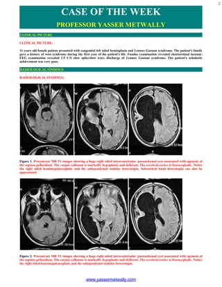
  • 1. 2 CASE OF THE WEEK PROFESSOR YASSER METWALLY CLINICAL PICTURE CLINICAL PICTURE: 11 years old female patient presented with congenital left sided hemiaplasia and Lennox Gastaut syndrome. The patient's family gave a history of west syndrome during the first year of the patient's life. Fundus examination revealed chorioretinal lacunae. EEG examination revealed 1.5 C/S slow spike/slow wave discharge of Lennox Gastaut syndrome. The patient's scholastic achievement was very poor. RADIOLOGICAL FINDINGS RADIOLOGICAL FINDINGS: Figure 1. Precontrast MR T1 images showing a huge right sided intraventricular /parenchymal cyst associated with agenesis of the septum pellucidum. The corpus callosum is markedly hypoplastic and deficient. The cerebral cortex is lissencephalic. Notice the right sided hemimegalencephaly and the subependymal nodular heterotopia. Subcortical band heterotopia can also be appreciated. Figure 2. Precontrast MR T1 images showing a huge right sided intraventricular /parenchymal cyst associated with agenesis of the septum pellucidum. The corpus callosum is markedly hypoplastic and deficient. The cerebral cortex is lissencephalic. Notice the right sided hemimegalencephaly and the subependymal nodular heterotopia. www.yassermetwally.com
  • 2. 3 Figure 3. MR T2 images showing a huge right sided intraventricular /parenchymal cyst associated with agenesis of the septum pellucidum. The corpus callosum is markedly hypoplastic and deficient. The cerebral cortex is lissencephalic. Notice the right sided hemimegalencephaly and the subependymal nodular heterotopia. Subcortical band heterotopia can also be appreciated. Figure 4. Precontrast MR T1 images showing A huge right sided intraventricular /parenchymal cyst. The cerebral cortex is lissencephalic. Notice the right sided hemimegalencephaly and the subependymal nodular heterotopia. Subcortical band heterotopia can also be appreciated. www.yassermetwally.com
  • 3. 4 Figure 5. Precontrast MR T1 images showing a huge right sided intraventricular /parenchymal cyst. The corpus callosum is markedly hypoplastic and deficient. The cerebral cortex is lissencephalic. Notice the subependymal nodular heterotopia and hypoplasia of the optic nerve. The cerebellum and brain stem are also hypoplastic. Figure 6. Precontrast MR T1 images showing a huge right sided intraventricular /parenchymal cyst. The cerebral cortex is lissencephalic. Notice the right sided hemimegalencephaly and the subependymal nodular heterotopia. Figure 7. Precontrast MR T1 images showing marked hypoplasia of the cerebellum and brain stem. www.yassermetwally.com
  • 4. 5 Figure 8. Chorioretinal lacunae  Criteria that are highly suggestive of Aicardi syndrome  Partial or complete callosal agenesis  Cortical dysplasia  Gross asymmetry of the hemispheres  Periventricular or subcortical heterotopias  Cysts of the choroid plexus or around the third ventricle is highly suggestive of AS DIAGNOSIS: DIAGNOSIS: AICARDI SYNDROME ASSOCIATED WITH MULTIPLE CORTICAL DYSPLASIAS THAT INCLUDE HEMIMEGALENCEPHALY, LISSENCEPHALY, HETEROTOPIAS, AND SEPTO-OPTIC DYSPLASIA. DISCUSSION DISCUSSION: The Aicardi syndrome (AS) is classically defined as a triad of abnormalities that includes agenesis of the corpus callosum, infantile spasms, and chorioretinal lacunae (1,2). Other eye defects and costovertebral and other malformations occur frequently. The syndrome has been observed exclusively in individuals with two X chromosomes, and only one familial case is known (3,4). Progress in neuroimaging has revealed that the central nervous system malformation in AS is not limited to agenesis of the corpus callosum but consists of a complex of abnormalities characterized by severe neuronal migration defects with periventricular and subcortical heterotopias, cortical polymicrogyria, and a tendency toward the development of cystic formations in the choroid plexuses and in other parts of the brain (3-5). The migration anomalies may even be more important for the definition of AS than callosal agenesis, which is a non- specific finding. Similarly, the eye abnormalities are often complex, and defects of closure of the primitive cupula are commonly present (6-8). The incidence of AS is unknown. More than 300 cases are known to this writer, and at least 170 cases have been published. In series of infantile spasms, AS may account for up to 4% of cases (9), but a selection bias is probable in series originating from tertiary referral centers. CLASSICAL FINDINGS IN AICARDI SYNDROME The classical picture of AS has been outlined in several articles (3,6, 1 0, I 1). The seizures are typically infantile spasms. These have been the only seizure type in 86 of 184 patients (47%) and have been associated with other seizures, especially partial motor attacks, in 65 of 184 patients (35%) (6). The partial seizures often begin before the infantile spasms, at times as early as the first few days of life (6,12). The age at first seizure was less than 3 months in 68% of 146 patients and less than I month in 23%. The age at the first spasm in 137 patients was less than I month in 18% and less than 3 months in 56%. Typical tonic spasms have been reported even during the neonatal period, and AS is one possible cause of the "early infantile epileptic encephalopathy" described by Ohtahara et al. (13). Partial seizures and infantile spasms often occur in association, a focal or lateralized tonic or clonic seizure being followed by a similarly lateralized cluster of spasms. The focal seizure is manifested on the EEG by a localized discharge of repetitive spikes lasting 10-30 s. The spasms follow immediately after the spikes and are associated with a series of slow complexes, often with a superimposed fast rhythm that is usually of higher amplitude on the side of the initial www.yassermetwally.com
  • 5. 6 partial discharge. The complexes occur 6- 20 s apart, and no paroxysmal activity appears between them. The initial partial seizure and the spasms that follow appear to represent a single attack that usually occurs on awakening or on changing from slow wave to REM sleep (12,14). Such periodic spasms are also associated with other brain malformations (14). Typical hypsarrhythmia is rare in AS (18% of 137 cases). The most common interictal EEG abnormality is an asymmetrical pseudo-periodic tracing with bursts of paroxysmal irregular slow and sharp waves of 3- to 6-s duration, separated by a relatively flat EEG for 5-20 s. Such bursts may occur independently over each hemisphere or may remain unilateral, with various abnormalities over the other hemisphere, the so-called "split-brain" EEG (6). The tracings more commonly evolve into multifocal paroxysmal abnormalities than into typical hypsarrhythmia. At a late stage, spike-and- wave complexes are unusual (6,9). The choroidal lacunae are multiple rounded whitish or pinkish areas ranging in size from one-tenth to several disc diameters. They are in the same plane as the retina, so that vessels do not bend on crossing their border. Pigment deposits may be visible at their periphery and may increase with age (6,15). The largest lacunae tend to cluster around the disc, whereas small pinkish lesions tend to be more peripheral. They are usually bilateral, but unilateral lacunae can be seen even when the opposite eye is not microphthalmic. Typical lacunae are probably pathognomonic of AS. Colobomata are a very common finding and are of significance for the diagnosis, especially when lacunae are few or atypical. Unlike the lacunae, they are frequently unilateral. Colobomatous discs are surrounded by pigmented rings in many cases. Other eye abnormalities, including persistence of the primary vitreous, anterior synechiae, and microphthalmia, are not uncommon. In one patient, the appearance was that of retinal detachment suggestive of the Walker- Warburg syndrome. Interestingly, despite the very abnormal ophthalmoscopic aspect, useful vision is often preserved as far as can be assessed. The ERG has been found normal or only mildly altered in several patients. The visual evoked potentials are usually present but abnormal, probably as a result of the cortical abnormalities. The fundoscopic appearance results from thinning of the choroid and sclera in the areas corresponding to the lacunae where the pigment epithelium is depigmented or hypopigmented (7), with degeneration of the rods and cones (8). There is no evidence of inflammatory lesions, and the appearance is suggestive of an early developmental disorder. Agenesis of the corpus callosum was complete in 137 of 152 patients for whom the information was available and was partial in 15 (10%). Partial agenesis is usually posterior (6), but cases of agenesis of both the genu and the splenium, with preservation of the body of the callosum, are known (16). The ventricular contour is not smooth as it is in most cases of callosal agenesis and the ventricles, including the third ventricle, have markedly irregular contours. In most cases there is also marked asymmetry between the two hemispheres, the larger one commonly but not consistently contralateral to a hemiplegia when present or to the side most involved by the spasms. Various abnormalities of the posterior fossa have also been reported (6). The neurologic and mental impairment in AS is almost always very marked. The estimated survival rate in one study (17) was 75% at 6 years and 40% at 15 years. Epilepsy persists in most patients as infantile spasms, which is unusual with other causes. Approximately one-third of patients are unable to feed themselves and only one-quarter are able to walk. Only rare patients can use two-word sentences (3). One patient had relatively well-developed language at age 14 years (17). According to McGregor et al. (17), a worse prognosis was correlated with larger lacunar size. Hemiplegia is commonly present but it is seldom isolated, and some degree of contralateral involvement is the rule. Mild microcephaly is common but is never present at birth. Indeed, some infants have large heads that may result from hydrocephalus or from the development of large intracranial cysts. Although an occasional patient has been operated on for shunting of hydrocephalus due to aqueductal stenosis or for drainage of a cyst, this is seldom justified because the cysts do not usually continue to increase in postnatal life and ventricular dilatation usually remains static even when marked. NEW FINDINGS IN AICARDI SYNDROME Over the past few years the diagnosis of AS has been facilitated by modern imaging techniques. These have shown that agenesis of the corpus callosum is virtually never an isolated finding but is part of a complex of developmental abnormalities. Migration anomalies are probably present in all cases. They include periventricular and/or subcortical nodular heterotopias, which are responsible for the irregular contours of the lateral ventricles, and cortical dysplasia with thickening of the cortical plate and abnormal rectilinear or blurred interface between gray and white matter involving one or both hemispheres to a variable degree. These dysplastic areas probably correspond pathologically to polymicrogyria (3, 5,15,18,19). Periventricular heterotopias are easily detectable, but the diagnosis of subcortical heterotopias and cortical dysplasia requires high-quality MRI. Cysts are frequently revealed on MRI. They can involve the glomus of the choroid plexus on one or both sides and/or the region of the third ventricle and pineal gland, where they may be single or multiple and may be quite large (Fig. 1). The cysts give a slightly more intense signal than CSF and their walls may enhance with gadolinium contrast. Cysts around the third ventricle were probably responsible for the distorted appearance of this ventricle on MRI or CT scan, which could not result from heterotopias. Cysts of the cerebral hemispheres are uncommon. Cysts of the posterior fossa have been reported (3,6). Most of them correspond to partial agenesis of the cerebellar vermis, to a megacisterna magna, or to arachnoid cysts. True intraparenchymal cysts are uncommon. The combination of cysts of the choroid plexus in association with agenesis of the corpus callosum permits the antenatal diagnosis of AS (20). Solid tumors, especially papillomas of the choroid plexus, have been reported in several patients (3,6) and may be multiple (21). Peripheral embryonic tumors have also been described (22,23). www.yassermetwally.com
  • 6. 7 Figure 1. Huge cyst in a case of Aicardi syndrome. The cyst probably arises from the pineal area and develops into the left parietooccipital lobe, displacing and compressing the lateral ventricle. Note also agenesis of the corpus callosum and abnormal appearance of the posterior cortex on both sides, suggestive of migration disorder. The combination of partial or complete callosal agenesis, cortical dysplasia, gross asymmetry of the hemispheres, periventricular or subcortical heterotopias, and cysts of the choroid plexus or around the third ventricle is highly suggestive of AS (3). Incomplete forms in which one or several components are lacking are not uncommon. Such forms are confirmed as AS by the presence of other typical manifestations, especially choroidal lacunae. There is probably no good reason to single out one component of the malformation complex so that cases without callosal agenesis but with heterotopias and/or cortical dysplasia can be diagnosed when other cardinal features are present. Two such cases (both with lacunae and one with vertebral abnormalities) are known to this author. Criteria that are highly suggestive of Aicardi syndrome  Partial or complete callosal agenesis  Cortical dysplasia  Gross asymmetry of the hemispheres  Periventricular or subcortical heterotopias  Cysts of the choroid plexus or around the third ventricle is highly suggestive of AS Figure 2. Image shows a cross-section of an eye in a patient with Aicardi syndrome. The arrow indicates chorioretinal lacunae. PATHOLOGY Pathologic data on AS are scanty, and detailed microscopic examination of the brain is available for only two cases (5,18). Total or partial absence of the corpus callosum was found in all verified cases, usually with the presence of Probst bundles. Other structures may be lacking, such as the first cranial nerve and the mammary bodies. Other commissures, e.g., the fornix or the anterior commissure (6), may be absent, but this is inconstant. Abnormalities of gyration were found in all studied cases (5-7,15,18). Their macroscopic aspect is variable, but microgyria is found microscopically and is of the unlayered type (18), taking the form of a thin, undulating cellular ribbon without any laminar organization. Fusion of the molecular layers of facing convolutions may result in the appearance of a pachygyric cortex (5). Heterotopias include subcortical neurons scattered in the white matter. Cysts have been reported in several cases (3,18,24) and were of ependymal origin. Pathologic findings are consistent with an etiologic factor acting before the end of the migration period and the development of the corpus callosum, which is complete by 14 weeks of gestational age (25). NOSOLOGIC LIMITS OF AS: THE PROBLEM OF INCOMPLETE FORMS www.yassermetwally.com
  • 7. 8 Because there is no laboratory marker specific for AS, the classical triad remains the cornerstone of diagnosis. The existence of incomplete forms, however, seems likely, and cases without callosal agenesis have been already discussed. In addition to the two cases above, six cases of possible AS without callosal agenesis are known (6). All six had infantile spasms and choroidal lacunae, and four had irregular ventricular contours on CT. None of these patients underwent an MRI. Cases without infantile spasms have been reported (6) and may not be rare. Recognition of the spasms may be difficult because they are often quite asymmetrical and atypical and are frequently associated with partial seizures, which may be the predominant seizure type. Isolated partial seizures are not unusual at onset or late in the course, but they may be absent altogether. The existence of AS cases without lacunae is particularly difficult to accept, because these are considered pathognomonic for the syndrome. There are, however, female patients with a suggestive brain malformation,, infantile spasms, and other abnormalities who might represent atypical forms. I know of three such patients who presented with infantile spasms and agenesis of the corpus callosum. Two of these had periventricular heterotopias on CT scan and an asymmetrical burst-suppression pattern on EEG. One girl had bilateral colobomata of the disk and another a small pigmented retinal area on one side. None of them had undergone MRI. GENETIC AND CYTOGENETIC DATA The original hypothesis that AS is an X-linked dominant disorder that is lethal at an early stage of gestation for affected hemizygous male conceptuses appears most compatible with the observed data (2,3,6). Only females are affected, with the exception of two phenotypical males with two X chromosomes (26). The male child with an XY karyotype reported by Curatolo et al. (27) is too atypical to be included (6,28, 29). This hypothesis is also consistent with the sporadic occurrence of the syndrome. Only one instance of familial recurrence in two sisters is reported (4) and this remains difficult to explain because no chromosomal abnormality was found in these patients. Another finding difficult to reconcile with the hypothesis is the occurrence of AS in only one of a pair of monozygotic twins, the co-twin being completely normal as a young adult (30). If this report is confirmed, it might be explained by extreme nonrandom inactivation of the abnormal X chromosome in the normal twin or, less probably, by a postzygotic mutation during early embryonic development. The strong suspicion of an abnormality of an X chromosome in patients with AS has been reinforced by the finding of skewed X inactivation in the lymphocytes of some patients (31). Patients with nonrandom inactivation were found to be more severely affected than those with a random pattern, suggesting that selection against abnormal cells in the developing neural tissue led to aberrant brain development. However, a normal inactivation pattern has been found in other cases (30,32). A possible locus for AS on the short arm of the X chromosome has been suggested by several case reports of eye abnormalities with callosal agenesis or other brain abnormalities (33-36) associated with translocations or other chromosomal abnormalities at Xp22.3. However, none of the reported patients had the typical triad of AS, even though microphthalmia (3), chorioretinal lesions reminiscent of the lacunae (Aughton et al., personal communication, 1991), other eye abnormalities (33,34), agenesis of the corpus callosum (36) or other brain defects (35), and costovertebral malformations (33) were present in variable associations. Four of these children also had focal dermal hypoplasia or Golz disease, a sex linked disorder with lethality for hemizygous males mapping at Xp22.3 (36). Such cases suggest that the Xp22.3 region is involved in the genesis of both Golz syndrome and AS. They could represent contiguous gene syndromes involving both loci. Intensive search for an AS gene in the Xp22.3 region is being pursued. SUMMARY SUMMARY AS is an uncommon malformation complex that affects mostly the eyes and the central nervous system. Brain malformation constitutes the core of the syndrome. Agenesis of the corpus callosum is the most easily detectable but probably not the most characteristic feature. Migration anomalies, including periventricular and subcortical nodular heterotopias and cortical dysplasia, and a tendency towards the formation of ependymal cysts in the glomus of the choroid plexuses and/or near of the third ventricle, are major components of the complex and may occur even in the presence of a complete corpus callosum. The resulting seizures, usually asymmetrical infantile spasms, and mental retardation constitute a severe disability with a much reduced life expectancy. The syndrome is probably due to a chromosomal accident involving one X chromosome. The Xp22.3 region is a prime candidate for location of one or several responsible genes, and demonstration of a DNA abnormality in this region will permit a better definition of the limits of the syndrome and perhaps help our understanding of some aspects of the development of the central nervous system. AS is an uncommon malformation complex that affects mostly the eyes and the central nervous system. Brain malformation constitutes the core of the syndrome. Agenesis of the corpus callosum is the most easily detectable but probably not the most characteristic feature. Migration anomalies, including periventricular and subcortical nodular heterotopias and cortical dysplasia, and a tendency towards the formation of ependymal cysts in the glomus of the choroid plexuses and/or near of the third ventricle, are major components of the complex and may occur even in the presence of a complete corpus callosum. The www.yassermetwally.com
  • 8. 9 resulting seizures, usually asymmetrical infantile spasms, and mental retardation constitute a severe disability with a much reduced life expectancy. The syndrome is probably due to a chromosomal accident involving one X chromosome. The Xp22.3 region is a prime candidate for location of one or several responsible genes, and demonstration of a DNA abnormality in this region will permit a better definition of the limits of the syndrome and perhaps help our understanding of some aspects of the development of the central nervous system. The diagnosis of AS is based upon the classic triad of corpus callosal agenesis, chorioretinal lacunae and infantile spasm. But there is a range of costovertebral, ocular and cerebral abnormalities associated with this disorder.(2) The cerebral gray-matter heterotopias and other cortical malformations act as epileptogenic foci.(2) Their seizures typically start in early childhood and are usually intractable. Besides infantile spasm, other seizure types are also demonstrated. Dissociated burst-suppression or burst-suppression pattern appearing asymmetrically in either cerebral hemisphere is a characteristic EEG finding in AS.(8) The developmental delay in AS is generally profound, involving both motor and language skills. Chevrie and Aicardi in their analysis of 184 patients of AS observed that none had acquired meaningful speech.(9) But of late a larger spectrum of the disease has been recognized and it had been found that higher functioning AS individuals do exist.(2) Most of the AS cases die at an early age primarily due to aspiration pneumonitis. But some do live into their adolescent years and even in their twenties.(2) Good visual function in AS patients do occur if the fovea is uninvolved with chorioretinal lacunae. Cerebral heterotopias, interhemispheric cysts, optic nerve coloboma, microphthalmia, thoracolumbar kyphoscoliosis are the known associated features in AS. Severe psychomotor impairment and absence of meaningful speech had also been noted. (3,4,5,6)  Addendum  A new version of this PDF file (with a new case) is uploaded in my web site every week (every Saturday and remains available till Friday.)  To download the current version follow the link "http://pdf.yassermetwally.com/case.pdf".  You can also download the current version from my web site at "http://yassermetwally.com".  To download the software version of the publication (crow.exe) follow the link: http://neurology.yassermetwally.com/crow.zip  The case is also presented as a short case in PDF format, to download the short case follow the link: http://pdf.yassermetwally.com/short.pdf  At the end of each year, all the publications are compiled on a single CD-ROM, please contact the author to know more details.  Screen resolution is better set at 1024*768 pixel screen area for optimum display.  For an archive of the previously reported cases go to www.yassermetwally.net, then under pages in the right panel, scroll down and click on the text entry "downloadable case records in PDF format"  Also to view a list of the previously published case records follow the following link (http://wordpress.com/tag/case-record/) or click on it if it appears as a link in your PDF reader REFERENCES References 1. Aicardi J, Lefebvre J, Lerique-Koechlin A. A new syndrome: spasms in flexion, callosal agenesis, ocular abnormalities. Electroencephalogr Clin Neurophysiol 1965;19:609-10. 2. Aicardi J, Chevrie JJ, Rousselie F. Le syndrome agdndsie calleuse, spasmes en flexion, lacunes chorioretiniennes. Arch Franc Pgdiatr 1969;26: 1103-20. 3. Aicardi J, Chevrie JJ. The Aicardi syndrome. In: Lassonde M, Jeeves MA, eds. Callosal agenesis: the natural split brain. New York: Plenum Press, 1995. 4. Molina JA, Mateos F, Merino M, Epifanio JL, Gorrono M. Aicardi syndrome in two sisters. J Pediatr 1989;115:282-3. 5. Billette de Villemeur T, Robain 0, Chiron C. Unlayered polymicrogyria and agenesis of the corpus callosum: a relevant association? Acta Neuropathol (Berl) 1992;83:265-70. 6. Chevrie JJ, Aicardi J. The Aicardi syndrome;, In: Meldrum BS, ed. Recent advances in epilepsy, Vol. 3. Edinburgh: Churchill Livingstone, 1986: 189-210. 7. McMahon RG, Bell RA, Moore RW, Ludwin SK. Aicardi syndrome. A clinicopathological study. Arch Ophthalmol 1984;102:250-3. 8. Del Pero RA, Mets MB, Tripathy RC, Torezynski E. Anomalies of retinal architecture in Aicardi syndrome. Arch Ophthalmol 1986;104:1659-64. www.yassermetwally.com
  • 9. 10 9. Aicardi J. Epilepsy in children, 2nd ed. New York: Raven Press, 1994:l-3. 10. Donnenfeld AE, Packer RJ, Zackai EH, Chee CM, Sellinger B, Emmanuel BS. Clinical, cytogenetic and pedigree findings in 18 cases of Aicardi syndrome. Am J Med Genet 1989;32:461-7. 11. Yamagata T, Momoi M, Miyamoto S, Kobayashi S, Kamoshita S. Multi-institutional survey of the Aicardi syndrome in Japan. Brain Dev 1990;12: 760-5. 12. Bour F, Chiron C, Dulac 0, Plouin P. Caractres electrocliniques des crises dans le syndrome d'Aicardi. Rev EEG Neurophysiol Clin 1986;16: 341-53. 13. Ohtahara S, Ohtsuka Y, Yamatogi Y, Oka E. The early infantile epileptic encephalopathy with suppression burst: developmental aspects. Brain Dev 1987;9:371-6. 14. Gobbi G, Bruno L, Pini A, Rossi PG, Tassinari CA. Periodic spasms: an unclassified type of epi- leptic seizure in childhood. Dev Med Child Neural 1987;29:766-75. 15. De Jong JGY, Delleman JW, Houben M, et al. Agenesis of the corpus callosum, infantile spasms, ocular anomalies (Aicardi's syndrome): clinical and pathological findings. Neurology 1976;26: 1152-8. 16. Aicardi J, Chevrie JJ, Baraton J. Agenesis of the corpus callosum. In: Vinken PJ, Bruyn GW, eds. Handbook of clinical neurology, Vol. 6. Brain malformations. Amsterdam: North-Holland, 1987:149-73. 17. McGregor DL, Menezes A, Buncic JR. Aicardi syndrome (AS): natural history and predictors of severity. Can J Neurol Sci 1993;20(suppl 2):S36. 18. Ferrer 1, Cusi MV, Liarte A, Campistol J. A Golgi study of the polymicrogyric cortex in Aicardi syndrome. Brain Dev 1986;8:518-25. 19. Baieari P, Marki A, Thelen M, Laub MC. MR imaging in Aicardi,s syndrome. Am J Neuroradiol 1988;9:805-6. 20. Roland EH, Flodmark 0, Hill A. Neurosonographic features of Aicardi syndrome. J Child Neurol 1989;4:307-10. 21. Hamano K, Matsubara T, Shibata S, et al. Aicardi syndrome accompanied by auditory disturbances and multiple brain tumors. Brain Dev 199 1; 13:438- 41. 22. Tanaka T, Takahura H, Takashima S, Kodama T, Hasegawa H. A rare case of Aicardi syndrome with severe brain malformation and hepatoblastoma. Brain Dev 1985;7:507-12. 23. Togawa T, Mimaki T, Ono J. Aicardi syndrome associated with embryonal carcinoma. Pediatr Neural 1989;5:45-7. 24. Brihaye L, Gillet P, Parmentier R, Peetrons A. Agenesie de la commissure calleuse associee A un kyste dpendymaire. Arch Suisses Neurol Psychi- atr 1956;77:415-31. 25. Barkovich A, Norman D. Anomalies of the corpus callosum: correlation with further anomalies of the brain. Am J Neuroradiol 1988;9:493-501. 26. Hopkins IJ, Humphrey 1, Keith CG, Susman M, Webb GC, Turner EK. The Aicardi syndrome in a 47XXY male. Aust Paediatr J 1979;15:278-80. 27. Curatolo P, Libutti G, Dalla Piccola B. Aicardi syndrome in a male infant. J Pediatr 1980;96: 286-7. 28. Aicardi J. The Aicardi syndrome in a male infant [Letter]. J Pediatr 1980;97:1040-41. 29. Hunter AGW. Aicardi syndrome in a male infant [Reply]. J Pediatr 1980;97:1041. 30. Costa T, Greer W, Duckworth M, Rysiecki M, Musarella M, Ray P. Monozygotic twins discordant for Aicardi syndrome [Abstract]. Am J Hum Genet 1990;47(suppl 5)202:14. 31. Neidich JA, Nussbaum RL, Packer RJ, Emanuel BS, Puck JM. Heterogeneity of clinical severity and molecular lesions in Aicardi syndrome. J Pediatr 1990; 1 16:911-7. 32. Wieacker P, Zimmer J, Ropeers HH. X-inactivation pattern in two syndromes with probable X-linked dominant, male lethal inheritance. Clin Genet 1985;28:238-42. 33. Ropers HH, Zuffardi 0, Biancai E, Tiepolo L. Agenesis of corpus callosum, ocular and skeletal anomalies (X-linked dominant Aicardi's syndrome) in a girl with balanced X/3 translocation. Hum Genet 1982;61:364-8. 34. Donnenfeld AE, Graham JM, Packer RJ, Aquino R, Berg SZ, Emanuel BS. Microphthalmia and chorioretinal lesions in a girl www.yassermetwally.com
  • 10. 11 with an Xp22.2 pter deletion and partial 3p trisomy: clinical observations relevant to Aicardi syndrome gene localization. Am J Med Genet 1990;37:182-6. 35. Al-Gazali LI, Muller RF, Caine A, et al. An XX male and two (X;Y) females with linear skin defects and congenital microphthalmia: a new syndrome at Xp22.3. J Med Genet 1988;25:638-9. 36. Friedman PA, Rao KW, Jeplin SW, Aylsworth AS. Provisional deletion mapping of the focal dermal hypoplasia (FDH) gene to Xp22.31 [Abstract]. Am J Hum Genet 1988;43:A50. 32- Metwally, MYM: Textbook of neuroimaging, A CD-ROM publication, (Metwally, MYM editor) WEB-CD agency for electronic publication, version 9.4a October 2008 www.yassermetwally.com
  • 11. 12 CASE OF THE WEEK PROFESSOR YASSER METWALLY CLINICAL PICTURE CLINICAL PICTURE: A 7 years old female patient presented clinically with Lennox Gastaut syndrome. RADIOLOGICAL FINDINGS RADIOLOGICAL FINDINGS: Figure 1. Cortical dysplasia. Precontrast MRI T1 images showing lissencephaly, microgyria, pachygyria, hypointense cystic white matter changes specially affecting the right head of caudate nucleus and the globus pallidus on the right side. The lissencephalic changes are most marked in the bifrontal regions. Notice the subependymal nodular heterotopia specially involving the frontal horns bilaterally. There is also reduction of the brain volume and moderate degree of central atrophy. www.yassermetwally.com
  • 12. 13 Figure 2. Cortical dysplasia. MRI FLAIR images showing lissencephaly, microgyria, pachygyria, hyperintense cystic white matter changes specially affecting the right head of caudate nucleus and the globus pallidus on the right side. The lissencephalic changes are most marked in the bifrontal regions. Notice the subependymal nodular heterotopia specially involving the frontal horns bilaterally, nodules are also seen subependymally in the left body of the lateral ventricles (C). There is also reduction of the brain volume and moderate degree of central atrophy. Figure 3. Cortical dysplasia. MRI T2 images showing lissencephaly, microgyria, pachygyria, hyperintense cystic white matter changes specially affecting the right head of caudate nucleus and the globus pallidus on the right side. The lissencephalic changes are most marked in the bifrontal regions. Notice the subependymal nodular heterotopia specially involving the frontal horns bilaterally. There is also reduction of the brain volume and moderate degree of central atrophy. www.yassermetwally.com
  • 13. 14 Figure 4. Cortical dysplasia. MRI FLAIR images showing lissencephaly, microgyria, pachygyria, hyperintense cystic white matter changes specially affecting the right head of caudate nucleus and the globus pallidus on the right side. The lissencephalic changes are most marked in the bifrontal regions. Notice the subependymal nodular heterotopia specially involving the frontal horns bilaterally, nodules are also seen subependymally in the left body of the lateral ventricles (C). There is also reduction of the brain volume and moderate degree of central atrophy. The hippocampi are atrophic and hyperintense bilaterally (possible mesial temporal sclerosis). DIAGNOSIS: DIAGNOSIS: CORTICAL DYSPLASIA DISCUSSION DISCUSSION: The brain is a seemingly nonsegmented organ that is, however, formed in a segmented fashion by the overlap of genes that define anatomic and probably functional components of the brain. Other genes and their encoded proteins regulate the processes of cell proliferation and migration; many of these genes have been identified based upon discoveries of human and mouse disease-causing genes. Human brain developmental disorders represent clinical challenges for the diagnosing clinician as well as for the treating physician. Some disorders represent well-defined clinical and genetic entities for which there are specific tests; others have ill-defined genetic causes, while others can have both genetic and destructive causes. In most cases the recognition of a disorder of brain development portends certain developmental disabilities and often seizure disorders that can be very difficult to treat. In addition, it now bears upon the treating physician to recognize the genetic causes, and to properly advise patients and their families of the risks of recurrence or refer them to the proper specialist who can do so. The genetics of some of these disorders are not all well defined at present, and the recognition of some disorders is variable; what is known is presented herein. The genetics and signaling utilized in brain development is briefly reviewed to provide the framework for the understanding of human brain developmental disorders. The well-defined genetic disorders of brain development are discussed, and a brief suggested algorithm for evaluation and for counseling of patients is provided. BRAIN DEVELOPMENT  Overview General mechanisms tend to recur in all phases of brain development, and these include induction, cell proliferation, cell fate determination (differentiation), cell process formation and targeting (synapse formation), and www.yassermetwally.com
  • 14. 15 cell movement (migration). Induction is the process by which one group of cells or tissue determines the fate of another by the release of soluble factors or inducers. Cell fate or differentiation is dependent upon this process of induction, and probably can best be understood as the initiation of a genetic program by the recognition of an inducing molecule and/or expression of a transcriptional regulator. In general, it is rare that a cell in the nervous system is born and differentiates in the same location that it finally resides. Rather, cells migrate over long distances to reach their final locations. Similarly, cells in the nervous system must extend processes over long distances to reach their synaptic targets.  Neural tube formation The human brain is formed from the neuroectoderm, a placode of cells that are induced to differentiate from the surrounding ectoderm by the presence of the notochord at about 18 days gestation. Candidate inducing factors include the retinoids, follistatin, and Noggin [1-4]. The neuroectoderm develops folds in the lateral aspects that begin to approximate in the region of the future medulla and fuse at 22 days gestation. This closure is known as neurulation, and results in the formation of a tube termed the neural tube [5]. The anterior neural tube closes by about 24 days gestation and serves as the foundation for further brain development; the posterior neural tube closes by about 26 days gestation and serves as the foundation for further spinal cord development. Defects in the closure of the neural tube lead to encephaloceles or myelomeningoceles.  Nervous system segmentation At the rostral end of the newly closed neural tube flexures delineate the primary vesicles, which are designated as the hindbrain (rhombencephalon), mesencephalon, and forebrain (prosencephalon). The primary vesicles can be further subdivided into secondary vesicles that will form adult brain structures. The hindbrain can be divided into the metencephalon and myelencephalon, which will become the pons, cerebellum, and medulla oblongata of the adult. The mesencephalon will be the midbrain, and the prosencephalon divides into the telencephalon (two telencephalic vesicles) and diencephalon. The telencephalic vesicles will become the cerebral hemispheres; the diencephalon will become the thalamus and hypothalamus. Regional specification of the developing telencephalon is an important step in brain development, and is likely under control of a number of genes that encode transcription -regulators. In the fruit fly, Drosophila, these genes are involved in segmentation of this animal and define structures such as hair-like spiracles. Not surprisingly, the role of these genes in human brain development differs, yet it appears that the general role of these proteins is that of regional specification of clones of cells destined to form specific brain structures. Homeobox and other transcription genes encode some of these transcriptional regulators and these "turn on" genes by binding to specific DNA sequences, and in so doing initiate genetic programs that lead to cell and tissue differentiation. EMX2, a transcriptional regulator, has a homolog in Drosophila that defines the hair spiracles and has been implicated in human brain malformations. DISORDERS OF SEGMENTATION  Schizencephaly Schizencephaly (cleft in brain) has been regarded by many as a migration abnormality; however, it is best understood as a disorder of segmentation because one of the genes that is abnormal in the more severe and familial forms is EMX2 [6,7]. Thus, this developmental disorder, at least in the more severe cases, appears to be the result of failure of regional specification of a clone of cells that are destined to be part of the cortex. Clinically, these patients vary depending upon the size of the defect and upon whether bilateral disease is present [8]. The clefts extend from the pia to the ventricle and are lined with a polymicrogyric gray matter (see the discussion in Polymicrogyria) [9]. The pia and ependyma are usually in apposition, especially in severe cases. The defect is termed open-lipped if the cleft walls are separated by cerebrospinal fluid, and closed-lipped if the walls are in contact with one another. Bilateral schizencephaly is associated with mental retardation and spastic cerebral palsy; affected patients often are microcephalic. Seizures almost always accompany severe lesions, especially the open-lipped and bilateral schizencephalies. The exact frequency of seizures in patients with the less severe lesions is uncertain. Most patients in whom schizencephaly is diagnosed undergo neuroimaging because of seizures. Therefore, a bias in favor of a universal occurrence of seizures in this disorder is noted. Hence, patients with schizencephaly who do not have epilepsy might exist, but the malformation remains undetected because no imaging is performed. www.yassermetwally.com
  • 15. 16 Figure 1. Closed-lip schizencephaly. Sagittal T1-weighted MRI shows gray matter (arrows) extending from cortex to a dimple in the surface of the left lateral ventricle. The lips of the schizencephaly are in apposition, making this a "closed-lip" schizencephaly. Figure 2. Bilateral open-lip schizencephaly. A,B: Axial T2-weighted images show open-lip schizencephalies in both hemispheres. Both images show vessels in the gray matter-lined clefts and large vessels (arrows) run at the outer surface of the right hemispheric cleft. This does not represent a vascular malformation. Seizure type and onset may also vary in this disorder. Patients may experience focal or generalized seizures, and some will present with infantile spasms. The onset varies from infancy to the early adult years. Seizures may be easily controlled or may be recalcitrant to standard anticonvulsant therapy. Figure 3a. MRI T1 (A,B) and CT scan (D) showing open-lip schizencephaly with pachygyria. Notice the associated encephalocele that is sometimes associated with cortical dysplasias www.yassermetwally.com
  • 16. 17 Figure 3b. Open-lip schizencephaly with cortical dysplasia Improvements in neuroimaging have enhanced the recognition of schizencephalic lesions [9-13]. The lesions may occur in isolation or may be associated with other anomalies of brain development such as septo-optic dysplasia (see Disorders of prosencephalic cleavage) [14]. Disorders of segmentation likely represent a heterogeneous set of abnormalities of varying etiologies. One theory holds that an early (first-trimester) destructive event disturbs subsequent formation of the cortex. Another theory is that segmental failure occurs in the formation of a portion of the germinal matrix or in the migration of primitive neuroblasts. Certainly, the finding of mutations of the EMX2 gene in some patients with the open-lipped form of schizencephaly supports the latter hypothesis.  Prosencephalon cleavage At about 42 days of gestation, the prosencephalon undergoes a division into two telencephalic vesicles that are destined to become the cerebral hemispheres. The anterior portion of this cleavage is induced by midline facial structures and the presence of the notochord. Abnormalities of this process are thought to result in holoprosencephaly, septo-optic dysplasia, and agenesis of the corpus callosum [I 5]. One of the important molecules responsible for the induction of this cleavage is Sonic hedgehog [16]. This protein is produced by the notochord, ventral forebrain, and the floor plate of the neural tube [17]. It interacts through at least one receptor, PTCH-A human homolog of patched, and alters the expression of transcription factors [18,19]. Furthermore, in an interesting link between these ventral inductive events and segmentation, Sonic hedgehog can alter the expression of the transcriptional regulating genes when applied to proliferating cells at critical times in development [21]. This ties the inductive proteins to the expression of transcriptional regulating genes and gives a hint as to the mechanisms involved in inductive processes. www.yassermetwally.com
  • 17. 18 Figure 4a. Agenesis of the corpus callosum Other molecules of interest in this inductive process are the retinoids, which are lipids capable of crossing membranes and that have been shown to exist in posterior to anterior gradients across embryos [3,20]. Retinoic acid can alter the pattern of transcriptional factors in neuroepithelial cells [3] and can downregulate Sonic hedgehog, perhaps explaining some of the head defects seen in retinoid embryopathy [17,22]. Agenesis of the corpus callosum Etiology •Both genetic and sporadic Pathogenesis •Unknown, •Agenesis of the corpus callosum may be part of an extensive malformation complex or the callosum may be partially or completely absent or hypoplastic Epidemiology in an otherwise normal brain. •The malformation is relatively rare. •The brain in agenesis of the corpus callosum shows batwing shaped ventricles as well as loss of the corpus callosum and there is no cingulate gyrus. General Gross •The remainder of the abnormalities depend on what syndrome or other Description malformations are associated with the defect. In most cases there is a bundle of white matter processes on both cerebral hemispheres in the area where the corpus callosum should be, called the bundle of Probst. In some patients there is a lipoma or other tumor in the area where the corpus callosum should be. General Microscopic •None Description •Patients with agenesis of the corpus callosum may be normal or may have Clinical neurological abnormalities dependent on the other accompanying Correlation malformations. Figure 4b. Agenesis of the corpus callosum DISORDERS OF PROSENCEPHALIC CLEAVAGE  Holoprosencephaly Holoprosencephaly is a heterogeneous disorder of prosencephalon cleavage that results from a failure of the www.yassermetwally.com
  • 18. 19 prosencephalic vesicle to cleave normally. Three forms of this disorder have been described: alobar, semilobar, and lobar [23,24]. In the alobar form, the telencephalic vesicle completely fails to divide, producing a single horseshoe- shaped ventricle, sometimes with a dorsal cyst, fused thalami, and a malformed cortex. In the semilobar form, the interhemispheric fissure is present posteriorly, but the frontal and, sometimes, parietal lobes, continue across the midline [25]; in some cases just ventral fusion is noted. In the lobar form, only minor changes may be seen: the anterior falx and the septum pellucidum usually are absent, the frontal lobes and horns are hypoplastic, and the genu of the corpus callosum may be abnormal. Figure 5. This is holoprosencephaly in which there is a single large ventricle with fusion of midline structures, including thalami. The affected fetuses and neonates typically have severe facial defects, such as cyclopia, as well. Underlying chromosomal abnormalities, such as trisomy 13, or maternal diabetes mellitus are possible causes, but some cases are sporadic. Holoprosencephaly is associated with a spectrum of midline facial defects. These include cyclopia, a supraorbital proboscis, ethmocephali, in which the nose is replaced by a proboscis located above hypoteloric eyes; cebocephaly, in which hypotelorism and a nose with a single nostril are seen; and premaxillary agenesis, with hypotelorism, a flat nose, and a midline cleft lip [26]. www.yassermetwally.com
  • 19. 20 Figure 6. (left three images) A,B semilobar holoprosencephaly from the same patient, C a patient with lobar holoprosencephaly. Arrowhead in A points to lack of ventral interhemispheric cleavage. Arrowhead in B points to fused thalami. The septum pellucidum is absent in B. In C notice the horseshoe or mushroom shaped single ventricle designated by * , arrowhead in C points to lack of interhemispheric fissure. (right image) MRI - Holoprosencephaly: This 6-day-old girl presented with laryngeal malacia and a diminished level of arousal. This proton density axial MR image shows an absence of the anterior horns of the lateral ventricles, fused thalami and absence of the corpus callosum anteriorly. Only children who have the lobar and semilobar forms are known to survive for more than a few months. An infant affected with the severe form is microcephalic, hypotonic, and visually inattentive [25]. In infants with the less severe forms of holoprosencephaly, myoclonic seizures frequently develop and, if the infant survives, autonomic dysfunction, failure to thrive, psychomotor retardation, and atonic or spastic cerebral palsy often are present. Some infants with the lobar form may be only mildly affected and, for example, present as a relatively mild spastic diplegia. Pituitary defects may be associated with these malformations, and may result in neuroendocrine dysfunction [27]. One, therefore, has to wonder how much genetic overlap exists between this condition and septo- optic dysplasia to be described below. Holoprosencephaly has been associated with maternal diabetes [28], retinoic acid exposure, cytomegalovirus, and rubella [29]. Chromosome abnormalities associated with this disorder include trisomies 13 and 18; duplications of 3p, 13q, and 18q; and deletions in 2p, 7q, 13q, and 18q [30]. Of particular concern to the clinician is the existence of an autosomal dominant form in which mutations in Sonic Hedgehog lead to variable expression of holoprosencephaly. In the mildest form of this genetic disorder, patients may have a single central incisor, a choroid fissure coloboma, or simply attention deficit disorder; a parent of a child with holoprosencephaly manifesting these features should be considered to be at high risk for recurrence of holoprosencephaly in their children (up to 50% risk) [31,32]. A number of other genes (HPE] (21 q22.3), HPE2 (2p2 1), HPE3 (7q36), HPE4 (18p), ZIC2, SIX3 (2p2l), and PATCHED) have been associated with holoprosencephaly, and although potentially inherited in an autosomal recessive fashion, most occurrence seems to be random [33,34].  Septo-optic dysplasia Septo-optic dysplasia (de Morsier syndrome) is a disorder characterized by the absence of the septum pellucidum, optic nerve hypoplasia, and hypothalamic dysfunction. It may be associated with agenesis of the corpus callosum. This disorder should be considered in any patient who exhibits at least two of the above abnormalities and perhaps even solely hypothalamic dysfunction [35]. Septo-optic dysplasia also appears to involve prosencephalic cleavage and development of anterior telencephalic structures [36]. About 50% of patients with septo-optic dysplasia have schizencephaly [14]. www.yassermetwally.com
  • 20. 21 Figure 7. Septo-optic dysplasia associated with schizencephaly. Arrows in A,B point to absent septum pellucidum, arrow in C, and black arrowhead in D point to open lip schizencephaly, white arrow head point to polymicrogyria in D Patients may present with visual disturbance, seizures, mental retardation, hemiparesis (especially if associated with schizencephaly), quadriparesis, or hypothalamic dysfunction. Endocrine abnormalities may include growth hormone, thyroid hormone, or antidiuretic hormone function or levels. The consideration of septo-optic dysplasia necessitates an evaluation of the hypothalamic-pituitary axis because as many as 60% of the children with this disorder might exhibit evidence of a disturbance of endocrine function [37]. This evaluation can include thyroid function studies and electrolytes; these patients are at high risk for growth retardation. The recent identification of patients with this condition that harbor mutations in the transcriptional regulator gene HESX1, suggest that the mechanism of this disorder is likely genetic and a patterning or segmental abnormality [38]. Even though the genetic abnormality has been identified for a minority of patients, there exists the possibility that this may not represent an entirely genetic disorder because associations have been made with young maternal age, diabetes, the use of anticonvulsants, phencyclidine, cocaine, and alcohol [39], DISORDERS OF CELL PROLIFERATION  Normal cell proliferation Following telencephalic cleavage, a layer of proliferative pseudostratified neuroepithelium lines the ventricles of the telencephalic vesicles. These cells will give rise to the neurons and glia of the mature brain. The generation of the proper complement of cells is a highly ordered process that results in the generation of billions of neurons and glia. Neuroepithelial processes extend from the ventricular surface to the pial surface, and the nuclei of the primitive neuroepithelial cells move from the cortical surface in a premitotic phase to a mitotic phase near the ventricle. Cells divide at the most ventricular aspects of the developing telencephalon, and after division move back toward the pial surface. The pial processes of neuroepithelial cells near the ventricle often will detach from the cortical surface before a new cycle begins. Neuroepithelial cells divide in so-called proliferative units such that each unit will undergo a specific number of divisions resulting in the appropriate number of cells for the future cortex. Abnormalities in the number of proliferative units or in the total number of divisions can lead to disorders of the brain manifested by abnormal brain size and, therefore, an unusually small or large head circumference. Two such disorders resulting in small head size -radial microbrain and microcephaly vera- are believed to result from abnormalities of this phase of neurodevelopment [40]. Disorders in which too many cells are generated in the proliferative phase result in megalencephaly (large brain) or, if proliferative events go awry on only one side of the developing cortex, hemimegalencephaly. The genes and molecules involved in regulating the proliferative cycles in human brain formation are likely similar www.yassermetwally.com
  • 21. 22 to those involved in other species. This cell cycle in the brain can be divided into a number of distinct phases: mitosis (M), first gap (GI), deoxyribonucleic acid synthesis (S), and second gap (G2) [41]. These phases appear to be regulated by key molecules to check the advancement of proliferation. Some cells enter a resting state (GO) that they maintain throughout life. Others temporarily enter this phase to await a specific signal to proliferate later. Probably the GI-S transition regulation determines the number of cell cycles and, therefore, the complement of cells that will make brain [40]. Cyclins are proteins that appear to be involved in cell cycle control. These proteins are activating subunits of cyclin-dependent kinases. Cyclins DI, D2, D3, C, and E seem to control the key transition of a cell to the GI S interface; this transition is regarded as important because it commits a cell to division [42-44]. Cyclin E seems to be the gatekeeper for this transition, and is essential for movement from the GI to the S phase [42,45]. The number of cells that finally make up the mature nervous system is less than that generated during proliferation. Cells appear not only to be programmed to proliferate during development but to contain programs that lead to cell death [46,47]. The term apoptosis (from the Greek, meaning "a falling off ") has been applied to this programmed loss of cells [48].  Non-neoplastic proliferative disorders  Microcephaly Although primary microcephaly may be a normal variant, in the classic symptomatic form, clinical and radiologic examinations reveal a receding forehead, flat occiput, early closure of fontanelles, and hair anomalies such as multiple hair whirls and an anterior cowlick. Neuroimaging may show small frontal and occipital lobes, open opercula, and a small cerebellum [24]. The cortex may appear thickened and the white matter reduced. Histologic examination may show a reduction of cell layers in some areas and an increase in others [49]. Neurologic findings also vary. Only mild psychomotor retardation may be noted, sometimes associated with pyramidal signs, or more severe retardation, seizures, and an atonic cerebral palsy might be evidenced. Primary microcephaly is seen in many genetic syndromes and, in its isolated form, may be autosomal recessive, autosomal dominant, or X-linked [50-52]. Microcephaly vera is the term most often alyplied to this genetic form of microcephaly. Affected children present with a head circumference that is usually more than 4 standard deviations below the mean, hypotonia, and psychomotor retardation. They later show mental retardation, dyspraxias, motor incoordination and, sometimes, seizures. On histologic examination, neurons in layers 11 and III are depleted [53]. Destructive lesions of the forming brain, such as those caused by teratogens and by infectious agents, also may result in microcephaly. Teratogens of note are alcohol, cocaine, and hyperphenylalaninemia (maternal phenylketonuria) [54]. Intense radiation exposure (such as that from a nuclear explosion) in the first trimester, can cause microcephaly [55]. Microcephaly and intracranial calcifications are likely due to well-recognized in utero infections caused by cytomegalovirus, toxoplasmosis, or the human immunodeficiency virus.  Megalencephaly and hemimegalencephaly The terms megalencephaly and hemimegalencephaly refer to disorders in which the brain volume is greater than normal (not owing to the abnormal storage of material); usually, the enlarged brain is accompanied by macrocephaly, or a large head. Although considered by some to be a migration disorder, the increase in brain size in these disorders appears to be attributable to errors in neuroepithelial proliferation, as the microscopic appearance of the brain is that of an increase in number of cells (both neurons and glia) and in cell size [56-59]. Typically, patients are noted to have large heads at birth, and may manifest an accelerated head growth in the first few months of life [60,61]. Children with megalencephaly or hemimegalencephaly may come to medical attention when presenting with seizures, a developmental disorder (mental retardation), hemihypertrophy, or a hemiparesis (opposite the affected hemisphere). Seizures vary both in onset and in type, and usually are the most problematic symptom. sometimes necessitating hemispherectomy or callosotomy [58]. Approximately 50% of patients with linear sebaceous nevus syndrome have hemimegalencephaly [62,63]. Many patients with hypomelanosis of Ito also have hemimegalencephaly [64]. The neuropathologic and clinical pictures of these associations appear to be identical to the isolated hemimegalencephalies.  Neoplastic proliferative disorders www.yassermetwally.com
  • 22. 23 Lesions of proliferation after an abnormal induction event during brain development may be malformative, hamartomatous, neoplastic, or a combination. Malformative disorders consisting of neoplasias on a background of disordered cortex or in association with focal cortical dysplasia include dysembryoplastic neuroepithelial tumor and ganglioglioma. [103,104]  Dysembryoplastic neuroepithelial tumours Dysembryoplastic neuroepithelial tumors are supratentorial, predominantly temporal lobe tumors that are typically multinodular with a heterogeneous cell composition, including oligodendrocytes, neurons, astrocytes, and other cells. These lesions are typically fairly well-demarcated, wedge-shaped lesions extending from the cortex to the ventricle. Calcification, enhancement, and peritumoral edema are lacking on neuroimaging studies. These low- attenuation lesions may suggest an infarct on computed tomography (CT), although there is no volume loss over time, and scalloping of the inner table or calvarial bulging suggests slow growth. Lesions are low in signal on Tl- weighted images and high in signal on T2- weighted images and often have a multinodular or pseudocystic appearance. There is a spectrum of pathology in dysembryoplastic neuroepithelial tumors. On one end of the spectrum are multinodular lesions with intervening malformed cortex, in which there is some hesitation to use the designation tumor. On the other end are lesions, which are clearly neoplastic and have clinically demonstrated some growth potential. Because the term dysembryoplastic neuroepithelial tumor has only recently been introduced, such malformative and neoplastic lesions were previously labeled as hamartomas, gangliogliomas, or mixed gliomas. [100,101,102]  Gangliogliomas Gangliogliomas are typically demonstrated within the temporal lobe. In one large series of 51 gangliogliomas, 84% were found in the temporal lobe, 10% were found in the frontal lobe, 2% were found in the occipital lobe, and 4% were found in the posterior fossa. These lesions are typically hypodense (60% to 70%) on CT, with focal calcifications seen in 35% to 40%, contrast enhancement in 45% to 50%, and cysts in nearly 60%. The reported incidence of calcifications demonstrated on imaging in pediatric gangliogliomas is higher, seen in 61% of one series of 42 children. Features on MR imaging are less specific, with solid components isointense on TI-weighted images, bright on proton density images, and slightly less bright on T2-weighted images. Although imaging features are not specific, an enhancing, cystic temporal lobe lesion with focal calcification should suggest the diagnosis of ganglioglioma. The pathologic features that suggest the diagnosis of ganglioglioma include a neoplastic glial and neoronal component and calcification. Because calcifications are often poorly demonstrated on MR imaging and because they increase specificity of imaging findings, documentation of calcium should be sought on CT after MR imaging demonstration of a temporal lobe tumor. [97,98,99] DISORDERS OF NEURONAL DIFFERENTIATION  Normal differentiation At the time of neuronal differentiation the neural tube consists of four consecutive layers: (1) the ventricular zone, the innermost layer, which gives rise to neurons and all of the glia of the central nervous system; (2) the subventricular zone, which is the adjacent, more superficial layer and is the staging area from which postmitotic neurons begin to differentiate and to migrate; (3) the intermediate zone, which is the contiguous, more superficial zone, and which is destined to become the cortical plate and the future cerebral cortex; and (4) the marginal zone, which is the outermost zone and is composed of the cytoplasmic extensions of ventricular neuroblasts, corticopetal fibers, and the terminal processes of radial glia (which, at this time, are completely spanning the neural tube). Differentiation of neuroepithelial cells begins in the subventricular layer at approximately gestational day 26. The older, larger pyramidal cells are the first cells to be born and probably differentiate early to act as targets in the migration of the nervous system.  Tuberous sclerosis Disorders such as tuberous sclerosis, in which both tumor development and areas of cortical dysplasia are seen, might be a differentiation disorder. The brain manifestations of this disorder include hamartomas of the subependymal layer, areas of cortical migration abnormalities (tubers, cortical dysgenesis), and the development of giant-cell astrocytomas in upwards of 5% of affected patients. Two genes for tuberous sclerosis have been identified: TSCI (encodes for Hamartin) has been localized to 9q34 [65], and TSC2 (encodes for Tuberin) has been localized to 16pl3.3 [65]. www.yassermetwally.com
  • 23. 24 Figure 8. Postmortem specimens showing cortical tubers in (A) and subependymal tubers in (B) Figure 9. Postmortem specimen showing cortical tubers, the affected gyri are abnormally broad and flat. DISORDERS OF MIGRATION  Normal migration At the most rostral end of the neural tube in the 40- to 41 -day-old fetus, the first mature neurons, Cajal-Retzius cells, begin the complex trip to the cortical surface. Cajal-Retzius cells, subplate neurons, and corticopetal nerve fibers form a preplate [66]. The,neurons generated in the proliferative phase of neurodevelopment represent billions of cells poised to begin the trip to the cortical surface and to form the cortical plate. These neurons accomplish this task by attaching to and migrating along radial glial in a process known as radial migration or by somal translocation in a neuronal process [67]. The radial glia extend from the ventricle to the cortical surface. In the process of migration, the deepest layer of the cortical plate migrates and deposits before the other layers. Therefore, the first neurons to arrive at the future cortex are layer VI neurons. More superficial layers of cortex then are formed-the neurons of layer V migrate and pass the neurons of layer VI; the same process occurs for layers IV, 111, and 11. The cortex therefore is formed in an inside-out fashion [67-69]. A possible mode of movement in neuronal migration on glia would be the attachment of the neuroblast to a matrix secreted by either the glia or the neurons. The attachment of the neuron would be through integrin receptors, cytoskeletal-linking membrane-bound recognition sites for adhesion molecules. That attachment serves as a stronghold for the leading process and soma of the migrating neuron. Neuron movement on radial glia involves an extension of a leading process, neural outgrowth having an orderly arrangement of microtubules. Shortening of the leading process owing to depolymerization or shifts of microtubules may result in movement of the soma relative to the attachment points. This theory of movement of neurons also must include a phase of detachment from the matrix at certain sites, so that the neuron can navigate successfully along as much as 6 cm of developing cortex (the maximum estimated distance of radial migration of a neuron in the human). Finally, the movement of cells must stop at the appropriate location, the boundary between layer I and the forming cortical plate. Therefore, some stop signal must be given for the migrating neuron to detach from the radial glia and begin to differentiate into a cortical neuron. Perhaps that signal is REELIN, a protein that is disrupted in the mouse mutant Reeler and is expressed www.yassermetwally.com
  • 24. 25 solely in the Caial Retizius cells at this phase of development [66,70-73].  Migration disorders Advanced neuroimaging techniques, particularly magnetic resonance imaging, have allowed the recognition of major migration disorders and of the frequency of more subtle disorders of migration. Some of these disorders are associated with typical clinical features that might alert the clinician to the presence of such malformations even before imaging is obtained. In other disorders, the clinical features are so varied that a strong correlation between imaging and the clinical presentation points to a specific genetic syndrome.  Lissencephaly Lissencephaly (smooth brain) refers to the external appearance of the cerebral cortex in those disorders in which a neuronal migration aberration leads to a relatively smooth cortical surface. One should not consider only agyria in making this diagnosis, rather, the full spectrum includes agyria and pachygyria. Gyri and sulci do not form in this disorder because the lack of cortical-cortical attractive forces owing to improper axon pathways. At least two types have been identified: classic lissencephaly, and cobblestone lissencephaly. The distinction is based upon the external appearance and upon the underlying histology, and can be made with neuroimaging. Figure 10. Pachygyric (A), and agyric (B) lissencephalic brain  Classic lissencephaly Classic lissencephaly may occur in isolation, owing to LIS1 or Doublecortin aberrations or in combination with somatic features and LIS1 deletions in the Miller-Dieker syndrome. The hallmarks on imaging are a lack of opercularization (covering of the sylvian fissure), large ventricles or colpocephaly (dilated posterior horns), and agyria or pachygyria. The corpus callosum is almost always present, and the posterior fossa is usually normal, although a form of lissencephaly does exist that includes cerebellar hypoplasia. The LIS1 protein forms complexes with other cellular proteins that are crucial for cell division, migration, and intracellular transport. Complete loss of LIS1 is fatal. Deletion of one copy of the gene is causes lissencephaly. The LIS1 gene is found in 17q13.3 location. Doublecortin (DCX), a gene on Xq22.3-q23 that codes for a microtubule associated protein, is responsible for migration of neurons. Mutation of this gene results in band heterotopia www.yassermetwally.com
  • 25. 26 Figure 11. Classical lissencephaly with patchy agyria/agyria, thick Cortex and band heterotopia  LIS1 genetic syndromes The Miller-Dieker phenotype consists of distinct facial features that include bitemporal hallowing, upturned nares, and a peculiar burying of the upper lip by the lower lip at the corners of the mouth. The lissencephaly is usually more severe than isolated lissencephaly, and the prognosis is worse. Most affected patients die in the first few years of life. By both molecular and cytogenetic techniques, deletions in the terminal portion of one arm of chromosome 17 can be found in approximately 90% of Miller-Dicker lissencephaly cases [74]. The deletions of the terminal part of chromosome 17 in these cases have included microdeletions [74], ring 17 chromosome [75], pericentric inversions [76], and a partial monosomy of 17pl3.3 [77]. The most appropriate genetic test is a fluorescent in situ hybridization (FISH) for LIS1; this test involves marking chromosome 17 at the centromere and LIS1 with fluorescent probes. The greatest risk to future offspring exists when a parent harbors a balanced translocation involving this region of chromosome 17. In families that are affected in this manner, screening by amniocentesis can be performed in subsequent pregnancies. Therefore, it is recommended that both parents have screening for chromosome 17 rearrangements by FISH for LIS1. Should a translocation be present in a parent, then the LIS1 fluorescence will be on another chromosome. www.yassermetwally.com
  • 26. 27 Figure 12. A, Type I lissencephaly, agyria type. Axial T2-weighted image shows a brain with a "figure-8" configuration secondary to the immature Sylvian fissures. A band of neurons (arrows) that was arrested during migration lies between the thin cortex and the lateral ventricles. B, Type 1 lissencephaly, pachygyria type. Axial T2- weighted image is similar to that shown in (A), with the exception that a few broad gyri with shallow sulci are present. C, Type 1 lissencephaly, pachygyria type. Axial T2-weighted image shows broad, flat gyri with shallow sulci throughout the cerebrum.  Isolated lissencephaly Classic lissencephaly without somatic or facial features represents a distinct genetic syndrome from Miller-Dieker, but it involves the same gene LIS1. Approximately 40% of patients with isolated lissencephaly have FISH detectable deletions of LIS1, and about 20% of additional patients harbor mutations of this gene [78-80]. The remaining patients may have mutations involving promoter regions of LIS1 or abnormalities of other genes such as Doublecortin or the involvement of other genes that have not been recognized. The genetic risk of recurrence is highest when rearrangements of chromosome 17 exist in one parent. This is rare in isolated lissencephaly, but could occur. Therefore, it would be prudent to perform FISH for LIS1 in the parents of children with isolated lissencephaly who have FISH proven deletions for the LIS1 region. The prognosis for this disorder is better than that for Miller-Dieker syndrome, but it is not consistent with long- term survival. These patients typically present in the first few months of life with hypotonia, lack of visual fixation, and often seizures. Patients with lissencephaly will uniformly have seizures and profound mental retardation. Often, seizures are very difficult to control and require multiple anticonvulsant drugs.  X-linked lissencephaly The imaging of X-linked lissencephaly looks nearly identical to the images of lissencephaly involving LIS1. Patients have classic lissencephaly, and the neurologic presentation described above. However, the skeletal and other anomalies seen in the Miller-Dieker are not noted in this form of lissencephaly. When viewing the images from patients with lissencephaly owing to abnormalities of LIS1 and of Doublecortin it is apparent that differences in an anterior to posterior gradient of severity exists [81-83]. Doublecortin mutations result in anterior greater than posterior severity, whereas LIS1 mutations result in posterior greater than anterior severity [84]. In addition, X-linked lissencephaly occurs mostly in boys; girls who are heterozygous for Doublecortin mutations have band heterotopia [80,81,85]. Women with band heterotopia have been known to give birth to boys with lissencephaly. In female patients, the less severe phenotype probably is attributed to random lyonization of the X chromosome, such that in a variable number of cells, normal gene expression is seen and, in the remaining cells, the Doublecortin mutation-containing X chromosome is expressed. It is presumed that those cells expressing the abnormal X chromosome will be arrested in the migration to the surface of the brain and reside in a subcortical band. www.yassermetwally.com
  • 27. 28 Figure 13. Gross specimen showing lissencephaly with pachygyria and agyria Figure 14. Lissencephaly with abnormally smooth agyric, pachygyric cortex Figure 15. Lissencephaly with abnormally smooth agyric cortex www.yassermetwally.com
  • 28. 29 When viewing images from patients with this disorder, a thick band of tissue that is isointense with cerebral gray matter is seen within what should be the white matter of the hemispheres. The overlying gyral appearance may vary from normal cortex to a pachygyria. A brain biopsy performed in a patient demonstrated well-preserved lamination in cortical layers I-IV [86]. Layers V-VI were not clearly separated and merged with underlying white matter. Beneath the white matter was a coalescent cluster of large, well-differentiated neurons. Figure 16. A, Band heterotopia with pachygyria. B, band heterotopia with mild periventricular nodular heterotopia  Lissencephaly with cerebellar hypoplasia The association of lissencephaly with cerebellar hypoplasia represents a distinct malformation from both a genetic and clinical standpoint to those described above. The cerebellar hypoplasia is usually extreme, and the brainstem may be small. Patients may or may not have an associated microcephaly. This disorder is often inherited in an autosomal recessive fashion and may be due to mutations in REELIN in some families [87]. Figure 17. Lissencephalic brain with hydrocephalus and cerebellar hypoplasia www.yassermetwally.com
  • 29. 30 Figure 18. MRI showing a mild form of lissencephaly (pachygyria), the brain have a few broad, flat gyri with thick cortex and separated by shallow sulci (pachygyria). The cerebellum and the brain stem are hypoplastic, the brain volume is also reduced especially the temporal lobes.  Cobblestone lissencephaly Cobblestone lissencephalies are disorders in which a smooth configuration of cortex is noted, but the distinction from classic lissencephaly is made based upon the clinical association of eye abnormalities, muscle disease, and progressive hydrocephalus. The term "cobblestone" refers to the appearance of the cortical surface upon pathologic examination. In these disorders, cells pass their stopping point and erupt over the surface of the cortex into the subarachnoid spaces. This results in a cobblestone street appearance to the surface, and therefore, the name. Figure 19. A, Type II lissencephaly, Cobblestone lissencephaly (Walker-Warburg syndrome). The cortex is lissencephalic and thickened, with an irregular gray matter-white matter junction that probably represents the bundles of disorganized cortex surrounded by fibroglial tissue. The patient has been shunted for hydrocephalus. The brain is hypomyelinated. B, Presumed microcephalia vera. The brain is completely smooth (lissencephaly) with a very thin cortex. No layer of arrested neurons is present in the white matter. C, Presumed radial microbrain. Axial T2- weighted image shows an immature gyral pattern and hypomyelination. Cortical thickness is normal. This patient was profoundly microcephalic (head circumference 19 cm). www.yassermetwally.com
  • 30. 31 The Walker-Warburg, muscle-eye-brain, and HARD +/- syndromes are likely all varying degrees of the same entity. Abnormalities that may or may not be seen in these disorders include muscular dystrophy, ocular anterior chamber abnormalities, retinal dysplasias (evidenced by abnormal electroretinogram and visual evoked responses), hydrocephalus (usually of an obstructive type), and encephaloceles. The Walker-Warburg syndrome might be diagnosed even if the ocular examination and muscle biopsies are normal if on MRI, an abnormal white matter signal and a thickened falx suggest the diagnosis. Neuroimaging of the muscle-eye-brain disorders often reveals focal white matter abnormalities. Figure 20. Cobblestone lissencephaly Fukuyama muscular dystrophy is distinguished from the Walker Warburg-like syndromes by the severity of the muscular dystrophy [88-91]. This disorder is seen more often in Japan than in the Western hemisphere, probably because it is the result of a founder mutation. Patients typically present with evidence of a neuronal migration defect, hypotonia, and depressed reflexes. Recent identification of Fukutin as the causative gene in this disorder should provide insight into the pathogenesis of the cobblestone lissencephalies [92]. This disorder is inherited as an autosomal recessive disorder. The cobblestone lissencephalies often have an associated cerebellar and brainstem hypoplasia, and therefore may be difficult to distinguish from lissencephaly with cerebellar hypoplasia described above. The presence of eye abnormalities, elevated CPK, or other evidence for the presence of muscle disease and progressive hydrocephalus distinguish this disorder. These disorders may be inherited in an autosomal recessive manner. Table 1. The lissencephalies syndromes TYPE DESCRIPTION GENE LIS1 genetic The Miller-Dieker phenotype consists of By both molecular and cytogenetic techniques, syndromes distinct facial features that include deletions in the terminal portion of one arm of bitemporal hallowing, upturned nares, and a chromosome 17 can be found in approximately   peculiar burying of the upper lip by the lower 90% of Miller-Dicker lissencephaly cases [74]. lip at the corners of the mouth. The The deletions of the terminal part of lissencephaly is usually more severe than chromosome 17 in these cases have included isolated lissencephaly, and the prognosis is microdeletions [74], ring 17 chromosome [75], worse. Most affected patients die in the first pericentric inversions [76], and a partial www.yassermetwally.com
  • 31. 32 few years of life. monosomy of 17pl3.3 [77]. The most appropriate genetic test is a fluorescent in situ hybridization (FISH) for LIS1; this test involves marking chromosome 17 at the centromere and LIS1 with fluorescent probes. Isolated Classic lissencephaly without somatic or It involves the same gene LIS1. Approximately lissencephaly facial features represents a distinct genetic 40% of patients with isolated lissencephaly syndrome from Miller-Dieker, have FISH detectable deletions of LIS1, and   about 20% of additional patients harbor mutations of this gene [78-80]. The remaining patients may have mutations involving promoter regions of LIS1 or abnormalities of other genes such as Doublecortin or the involvement of other genes that have not been recognized.  X-linked  The imaging of X-linked lissencephaly  LIS1 gene mutation results in lissencephaly- looks nearly identical to the images of lissencephaly subcortical band lissencephaly involving LIS1. Patients heterotopia have classic lissencephaly, and the  Doublecortin gene mutation results in (XLIS-SBH) neurologic presentation described band heterotopia (Doublecortin (DCX), a above. However, the skeletal and other gene on Xq22.3-q23 that codes for a anomalies seen in the Miller-Dieker are microtubule associated protein, is not noted in this form of lissencephaly. responsible for migration neurons. Mutation of this gene results in band  In addition, X-linked lissencephaly heterotopia) occurs mostly in boys; girls who are heterozygous for Doublecortin mutations have band heterotopia [80,81,85] Lissencephaly The association of lissencephaly with This disorder is often inherited in an with cerebellar cerebellar hypoplasia represents a distinct autosomal recessive fashion and may be due to hypoplasia malformation from both a genetic and clinical mutations in REELIN in some families [87]. standpoint to those described above. The   cerebellar hypoplasia is usually extreme, and   the brainstem may be small. Patients may or may not have an associated microcephaly. Cobblestone are disorders in which a smooth ? configuration of cortex is noted, but the lissencephalies distinction from classic lissencephaly is made based upon the clinical association of eye abnormalities, muscle disease, and progressive hydrocephalus. The term "cobblestone" refers to the appearance of the cortical surface upon pathologic examination. In these disorders, cells pass their stopping point and erupt over the surface of the cortex into the subarachnoid spaces. This results in a cobblestone street appearance to the surface, and therefore, the name.  Polymicrogyria Polymicrogyria (many small gyri) is a disorder often considered to be a neuronal migration disorder, but alternate theories exist regarding its pathogenesis, The microscopic appearance of the lesion is that of too many small abnormal gyri. The gyri may be shallow and separated by shallow sulci, which may be associated with an apparent increased cortical thickness on neuroimaging. The multiple small convolutions may not have intervening sulci, or the sulci may be bridged by fusion of overlying molecular layer, which may give a smooth appearance to the brain's surface. The interface of white matter with gray matter is not distinct and often this observation serves as the www.yassermetwally.com
  • 32. 33 confirmation of the presence of polymicrogyria. Figure 21. Polymicrogyria Figure 22. A, Polymicrogyria. B, pachygyria with polymicrogyria, notice the subependymal nodular heterotopia Polymicrogyria has also been associated with genetic and chromosomal disorders. It is found in disorders of peroxisomal metabolism such as Zellweger syndrome and neonatal adrenal leukodystrophy . Familial bilateral frontal polymicrogyria and bilateral perisylvian polymicrogyria have been reported. Therefore, if no identifiable cause of the polymicrogyric malformation is found, the recurrence risk may be that of an autosomal recessive disorder. A bilateral parasagittal parieto-occipital polymicrogyria has also been described. The clinical picture varies depending on the location, extent, and cause of the abnormality. Microcephaly with severe developmental delay and hypertonia may result when polymicrogyria is diffuse. When polymicrogyria is unilateral, focal deficits might be seen. Epilepsy often is present, characterized by partial complex seizures or partial seizures that secondarily generalize. The age at presentation and severity of seizures depends on the extent of the associated pathology. www.yassermetwally.com
  • 33. 34 Figure 23. A, pachygyria with polymicrogyria. B, pachygyria with polymicrogyria, notice the subependymal nodular heterotopia Bilateral perisylvian dysplasia is a disorder of perisylvian polymicrogyria resulting in an uncovered sylvian fissure on neuroimaging and on sagittal imaging an extension of the sylvian fissure to the top of the convexity. Patients with bilateral perisylvian dysplasia have a pseudobulbar palsy, and often dysphagia can impair proper nutrition. The majority of patients have epilepsy with early onset; infantile spasms are common.   Figure 24. Polymicrogyria with pachygyria www.yassermetwally.com
  • 34. 35 Figure 25. Polymicrogyri. In B the patient also had meningomyelocele, obstructive hydrocephalic and Arnold - Chiari type II malformation The cause of this syndrome remains unknown, although hints of a genetic mechanism exist. Detailed chromosomal analyses have revealed deletions of chromosomes 1, 2, 6, 21, and 22 [93], and an X-linked form has been also described [94,95]. Monozygotic twins and siblings with this disorder have been described, suggesting a possible autosomal recessive mechanism. Some speculate that this is a disorder of regional specification, given the bilateral, symmetric nature of the lesions.  Heterotopias Heterotopias are collections of normal-appearing neurons in an abnormal location, presumably secondary to a disturbance in migration. The exact mechanism of the migration aberration has not been established, although various hypotheses have been proposed. These include damage to the radial glial fibers, premature transformation of radial glial cells into astrocytes, or a deficiency of specific molecules on the surface of neuroblasts or of the radial glial cells (or the receptors for those molecules) that results in disruption of the normal migration process [96]. Heterotopias often occur as isolated defects that may result in only epilepsy. However, when they are multiple, heterotopias might also be associated with a developmental disorder and cerebral palsy (usually spastic). In addition, if other migration defects such as gyral abnormalities are present, the clinical syndrome may be more profound. Usually, no cause is apparent. Occasionally, heterotopias may be found in a variety of syndromes, including neonatal adrenal leukodystrophy, glutaric aciduria type 2, GMI gangliosidosis, neurocutaneous syndromes, multiple congenital anomaly syndromes, chromosomal abnormalities, and fetal toxic exposures. www.yassermetwally.com
  • 35. 36 Figure 26. Subependymal nodular heterotopia Figure 27. CT scan showing Subependymal nodular heterotopia with cerebellar hypoplasia Heterotopias may be classified by their location: subpial, within the cerebral white matter, and in the subependymal region. When subependymal, one must consider the X-linked dominant disorder associated with Filamin mutations (Xq28). Leptomeningeal heterotopias often contain astrocytes mixed with ectopic neurons and may resemble a gliotic scar. They may be related to discontinuities in the external limiting membrane and often are associated with cobblestone lissencephaly. These subarachnoid heterotopias are responsible for the pebbled appearance of the surface of the brain. White matter heterotopias may be focal, subcortical, or diffuse. They may cause distortion of the ventricles and may be associated with diminished white matter in the surrounding area. www.yassermetwally.com