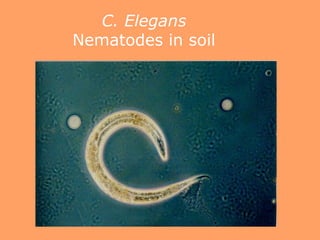
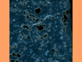
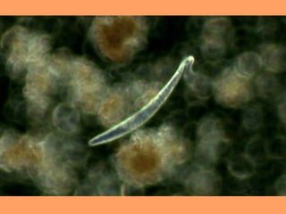
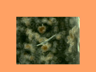
Microscopy Techniques

            By
    Carolina Montañez
Objective
• C. elegans
         Transparent nematode
         Found in soil
         About 1mm in length


• Phase Contrast Technique
         bright-field
         details in the image appear darker/brighter against a
            background
         transparent specimen, such as living cells and
        microorganisms
Methodology
1. Soil in the botany garden
2. Petridish
3. Dissecting microscope
4. Slide
5. Light microscope
6. Took some pictures
C. Elegans
Nematodes in soil
100x
400X
Carolina montanez mt

Carolina montanez mt

Editor's Notes

  • #2 Good afternoon everyone Today I will be talking about what I could accomplish with the usage of microscopy techniques
  • #3 Caenorhabditis elegans -use with brightfield - details in the image appear darker/brighter against a background -used on transparent specimen, such as living cells and microorganisms
  • #4 Found soil in the botany garden. Put soil on a Petri-dish and add water. Look for the nematode in the dissecting microscope Once I found it, put it on a slide Use the light microscope Finally took some pictures
  • #6 This is a picture of a c.elegans at 100x