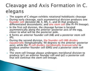
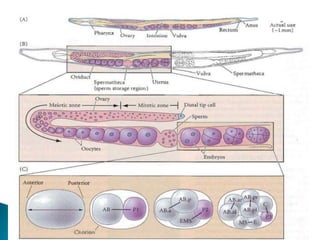
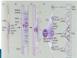
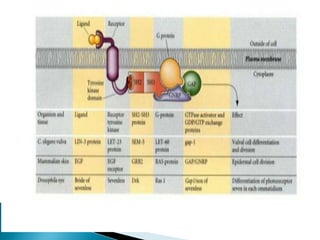
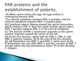
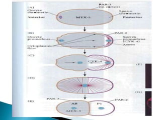
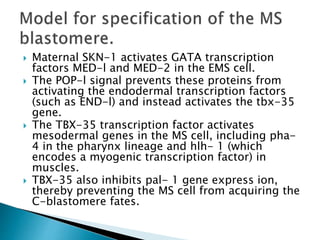
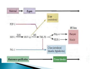
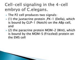
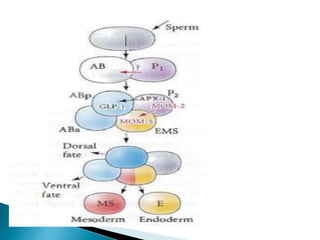
Presented by :-lovyansh lifescience
 The zygote of C elegan exhibits rotational holoblastic cleavage
 During early cleavage, each asymmetrical division produces one
founder cell (denoted AB, E, MS, C, and D) that produces
differentiated descendants; and one stem cell (the Pl-P4 Iineage).
 In the first cell division, the cleavage furrow is located
asymmetrically along the anterior-posterior axis of the egg,
closer to what will be the posterior pole.
 It forms an anterior founder cell (AB) and a posterior stem cell
(Pl).
 During the second division, the founder cell (AB) divides
equatorially (longitudinally; 90 degrees to the anterior-posterior
axis), while the PI cell divides meridionally (transversely) to
produce another founder cell (EMS) and a posterior stem cell
(P2).
 The stem cell lineage always undergoes meridional division to
produce (p1) an anterior founder cell and (p2) a posterior cell
that will continue the stem cell lineage.
 The formation of the vulva in C. elgans represents a case in which one
inductive signal generates a variety of cell types.
 This organ forms during the larval stage from six cells called the vulval
precursor cells (Vpes).
 The cell connecting the overlying gonad to the vulval precursor cells is called
the anchor cell
 The anchor cell secretes the LlN-3 protein, a paracrine factor (Similar to
mammalian epidermal growth factor, or EGF) that activates the RTK pathway
 If the anchor cell is destroyed (or if the Iin-3 gene is mutated), the VPCs will
not form a vuIva, but instead become part of the hypodermis (skin)
 The six VPCs influenced by the anchor cell form an equivalence group.
 Each member of this group is competent to become induced by the anchor
cell and can assume any of three fates, depending on its proximity to the
anchor cell.
 The cell directly beneath the anchor cell divides to form the central vulval
cells. The two cells flanking that central cell divide to become the lateral
vulval cells, while the three cells farther away from the anchor cell generate
hypodermal cells. If the anchor cell is destroyed, all six cells of the
equivalence group. divide once and contribute to the hypodermal tissue.
 If the three central VPCs are destroyed, the three outer cells, which normally
form hypodermis, generate vulval cells instead.
 The LlN-3 protein is received by the LET-23 receptor tyrosine kinase on
the VPCs, and the signal is transferred to the nucleus through the RTK
pathway.
 The target of the kinase cascade is the LlN-31 protein
 When this protein is phosphorylated in the nucleus, it loses its inhibitory
protein partner and is able to function as a transcription factor, promoting
vulval cell fates.
 Two mechanisms coordinate the formation of the vulva through this
induction
 1. The LIN-3 protein forms a concentration gradient. Here, the VPC closest
to the anchor cell (i.e., the P6.p cell) receives the highest concentration of
LlN-3 protein and generates the central vulval cells. The two VPCs adjacent
to it (PS.p and P7.p) receive a lower amount of LlN-3 and become the lateral
vulval cells.
 The VPCs farther away from the anchor cell do not receive enough LlN-3 to
have a n effect, so they become hypodermis.
 2. In addition to forming the central vulval lineage, theVPC closest to the
anchor cell also signals laterally to the two adjacent (P5.p and P7.p) cells
and instructs them not to generate the central vulval lineages. The P5.p and
P7.p cells receive the signal through the LlN-12 (Notch) proteins on their
cell membranes.
 The Notch signal activates a microRNA, mir-61, which represses the gene
that would specify central vulval fate, as well as promoting those genes that
are involved in forming the lateral vulval cells).
 The lateral cells do not instruct the peripheral VPCs to do anything, so they
become hypodermis.
 The formation of the anchor cell is mediated by lin-12, the C.efegans
homologue of the Notch gene.
 In wild-type C. elegans hermaphrodites, two adjacent cells, Zl .ppp and
Z4.aaa, have the potential to become the anchor cell. They interact in a
manner that causes one of them to become the anchor cell while the
other one becomes the precursor of the uterine tissue.
 In loss of function Iin-12 mutants, both cells become anchor cells,
 whereas in gain of-function mutations, both cells become uterine
precursors (Greenwald et al. 1 983).
 the lin-I2 gene needs to (unction only in that cell destined to become
the uterine precursor cell.
 The presumptive anchor cell does not need it.
 During a particular time in larval development, the cell that, by chance,
is secreting more LAG-2 causes its neighbor to cease its production of
this differentiation signal and to increase its production of LIN-12
protein.
 The cell secreting LAG-2 becomes the gonadal anchor cell, while the cell
receiving the signal through its LIN-12 protein becomes the ventral
uterine precursor cell
 Model for the generation of two cell types (anchor cell and
ventral uterine precursor) from two equivalent cells (ZI
.ppp and Z4.aaa) in C. e/egans.
 (A) The cells start off as equivalent, producing fluctuating
amounts of signal and receptor (inverted arrow). The lag-2
gene is thought to encode the signal; the Iin-12 gene is
thought to encode the receptor. Reception of the signal
turns down LAG-2 (Della) production and up-regulate,
LlN-12 (Notch).
 (B) A stochastic (chance) event causes one cell to produce
more LAG-2 than The other cell at some particular critical
time. This stimulates more LIN-12 production in the
neighboring cell.
 (C) This difference is amplified, since The cell producing
more LlN-12 produces less LAG-2. Eventually, just one cell
is delivering the LAG-2 signal, and the other cell is
receiving it.
 (D) The signaling cell becomes the anchor Self; the
receiving cell becomes the ventral uterine precursor.
(A) When sperm enters the egg, the egg nucleus is
undergoing meiosis (left).
The cortical cytoplasm contains PAR-3 (orange), and the
internal cytoplasm contains MEX-5 (shaded gray),
(B) Cytoplasm begins flowing toward the sperm pronucleus,
which contains CYK-4. Near the newly formed sperm asters,
PAR-2 (purple) replaces PAR-3 in the cortical cytoplasm.
(C) The domain of PAR-2 expression expands as the sperm
nucleus migrates toward the center of the cell.
(D) At the first division, about half the PAR-2, PAR-3, and
MEX-5 polarity remains. (EI At the end of the first division,
the AB blastomere has MEX-5 and PAR-3, while most of the
Pl blastomere has very little MEX-5 and has a cortex of PAR-2
(except where the two blastomeres meet). (F) In this dividing
C. elegans
 Maternal SKN-1 activates GATA transcription
factors MED-l and MED-2 in the EMS cell.
 The POP-l signal prevents these proteins from
activating the endodermal transcription factors
(such as END-l) and instead activates the tbx-35
gene.
 The TBX-35 transcription factor activates
mesodermal genes in the MS cell, including pha-
4 in the pharynx lineage and hlh- 1 (which
encodes a myogenic transcription factor) in
muscles.
 TBX-35 also inhibits pal- 1 gene express ion,
thereby preventing the MS cell from acquiring the
C-blastomere fates.
 The P2 cell produces two signals:
 (1) the juxtacrine protein ,PX-1 (Della), which
is bound by GLP-1 (Notch) on the ABp cell,
and
 (2) the paracrine protein MOM-2 (Wnt), which
is bound by the MOM-S (Frizzled) protein on
the EMS cell
c elegan.pptx
c elegan.pptx
c elegan.pptx

c elegan.pptx

  • 1.
  • 2.
     The zygoteof C elegan exhibits rotational holoblastic cleavage  During early cleavage, each asymmetrical division produces one founder cell (denoted AB, E, MS, C, and D) that produces differentiated descendants; and one stem cell (the Pl-P4 Iineage).  In the first cell division, the cleavage furrow is located asymmetrically along the anterior-posterior axis of the egg, closer to what will be the posterior pole.  It forms an anterior founder cell (AB) and a posterior stem cell (Pl).  During the second division, the founder cell (AB) divides equatorially (longitudinally; 90 degrees to the anterior-posterior axis), while the PI cell divides meridionally (transversely) to produce another founder cell (EMS) and a posterior stem cell (P2).  The stem cell lineage always undergoes meridional division to produce (p1) an anterior founder cell and (p2) a posterior cell that will continue the stem cell lineage.
  • 5.
     The formationof the vulva in C. elgans represents a case in which one inductive signal generates a variety of cell types.  This organ forms during the larval stage from six cells called the vulval precursor cells (Vpes).  The cell connecting the overlying gonad to the vulval precursor cells is called the anchor cell  The anchor cell secretes the LlN-3 protein, a paracrine factor (Similar to mammalian epidermal growth factor, or EGF) that activates the RTK pathway  If the anchor cell is destroyed (or if the Iin-3 gene is mutated), the VPCs will not form a vuIva, but instead become part of the hypodermis (skin)  The six VPCs influenced by the anchor cell form an equivalence group.  Each member of this group is competent to become induced by the anchor cell and can assume any of three fates, depending on its proximity to the anchor cell.  The cell directly beneath the anchor cell divides to form the central vulval cells. The two cells flanking that central cell divide to become the lateral vulval cells, while the three cells farther away from the anchor cell generate hypodermal cells. If the anchor cell is destroyed, all six cells of the equivalence group. divide once and contribute to the hypodermal tissue.  If the three central VPCs are destroyed, the three outer cells, which normally form hypodermis, generate vulval cells instead.
  • 6.
     The LlN-3protein is received by the LET-23 receptor tyrosine kinase on the VPCs, and the signal is transferred to the nucleus through the RTK pathway.  The target of the kinase cascade is the LlN-31 protein  When this protein is phosphorylated in the nucleus, it loses its inhibitory protein partner and is able to function as a transcription factor, promoting vulval cell fates.  Two mechanisms coordinate the formation of the vulva through this induction  1. The LIN-3 protein forms a concentration gradient. Here, the VPC closest to the anchor cell (i.e., the P6.p cell) receives the highest concentration of LlN-3 protein and generates the central vulval cells. The two VPCs adjacent to it (PS.p and P7.p) receive a lower amount of LlN-3 and become the lateral vulval cells.  The VPCs farther away from the anchor cell do not receive enough LlN-3 to have a n effect, so they become hypodermis.  2. In addition to forming the central vulval lineage, theVPC closest to the anchor cell also signals laterally to the two adjacent (P5.p and P7.p) cells and instructs them not to generate the central vulval lineages. The P5.p and P7.p cells receive the signal through the LlN-12 (Notch) proteins on their cell membranes.  The Notch signal activates a microRNA, mir-61, which represses the gene that would specify central vulval fate, as well as promoting those genes that are involved in forming the lateral vulval cells).  The lateral cells do not instruct the peripheral VPCs to do anything, so they become hypodermis.
  • 8.
     The formationof the anchor cell is mediated by lin-12, the C.efegans homologue of the Notch gene.  In wild-type C. elegans hermaphrodites, two adjacent cells, Zl .ppp and Z4.aaa, have the potential to become the anchor cell. They interact in a manner that causes one of them to become the anchor cell while the other one becomes the precursor of the uterine tissue.  In loss of function Iin-12 mutants, both cells become anchor cells,  whereas in gain of-function mutations, both cells become uterine precursors (Greenwald et al. 1 983).  the lin-I2 gene needs to (unction only in that cell destined to become the uterine precursor cell.  The presumptive anchor cell does not need it.  During a particular time in larval development, the cell that, by chance, is secreting more LAG-2 causes its neighbor to cease its production of this differentiation signal and to increase its production of LIN-12 protein.  The cell secreting LAG-2 becomes the gonadal anchor cell, while the cell receiving the signal through its LIN-12 protein becomes the ventral uterine precursor cell
  • 10.
     Model forthe generation of two cell types (anchor cell and ventral uterine precursor) from two equivalent cells (ZI .ppp and Z4.aaa) in C. e/egans.  (A) The cells start off as equivalent, producing fluctuating amounts of signal and receptor (inverted arrow). The lag-2 gene is thought to encode the signal; the Iin-12 gene is thought to encode the receptor. Reception of the signal turns down LAG-2 (Della) production and up-regulate, LlN-12 (Notch).  (B) A stochastic (chance) event causes one cell to produce more LAG-2 than The other cell at some particular critical time. This stimulates more LIN-12 production in the neighboring cell.  (C) This difference is amplified, since The cell producing more LlN-12 produces less LAG-2. Eventually, just one cell is delivering the LAG-2 signal, and the other cell is receiving it.  (D) The signaling cell becomes the anchor Self; the receiving cell becomes the ventral uterine precursor.
  • 13.
    (A) When spermenters the egg, the egg nucleus is undergoing meiosis (left). The cortical cytoplasm contains PAR-3 (orange), and the internal cytoplasm contains MEX-5 (shaded gray), (B) Cytoplasm begins flowing toward the sperm pronucleus, which contains CYK-4. Near the newly formed sperm asters, PAR-2 (purple) replaces PAR-3 in the cortical cytoplasm. (C) The domain of PAR-2 expression expands as the sperm nucleus migrates toward the center of the cell. (D) At the first division, about half the PAR-2, PAR-3, and MEX-5 polarity remains. (EI At the end of the first division, the AB blastomere has MEX-5 and PAR-3, while most of the Pl blastomere has very little MEX-5 and has a cortex of PAR-2 (except where the two blastomeres meet). (F) In this dividing C. elegans
  • 15.
     Maternal SKN-1activates GATA transcription factors MED-l and MED-2 in the EMS cell.  The POP-l signal prevents these proteins from activating the endodermal transcription factors (such as END-l) and instead activates the tbx-35 gene.  The TBX-35 transcription factor activates mesodermal genes in the MS cell, including pha- 4 in the pharynx lineage and hlh- 1 (which encodes a myogenic transcription factor) in muscles.  TBX-35 also inhibits pal- 1 gene express ion, thereby preventing the MS cell from acquiring the C-blastomere fates.
  • 17.
     The P2cell produces two signals:  (1) the juxtacrine protein ,PX-1 (Della), which is bound by GLP-1 (Notch) on the ABp cell, and  (2) the paracrine protein MOM-2 (Wnt), which is bound by the MOM-S (Frizzled) protein on the EMS cell